clinical study early clinical outcomes associated with a...
TRANSCRIPT

Clinical StudyEarly Clinical Outcomes Associated with a Novel OsteochondralAllograft Transplantation System in the Knee
William J. Long,1,2,3,4 Joseph W. Greene,5,6 and Fred D. Cushner7,8
1 Insall Scott Kelly Institute, New York, NY 10065, USA2New York University, New York, NY 10065, USA3Hospital for Joint Disease, NYU, New York, NY 10065, USA4St. Francis Hospital, Roslyn, NY 11548, USA5Norton Healthcare, Louisville, KY 40207, USA6Department of Orthopaedic Surgery, University of Louisville, Louisville, KY 40202, USA7Southside Hospital, Bay Shore, NY 11706, USA8Lenox Hill Hospital, Northwell Health System, New York, NY 10065, USA
Correspondence should be addressed to William J. Long; doctor [email protected]
Received 8 November 2015; Revised 7 March 2016; Accepted 10 March 2016
Academic Editor: Werner Kolb
Copyright © 2016 William J. Long et al.This is an open access article distributed under the Creative CommonsAttribution License,which permits unrestricted use, distribution, and reproduction in any medium, provided the original work is properly cited.
Background. Osteochondral defects of the knee are a common finding at the time of arthroscopic intervention. Purpose/Hypothesis.To report our outcomes after utilizing a new technique of osteochondral allograft transplantation for focal cartilage defects. StudyDesign. Case series. Methods. All patients treated with osteochondral allograft transplantation with a Zimmer Chondrofix plug(Zimmer Inc.,Warsaw, IN) for focal cartilage defects over a 12-month period were followed up at aminimumof 24months. Failureswere documented and radiographs were evaluated. Results. 61 knees (58 patients) underwent grafting. Three cases were lost tofollow-up. In the remaining 58 cases the average age was 40 (range 18–59). At a mean follow-up of 28 months (range 24–36), therewere 5 failures requiring further surgery. Mean KOOS scores in the Pain, Symptoms, ADL, Sports, and Quality of Life dimensionswere 82, 79, 84, 66, and 58, respectively. Radiographs demonstrated maintenance of the subchondral bone without graft absorptionor subsidence. Conclusions. Our observations suggest that osteochondral allograft transplantation leads to a satisfactory activitylevel and function at early follow-upwhile avoiding the inherent complexities associatedwith other cartilage restoration techniques.Longer follow-up is warranted to monitor the subchondral bone, articular surface, and patient outcome measures.
1. Introduction
Each year it is estimated that chondral defects of the kneeaffect more than 900,000 people in the United States andresult inmore than 200,000 procedures [1]. In 34% of all kneearthroscopies, one or more focal full-thickness or nearly full-thickness cartilage defects are seen [2–4]. It is also estimatedthat 36% of all athletes have a cartilage defect in the knee [5].Human articular cartilage has limited ability to self-repair, sodamage to the articular cartilage in the knee is a potential riskfactor in the development of early-onset osteoarthritis andcan result in loss of movement and pain [6–8].
Full-thickness articular cartilage defects, defined as Out-erbridge grades III–IV [9], are likely to progress to early
degenerative wear and become increasingly symptomatic[10]; thus surgical treatment is recommended [11]. A varietyof surgical options exist for the treatment of full-thicknesschondral defects in the knee such as debridement, microfrac-ture, osteochondral autograft transfer (OAT), osteochondralallograft transplantation (OCA), and autologous chondrocyteimplantation (ACI). Each of these techniques presents its ownunique set of limitations.
Microfracture is the most commonmethod employed forcartilage restoration in the knee [12]. It relies onmarrow stim-ulation and the influx of marrow products into the cartilagedefect. The mature fibrocartilage formed is less durable thannative type II collagen, resulting in some degradation overtime [13]. Some surgical algorithms use approximately 2 cm2
Hindawi Publishing CorporationAdvances in Orthopedic SurgeryVolume 2016, Article ID 1979348, 6 pageshttp://dx.doi.org/10.1155/2016/1979348

2 Advances in Orthopedic Surgery
as the upper threshold for microfracture and beyond thismove onto more advanced cartilage restoration techniques[14–16].
OATS and mosaicplasty are techniques involving thetransport of osteochondral plugs from another area of theknee to the defect. Disadvantages associated with these tech-niques include leaving an articular cartilage defect in anotherlocation of the knee, progression of osteoarthritic changes atthe donor site, the relative size limitation of <20mm2 [17],and the fact that an arthrotomy may be required. As largerareas of OATs are required, more donor site complaints andsymptoms occur [18].
OCA eliminates the donor site complications and sizelimitations of OATS by obtaining the donor tissue from acadaver. It allows treatment of both cartilage and underlyingbony defects to a greater degree than ACI. This techniquehas shown promising results at long-term follow-up [19].Limitations to OCA involve the need for donor tissue andthe timing of transplantation, which is best achieved between14 and 28 days to allow testing of the tissue, without thesignificant loss of cell viability that occurs over time [20].
As first proposed in 1994 ACI allows the treatmentof large cartilage defects in the knee. Unfortunately, it isexpensive, costing approximately 66,000 dollars per case [21].The technique requires a 2-stage procedure (harvesting andimplantation) and is less successful with bone loss at the baseof the lesion [22].
In an effort to avoid the individual complications andlimitations associated with each of these techniques, wehave been using a relatively new technique of osteochondralallograft transplantation, Chondrofix� Osteochondral Allo-graft (Zimmer Inc., Warsaw, IN). These allografts consistof decellularized hyaline cartilage and cancellous bone thatmaintain the mechanical properties of unprocessed osteo-chondral grafts.
Similar to OCA, Chondrofix transplantation is single-stage and guarantees an immediate reliable tissue transfer ofan osteochondral unit; however the grafts are readily availableon the shelf, thus eliminating the narrow timing aspects toOCA.The aim of this study was to evaluate the results of thistechnique retrospectively in a selected group of patients withthe goal of analyzing patient outcomes and understandingwhich factors could influence the clinical outcome in orderto clarify the correct indication of this treatment option.
2. Methods
This study was approved by our Health System InstitutionalReview Board. All patients between the ages of 18 and 60who had received a Chondrofix plug in the twelve-monthperiod between February 2012 and February 2013 wereretrospectively included in the study.
The procedures were performed by one of two fellowship-trained, board-certified orthopaedic surgeons. Inclusion cri-teria included a symptomatic full-thickness cartilage lesionidentified preoperatively by advanced imaging or priorarthroscopy, though the size of the lesion was consis-tently underestimated. Patients with inflammatory arthritis
Table 1: Primary diagnosis.
DJD 27PF DJD 15ACL tear 5OCD lesion 4PF instability 3DJD: degenerative joint disease; PF: patellofemoral; ACL: anterior cruciateligament; OCD: osteochondritis dissecans.
or significant uni- or tricompartmental arthritis were notconsidered candidates for transplantation.
A tourniquet was used, and the knee was examinedarthroscopically. Once the final decision to perform osteo-chondral allograft transplantation was taken, the lesion wassized, prepared, and graftedwith instrumentation provided inthe Chondrofix set. One or more plugs were placed depend-ing on the size and shape of the lesion. The instrumentationwas introduced through an appropriately sized accessoryportal, and the procedure was visualized arthroscopically.An arthrotomy was employed only in the case of a patellarlesion, allowing the patella to be everted for perpendicularpreparation and grafting of the defect.
Postoperatively all patients were allowed to weight-bearas tolerated and encouraged to progress to full range ofmotion.Abracewas only used if required due to an associatedprocedure (e.g., cruciate ligament reconstruction). Patientswere instructed to avoid high impact activities for 6 weeks.Follow-up visits occurred at 3 weeks, 6 weeks, 3 months,6 months, and yearly thereafter. Radiographs of the kneewere obtained (weight bearing anteroposterior, lateral, andskyline) at the 3-week, six-month, and yearly follow-ups.
Clinical evaluation included range of motion (ROM),presence of an effusion, and radiographic changes. Theycompleted the Tegner score [23], and the Knee Injury andOsteoarthritis Outcome Score (KOOS) patient-derived out-come tool [24]. Patients who were unable to be evaluated inperson completed the scores over the telephone.
3. Results
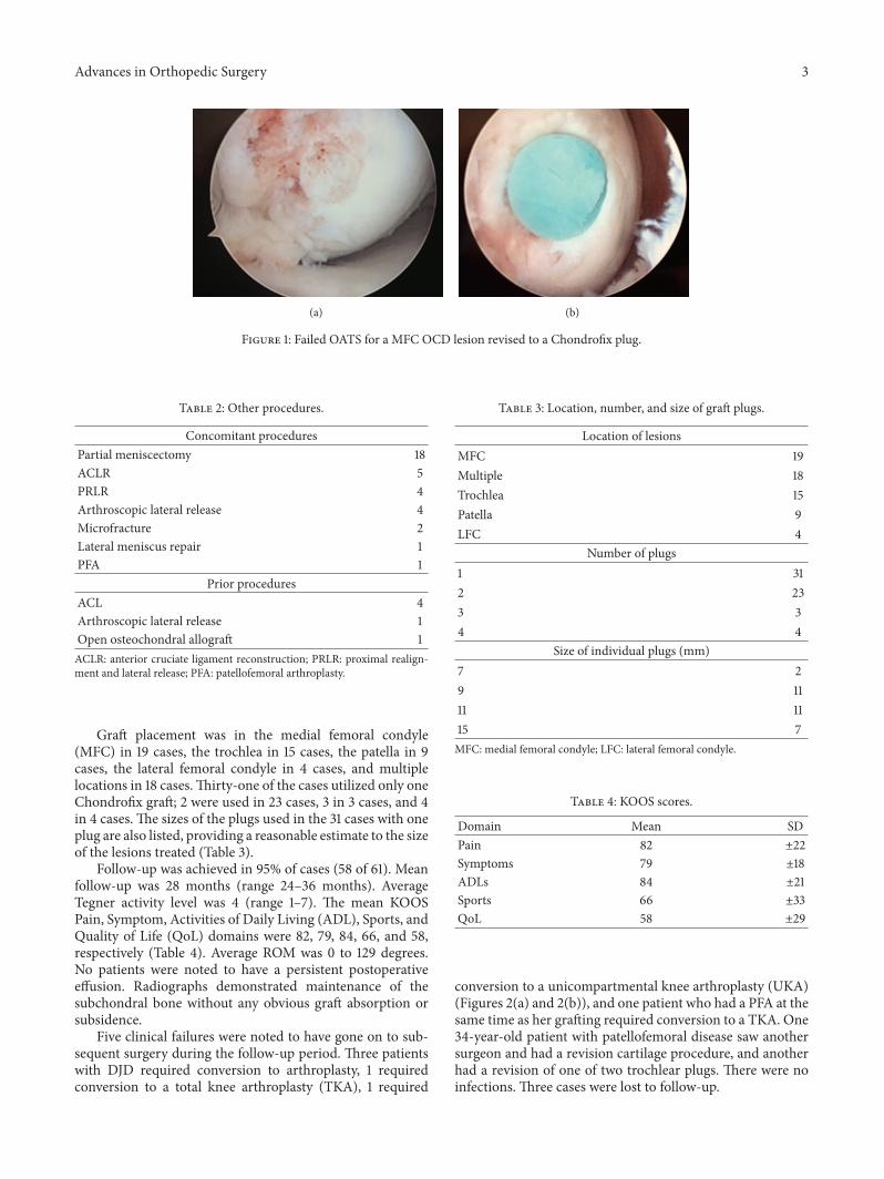
61 cases in 58 patients were eligible for inclusion.The averageage was 40.0 (range 18–59) and 59% of cases were in malepatients. The primary diagnoses (Table 1) were degenerativejoint disease (DJD) in 27 cases, patellofemoral (PF) DJD in15 cases, anterior cruciate ligament (ACL) tear and resultantinstability in 5 cases, osteochondritis dissecans (OCD) lesionin 4 cases (Figures 1(a) and 1(b)), and PF instability and DJDin 3 cases.
Procedures performed concomitantly (Table 2) includearthroscopic partial meniscectomy in 18 cases, ACL recon-struction in 5 cases, open proximal realignment and lat-eral release in 4 cases, arthroscopic lateral release in 4cases, microfracture in 2 cases, and lateral meniscus repair,patellofemoral arthroplasty in 1 case each. Four knees hadundergone prior ACL reconstruction, 1 knee had priorarthroscopic lateral release, and 1 knee had previous openosteochondral allografting.
Advances in Orthopedic Surgery 3
(a) (b)
Figure 1: Failed OATS for a MFC OCD lesion revised to a Chondrofix plug.
Table 2: Other procedures.
Concomitant proceduresPartial meniscectomy 18ACLR 5PRLR 4Arthroscopic lateral release 4Microfracture 2Lateral meniscus repair 1PFA 1
Prior proceduresACL 4Arthroscopic lateral release 1Open osteochondral allograft 1ACLR: anterior cruciate ligament reconstruction; PRLR: proximal realign-ment and lateral release; PFA: patellofemoral arthroplasty.
Graft placement was in the medial femoral condyle(MFC) in 19 cases, the trochlea in 15 cases, the patella in 9cases, the lateral femoral condyle in 4 cases, and multiplelocations in 18 cases.Thirty-one of the cases utilized only oneChondrofix graft; 2 were used in 23 cases, 3 in 3 cases, and 4in 4 cases. The sizes of the plugs used in the 31 cases with oneplug are also listed, providing a reasonable estimate to the sizeof the lesions treated (Table 3).
Follow-up was achieved in 95% of cases (58 of 61). Meanfollow-up was 28 months (range 24–36 months). AverageTegner activity level was 4 (range 1–7). The mean KOOSPain, Symptom, Activities of Daily Living (ADL), Sports, andQuality of Life (QoL) domains were 82, 79, 84, 66, and 58,respectively (Table 4). Average ROM was 0 to 129 degrees.No patients were noted to have a persistent postoperativeeffusion. Radiographs demonstrated maintenance of thesubchondral bone without any obvious graft absorption orsubsidence.
Five clinical failures were noted to have gone on to sub-sequent surgery during the follow-up period. Three patientswith DJD required conversion to arthroplasty, 1 requiredconversion to a total knee arthroplasty (TKA), 1 required
Table 3: Location, number, and size of graft plugs.
Location of lesionsMFC 19Multiple 18Trochlea 15Patella 9LFC 4
Number of plugs1 312 233 34 4
Size of individual plugs (mm)7 29 1111 1115 7MFC: medial femoral condyle; LFC: lateral femoral condyle.
Table 4: KOOS scores.
Domain Mean SDPain 82 ±22Symptoms 79 ±18ADLs 84 ±21Sports 66 ±33QoL 58 ±29
conversion to a unicompartmental knee arthroplasty (UKA)(Figures 2(a) and 2(b)), and one patient who had a PFA at thesame time as her grafting required conversion to a TKA. One34-year-old patient with patellofemoral disease saw anothersurgeon and had a revision cartilage procedure, and anotherhad a revision of one of two trochlear plugs. There were noinfections. Three cases were lost to follow-up.

4 Advances in Orthopedic Surgery
(a) (b)
Figure 2: (a) Intact Chondrofix plugs in the MFC at the time of UKA. (b) Stable underlying bony integration of the bony portion of the plugfor femoral component seating.
4. Discussion
Anumber of arthroscopic repair or reconstructive techniquescan be performed in the setting of full-thickness cartilagedefects in the knee. Variousmaterials, such as allografts, auto-grafts, synthetic polymers, and periosteal and perichondralflaps, have been proposed, but the 4 techniques that havegained widespread use and interest during the last decadein North America are microfracture, OCA, OATS, and ACIprocedures.
Currently, the important step of determining the area ofthe defect is accomplished either by attempting to interpretradiographic images prior to surgery or by using mechanicalinstrumentation during an arthroscopic diagnostic examina-tion. Unfortunately, the sensitivity of conventional magneticresonance imaging (MRI) is still limited. Improved diagnosticperformance has been seen with 3-Tesla (T) MRIs comparedwith 1.5 T protocols [25, 26]. In a study comparing 3 TMRI toarthroscopy, the ability of MRI to predict articular cartilagelesions was examined. When using the Outerbridge classi-fication the values for sensitivity, specificity, and accuracywere 57%, 71%, and 63%, respectively [27]. It is particularlydifficult to determine the depth of the lesion [28]. Therefore,surgeons cannot rely on preoperative imaging to accuratelydetermine treatment choice, leaving much of the decision-making process to the arthroscopic evaluation of the defectgrade and size.
Thus both the complexity of existing techniques andthe difficulty in predicting the size of the lesion lead usto consider this novel Chondrofix option. We were fairlyfamiliar with this technique, having used it with Tru-Fitplugs for the preceding five years [29]. With the Chondrofixsystem, multiple graft sizes (7–15mm) are available for off-the-shelf use. Single-stage implantation using an arthroscopicapproach with a small incision for graft insertion can beused for all lesions with the exception of the patella, wherea miniarthrotomy is required for access.
Figure 3: MRI evaluation of a Chondrofix plug at 3 months afterimplantation.
Figure 4: MRI evaluation of a plug at 8 months after implantationwith further bony incorporation.

Advances in Orthopedic Surgery 5
(a) (b)
(c) (d)
Figure 5: Cartilage surface delamination of a plug overlying a stable bony base with successful revision to a similarly sized plug.
Our primary concerns with this new graft option werewith respect to its incorporation and durability as the graftsare acellular and treated in a novel fashion, prior to implan-tation. Our radiographic review did not demonstrate anysignificant changes. A more sensitive MRI evaluation wasavailable for two cases that had follow-up imaging for newinjuries to the knee. The first (Figure 3) was performed at 3months and the second (Figure 4) at 8 months. They bothdemonstrate incorporation of the bony portion of the graft,with some changes to the overlying cartilage surface, whichwill have to be followed up carefully over the longer-term.
Outside the cohort in this paper, we have seen onenovel failure mechanism. In this case the cartilage surfacedelaminated from the underlying bone on the plug. Itoccurred in a labourer during a squat at lift of a 100 lbpiece of equipment. Arthroscopic evaluation demonstrateda well-incorporated bone base separated from the overlyingcartilage (Figures 5(a)–5(d)). A successful revision to a newplug was performed, and the patient has done well to a yearfollowing revision.
The failures in this series were evaluated. Four of the fivefailures had two plugs in a single compartment. The plugsused were all at least 9mm, consistent with larger defects.Thus the failure rate was 1/31 for single-plug cases and 4/23for two-plug cases. Interestingly, none of the even larger 3-and 4-plug cases failed. Location of the lesion did not predict
failure, with 3 failures in MFC lesions and 2 in trochlearlesions.
5. Conclusions
While it is generally accepted that focal chondral lesions oftenprogress towards osteoarthritis, a review of the literaturepresents compelling evidence that between 10 and 40% of allpatients aged <40 years undergoing arthroscopic surgery forother reasons have treatable chondral injuries thatwill remainunaddressed [2–4].The current study demonstrates a simple,arthroscopic, off-the-shelf solution that appears to providereasonable clinical outcomes at short-term follow-up.
There are a number of obvious limitations to this study:it is a short-term retrospective follow-up; preoperative scoreswere not obtained; therewere a number of different diagnosesand concomitant procedures performed; and there was nocomparative group. In the future, we are planning morecomprehensive and rigorous prospective comparative studiesinvolving both preoperative and postoperative evaluationswith advanced imaging, to better compare outcomes withthese cartilage lesions. Based on this study, Chondrofix plugsappear to be a reasonable on-demand option for addressingfull-thickness cartilage lesions encountered at the time ofarthroscopy, with acceptable short-term patient satisfactionand function.

6 Advances in Orthopedic Surgery
Competing Interests
The authors declare that they have no competing interests.
References
[1] W. W. Curl, J. Krome, E. S. Gordon, J. Rushing, B. P. Smith,and G. G. Poehling, “Cartilage injuries: a review of 31,516 kneearthroscopies,” Arthroscopy, vol. 13, no. 4, pp. 456–460, 1997.
[2] A. Aroøen, S. Løken, S. Heir et al., “Articular cartilage lesionsin 993 consecutive knee arthroscopies,” The American Journalof Sports Medicine, vol. 32, no. 1, pp. 211–215, 2004.
[3] K. Hjelle, E. Solheim, T. Strand, R. Muri, and M. Brit-tberg, “Articular cartilage defects in 1,000 knee arthroscopies,”Arthroscopy, vol. 18, no. 7, pp. 730–734, 2002.
[4] W. Widuchowski, J. Widuchowski, and T. Trzaska, “Articularcartilage defects: study of 25,124 knee arthroscopies,” Knee, vol.14, no. 3, pp. 177–182, 2007.
[5] D. C. Flanigan, J. D. Harris, T. Q. Trinh, R. A. Siston, and R.H. Brophy, “Prevalence of chondral defects in Athletes’ Knees: asystematic review,”Medicine and Science in Sports and Exercise,vol. 42, no. 10, pp. 1795–1801, 2010.
[6] E. Arendt and R. Dick, “Knee injury patterns among men andwomen in collegiate basketball and soccer. NCAA data andreview of literature,” The American Journal of Sports Medicine,vol. 23, no. 6, pp. 694–701, 1995.
[7] S. Drawer and C. W. Fuller, “Propensity for osteoarthritis andlower limb joint pain in retired professional soccer players,”British Journal of Sports Medicine, vol. 35, no. 6, pp. 402–408,2001.
[8] H. Roos, “Are there long-term sequelae from soccer?” Clinics inSports Medicine, vol. 17, no. 4, pp. 819–831, 1998.
[9] R. E. Outerbridge, “The etiology of chondromalacia patellae,”The Journal of Bone & Joint Surgery—British Volume, vol. 43, pp.752–757, 1961.
[10] H. J. Mankin, “The response of articular cartilage tomechanicalinjury,”The Journal of Bone & Joint Surgery—American Volume,vol. 64, no. 3, pp. 460–466, 1982.
[11] N. A. Sgaglione, A. Miniaci, S. D. Gillogly, and T. R. Carter,“Update on advanced surgical techniques in the treatmentof traumatic focal articular cartilage lesions in the knee,”Arthroscopy, vol. 18, no. 2, pp. 9–32, 2002.
[12] A. G. McNickle, M. T. Provencher, and B. J. Cole, “Overviewof existing cartilage repair technology,” Sports Medicine &Arthroscopy Review, vol. 16, no. 4, pp. 196–201, 2008.
[13] K. Mithoefer, T. Mcadams, R. J. Williams, P. C. Kreuz, and B. R.Mandelbaum, “Clinical efficacy of the microfracture techniquefor articular cartilage repair in the knee: an evidence-basedsystematic analysis,” American Journal of Sports Medicine, vol.37, no. 10, pp. 2053–2063, 2009.
[14] J. W. Alford and B. J. Cole, “Cartilage restoration, part 2:techniques, outcomes, and future directions,” American Journalof Sports Medicine, vol. 33, no. 3, pp. 443–460, 2005.
[15] E. L. Cain and W. G. Clancy, “Treatment algorithm for osteo-chondral injuries of the knee,” Clinics in Sports Medicine, vol.20, no. 2, pp. 321–342, 2001.
[16] H. Clarke, F. Cushner, and W. Scott, “Clinical algorithm fortreatment of chondral injuries,” in Insall & Scott Surgery ofthe Knee, W. N. Scott, Ed., pp. 433–437, Churchill Livingstone,Philadelphia, Pa, USA, 4th edition, 2005.
[17] A. J. Krych, H. W. Harnly, S. A. Rodeo, and R. J. Williams III,“Activity levels are higher after osteochondral autograft transfermosaicplasty than after microfracture for articular cartilagedefects of the knee: a retrospective comparative study,” TheJournal of Bone & Joint Surgery—American Volume, vol. 94, no.11, pp. 971–978, 2012.
[18] L. Hangody and P. Fules, “Autologous osteochondral mosaic-plasty for the treatment of full-thickness defects of weight-bearing joints: Ten years of experimental and clinical experi-ence,” The Journal of Bone & Joint Surgery—American Volume,vol. 85, no. 1, pp. 25–32, 2003.
[19] A. E. Gross, W. Kim, F. Las Heras, D. Backstein, O. Safir, and K.P. H. Pritzker, “Fresh osteochondral allografts for posttraumaticknee defects: long-term followup,” Clinical Orthopaedics andRelated Research, vol. 466, no. 8, pp. 1863–1870, 2008.
[20] R. F. LaPrade, J. Botker, M. Herzog, and J. Agel, “Refrigeratedosteoarticular allografts to treat articular cartilage defects of thefemoral condyles. A prospective outcomes study,”The Journal ofBone & Joint Surgery—American Volume, vol. 91, no. 4, pp. 805–811, 2009.
[21] E. M. Samuelson and D. E. Brown, “Cost-effectiveness analysisof autologous chondrocyte implantation: a comparison ofperiosteal patch versus type I/III collagen membrane,” Amer-ican Journal of Sports Medicine, vol. 40, no. 6, pp. 1252–1258,2012.
[22] M. Brittberg, A. Lindahl, A. Nilsson, C. Ohlsson, O. Isaksson,and L. Peterson, “Treatment of deep cartilage defects in the kneewith autologous chondrocyte transplantation,” New EnglandJournal of Medicine, vol. 331, no. 14, pp. 889–895, 1994.
[23] Y. Tegner and J. Lysholm, “Rating systems in the evaluationof knee ligament injuries,” Clinical Orthopaedics and RelatedResearch, vol. 198, pp. 43–49, 1985.
[24] E. M. Roos, H. P. Roos, L. S. Lohmander, C. Ekdahl, andB. D. Beynnon, “Knee Injury and Osteoarthritis OutcomeScore (KOOS)—development of a self-administered outcomemeasure,” Journal of Orthopaedic and Sports Physical Therapy,vol. 28, no. 2, pp. 88–96, 1998.
[25] R. Kijowski, D. G. Blankenbaker, K. W. Davis, K. Shinki, L. D.Kaplan, and A. A. De Smet, “Comparison of 1.5- And 3.0-T MRimaging for evaluating the articular cartilage of the knee joint,”Radiology, vol. 250, no. 3, pp. 839–848, 2009.
[26] S. Wong, L. Steinbach, J. Zhao, C. Stehling, C. B. Ma, and T. M.Link, “Comparative study of imaging at 3.0 T versus 1.5 T of theknee,” Skeletal Radiology, vol. 38, no. 8, pp. 761–769, 2009.
[27] M. E. Reed, D. C. Villacis, G. F. Hatch III et al., “3.0-teslaMRI and arthroscopy for assessment of knee articular cartilagelesions,” Orthopedics, vol. 36, no. 8, pp. e1060–e1064, 2013.
[28] M. L. Gray, D. Burstein, Y.-J. Kim, and A. Maroudas, “Magneticresonance imaging of cartilage glycosaminoglycan: basic prin-ciples, imaging technique, and clinical applications,” Journal ofOrthopaedic Research, vol. 26, no. 3, pp. 281–291, 2008.
[29] P.Hindle, J. L. Hendry, J. F. Keating, and L. C. Biant, “Autologousosteochondral mosaicplasty or TruFit plugs for cartilage repair,”Knee Surgery, Sports Traumatology, Arthroscopy, vol. 22, no. 6,pp. 1235–1240, 2014.

Submit your manuscripts athttp://www.hindawi.com
Stem CellsInternational
Hindawi Publishing Corporationhttp://www.hindawi.com Volume 2014
Hindawi Publishing Corporationhttp://www.hindawi.com Volume 2014
MEDIATORSINFLAMMATION
of
Hindawi Publishing Corporationhttp://www.hindawi.com Volume 2014
Behavioural Neurology
EndocrinologyInternational Journal of
Hindawi Publishing Corporationhttp://www.hindawi.com Volume 2014
Hindawi Publishing Corporationhttp://www.hindawi.com Volume 2014
Disease Markers
Hindawi Publishing Corporationhttp://www.hindawi.com Volume 2014
BioMed Research International
OncologyJournal of
Hindawi Publishing Corporationhttp://www.hindawi.com Volume 2014
Hindawi Publishing Corporationhttp://www.hindawi.com Volume 2014
Oxidative Medicine and Cellular Longevity
Hindawi Publishing Corporationhttp://www.hindawi.com Volume 2014
PPAR Research
The Scientific World JournalHindawi Publishing Corporation http://www.hindawi.com Volume 2014
Immunology ResearchHindawi Publishing Corporationhttp://www.hindawi.com Volume 2014
Journal of
ObesityJournal of
Hindawi Publishing Corporationhttp://www.hindawi.com Volume 2014
Hindawi Publishing Corporationhttp://www.hindawi.com Volume 2014
Computational and Mathematical Methods in Medicine
OphthalmologyJournal of
Hindawi Publishing Corporationhttp://www.hindawi.com Volume 2014
Diabetes ResearchJournal of
Hindawi Publishing Corporationhttp://www.hindawi.com Volume 2014
Hindawi Publishing Corporationhttp://www.hindawi.com Volume 2014
Research and TreatmentAIDS
Hindawi Publishing Corporationhttp://www.hindawi.com Volume 2014
Gastroenterology Research and Practice
Hindawi Publishing Corporationhttp://www.hindawi.com Volume 2014
Parkinson’s Disease
Evidence-Based Complementary and Alternative Medicine
Volume 2014Hindawi Publishing Corporationhttp://www.hindawi.com