clinical study clinical outcomes of primary palatal...
TRANSCRIPT
Clinical StudyClinical Outcomes of Primary Palatal Surgery in Children withNonsyndromic Cleft Palate with and without Lip
Seunghee Ha,1 Kyung S. Koh,2 Heewon Moon,2 Seungeun Jung,2 and Tae Suk Oh2
1Division of Speech Pathology and Audiology, Audiology and Speech Pathology Research Institute, Hallym University,Chuncheon 200-702, Republic of Korea2Department of Plastic Surgery, Seoul Asan Medical Center, University of Ulsan College of Medicine, Seoul 138-736, Republic of Korea
Correspondence should be addressed to Kyung S. Koh; [email protected]
Received 16 December 2014; Accepted 23 January 2015
Academic Editor: Antonio Ysunza
Copyright © 2015 Seunghee Ha et al. This is an open access article distributed under the Creative Commons Attribution License,which permits unrestricted use, distribution, and reproduction in any medium, provided the original work is properly cited.
This study presents clinical outcomes of primary cleft palate surgery, including rate of oronasal fistula development, rate ofvelopharyngeal insufficiency (VPI) requiring secondary surgery, and speech outcomes. We examined the effect of cleft type on theclinical outcomes. Retrospective analysis was performed using clinical records of all patients who received a primary palatoplastyat the Cleft Palate Clinic at Seoul Asan Medical Center, South Korea, between 2007 and 2012. The study included 292 patientswith nonsyndromic overt cleft palate (±cleft lip). The results revealed that the rate of oronasal fistula was 7.9% and the incidenceof VPI based on the rate of secondary palatal surgery was 19.2%. The results showed that 50.3% of all the patients had receivedspeech therapy and 28.8% and 51.4% demonstrated significant hypernasality and articulatory deficits, respectively. The results ofthe rate of VPI and speech outcomes were significantly different in terms of cleft type. Except for the rate of oronasal fistula, patientswith cleft palate generally exhibited better clinical outcomes compared to those with bilateral or unilateral cleft lip and palate. Thisstudy suggests that several factors, including cleft type, should be identified and comprehensively considered to establish an optimaltreatment regimen for patients with cleft palate.
1. Introduction
Cleft palate is the most common type of innate cranio-facial anomaly, which requires multidisciplinary treatmentapproach, including physical palatal correction, feedingman-agement, orthodontic management, and speech-languageservices. Primary surgical correction of the cleft palate istypically performed by 12 months of age, and it ultimatelyaims to restore a mechanism for normal speech production.The criteria for successful primary palatal surgery includerates of occurrence of oronasal fistula, rates of persistentvelopharyngeal insufficiency (VPI), and the achievement ofnormal speech. Over the past several years, advances havebeen made in surgical management of cleft palate in termsof surgical techniques and timing of palatal surgery [1–5], which has decreased the postoperative rate of oronasalfistula, decreased the rate of persistent VPI, and improved
speech outcomes. Surgical palatal techniques have focusedon a proper muscle repair (e.g., Furlow double-opposingZ-plasty), and the timing of palatal surgery has dramati-cally decreased from 18–24 months before the 1980s to 9–12 months in the present [6]. These changes in surgicalmanagement of cleft palate have led to improved clinicaloutcomes.
A number of studies reviewed cleft palate managementemployed by various centers worldwide. Clinical outcomesfollowing primary palatal surgery tend to vary significantly,although the results have generally improved compared tothose in the past. The incidence of oronasal fistula has beendocumented to range from 0% to 12.8% in the recent studies[5, 7–12]. A meta-analysis using 11 studies published between2000 and 2012 comprising 2505 children found that the rateof fistula formation following primary palatal surgery was4.9% [11].The study also reported that patients with complete
Hindawi Publishing CorporationBioMed Research InternationalVolume 2015, Article ID 185459, 5 pageshttp://dx.doi.org/10.1155/2015/185459

2 BioMed Research International
bilateral clefts (Veau IV) would be more likely to develop afistula. The reports of incidence of persistent VPI followingprimary surgery vary across the literature, with some studiesreporting incidence as high as 30% [5–7, 13–15]. In general,the rate of VPI reportedly decreased in the condition usingsurgical palatal techniqueswith emphasis on a propermuscle,although the rate was higher in patients with more severeand wider clefts [7]. The relationship between the rate of VPIand cleft type was not statistically significant in some studies[5, 7, 16]. However, some other studies reported that VPI ismore frequent in cleft lip and palate than in isolated cleftpalate [16].
Furthermore, the existing studies reported awide range ofspeech outcomes following primary surgery in terms of dif-ferent speech aspects and assessment methods. Most studiesreported speech outcomes based on the presence or severityof hypernasal speech. Some studies also reviewed, in detail,articulatory issues that negatively affectedmany childrenwithcleft palate. It is reported that up to approximately 25% to30% of the children with cleft palate still tend to exhibitspeech problems throughout most of their important forma-tive preschool age and school-age years [17]. Hardin-Jonesand Jones (2005) found that 37% of 212 preschoolers withrepaired cleft palate demonstrated significant hypernasalityor received secondary surgical management for velopharyn-geal insufficiency.The study also reported that approximatelytwo-thirds of the children demonstrated significant speechproduction problems and therefore were enrolled in directspeech therapy. In addition, the study indicated that a smallerpercentage of children with clefts of the soft palate requiredspeech therapy for articulatory problems and showed signif-icant hypernasality compared to children with bilateral orunilateral cleft lip and palate and clefts of the hard and softpalate [6].
It is important to evaluate clinical outcomes of primarypalatal surgery and to identify factors related to clinicaloutcomes in order to improve cleft care and achieve theultimate goal of individuals with cleft palate, that is, to restorea mechanism for normal speech production. The purpose ofthe study was to investigate the clinical outcomes of primarypalatal surgery in patients with nonsyndromic overt cleftpalate (±cleft lip) treated in the cleft palate-craniofacial clinicof the Seoul Asan Medical Center, South Korea. The studypresents an analysis of the results of cleft palate surgery,including postoperative rate of oronasal fistula development,rate of VPI necessitating secondary surgery, and speechoutcomes. Moreover, we evaluate the effect of the cleft typeon the clinical outcomes.
2. Materials and Methods
2.1. Participants. The Seoul Asan Medical Center Institu-tional Review Board approved this study. The study involveda retrospective analysis of 459 patients who received primarypalatal surgery in the Cleft Palate-Craniofacial Clinic of theSeoul Asan Medical Center, South Korea, between 2007and 2012. All patients had received primary palatal surgeryperformed by one plastic surgeon (the second author, K. S.Koh) in the clinic. Primary palatal surgery in the clinic is
implemented at around 12 months of age using a Furlowdouble opposing Z-plasty, two-flap palatoplasty, intravelarveloplasty, or von Langenbeck flaps. A two-stage approach(soft palate closure at 9–12 months of age and hard palateclosure around 18 months of age) has also been performedin some cases involving wide clefting of the palate. Choiceof primary palatal surgical techniques was based on preop-erative cleft anatomy. In addition, an orthodontist, a memberof the cleft palate team at the medical center who works at alocal dental clinic outside of the medical center, treated mostbabies bornwith unilateral or bilateral cleft lip and palatewithpreoperative nasoalveolar molding (NAM).
For inclusion in the current study, patients should be bornwith overt cleft palate, and they must have been seen for rou-tine follow-up speech examinations at least until 36 monthsof age or for two years from the time of primary palatoplasty.Patients with submucous cleft palate were excluded from thisstudy. None of the patients had demonstrated syndrome,other congenital anomalies, sensorineural hearing impair-ment, cognitive deficits, or neurological involvement. Twohundreds ninety-two patients with nonsyndromic overt cleftpalate finally met the inclusion criteria. Of 292 patients intotal, 41 patients had unilateral cleft lip and palate (UCLP),94 had bilateral cleft lip and palate (BCLP), and 157 had cleftsof palate only (CPO).
2.2. Procedures. The data on cleft type, sex, age at palatalsurgery, and postoperative complications (e.g., oronasal fis-tula, VPI), along with secondary palatal surgery rates andthe results of follow-up speech examinations, were obtainedfrompatients’ electronicmedical records.This study includedonly clinically significant oronasal fistula developed at theanterior part of the hard palate (i.e., the hard palate anterioror posterior to incisive foramen) and in the region atthe junction of the soft and hard palate according to themedical records. The follow-up speech examinations wereadministered to each participant at 12 (one month afterprimary palatoplasty), 15, 24, and 36 months of age. Forthe purpose of this study, the data from the 36-monthfollow-up examination were considered as clinical decisions(i.e., speech therapy and/or secondary palatal surgery, ortermination of routine speech examination due to normalspeech-language development) made around 36 months ofage or 2 years after the time of primary surgery. At the follow-up speech examination, the presence and type of resonanceproblems and articulatory proficiencywere determined basedon patients’ speech samples. A speech-language pathologistin the clinic measured perceptual judgments of the severityof hypernasality and articulatory proficiency on a seven-pointrating scale (1 = normal; 2 = minimal; 3 = mild; 4 = mild tomoderate; 5 = moderate; 6 = moderate to severe; 7 = severe).
2.3. Statistical Analysis. Dependent variables included (1) rateof occurrence oronasal fistula, (2) percentage of patientswho had received secondary palatal surgery for VPI, (3)percentage of patients receiving speech therapy as recom-mended, (4) percentage of patients demonstrating significanthypernasality above mild to moderate, and (5) percentage
BioMed Research International 3
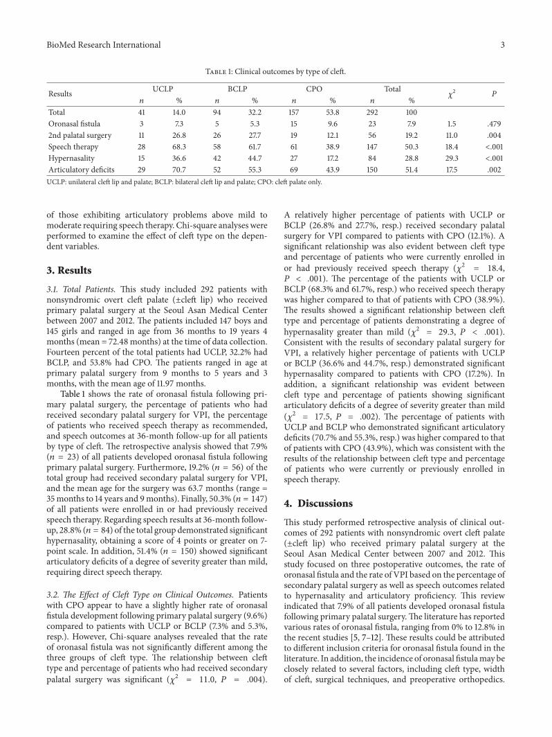
Table 1: Clinical outcomes by type of cleft.
Results UCLP BCLP CPO Total𝜒2
𝑃
𝑛 % 𝑛 % 𝑛 % 𝑛 %Total 41 14.0 94 32.2 157 53.8 292 100Oronasal fistula 3 7.3 5 5.3 15 9.6 23 7.9 1.5 .4792nd palatal surgery 11 26.8 26 27.7 19 12.1 56 19.2 11.0 .004Speech therapy 28 68.3 58 61.7 61 38.9 147 50.3 18.4 <.001Hypernasality 15 36.6 42 44.7 27 17.2 84 28.8 29.3 <.001Articulatory deficits 29 70.7 52 55.3 69 43.9 150 51.4 17.5 .002UCLP: unilateral cleft lip and palate; BCLP: bilateral cleft lip and palate; CPO: cleft palate only.
of those exhibiting articulatory problems above mild tomoderate requiring speech therapy. Chi-square analyses wereperformed to examine the effect of cleft type on the depen-dent variables.
3. Results
3.1. Total Patients. This study included 292 patients withnonsyndromic overt cleft palate (±cleft lip) who receivedprimary palatal surgery at the Seoul Asan Medical Centerbetween 2007 and 2012. The patients included 147 boys and145 girls and ranged in age from 36 months to 19 years 4months (mean = 72.48 months) at the time of data collection.Fourteen percent of the total patients had UCLP, 32.2% hadBCLP, and 53.8% had CPO. The patients ranged in age atprimary palatal surgery from 9 months to 5 years and 3months, with the mean age of 11.97 months.
Table 1 shows the rate of oronasal fistula following pri-mary palatal surgery, the percentage of patients who hadreceived secondary palatal surgery for VPI, the percentageof patients who received speech therapy as recommended,and speech outcomes at 36-month follow-up for all patientsby type of cleft. The retrospective analysis showed that 7.9%(𝑛 = 23) of all patients developed oronasal fistula followingprimary palatal surgery. Furthermore, 19.2% (𝑛 = 56) of thetotal group had received secondary palatal surgery for VPI,and the mean age for the surgery was 63.7 months (range =35months to 14 years and 9months). Finally, 50.3% (𝑛 = 147)of all patients were enrolled in or had previously receivedspeech therapy. Regarding speech results at 36-month follow-up, 28.8% (𝑛 = 84) of the total group demonstrated significanthypernasality, obtaining a score of 4 points or greater on 7-point scale. In addition, 51.4% (𝑛 = 150) showed significantarticulatory deficits of a degree of severity greater than mild,requiring direct speech therapy.
3.2. The Effect of Cleft Type on Clinical Outcomes. Patientswith CPO appear to have a slightly higher rate of oronasalfistula development following primary palatal surgery (9.6%)compared to patients with UCLP or BCLP (7.3% and 5.3%,resp.). However, Chi-square analyses revealed that the rateof oronasal fistula was not significantly different among thethree groups of cleft type. The relationship between clefttype and percentage of patients who had received secondarypalatal surgery was significant (𝜒2 = 11.0, 𝑃 = .004).
A relatively higher percentage of patients with UCLP orBCLP (26.8% and 27.7%, resp.) received secondary palatalsurgery for VPI compared to patients with CPO (12.1%). Asignificant relationship was also evident between cleft typeand percentage of patients who were currently enrolled inor had previously received speech therapy (𝜒2 = 18.4,𝑃 < .001). The percentage of the patients with UCLP orBCLP (68.3% and 61.7%, resp.) who received speech therapywas higher compared to that of patients with CPO (38.9%).The results showed a significant relationship between clefttype and percentage of patients demonstrating a degree ofhypernasality greater than mild (𝜒2 = 29.3, 𝑃 < .001).Consistent with the results of secondary palatal surgery forVPI, a relatively higher percentage of patients with UCLPor BCLP (36.6% and 44.7%, resp.) demonstrated significanthypernasality compared to patients with CPO (17.2%). Inaddition, a significant relationship was evident betweencleft type and percentage of patients showing significantarticulatory deficits of a degree of severity greater than mild(𝜒2 = 17.5, 𝑃 = .002). The percentage of patients withUCLP and BCLP who demonstrated significant articulatorydeficits (70.7% and 55.3%, resp.) was higher compared to thatof patients with CPO (43.9%), which was consistent with theresults of the relationship between cleft type and percentageof patients who were currently or previously enrolled inspeech therapy.
4. Discussions
This study performed retrospective analysis of clinical out-comes of 292 patients with nonsyndromic overt cleft palate(±cleft lip) who received primary palatal surgery at theSeoul Asan Medical Center between 2007 and 2012. Thisstudy focused on three postoperative outcomes, the rate oforonasal fistula and the rate of VPI based on the percentage ofsecondary palatal surgery as well as speech outcomes relatedto hypernasality and articulatory proficiency. This reviewindicated that 7.9% of all patients developed oronasal fistulafollowing primary palatal surgery.The literature has reportedvarious rates of oronasal fistula, ranging from 0% to 12.8% inthe recent studies [5, 7–12]. These results could be attributedto different inclusion criteria for oronasal fistula found in theliterature. In addition, the incidence of oronasal fistulamay beclosely related to several factors, including cleft type, widthof cleft, surgical techniques, and preoperative orthopedics.

4 BioMed Research International
Among the influential factors, this study examined the effectof cleft type on the incidence of oronasal fistula. We found nosignificant effect of cleft type on the rate of oronasal fistula.In general, the literature reports that patients with completebilateral clefts (Veau IV) are more likely to develop a fistula[10, 11]. In this study, patients with UCLP or BCLP had arelatively lower rate of oronasal fistula development followingprimary palatal surgery compared to patients with CPO,although the results did not reach statistical significance.The low incidence of oronasal fistula in patients with UCLPor BCLP in this study might be related to preoperativeNAM. Approximately 90% of babies born with UCLP orBCLP in our institution were treated with preoperativeNAM, but only few babies with CPO underwent preoperativeprocedure. Recently, preoperative orthopedics, such as theNAMprocedure, was reported to contribute to the narrowingof the cleft width and therefore reducing the rate of oronasalfistula developed following primary palatal surgery [10, 17,18]. However, we could not obtain valid medical records onNAM, as the preoperative procedure was performed at a localdental clinic outside the institution. Future research shoulduse objective and systematic data to examinewhether patientswho received the preoperative orthopedic procedure showbetter clinical outcomes after primary cleft repair.
The study also reported that 19.2% of the total group hadreceived secondary palatal surgery for VPI. The incidencerates of persistent VPI following primary surgery also varyin the literature, being as high as 30% [5–7, 13–15]. Suchvaried outcomes concerning VPI following primary surgerymight be associated with several factors, including variabilityin the definition of or criteria defining VPI, different surgicaltechniques, cleft type, and extent of clefting. Although manystudies usedmainly secondary corrective surgery as inclusioncriteria of VPI, some studies included results evidenced byperceptual assessment of hypernasal speech and assessmentsusing nasoendoscopy and/or videofluoroscopy. Regardinghypernasality, which is a speech problem associated withVPI, 28.8% of the total group demonstrated significanthypernasality requiring secondary palatal surgery or speechtherapy. In general, secondary palatal surgery has beenperformed at our institution in cases where anatomic deficitsof the velopharyngeal mechanism following primary repairappear to show persistently moderate or severe degrees ofhypernasality. Patients who exhibit amild tomoderate degreeof hypernasality and simultaneous articulatory deficits arereferred to speech therapy. Therefore, this clinical decision-making process results in differences between the percentageof patients demonstrating significant hypernasality and therate of secondary palatal surgery. In addition, the resultsshowed that cleft type has a significant effect on the incidenceof VPI, that is, the percentage of patients who had receivedsecondary palatal surgery. A relatively higher percentage ofpatients with UCLP or BCLP received secondary palatalsurgery for VPI compared to patients with CPO.
The percentage of patients enrolled in speech therapyor demonstrating significant articulatory deficits requiringspeech therapy was high in this study. Approximately half ofall the patients had enrolled in or received speech therapy,and 51.4% showed significant articulatory deficits. These
results of speech outcomes in this study were consistentwith the previous study [6], which examined speech pro-duction of preschoolers with cleft palate. Hardin-Jones andJones concluded that the majority of preschoolers with cleftpalate continue to demonstrate speech problems that requiredirect speech therapy, despite advances in management ofcleft palate [6]. A significant relationship was also foundbetween cleft type and speech outcomes. More patients withUCLP and BCLP were enrolled in speech therapy and moredemonstrated significant articulatory deficits compared topatients with CPO. This result might be associated with therelatively higher rate of VPI in patients with UCLP and BCLPcompared to those with CPO. That is, more patients withUCLP and BCLP showed significant hypernasal speech andarticulatory problems due to VPI and therefore needed toreceive speech therapy.
To implement a better clinical service approach, it isimportant to evaluate complications and surgical outcomes.It is also necessary to identify factors that influence surgicaloutcomes and successful clinical management of individualswith cleft palate. Clinical outcomes of primary cleft palaterepair are related to several factors, including cleft type, theextent of innate clefting, surgical repair techniques, expertiseof the operating surgeon, preoperative orthopedics, and tim-ing of primary palatal repair. This study examined the effectof only one influential factor on clinical outcomes. Futureresearch should investigate the relationship between severalinfluential factors and clinical outcomes comprehensively.
A final comment on the limitations of this study appearswarranted. Several limitations arose in the retrospectivereview of patient records, which is subject to confoundingfactors. Especially, the limitation was evident in classifyingcleft type and comparing the results with those of previ-ous studies. We found that the cleft classification systemof patients’ electronic medical records was not consistentand sometimes insufficient, as several junior doctors wereinvolved in recording patients’ cleft type. Information oncleft type generally appeared to be described using the termssuch as unilateral (right or left sides), bilateral, complete,and incomplete. However, to classify all patients in the studyusing the existing medical records, we had to use simplecleft classification system, which does not accurately reflectthe magnitude of the defect and does not guarantee thehomogeneity of each cleft type group. The classificationsystem in this study, by the nature of its simplicity, mayreduce the sensitivity for the analysis of the effect of clefttype on clinical outcomes. More detailed cleft classificationsystem should be used or the exact size or width of cleftingshould be estimated in future research so that a large degree ofheterogeneity in cleft type group can be reduced and the effectof cleft type on clinical outcomes can be sensitively detected.Furthermore, continuous research efforts using prospectiveanalysis should attempt to identify presurgical risk factors forclinical outcomes in primary palatal surgery.
5. Conclusion
This study represents clinical outcomes of primary cleft palaterepairs based on large data gathered from a single institution
BioMed Research International 5
and reflects a single surgeon’s intensive experience over a 5-year period. This makes it possible to rule out influentialand confounding factors, such as expertise of surgeons. Thestudy showed that the incidence of oronasal fistula and theincidence of VPI following cleft palate surgery are compara-ble to those of other cleft centers worldwide, although careneeds to be taken when interpreting the results due to lackof standard definitions used for oronasal fistula and VPIin the literature and the weakness of retrospective analyses.The study also suggests that approximately half of patientsshow speech problems following primary palatal surgeryand require direct speech therapy. This result highlightsthe importance of routine follow-up speech examinationsand multidisciplinary team approach for this population. Inaddition, the study suggests that cleft type is one of theimportant factors related to clinical outcomes. This auditprovides a retrospective quality review of primary palatalsurgery at the Seoul Asan Medical Center and a basis forongoing research efforts to establish an optimal treatmentregimen for patients with cleft palate.
Conflict of Interests
The authors declare that there is no conflict of interestsregarding the publication of this paper.
Acknowledgment
This research was supported by Hallym University ResearchFund (HRF-201408-015).
References
[1] B. C. Sommerlad, “A technique for cleft palate repair,” Plasticand Reconstructive Surgery, vol. 112, no. 6, pp. 1542–1548, 2003.
[2] S. R. Sullivan, E. M. Marrinan, R. A. LaBrie, G. F. Rogers,and J. B. Mulliken, “Palatoplasty outcomes in nonsyndromicpatients with cleft palate: a 29-year assessment of one surgeon’sexperience,” Journal of Craniofacial Surgery, vol. 20, no. 5,supplement 1, pp. 612–616, 2009.
[3] P. Andrades, A. Espinosa-de-los-Monteros, D. H. Shell et al.,“The importance of radical intravelar veloplasty during two-flappalatoplasty,” Plastic and Reconstructive Surgery, vol. 122, no. 4,pp. 1121–1130, 2008.
[4] E. B. Katzel, P. Basile, P. F. Koltz, J. R. Marcus, and J. A. Girotto,“Current surgical practices in cleft care: cleft palate repairtechniques and postoperative care,” Plastic and ReconstructiveSurgery, vol. 124, no. 3, pp. 899–906, 2009.
[5] O. Jackson, C. A. Stransky, A. F. Jawad et al., “The children’shospital of philadelphia modification of the furlow double-opposing Z-palatoplasty: 30-year experience and long-termspeech outcomes,” Plastic and Reconstructive Surgery, vol. 132,no. 3, pp. 613–622, 2013.
[6] M. A. Hardin-Jones and D. L. Jones, “Speech production ofpreschoolers with cleft palate,”Cleft Palate-Craniofacial Journal,vol. 42, no. 1, pp. 7–13, 2005.
[7] S. P. Yun and T. de Chalain, “Incidence of oronasal fistulaeand velopharyngeal insufficiency after cleft palate repair: anaudit of 211 children born between 1990 and 2004,” Cleft Palate-Craniofacial Journal, vol. 45, no. 2, pp. 172–178, 2008.
[8] R. H. Lithovius, L. P. Ylikontiola, and G. K. Sandor, “Incidenceof palatal fistula formation after primary palatoplasty in north-ern Finland,” Oral Surgery, Oral Medicine, Oral Pathology andOral Radiology, vol. 118, no. 6, pp. 632–636, 2014.
[9] A. R. Muzaffar, H. Steve Byrd, R. J. Rohrich et al., “Incidenceof cleft palate fistula: an institutional experience with two-stagepalatal repair,” Plastic and Reconstructive Surgery, vol. 108, no. 6,pp. 1515–1518, 2001.
[10] A. Eberlinc and V. Kozelj, “Incidence of residual oronasalfistulas: a 20-year experience,” Cleft Palate-Craniofacial Journal,vol. 49, no. 6, pp. 643–648, 2012.
[11] M. R. Bykowski, S. Naran, D. G. Winger, and J. E. Losee, “Therate of oronasal fistula following primary cleft palate surgery: ameta-analysis,”The Cleft Palate-Craniofacial Journal, 2014.
[12] D. Bearn, S. Mildinhall, T. Murphy et al., “Cleft lip and palatecare in the United Kingdom—The Clinical Standards AdvisoryGroup (CSAG) study. Part 4: outcome comparisons, training,and conclusions,” The Cleft Palate-Craniofacial Journal, vol. 38,no. 1, pp. 38–43, 2001.
[13] D. S. Inman, P.Thomas, P.D.Hodgkinson, andC.A. Reid, “Oro-nasal fistula development and velopharyngeal insufficiencyfollowing primary cleft palate surgery—an audit of 148 childrenborn between 1985 and 1997,” British Journal of Plastic Surgery,vol. 58, no. 8, pp. 1051–1054, 2005.
[14] A. A. C. Webb, R. Watts, E. Read-Ward, J. Hodgkins, and A. F.Markus, “Audit of a multidisciplinary approach to the care ofchildren with unilateral and bilateral cleft lip and palate,” BritishJournal of Oral andMaxillofacial Surgery, vol. 39, no. 3, pp. 182–188, 2001.
[15] S. Zhao, Y. Xu, H. Yin et al., “Incidence of postoperativevelopharyngeal insufficiency in late palate repair,” The Journalof Craniofacial Surgery, vol. 23, no. 6, pp. 1602–1606, 2012.
[16] M.-H. Mahoney, M. C. Swan, and D. M. Fisher, “Prospectiveanalysis of presurgical risk factors for outcomes in primarypalatoplasty,” Plastic and Reconstructive Surgery, vol. 132, no. 1,pp. 165–171, 2013.
[17] W. Dec, P. R. Shetye, B. H. Grayson, L. E. Brecht, C. B. Cutting,and S. M. Warren, “Incidence of oronasal fistula formationafter nasoalveolar molding and primary cleft repair,” Journal ofCraniofacial Surgery, vol. 24, no. 1, pp. 57–61, 2013.
[18] B. H. Grayson and P. R. Shetye, “Presurgical nasoalveolarmoulding treatment in cleft lip and palate patients,” IndianJournal of Plastic Surgery, vol. 42, supplement 1, pp. S56–S61,2009.

Submit your manuscripts athttp://www.hindawi.com
Stem CellsInternational
Hindawi Publishing Corporationhttp://www.hindawi.com Volume 2014
Hindawi Publishing Corporationhttp://www.hindawi.com Volume 2014
MEDIATORSINFLAMMATION
of
Hindawi Publishing Corporationhttp://www.hindawi.com Volume 2014
Behavioural Neurology
EndocrinologyInternational Journal of
Hindawi Publishing Corporationhttp://www.hindawi.com Volume 2014
Hindawi Publishing Corporationhttp://www.hindawi.com Volume 2014
Disease Markers
Hindawi Publishing Corporationhttp://www.hindawi.com Volume 2014
BioMed Research International
OncologyJournal of
Hindawi Publishing Corporationhttp://www.hindawi.com Volume 2014
Hindawi Publishing Corporationhttp://www.hindawi.com Volume 2014
Oxidative Medicine and Cellular Longevity
Hindawi Publishing Corporationhttp://www.hindawi.com Volume 2014
PPAR Research
The Scientific World JournalHindawi Publishing Corporation http://www.hindawi.com Volume 2014
Immunology ResearchHindawi Publishing Corporationhttp://www.hindawi.com Volume 2014
Journal of
ObesityJournal of
Hindawi Publishing Corporationhttp://www.hindawi.com Volume 2014
Hindawi Publishing Corporationhttp://www.hindawi.com Volume 2014
Computational and Mathematical Methods in Medicine
OphthalmologyJournal of
Hindawi Publishing Corporationhttp://www.hindawi.com Volume 2014
Diabetes ResearchJournal of
Hindawi Publishing Corporationhttp://www.hindawi.com Volume 2014
Hindawi Publishing Corporationhttp://www.hindawi.com Volume 2014
Research and TreatmentAIDS
Hindawi Publishing Corporationhttp://www.hindawi.com Volume 2014
Gastroenterology Research and Practice
Hindawi Publishing Corporationhttp://www.hindawi.com Volume 2014
Parkinson’s Disease
Evidence-Based Complementary and Alternative Medicine
Volume 2014Hindawi Publishing Corporationhttp://www.hindawi.com