clinical presentation and investigations for breast carcinoma
TRANSCRIPT

Clinical Presentation
and Investigations for
Breast carcinoma
Dr.P.Viswakumar,M.S
Assistant professor,
Dept.of General surgery,
PSGIMSR.

Embryology and Functional Anatomy of the Breast
• Two ventral bands of thickened ectoderm appears at 5th or 6th week _ ‘milk line’.
• Each breast develops when an ingrowth of ectoderm forms a primary tissue bud in the mesenchyme.
• The primary bud, in turn, initiates the development of 15 to 20 secondary buds.
• Major (lactiferous) ducts develop, which open into a shallow mammary pit.
• During infancy, a proliferation of mesenchyme transforms the mammary pit into a nipple

• The breast remains undeveloped in the female until puberty, when it enlarges in response to ovarian estrogen and progesterone, which initiate proliferation of the epithelial and connective tissue elements
• However, the breasts remain incompletely developed until pregnancy occurs.

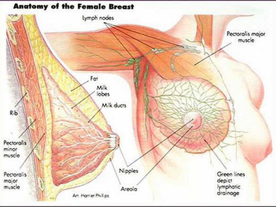
Functional anatomy
• The breast is composed of 15 to 20 lobes which are each composed of several lobules.
• Fibrous bands of connective tissue travel through the breast (Cooper's suspensory ligaments), insert perpendicularly into the dermis, and provide structural support.
• The upper outer quadrant of the breast contains a greater volume of tissue than do the other quadrants.
• The epidermis of the nipple-areola complex is pigmented and is variably corrugated.

Functional anatomy
• The dermal papilla at the tip of the nipple contains numerous sensory nerve endings and Meissner's corpuscles.
• This rich sensory innervation is of functional importance, because the sucking of the infant initiates a chain of neurohumoral events that results in milk letdown.


Blood supply,Innervation and Lymphatics
• Arteries (a) perforating branches of the internal mammary artery; (b) lateral branches of the posterior intercostal arteries; and (c) branches from the axillary artery, including the highest thoracic, lateral thoracic, and pectoral branches of the thoracoacromial artery .
• The three principal groups of veins are (a) perforating branches of the internal thoracic vein, (b) perforating branches of the posterior intercostal veins, and (c) tributaries of the axillary vein.
• Batson's vertebral venous plexus, which invests the vertebrae and extends from the base of the skull to the sacrum, may provide a route for breast cancer metastases to the vertebrae, skull, pelvic bones, and central nervous system.

Blood supply,Innervation and Lymphatics

Blood supply,Innervation and Lymphatics
• Lateral cutaneous branches of the third through sixth intercostal nerves provide sensory innervation of the breast (lateral mammary branches) and of the anterolateral chest wall.
• Six axillary lymph node group recognized by surgeons
1) Lateral 2)Anterior 3) Posterior 4) Central 5) Apical 6) Interpectoral


Epidemiology and Natural history
• Breast cancer is the most common site-specific cancer in women and is the leading cause of death from cancer for women aged 20 to 59 years.
• Collectively, US, India and China account for almost one third of the global breast cancer burden.


Indian Data
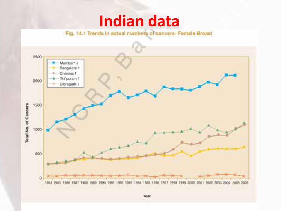
Indian data

Age Trend

Age Trend

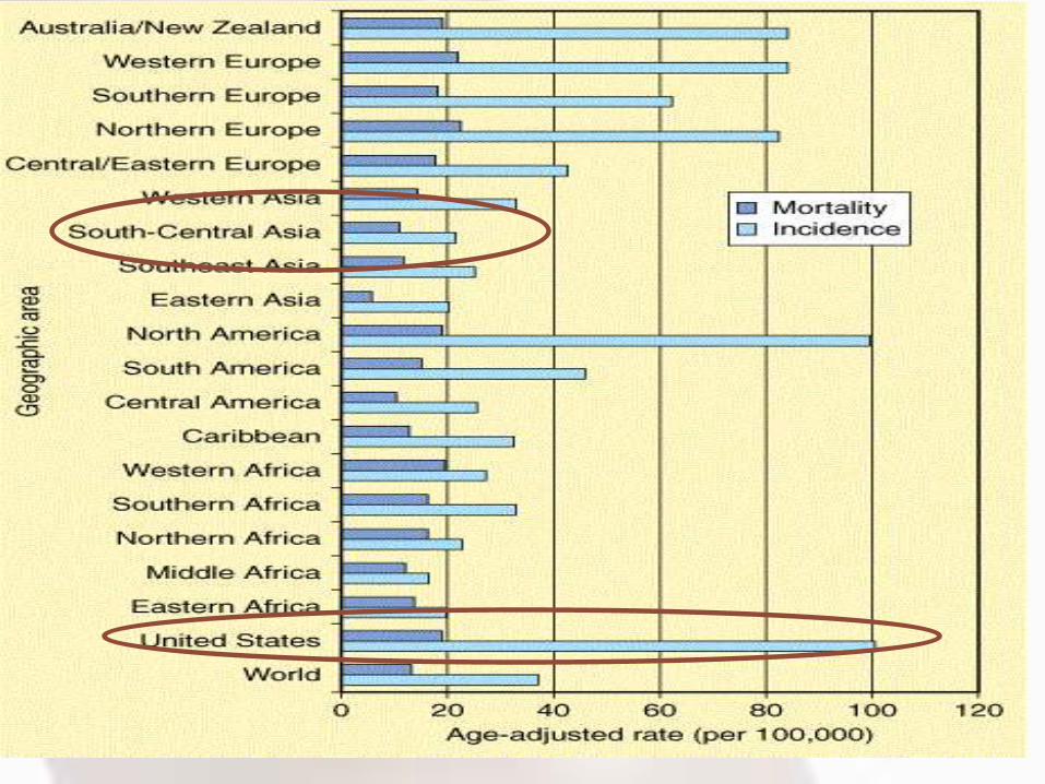
Peer Comparision

Peer comparision
• For the United States, for the year 2012:
For every 5 or 6 women newly diagnosed with breast cancer, one lady is dying of it.
• For China, for the year 2012:
For every 4 women newly diagnosed with breast cancer, one lady is dying of it.
• For India, for the year 2012:
For every 2 women newly diagnosed with breast cancer, one lady is dying of it.

Global Data

High Risk groups• The average lifetime risk of breast cancer for newborn
U.S. females is 12%.• A recent study of breast cancer risk in India revealed
that 1 in 28 women develop breast cancer during her lifetime.
• In India the average age of the high risk group is 43-46 years while it is 53-57 years in west.
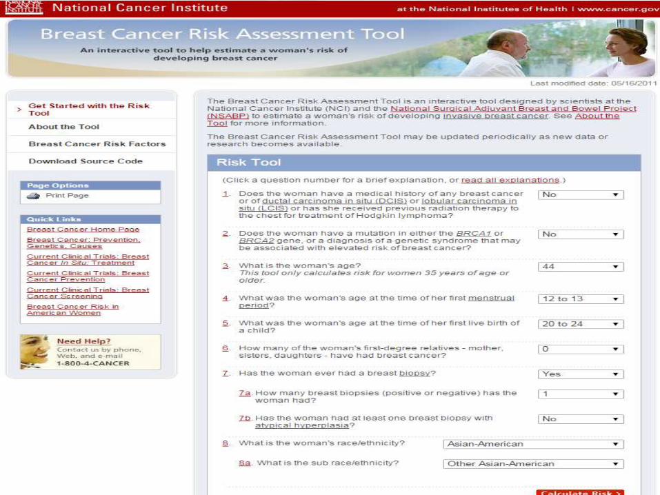
• Risk assessment tool commonly used now a days is Modified Gail model.
• It is a computer based model uses Age, race, ethnicity age@menarche, age@first live birth, no of first degree relatives with breast cancer, no of previous breast biopsies and their histological picture for future breast cancer risk.
• http://bcra.nci.nih.gov/brc

High Risk groups• Increased risk groups are categorized into 6
groups a)Women with prior h/o breast cancer
b) Women >35 years with 5 year risk of invasive breast ca >= 1.7% as per Gail model
c) Women with lifetime risk of 20 % for invasive malignancy calculated by models dependent on family history.
d) Prior therapeutic chest irradiation.
e) Women with LCIS.
f) Women with pedigree suggestive of or with a genetic predisposition (BRCA1/2,P53,PTEN and other genetic mutations )

Breast Cancer Screening
Breast cancer screening has to address two groups
1) Normal woman with no inherent risk factors.
2) High risk groups.

Breast Cancer Screening• Screening for Average Risk groups :
i) Women under 25-40 age group complete breast examination at every 1-3 years.
ii)Breast awareness to be encouraged.
iii)Women aged >40 yrs annual CBE and Mammogram,breast awareness.
iv)Controversy exist in age groups 50-74 for interval of screening(annually or every other year).
v) Mammography detects lesion 2 years prior to CBE (sojourn period of 2 yrs).

High Risk Screening
• Women with increased risk the screening should start at the age of 35 yrs.
• CBE should done at every 6-12 months.
• Annual mammography
• Women with genetic predisposition screening should begin by 30 yrs of age.

Clinical Presentation
Signs and Symptoms : New lump or mass(Most common)- 33%Other possible signs :• Swelling of all or part of a breast (even if no
distinct lump is felt),axillary lump.• A nipple discharge other than breast milk• Skin dimpling• Breast or nipple pain• Nipple retraction (turning inward)• Redness, scaliness, or thickening of the nipple or
breast skin

Complete Breast examination

Palpable Mass
How to proceed with palpable mass??

Palpable MassPalpable mass;Age >30 years
Diagnostic mammogram/USG
Birads 1-3 Birads 4-5
Solid; Simple; Core needle biopsy
complex cyst No abnormality Pathology/Image: concordant/
Short term follow up Observe Discordant: Surgical excision/
Reassessment Excision
Mammo every 6-12 mon
for 2 years

Palpable mass
Palpable mass; Age <30 yrs
Low clinical suspicion High clinical suspicion
Observe 1-2 menstrual cycles USG of Breast
No resolution
Solid Simple cyst complex cyst No abnormality

Breast Pain
• Mostly it is symptom of benign breast condition
• But malignancy is detected in 0.4% cases undergoing mammogram for mastalgia.
• 50 % of operable breast ca reported pain as one symptom.
• 7% has only pain as their symptom.
• So patient with unilateral breast pain and age>35 years –mammographic examination is necessary

Nipple Discharge
• Unilateral Single duct spontaneous and persistent discharge,
• Clear,Serous, Serosanguinous or bloody discharge are at increased risk of underlying malignancy.
• There is 5.9% to 20.2 % incidence of malignancy in such pathological discharge.
• Investigation such as USG > Mammogram done along with history and CBE to R/o underlying malignancy.
• Cytology of nipple discharge is of less or no value but can be highly specific but not recommended as standard of care.

Skin Changes
Clinical suspicion of inflammatory breast Ca
1) Paeu-de orange
2) Erythema
Clinical suspicion of Pagets disease or other manifestation of breast Ca
1) Nipple Excoriation
2) Scaling/ Eczema
3) Skin ulceration
Skin Changes
In Both above conditions diagnostic mammogram with or without USG to be done.
• In BIRADS 1-3 : Punch biopsy of skin/Nipple biopsy done.
If Benign : a) Clinical reassessment b) Pathological correlation c) MRI of breast d) repeat biopsy e) Consultation with breast specialist
If Malignant : Follow malignancy treatment protocol.

Skin Changes
• In BIRADS 4-5 : Core needle biopsy with or without punch biopsy of skin or surgical excision.
• If Benign : Punch/ Nipple biopsy if not done before benign; follow as said above.
• If Malignant : Follow malignancy treatment protocol.

Breast abscess and malignancy
• Malignancy was reported when the abscess wall cavity sent for HPE.
• A 10 year retrospective study published in American journal of Surgery reported the incidence is around 4.4 % (9/206 cases).
• Is it warrant biopsy as routine while doing
I&D ?? Age/lactational status should be considered?
• Since there is no clear guidelines its upto the institution to decide and warrants well designed study……

Investigational Tools
Mammography
Ultrasound
Stereotactic biopsy
MRI
Mammography• Conventional mammography delivers a radiation
dose of 0.1 cGy per study.• Screening mammography is used to detect
unexpected breast cancer in asymptomatic women.• Two views of the breast are obtained, the
craniocaudal (CC) view and the mediolateral oblique (MLO) view.
• In diagnostic mammogram additional views are incorporated such as spot compression technique,etc.
• Solid mass with or without stellate features, asymmetric thickening of breast tissues, and clustered microcalcifications.
• The presence of fine, stippled calcium in and around a suspicious lesion is suggestive of breast cancer.


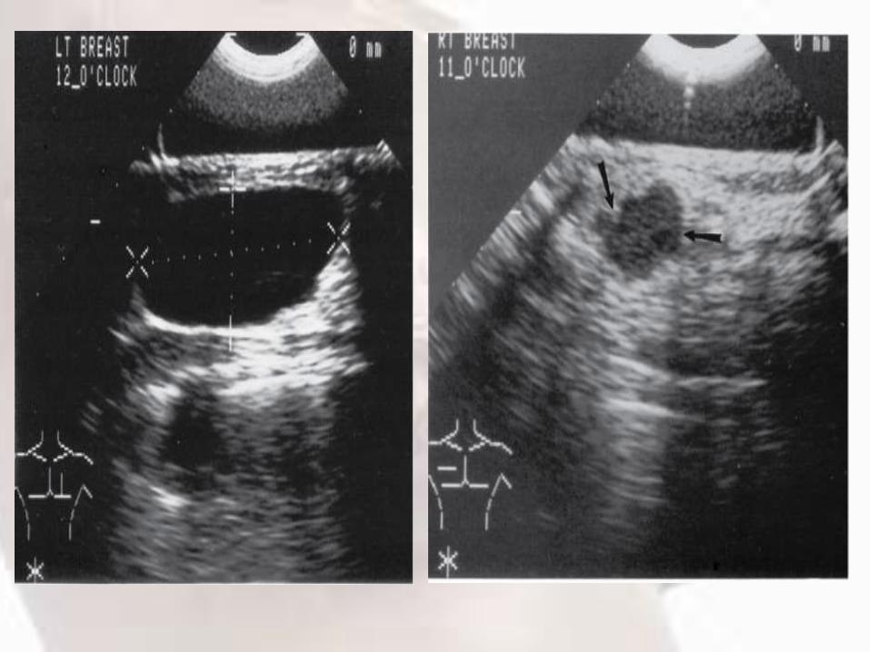
Ultrasonogram• USG is an important method of resolving equivocal
mammographic findings, defining cystic masses, and demonstrating the echogenic qualities of specific solid abnormalities.
• Recommended as initial imaging in women <30yrs with palpable mass or asymmetric thickening or nodularity.
• Women >30yrs diagnostic mammogram show BIRAD 1-3 for palpable mass.
• All age group USG should be considered as adjuvant to Mammography for women with skin changes and spontaneous nipple discharge with no mass.
• Lesions <1cm are difficult to diagnose in USG.

MRI
• Sensitivity of MRI is higher than mammography but got low specificity.
• Leads to high false positivity rate.Annual MRI recommended as screening tool ini) BRCA mutation.ii) First degree relative of BRCA carrier but untestediii) Lifetime risk >=20% defined by risk assesment
tools using family historyOther less evidence in 1)chest irradiation between 10-30 yrs 2) Li fraumeni,cowden and bannayan-Riley-Ruvalcaba syndrome and first degree relatives.

Diagnostic Breast MRI
• To whom we have to recommend MRI of Breast ???
Patient with skin changes with high clinical suspicion of serious breast disease but skin punch biopsy and mammographic/USG findings reported benign MRI should be considered.
Before recommending MRI:a)Dedicated breast coil availableb)Radiologist experience in MRI breast images.c)Ability to perform MRI guided procedure to localize to MRI detected finding.

Sterotactic Breast biopsy

BI-RADS• Breast Imaging-Reporting and Data System
• It is a widely accepted risk assessment and quality assurance tool in mammography, ultrasound or MRI0 Incomplete assessment; need additional imaging
evaluation
1 Negative; routine mammogram in 1 year recommended
2 Benign finding; routine mammogram in 1 year recommended
3 Probably benign finding; short-term follow-up suggested
4 Suspicious abnormality; biopsy should be considered
5 Highly suggestive of malignancy; appropriate action should be taken

Take Home message

Take Home message
• Breast cancer becoming leading cancer among Indian women.
• The incidence ,mortality ratio poor compared to our peers showing lacunae in management.
• Screening programs still inefficient in our country due to scarce resources and poor infrastructure.
• So adequate targeting high risk groups with the risk assessment tools is important.
• Hence every physician should aware of high screening recommendations.
• Palpable mass ,>30yrs first should undergo mammographic imaging before pathological evaluation.
• For <30 yrs females USG is recommended as initial choice of imaging.
• For skin changes and nipple discharge with high suspicion of underlying breast disease Mammogram can aided with USG.
• MRI can be used as screening and diagnostic tool only in certain conditions as described above.
• Stereotactic breast biopsy is excellent tool for adequately targeting the suspicious lesion that are not clinically palpable/detectable.


Hope You all got our point
Thank you
for your patience