breast carcinoma insitu
TRANSCRIPT

Nabeel Yahiya

DCIS
LCIS
PAGETS DISEASE OF NIPPLE
Its incidence is increasing due to over diagnosis by screening mammogram
Considerable controversy regarding optimal management
Ranges from observation to bcs to mastectomy with and without adjuvant treatment
neoplastic process that is confined to the ductalsystem of the breast and lacks histologic evidence of invasion
These cells neither disrupt the basement membrane nor involve the surrounding breast stroma.
lacks the ability to metastasize and is confined to the breast
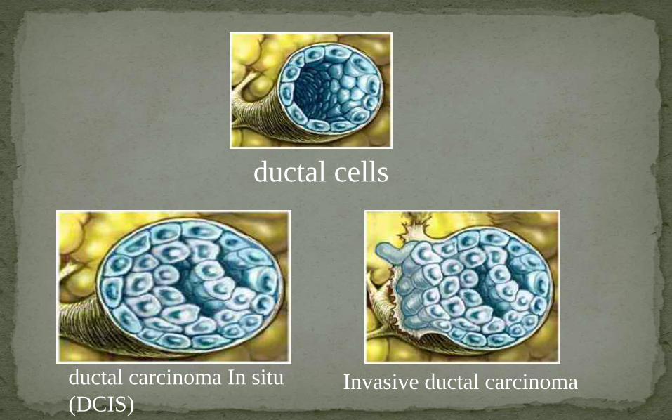
ductal cells
ductal carcinoma In situ
(DCIS)Invasive ductal carcinoma
Before the use of screening mammography, DCIS typically presented as a palpable mass or nipple discharge.
An invasive component commonly was found, and pure DCIS rarely was encountered.
The widespread use of mammography now routinely detects DCIS <1 cm in diameter
results in breast cancer-free survival rates that approach 100%
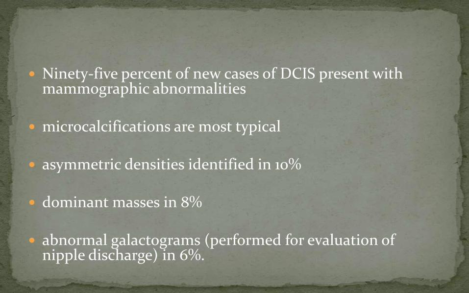
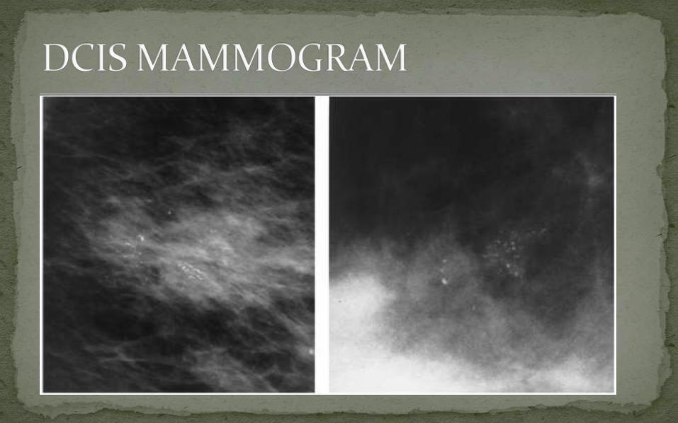
Ninety-five percent of new cases of DCIS present with mammographic abnormalities
microcalcifications are most typical
asymmetric densities identified in 10%
dominant masses in 8%
abnormal galactograms (performed for evaluation of nipple discharge) in 6%.

Linear and branching calcifications frequently are associated with high-grade DCIS and necrosis, whereas fine and granular calcifications are associated more commonly with low-grade
Initial evaluation should include magnification views that allow for complete characterization of mammographic findings and determination of the need for biopsy
The extent of the lesion as determined mammographically may be used as a guide for excision
Ultrasonography, digital mammography, and magnetic resonance imaging all have the potential to be helpful in the management of DCIS
but have yet to be proven as an acceptable substitute for mammography in screening
Traditionally, classification of DCIS has followed its architectural or morphologic appearance
The five subtypes of DCIS are
Comedo
Solid
Cribriform
Micropapillary
papillary

it is common to encounter a mixture of subtypes within the same specimen
Less common subtypes
Apocrine
Neuroendocrine
signet-cell cystic hypersecretory carcinoma
clinging DCIS

features that should be documented for each case of DCIS
nuclear grade, presence of necrosis, polarization, and architectural patterns
margin status, lesion size, extent of microcalcifications, and correlation between specimen x-ray and mammographic findings
Unicentric (one area only)
Multicentric (two distinct areas separated by more than 4 cm)
Continuous (extension along ductal system without gaps)
Discontinuous or multifocal (two or more areas separated by <4 cm).
recognized association between the presence of DCIS and the subsequent increased risk of developing an invasive breast cancer
presence of shared identical genetic abnormalities between DCIS and synchronous invasive breast cancer demonstrates a clonal relationship of biologic progression
The estrogen receptor is present in 70% of DCIS
Rate of expression is higher in low-grade lesions (90%) than in high-grade lesions (25%).
This association with histologic grade is reversed for the rate of overexpression of HER2/neu proto-oncogene and the p53 tumor suppression gene.
An occult microinvasive tumor (<1mm) may be seen with some cases of DCIS
Occult microinvasive tumors are most common in patients with DCIS
Lesions that are >2.5 cm in diameter
Presenting with palpable masses or nipple discharge,
High-grade DCIS or comedonecrosis
A primary consideration in the natural history of DCIS is the risk of progression to invasive carcinoma
The few published long-term follow-up studies of DCIS after only biopsy document an overall incidence of subsequent invasive carcinoma of more than 36%
Women with DCIS in one breast are at risk for a second tumor (either invasive or in situ) in the contralateral breast
The goal of treatment with DCIS
eradication of the initial cancer
prevention of local recurrence, with particular emphasis on the prevention of invasive breast cancer
The recommended workup and staging of DCIS includes:
history and physical examination
bilateral diagnostic mammography
MRI (optional)
pathology review
tumor ER determination

MASTECTOMY
BCS
BCS n RT
Mastectomy was the standard treatment of DCIS through the first four decades
Mastectomy is a highly effective treatment for DCIS, with a locoregional control rate of 96% to 100%
cancer-specific mortality rates of 4% or less

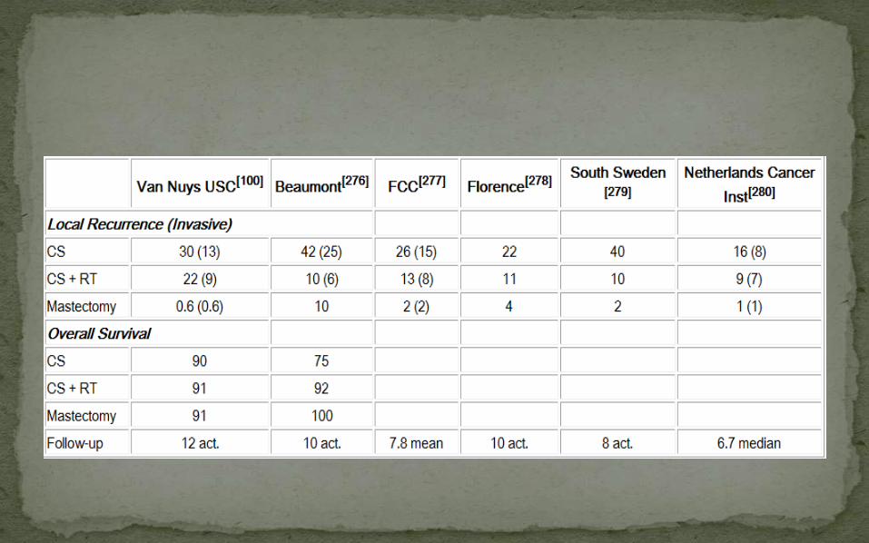
Many retrospective studies suggest that the rates of local or regional recurrence are significantly lower after mastectomy than after breast-conserving surgery
but there have been no significant differences in overall survival
No prospective randomized trials comparing mastectomy to breast-conserving surgery for DCIS
Prospective randomized trials have shown that the addition of whole breast irradiation to a margin-free excision of pure DCIS
decreases the rate of in-breast disease recurrence, but does not affect survival OR distant metastasis-free survival.
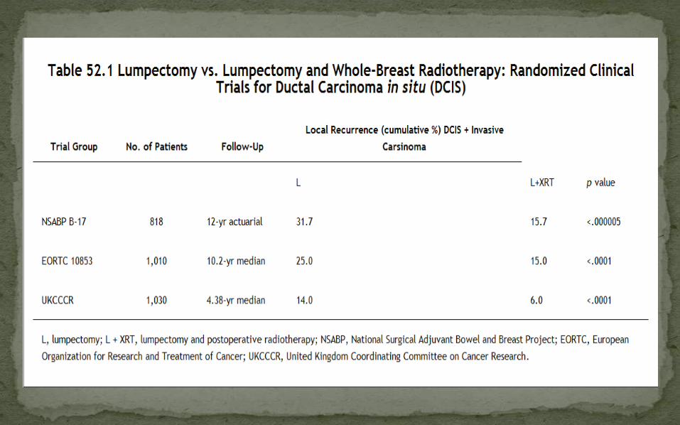
Whole breast irradiation after breast-conserving surgery reduces the relative risk of a local failure by approximately one half.
The current challenge is to identify women with DCIS whose risk of an ipsilateral breast tumor recurrence (primarily invasive) with breast-conserving surgery, with or without radiation
Age
Women 40 years of age or younger with DCIS have been reported to have ipsilateral breast tumor recurrence rates of approximately 50% in retrospective series n EORTC prospective randomized trial.
Most of the prospective randomized trials suggest that increasing age is associated with a decreased risk of ipsilateralbreast tumor recurrence
in patients treated with conservative surgery alone or conservative surgery and radiation.
Methode of detection
detection of DCIS solely by mammography was associated with a lower risk of ipsilateral breast tumor recurrence
when compared with clinical detection with symptoms such as a palpable mass or bloody nipple discharge
Size
Clinical assessment of tumor size includes measurements of a palpable mass, the dimensions of the mammographic abnormalities, including calcifications and/or a mass
In the EORTC and South Sweden prospective randomized trials, increasing clinical size was associated with an increased risk of ipsilateral breast tumor recurrence in patients treated with conservative surgery alone
but not those treated with conservative surgery and radiation
In the NSABP B-17 randomized trial the ipsilateralbreast tumor recurrence rate was correlated with the extent of calcifications on the mammogram, both in women treated with conservative surgery alone or conservative surgery and radiation
In patients having calcifications, a postexcisionmammogram before radiation or observation is essential to assure the removal of all malignant-appearing calcifications
Multifocality
Multifocal DCIS has been associated with an increased risk of ipsilateral breast tumor recurrence in different prospective randomized trials
when compared with unifocal disease in patients treated with conservative surgery alone or conservative surgery and radiation
Resection margin status
Positive margins of resection have been associated with an increased risk of ipsilateral breast tumor recurrence in the NSABP B-17, EORTC, and the NSABP B-24 trials
Negative margins greater than or equal to 2 mm were associated with a decreased risk of ipsilateral breast tumor recurrence when compared with those less than 2 mm
Critical margin < 1mm and >10mm

High nuclear grade and the presence of necrosis have been associated with an increased risk of ipsilateralbreast tumor recurrence in patients undergoing conservative surgery
These factors have had less of an impact on ipsilateralbreast tumor recurrence rates in patients undergoing conservative surgery and radiation
breast density is an increased risk of ipsilateral breast tumor recurrence in women with DCIS.

a scoring system with four categories:
Age
Size
Margins
nuclear grade combined with necrosis
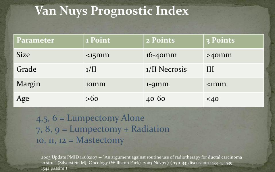
Van Nuys Prognostic Index
Parameter 1 Point 2 Points 3 Points
Size <15mm 16-40mm >40mm
Grade 1/II 1/II Necrosis III
Margin 10mm 1-9mm <1mm
Age >60 40-60 <40
4,5, 6 = Lumpectomy Alone7, 8, 9 = Lumpectomy + Radiation10, 11, 12 = Mastectomy
2003 Update PMID 14682107 -- "An argument against routine use of radiotherapy for ductal carcinoma in situ." (Silverstein MJ, Oncology (Williston Park). 2003 Nov;17(11):1511-33; discussion 1533-4, 1539, 1542 passim.)
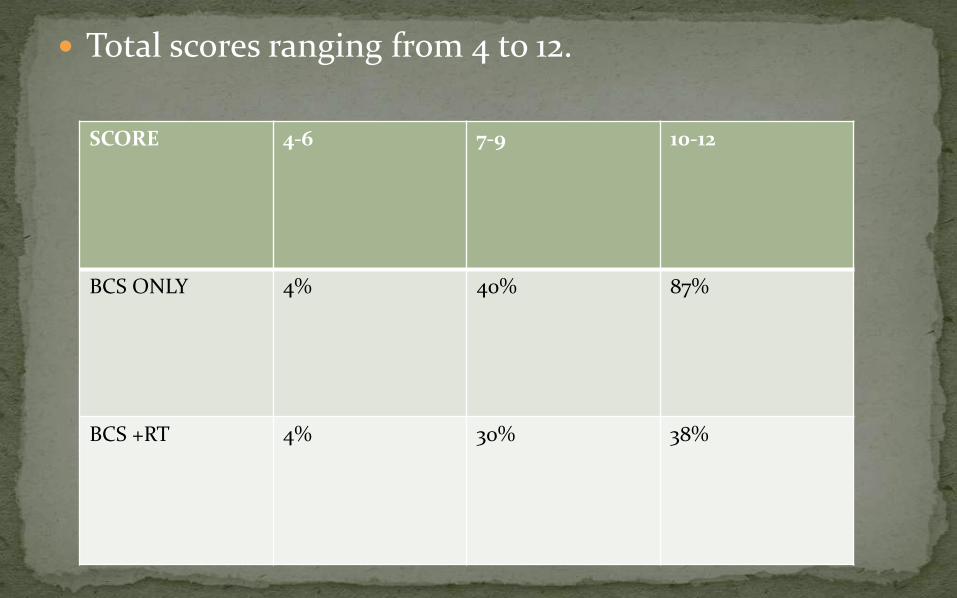
Total scores ranging from 4 to 12.
SCORE 4-6 7-9 10-12
BCS ONLY 4% 40% 87%
BCS +RT 4% 30% 38%
The investigators concluded that
patients with scores of 4 to 6 were candidates for wide excision alone
scores of 7 to 9 for excision and radiation
scores of 10 to 12 for mastectomy.

The reproducibility of this system has been questioned by a number of investigators in retrospective and prospective studies.
Inaccuracies in calculating the score could result in overtreatment or undertreatment
There is retrospective evidence suggesting that selected patients have a low risk of in-breast recurrence with excision alone without breast irradiation.
Retrospective study of 215 patients with DCIS treated with lumpectomy without radiation therapy, endocrine therapy, or chemotherapy, the
recurrence rate over 8 years was 0%, 21.5%, and 32.1% in patients with low-, intermediate- or high-risk DCIS
A multi-institutional, nonrandomized, prospective study of selected patients with low-risk DCIS treated without radiation was studied
Although an acceptably low ipsilateral recurrence rate was observed in the low-/intermediate-grade arm of the study at 5 years
7-year ipsilateral recurrence rate in this group of patients was considerably higher

An analysis of specimen margins and specimen radiographs should be performed to ensure that all mammographically detectable DCIS has been excised.
In addition, a post-excision mammogram should be considered

Axillary dissection is not recommended for patients with pure DCIS

NSABP B-24 trial
women with DCIS who were treated with breast-conserving therapy were randomized to receive placebo or tamoxifen.
13.6 years median follow-up, the women treated with tamoxifen had a 3.4% absolute reduction in ipsilateral in-breast tumor recurrence risk
No differences in overall survival (OS) were noted
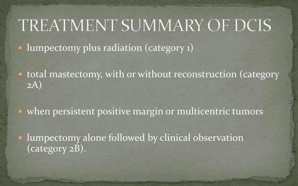
lumpectomy plus radiation (category 1)
total mastectomy, with or without reconstruction (category 2A)
when persistent positive margin or multicentric tumors
lumpectomy alone followed by clinical observation (category 2B).
history and physical examination every 6 to 12 months for 5 years and then annually
as well as yearly diagnostic mammography.

characterized by multicentric breast involvement
Consists of loose, discohesive epithelial cells that are large in size, variable in shape, and contain a normal cytoplasm to nucleus ratio
The extent of involvement of the lobular lumen ranges from simple filling to moderate-to-severe distention
with extension into the adjacent extralobular ducts
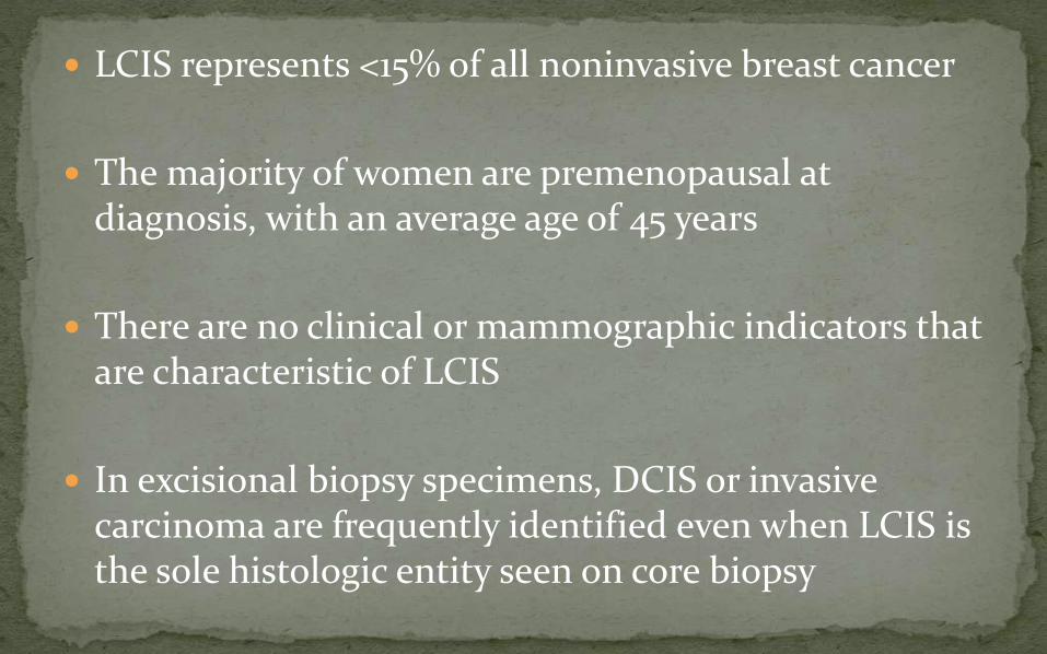
LCIS represents <15% of all noninvasive breast cancer
The majority of women are premenopausal at diagnosis, with an average age of 45 years
There are no clinical or mammographic indicators that are characteristic of LCIS
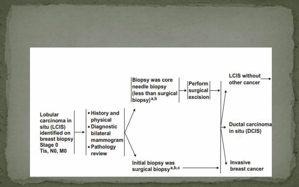
In excisional biopsy specimens, DCIS or invasive carcinoma are frequently identified even when LCIS is the sole histologic entity seen on core biopsy
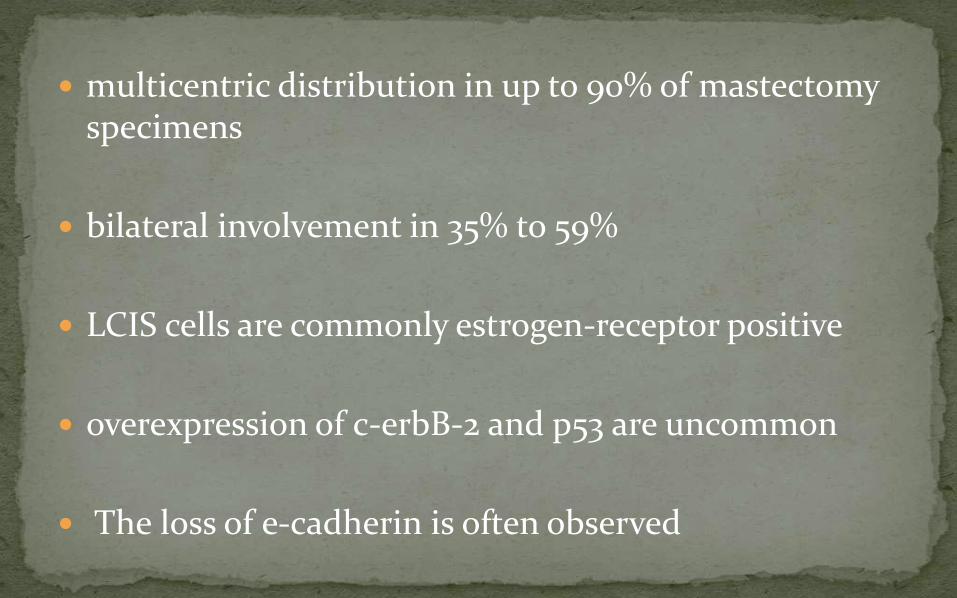
multicentric distribution in up to 90% of mastectomy specimens
bilateral involvement in 35% to 59%
LCIS cells are commonly estrogen-receptor positive
overexpression of c-erbB-2 and p53 are uncommon
The loss of e-cadherin is often observed

The presence of LCIS is considered a marker of increased risk for the subsequent development of invasive (usually ductal) carcinoma
greatest for high-grade or more extensive lesions
This risk appears to be nearly equal for both breasts
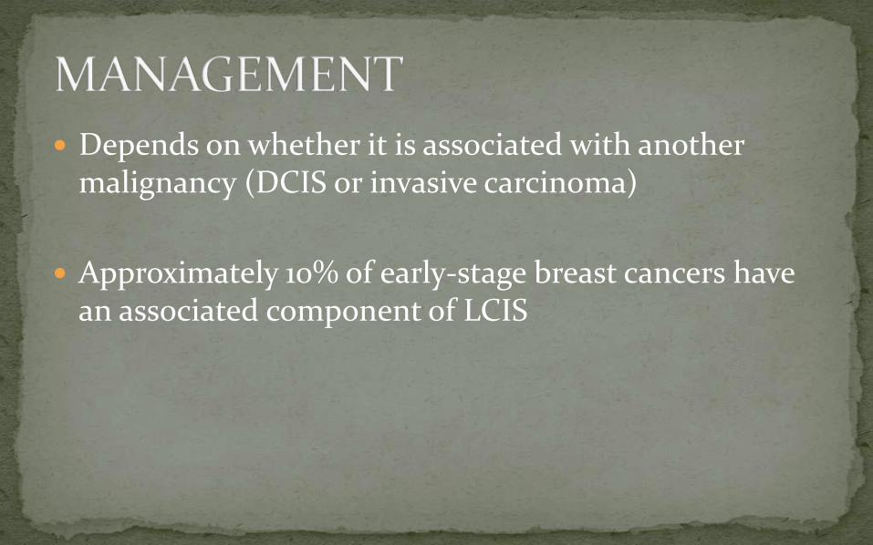
Depends on whether it is associated with another malignancy (DCIS or invasive carcinoma)
Approximately 10% of early-stage breast cancers have an associated component of LCIS
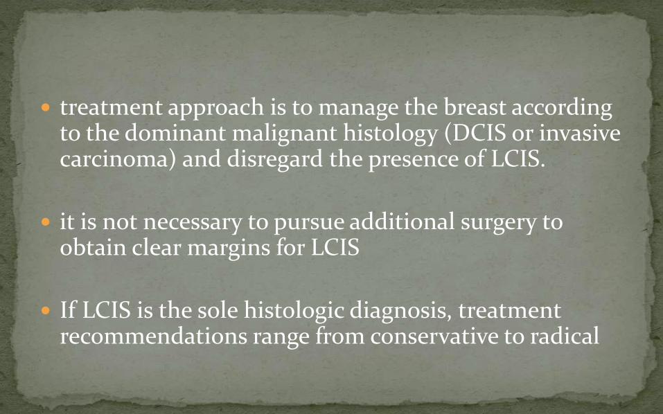
treatment approach is to manage the breast according to the dominant malignant histology (DCIS or invasive carcinoma) and disregard the presence of LCIS.
it is not necessary to pursue additional surgery to obtain clear margins for LCIS
If LCIS is the sole histologic diagnosis, treatment recommendations range from conservative to radical

Earlier days due to high frequency of contralateralbreast involvement it was justified to do contralateralbiopsy and even bilateral mastectomy
Observational studies after wide local excision alone have led to a better understanding of the natural history of this condition, and a more conservative approach is now commonly practiced

In patients with LCIS as the sole histologic diagnosis,
the most widely accepted clinical practice is close observation with regular physical examination and mammographic surveillance
There is no role for radiotherapy in the management of LCIS.
Unilateral mastectomy both inadequate and illogical.
Bilateral prophylactic mastectomy is likely excessive
prophylactic approach in high-risk patients is to consider the use of tamoxifen
characterized by the presence of Paget's cells that are located throughout the epidermis
Paget's disease is a rare entity representing <5% of all breast cancer cases)
typically diagnosed in the fifth or sixth decade.
Synchronous bilateral and male Paget's disease have been reported
Paget's disease is associated with an underlying malignancy in more than 95%

the disease originates from the underlying in situ or invasive disease
There is histologic evidence of intraepithelial extension, immunohistochemical studies, and evidence suggesting that the epidermal keratinocytes release a motility factor, heregulin
that results in the chemotaxis of Paget's cells that migrate to the overlying nipple epidermis
Itching and burning of the nipple and areola.
There is a slow progression toward a crusting eczematoidappearance that can extend to the periareolar skin.
If neglected, bleeding, pain, and ulceration can occur .
Alternatively, Paget's disease can be asymptomatic and present as a pathologic finding after incidental surgical removal of the nipple areolar complex
A palpable mass is detected in approximately 50% of patients at diagnosis
more than 90% of cases this will be an invasive carcinoma.
if no palpable mass is detected, 66% to 86% will have an underlying DCIS.
These associated malignancies are usually located centrally,
Mammographic findings are frequent in the presence of a palpable mass
but normal mammograms are reported in as many as 50% of cases
clinical evaluation includes bilateral breast examination
mammography, and biopsy to confirm the diagnosis of Paget's disease and to fully evaluate the extent of the associated malignancy
The prognosis does not dependent on the diagnosis of Paget's disease, but rather on the associated malignancy
Management of Paget's disease continues to evolve.
Mastectomy was employed in the past but this has been increasingly supplanted by breast-conserving treatment
The infrequent occurrence of this disease entity
the range of disease presentations
variable extent of surgical resection has made the evaluation of treatment options difficult

The combination of limited surgical resection and postoperative radiotherapy appears to be the most practical breast-conserving approach
Surgical resection should include the nipple areolarcomplex with microscopically clear margins surrounding both the Paget's disease and the associated malignancy

THANK YOU