clinical manifestations, diagnosis, and treatment of ... · philum and mycobacterium leprae. first,...
TRANSCRIPT

CLINICAL MICROBIOLOGY REVIEWS, Oct. 2011, p. 701–717 Vol. 24, No. 40893-8512/11/$12.00 doi:10.1128/CMR.00020-11Copyright © 2011, American Society for Microbiology. All Rights Reserved.
Clinical Manifestations, Diagnosis, and Treatment ofMycobacterium haemophilum Infections
Jerome A. Lindeboom,1 Lesla E. S. Bruijnesteijn van Coppenraet,2Dick van Soolingen,3,4 Jan M. Prins,5 and Eduard J. Kuijper6*
Department of Oral and Maxillofacial Surgery, Academic Medical Center, Amsterdam, The Netherlands1; Department of MedicalMicrobiology and Infectious Diseases, Isala Clinics, Zwolle, The Netherlands2; Mycobacterial Reference Laboratory,
National Institute for Public Health and the Environment, Bilthoven, The Netherlands3; Department ofPulmonary Diseases and Department of Microbiology, Radboud University Nijmegen Medical Centre,
P.O. Box 9101, 6500HB Nijmegen, The Netherlands4; Department of Internal Medicine, Division ofInfectious Diseases, Tropical Medicine and AIDS, Academic Medical Center, Amsterdam,
The Netherlands5; and Department of Medical Microbiology, Center ofInfectious Diseases, Leiden University Medical Center,
Leiden, The Netherlands6
INTRODUCTION .......................................................................................................................................................702GENERAL DESCRIPTION AND TAXONOMY ....................................................................................................702CLINICAL PRESENTATION ...................................................................................................................................702IMMUNOCOMPROMISED PATIENTS ................................................................................................................703
Cutaneous Manifestations .....................................................................................................................................703Pyomyositis ..............................................................................................................................................................703Disseminated and Pulmonary Infections.............................................................................................................703Ophthalmologic Manifestations............................................................................................................................705Osteomyelitis ...........................................................................................................................................................705Uncommon Clinical Presentations .......................................................................................................................705
Central catheter infections ................................................................................................................................705Epididymal abscess.............................................................................................................................................706Mixed infections..................................................................................................................................................706
IMMUNOCOMPETENT PATIENTS ......................................................................................................................706Adult Infections.......................................................................................................................................................706
Cervicofacial infections ......................................................................................................................................706“Other” skin lesions...........................................................................................................................................706
Pediatric M. haemophilum Infections ...................................................................................................................708Cervicofacial infections ......................................................................................................................................708Inguinal lymphadenitis ......................................................................................................................................708Pulmonary involvement......................................................................................................................................708
ANIMAL INFECTIONS.............................................................................................................................................708PATHOGENESIS........................................................................................................................................................709EPIDEMIOLOGY.......................................................................................................................................................709
Typing of M. haemophilum .....................................................................................................................................709Environmental Findings.........................................................................................................................................709
DIAGNOSTICS ...........................................................................................................................................................710Skin Testing.............................................................................................................................................................710Histopathology.........................................................................................................................................................710Microscopy ...............................................................................................................................................................710Culture......................................................................................................................................................................710Molecular Identification Methods ........................................................................................................................711Direct Detection Methods......................................................................................................................................711Diagnostic Approach ..............................................................................................................................................711
ANTIMICROBIAL SUSCEPTIBILITY....................................................................................................................712TREATMENT..............................................................................................................................................................712
Immunocompromised Patients .............................................................................................................................713Skin lesions..........................................................................................................................................................713Disseminated infection/pulmonary infection...................................................................................................713Pyomyositis ..........................................................................................................................................................713
* Corresponding author. Mailing address: Department of MedicalMicrobiology, Center of Infectious Diseases, Leiden University Med-ical Center, P.O. Box 9600, Leiden 2300 RC, The Netherlands. Phone:071 5269111. Fax: 071 5248148. E-mail: [email protected].
701
on March 12, 2020 by guest
http://cmr.asm
.org/D
ownloaded from
Skeletal infections/osteomyelitis .......................................................................................................................713Immunocompetent Patients...................................................................................................................................713
Immunocompetent adults ..................................................................................................................................713Immunocompetent children...............................................................................................................................713
Treatment Outcome................................................................................................................................................713RECOMMENDATIONS AND CONCLUSION ......................................................................................................714REFERENCES ............................................................................................................................................................714
INTRODUCTION
Mycobacterium haemophilum is an acid-fast bacillus (AFB)belonging to the group of nontuberculous mycobacteria (NTM)frequently found in environmental habitats, which can colonizeand occasionally infect humans and animals (98). M. haemo-philum can cause localized or disseminated disease in immu-nocompromised hosts and is a rare cause of disease in immu-nologically competent individuals. In 1996, Saubolle et al.(123) presented an overview of 64 cases reported in the liter-ature. Since that time, another 154 cases have been reported.The purpose of this review is to present an update of theclinical picture, diagnostic approach, and therapeutic optionsfor M. haemophilum infections.
GENERAL DESCRIPTION AND TAXONOMY
M. haemophilum, or the “blood-loving” mycobacterium, is aslowly growing AFB that differs from all other identified My-cobacterium species in preferring a lower growth temperatureand having a unique culture requirement for iron supplemen-tation. Thus, the classification of mycobacteria into severalRunyon groups based on growth characteristics and pigmentproduction may not be applicable to M. haemophilum. Manyinfections with M. haemophilum likely remain unrecognized,although suspicion should arise when AFB are visualized insmears and when cultures fail to yield an etiologic agent.
M. haemophilum was first described in 1978 as a pathogencausing skin infections most frequently in immunocompro-mised patients, which may explain its preferred growth tem-perature of 30°C (130). In 1981, Dawson and colleagues de-scribed a case of submandibular lymphadenitis due to M.haemophilum in an otherwise healthy child (31), and M. hae-mophilum has since been recognized as an emerging pathogenin a variety of syndromes. The microorganism is now alsoknown to cause cutaneous and subcutaneous infections, septicarthritis, osteomyelitis, and pneumonitis in immunocompro-mised patients. Cervicofacial lymphadenitis is the most com-mon manifestation in immunocompetent children. Reports ofsuch cases originate from all continents. However, althoughour understanding of M. haemophilum infections in humanshas increased considerably in recent years, the natural habitatand how an infection is acquired remain unknown.
M. haemophilum most resembles Mycobacterium marinumand M. ulcerans in regard to its role in skin infections. Therelatedness can also be observed for genomic traits, as all threespecies have a relatively low GC content compared to those ofmost other Mycobacterium species.
Some interesting similarities also exist between M. haemo-philum and Mycobacterium leprae. First, the fatty acid doco-sanoic acid is found in abundant quantities in both species.Second, M. haemophilum has also been shown to possess a
specific phenolic glycolipid antigen that closely resembles thecorresponding lipid in M. leprae (10). Third, M. leprae hasmajor membrane protein I (35 kDa), which is absent in mem-bers of the M. tuberculosis complex, but homologous sequenceshave been detected in M. avium, M. haemophilum, and M.smegmatis (159).
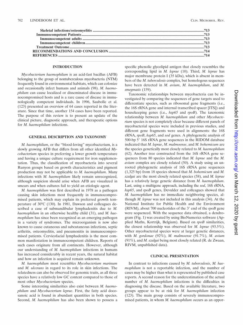
Taxonomic relationships between mycobacteria can be in-vestigated by comparing the sequences of gene targets used todifferentiate species, such as ribosomal gene fragments (i.e.,the 16S rRNA gene and internal transcribed spacer [ITS]) andhousekeeping genes (i.e., hsp65 and rpoB). The taxonomicrelationship between M. haemophilum and other Mycobacte-rium species is not completely clear because different panels ofmycobacterial species were included in previous studies, anddifferent gene fragments were used in alignments: the 16SrRNA, rpoB, hsp65, and sod genes. A phylogenetic analysis of500-bp 5� 16S rRNA gene sequences in the RIDOM databaseindicated that M. leprae, M. malmoense, and M. bohemicum arethe species genetically most closely related to M. haemophilum(62). Another tree constructed from the 16S rRNA gene se-quences from 80 species indicated that M. leprae and the M.avium complex are closely related (50). A study using an un-rooted phylogenetic analysis of 16S rRNA gene sequences(1,325 bp) from 18 species showed that M. bohemicum and M.szulgai are the most closely related species (58), and M. lepraehas a relatively large genetic distance from M. haemophilum.Last, using a multigene approach, including the sod, 16S rRNA,hsp65, and rpoB genes, Devulder and colleagues showed thatM. haemophilum has no immediate neighboring species, al-though M. leprae was not included in this analysis (34). At theNational Institute for Public Health and the Environment(RIVM), about 700 nucleotides of the 3� end of the rpoB genewere sequenced. With the sequence data obtained, a dendro-gram (Fig. 1) was created by using BioNumerics software (Ap-plied Maths, Kortrijk, Belgium). Based on rpoB similarities,the closest relationship was observed for M. leprae (93.5%).Other mycobacterial species were at larger genetic distances,with M. gordonae (92%), M. malmoense (91.7%), M. avium(91%), and M. szulgai being most closely related (R. de Zwaan,RIVM, unpublished data).
CLINICAL PRESENTATION
In contrast to infections caused by M. tuberculosis, M. hae-mophilum is not a reportable infection, and the number ofcases may be higher than what is represented by published casereports. A second reason for the underestimation of the actualnumber of M. haemophilum infections is the difficulties indiagnosing the disease. Based on the available literature, twogroups appear to be at risk for M. haemophilum infection(123). The main group consists of severely immunocompro-mised patients, in whom M. haemophilum occurs as an oppor-
702 LINDEBOOM ET AL. CLIN. MICROBIOL. REV.
on March 12, 2020 by guest
http://cmr.asm
.org/D
ownloaded from
tunistic infection (1, 2). M. haemophilum is being increasinglyrecognized in persons who are severely immunocompromisedby HIV infection; after renal, bone marrow, or cardiac trans-plantation; or after treatment for lymphoma or rheumatoidarthritis. The second at-risk group is otherwise healthy chil-dren, who typically develop cervical and perihilar lymphadeni-tis similar to that caused by infection with the Mycobacteriumavium complex (3, 90, 164).
IMMUNOCOMPROMISED PATIENTS
Cutaneous Manifestations
M. haemophilum causes mainly skin lesions in immunocom-promised patients (42, 150). Cutaneous infections with poten-tially pathogenic mycobacterial species are important for thedifferential diagnosis of skin lesions in these patients (36, 61,93, 106). M. haemophilum infections have been reported, es-pecially in patients with lymphoma or HIV and in organ trans-plant recipients (19, 66, 81, 84, 112, 153). The clinical spectrumof cutaneous infections caused by M. haemophilum appears tobe broad (19, 30), varying from localized disease to systemicdisease with cutaneous dissemination (49). Multiple skin le-sions tend to occur and can present as erythematous papules,plaques, nodules, necrotic abscesses, or chronic ulcers. Cuta-neous lesions are found most frequently on the extremities,particularly over joints, and less commonly on the trunk andface. Purpuric and annular lesions have also been described
(47). Skin lesions typically evolve from papules to asymptom-atic pustules and eventually to very painful deep-seated ulcers.The erythematous or violaceous papules and/or nodules areusually painless at first, but they can develop into potentiallyvery painful abscesses or ulcers. Patients with cutaneous andarticular manifestations have a more favorable prognosis thanthose with pulmonary involvement (126). An overview of theskin infections reported since the review by Saubolle et al.(123) is presented in Table 1. Thirty-three new cases have beenreported, with a median age of the patients of 48 years (range,14 months to 67 years). The sex distribution was equal. Most ofthe reports were from the United States (10 cases), followed byGermany (4 cases), Australia (4 cases), and Singapore (4cases). The majority of the patients had a history of solid-organtransplant or AIDS.
Cutaneous lesions have been rarely reported for children(21, 28), but the manifestations of the skin lesions are similarto those of immunosuppressed adults.
Pyomyositis
Mycobacterial infection of the skeletal muscle is very rare; inparticular, large muscles are involved, and the condition usu-ally presents as localized muscle involvement through directextension from a proximal focus of infection. Only four casesof pyomyositis caused by M. haemophilum have been reported(70, 82, 124, 127).
In a recent report by Lee et al. (82), a 23-year-old immuno-suppressed female patient with multiple, tender, erythematous,and palpable fluctuant abscesses on the left leg due to an M.haemophilum infection was described. In another case, thepatient had been on long-term steroid treatment for poly-myositis and presented with ulcerations over both thighs andthe left arm after a year of steroid therapy (127). A 24-year-oldfemale renal transplant recipient was described as having ten-der, erythematous, and palpable fluctuant swelling on the leftcalf (70). The patient had undergone kidney transplantation 8years earlier, after which she had been on immunosuppressivetreatment with cyclosporine and mycophenolate mofetil.
Disseminated and Pulmonary Infections
Several cases of septicemia and pneumonitis due to M. hae-mophilum have been documented (Tables 2 and 3).
The patients with disseminated disease in Table 2 include 11adults aged 30 to 67 years and 1 6-year-old child. Nine patientswere from the United States, one was from Germany, and onewas from Brazil. Five patients had AIDS, one had received arenal transplant, one had received a cardiac transplant, twohad received a bone marrow transplant, and one was under-going treatment for multiple myeloma. Only one case of apediatric disseminated infection has been described (11). A6-year-old child from The Netherlands with a history of B cellprecursor acute lymphoblastic leukemia presented with feverand painful suppurative skin lesions on the knees, elbows, andface. The patient later developed arthritis and osteomyelitis ofthe right knee in addition to several subcutaneous abscesses,and she remained febrile.
Nine patients have been reported to have M. haemophilumpulmonary infections. Six patients were male, and three were
FIG. 1. Dendrogram made by using the rpoB gene sequences of 29mycobacterial species. M. leprae was most closely associated (93.5%).
VOL. 24, 2011 MYCOBACTERIUM HAEMOPHILUM INFECTIONS 703
on March 12, 2020 by guest
http://cmr.asm
.org/D
ownloaded from

TA
BL
E1.
Rep
orte
dcu
tane
ous
man
ifest
atio
nsin
imm
unoc
ompr
omis
edpa
tient
s,19
96to
2011
a
Ref
eren
ceA
geof
patie
nt(y
r)/s
exU
nder
lyin
gdi
seas
e(s)
Cou
ntry
Tre
atm
ent
Out
com
eD
urat
ion
oftr
eatm
ent
848
/MIg
Ade
ficie
ncy
Ger
man
yC
LR
,RB
,ER
esol
ved
6m
o22
59/M
Ren
altr
ansp
lant
Bra
zil
CL
R,C
IR
esol
ved
1yr
114
52/M
Ren
altr
ansp
lant
Uni
ted
Stat
esC
I,C
LR
Res
olve
d1
yr11
962
/MH
eart
tran
spla
ntIs
rael
CI,
CL
R,R
Reg
ress
ion
with
in3
wk
1m
o72
65/F
CL
LU
nite
dSt
ates
R,C
LR
,CI
Res
olve
d6
mo
7217
/FSL
E,M
DS
Uni
ted
Stat
esR
,CL
R,G
Res
olve
dN
A28
45/F
Ren
altr
ansp
lant
Ven
ezue
laC
LR
Res
olve
d6
mo
2814
mo/
FU
nkno
wn
imm
unod
efici
ency
,C
D4�
�16
%V
enez
uela
CL
R,T
MS,
R,I
Res
olve
d6
mo
125
67/F
RA
Ger
man
y(i
)C
LR
,CI;
(ii)
RB
,E,C
LR
;(iii
)C
LR
mon
othe
rapy
afte
r8
wk
Res
olve
d�
6m
o,N
A
9438
/FA
utoi
mm
une
cirr
hosi
sU
nite
dSt
ates
CI,
CL
RR
esol
ved
6m
o94
47/F
Mya
sthe
nia
grav
is,c
ortic
oste
roid
sU
nite
dSt
ates
D,R
B,A
ZI
Impr
ovem
ent
8m
o20
37/M
AID
SSp
ain
I,R
,E,A
K,C
LR
,CI,
Min
Res
olve
d5
mo
2116
/MR
enal
tran
spla
ntU
nite
dSt
ates
E,R
,CL
R,C
I,A
KPa
rtia
lres
olut
ion
14m
o14
959
/MPo
lym
yalg
iarh
eum
atic
aG
erm
any
I,E
,RD
ied
5327
/FA
IDS
Uni
ted
Stat
esR
B,C
LR
,CI
Impr
ovem
ent
1m
o13
859
/FSL
ESi
ngap
ore
(i)
CL
R,C
I,R
,I,E
;(ii)
RB
,CL
RR
esol
ved;
recu
rren
cean
dre
solu
tion
afte
r2n
dco
urse
13m
o
138
64/F
Cut
aneo
usva
scul
itis
Sing
apor
eC
LR
,CI
Res
olut
ion
18m
o13
842
/FSj
ogre
n’s
synd
rom
e,C
rohn
’sdi
seas
eSi
ngap
ore
CL
R,D
NA
18m
o
9851
/MA
IDS
Ital
yR
B,E
,CL
R;l
ater
RB
,AZ
I,L
Res
olut
ion
5m
o85
44/M
Ren
altr
ansp
lant
Tai
wan
CI,
R,C
LR
Res
olut
ion
1yr
4959
/MD
iabe
tes
Uni
ted
Stat
esC
I,R
B,C
LR
Res
olut
ion
NA
139
25/F
SLE
Sing
apor
eC
LR
,E,I
,RR
esol
utio
n6
mo
108
30/F
AID
SG
erm
any
Onl
yan
tiret
rovi
ralt
hera
pyR
esol
utio
n14
mo
3851
/MA
IDS
Japa
nC
LR
,ER
esol
utio
n8
mo
126
29/F
AID
SU
nite
dSt
ates
R,C
I,C
LR
,DD
ied
11m
o47
62/M
AID
SSw
itzer
land
AZ
I,R
BR
esol
utio
n6
mo
5151
/MA
IDS
Uni
ted
Stat
esC
LR
,TM
S,C
IR
esol
utio
nN
A97
56/M
Lun
gtr
ansp
lant
Aus
tral
iaI,
CL
R,P
,D,C
IR
esol
utio
n42
mo
9749
/FL
ung
tran
spla
ntA
ustr
alia
CL
R,E
,RR
esol
utio
n17
mo
9753
/FL
ung
tran
spla
ntA
ustr
alia
CL
R,R
,CI,
DR
esol
utio
n31
mo
9739
/ML
ung
tran
spla
ntA
ustr
alia
CL
R,R
,CI,
DR
esol
utio
n18
mo
8030
/FA
IDS
The
Net
herl
ands
(i)
D,C
I,R
,CL
R;(
ii)M
in,R
B,
E;(
iii)
I,C
I,C
YR
esol
utio
n6
mo
635
/MA
IDS
Ger
man
yR
,E,I
,CL
RR
esol
utio
n7
wk,
rela
pse
and
retr
eatm
ent
with
sam
ere
gim
en
aI,
ison
iazi
d;R
,ri
fam
pin;
RB
,ri
fabu
tin;
E,
etha
mbu
tol;
CY
,cy
clos
erin
e;C
I,ci
profl
oxac
in;
AK
,am
ikac
in;
AZ
I,az
ithro
myc
in;
CL
R,
clar
ithro
myc
in;
TM
S,tr
imet
hopr
im-s
ulfa
met
hoxa
zole
;P,
pyra
zina
mid
e;D
,do
xycy
clin
e;M
in,m
inoc
yclin
e;G
,gat
iflox
acin
;L,l
evofl
oxac
in;R
A,r
heum
atoi
dar
thri
tis;M
DS,
mye
lody
spla
stic
synd
rom
e;SL
E,s
yste
mic
lupu
ser
ythe
mat
osus
;CL
L,c
hron
icly
mph
ocyt
icle
ukem
ia;N
A,d
ata
nota
vaila
ble;
M,m
ale;
F,f
emal
e.
704 LINDEBOOM ET AL. CLIN. MICROBIOL. REV.
on March 12, 2020 by guest
http://cmr.asm
.org/D
ownloaded from

female, with a median age of 38 years (range, 27 to 72 years)(Table 3). Eight reports were from the United States, and themost recent report was from The Netherlands. Despite severalmultidrug regimens (Table 3), a resolution of the infection wasobserved for less than half of the patients.
Ophthalmologic Manifestations
Two reports in the literature described primary ophthalmo-logic infections due to M. haemophilum (102, 104). Millar et al.(102) described a 55-year-old man with a history of acutemyeloid leukemia with chronic bilateral conjunctivitis and dryeyes for a period of 6 months. Skin lesions were also noted onthe patient’s face and arms. The clinical condition improvedwith moxifloxacin and clarithromycin with the addition of va-lacyclovir and clindamycin 1 week later. Modi et al. (104)presented a unilateral chronic granulomatous iridocyclitis in a66-year-old man with previous cardiac transplantation and cy-closporine and mycophenolate mofetil treatment. Antibiotictherapy did not prevent the progression of intraocular inflam-mation, and the patient developed a corneal ulcer that perfo-rated. Enucleation was performed 1 year after the initial pre-sentation. However, the skin lesions regressed with antibiotictherapy.
Osteomyelitis
A less common manifestation of M. haemophilum in immu-nocompromised patients is septic arthritis or osteomyelitis withor without cutaneous lesions. Osteomyelitis caused by M. hae-mophilum resembles that caused by other microorganisms(107, 163). On radiographs, bony resorption with clear mar-gins, cortical destruction, and adjacent soft tissue swelling areapparent. Magnetic resonance imaging (MRI) can reveal well-circumscribed medullary lesions with cortical disruption and alarge soft tissue component (83). Table 4 provides an overviewof the cases of skeletal M. haemophilum infections described inthe literature. Twenty-six cases have been reported, with amedian age of the patients of 45.5 years (range, 20 to 77 years).The underlying illnesses most frequently included AIDS (15cases) and organ transplantation (7 cases).
Uncommon Clinical Presentations
Central catheter infections. Ward et al. (152) described anM. haemophilum infection of the central venous catheter tun-nel in two young (26 and 29 years old) immunosuppressedpatients with hematological malignancy undergoing high-dosechemotherapy supported by bone marrow transplantation.The M. haemophilum infections occurred at the site of thetunneled catheter after the line had been removed. For onepatient, therapy consisted of amikacin, clarithromycin, cip-rofloxacin, and meropenem for 3 weeks, after which amika-cin and meropenem were ceased and ethambutol wasstarted. For the other patient, repeated surgical excisions werecombined with clarithromycin, amikacin, and meropenemtreatment. The drugs were later changed to rifampin, cipro-floxacin, and clarithromycin. For both patients, the woundeventually healed.
TA
BL
E2.
Reported
disseminated
infectionsin
imm
unocomprom
isedpatients
a
Reference(s)
Age
ofpatient
(yr)/sexC
ountryU
nderlyingdisease(s)
Initialpresentation(s)C
ulturesource(s)
Treatm
entO
utcome
13664/M
Germ
anyA
IDS
Skin,pulmonary
Blood,sputum
RB
,E,C
LR
�T
MS
Cure
116/F
The
Netherlands
AL
LSkin,pulm
onary,jointsSkin,bone
marrow
E,R
,CL
R,A
K,drainage
ofabscesses
Cure
3946/F
United
StatesC
ardiactransplant
Skin,pulmonary,joints
Skin,synovialfluidIm
i,CI,C
LR
,DC
ure121
67/MB
razilR
enaltransplantN
AB
loodN
AN
A126
32/MU
nitedStates
BM
T,M
DS
Pulmonary
infiltrateB
loodN
oneD
ied19
51/FU
nitedStates
Multiple
myelom
a,RA
Skindisease
Skin,bloodR
,E,C
LR
,CI
Cure
12333/M
United
StatesA
IDS
Synovialinvolvement
Synovialfluid,bloodE
,CL
R,C
I,AK
Relapse,died
ofA
IDS
75,76,135,15630/F
United
StatesB
MT
,APM
LSubcutaneous
nodulesSkin,blood
CI,C
LR
,D,I,R
,EC
ure75,76,79
36/MU
nitedStates
AID
SSkin
lesions,septicarthritis
Synovialfluid,bloodPA
S,RC
ure75,135,163
37/MU
nitedStates
AID
SSkin
lesions,septicarthritis
Skin,bloodA
K,C
I,I,CL
,D,E
,I,RC
ure75,117
34/MU
nitedStates
AID
SSubcutaneous
nodulesSkin,blood
E,I,R
Persisted23
NA
United
StatesN
AN
AB
loodN
AN
A
aI,isoniazid;Im
i,imipenem
;R,rifam
pin;RB
,rifabutin;E,etham
butol;CI,ciprofloxacin;A
K,am
ikacin;CL
R,clarithrom
ycin;TM
S,trimethoprim
-sulfamethoxazole;D
,doxycycline;PAS,p-am
inosalicylicacid;R
A,
rheumatoid
arthritis;BM
T,allogeneic
bonem
arrowtransplantation;M
DS,m
yelodysplasticsyndrom
e;APM
L,acute
promyelocytic
leukemia;C
ML
,chronicm
yelogenousleukem
ia;AL
L,acute
lymphocytic
leukemia;N
A,
datanot
available.
VOL. 24, 2011 MYCOBACTERIUM HAEMOPHILUM INFECTIONS 705
on March 12, 2020 by guest
http://cmr.asm
.org/D
ownloaded from

Epididymal abscess. Keller et al. (73) described an epidid-ymal abscess due to M. haemophilum infection in a renal trans-plant patient. A right orchidectomy was performed, combinedwith clarithromycin, rifabutin, and ethambutol treatment. Thesymptoms resolved over 5 months of follow-up.
Mixed infections. Dual infections with M. haemophilum andother NTM species are extremely rare but also difficult todiagnose. Since the first case report by Branger et al. (12),describing a mixed infection with M. haemophilum and M.xenopi, two new cases have been reported. Bekou and col-leagues (8) reported a skin infection with multifocal nodules ofvariable sizes arranged in a sporotrichoid-like manner on theextremities and back of a 48-year-old male patient with an IgAdeficiency. Both M. haemophilum and M. kansasii were cul-tured. Treatment with clarithromycin, rifabutin, and ethambu-tol for 6 months led to a complete clinical remission of the skinlesions. Phowthongkum et al. (111) described a 40-year-oldmale patient with AIDS who developed a spindle cell pseudo-brain tumor as a result of M. haemophilum and M. simiaeinfection. He was treated with isoniazid, rifampin, pyrazin-amide, ethambutol, and clarithromycin. One month after hos-pitalization, he commenced antiretroviral treatment, includingzidovudine, lamivudine, and efavirenz. He was dischargedhome, and was seen for the last time 3 months after the oper-ation.
IMMUNOCOMPETENT PATIENTS
Adult Infections
Cervicofacial infections. An outbreak of 12 cases of M. hae-mophilum skin infection with lymphadenitis after permanentmakeup on the eyebrows was described recently (52). The inkused by the tattoo artist was found to be contaminated with M.haemophilum. All 12 patients were female, with a median ageof 56 years, and none of the patients were immunosuppressed.The patients presented with an inflammatory lesion consistingof a few red papules or pustules or an erythematous plaque onone eyebrow. In all cases, the lesion was associated with ipsi-lateral lymphadenopathy in the parotid region, affecting one ormore lymph nodes (median, 2; range, 1 to 5). Eight patientspresented with an abscess, which later developed into a fistulain seven cases, whereas none of the patients reported systemicsymptoms.
Minani et al. (103) described a 27-year-old immunocompe-tent woman with a right buccal abscess and submandibularlymphadenitis. The patient was cured with surgical excisionaltherapy of the affected lymph nodes and drainage of the buccalabscess. A retrospective overview of another six patients withcervicofacial lymphadenitis (five females and one male, with anage range of 19 to 65 years) seen over a 15-year period inPhoenix, AZ, was also presented (103).
“Other” skin lesions. Skin lesions in immunocompetentadults due to M. haemophilum infection are rare and the resultof injury. Two cases have been reported (99, 128): a 61-year-old male who sustained several lacerations to the forearmwhen he was thrown against coral while surfing and a 65-year-old female who developed subcutaneous skin nodules aftercoronary artery bypass surgery.
TA
BL
E3.
Rep
orte
dpu
lmon
ary
man
ifest
atio
nsin
imm
unoc
ompr
omis
edpa
tient
sa
Ref
eren
ce(s
)A
geof
patie
nt(y
r)/s
exC
ount
ryU
nder
lyin
gdi
seas
e(s)
Initi
alpr
esen
tatio
n(s)
Cul
ture
sour
ce(s
)T
reat
men
tO
utco
me
137
72/F
The
Net
herl
ands
RA
/OSA
SPn
eum
onia
Sput
umR
,AZ
IR
esol
utio
n12
632
/MU
nite
dSt
ates
BM
T,M
DS
Pulm
onar
yin
filtr
ate
Blo
odN
one
Dea
th15
762
/FU
nite
dSt
ates
NA
Pulm
onar
yno
dule
Lun
gbi
opsy
spec
imen
R,E
Cur
e12
330
/FU
nite
dSt
ates
Ren
altr
ansp
lant
,su
bseq
uent
AID
SSk
inle
sion
s,se
ptic
arth
ritis
,su
bseq
uent
pulm
onar
yin
volv
emen
t
NA
E,I
,R,s
ubse
quen
tM
inIn
itial
reso
lutio
n,su
bseq
uent
deat
h
135
51/M
Uni
ted
Stat
esA
IDS
Bro
nchi
tisSp
utum
,bon
eC
I,D
,E,I
,P,R
Res
olut
ion
7642
/MU
nite
dSt
ates
BM
T,M
DS
Pulm
onar
yin
filtr
ate
BA
Lflu
id,s
putu
mR
,E,C
I,C
LR
,AK
,PD
eath
7637
/MU
nite
dSt
ates
AID
SSk
inle
sion
s,pn
eum
onia
Skin
,spu
tum
AK
,CI,
D,E
,I,R
Res
pond
edan
dth
enre
laps
edan
ddi
ed27
,75,
76,1
3535
/MU
nite
dSt
ates
AID
SSk
in,p
ulm
onar
yin
filtr
ate
Skin
,spu
tum
R,E
,CI,
AK
,D,E
ryD
eath
27,7
5,76
,135
,156
27/M
Uni
ted
Stat
esB
MT
,AA
Pulm
onar
yno
dule
sSp
utum
,BA
Lflu
id,l
ung
biop
sysp
ecim
enR
,E,A
K,P
,I,S
Dea
th
aI,
ison
iazi
d;R
,rifa
mpi
n;R
B,r
ifabu
tin;E
,eth
ambu
tol;
CI,
cipr
oflox
acin
;AK
,am
ikac
in;A
ZI,
azith
rom
ycin
;CL
R,c
lari
thro
myc
in;P
,pyr
azin
amid
e;D
,dox
ycyc
line;
Min
,min
ocyc
line;
Ery
,ery
thro
myc
in;S
,str
epto
myc
in;
RA
,rhe
umat
oid
arth
ritis
;BM
T,a
lloge
neic
bone
mar
row
tran
spla
ntat
ion;
MD
S,m
yelo
dysp
last
icsy
ndro
me;
AA
,apl
astic
anem
ia;C
ML
,chr
onic
mye
loge
nous
leuk
emia
;BA
L,b
ronc
hoal
veol
arla
vage
;OSA
S,ob
stru
ctiv
esl
eep
apne
asy
ndro
me;
NA
,dat
ano
tav
aila
ble.
706 LINDEBOOM ET AL. CLIN. MICROBIOL. REV.
on March 12, 2020 by guest
http://cmr.asm
.org/D
ownloaded from

TA
BL
E4.
Reported
septicarthritis/osteom
yelitisin
imm
unocomprom
isedpatients
a
Reference(s)
Age
ofpatient
(yr)/sexU
nderlyingdisease
Country
Area(s)
ofseptic
arthritisb
OM
bO
thersite(s)
Therapy
Outcom
e
10158/M
Lym
phoma
Australia
Foot
I,R,E
Partialresponse101
55/FR
enaltransplantA
ustraliaA
nkleI,R
,ED
ied26
NA
Cardiac
transplantSouth
Africa
Lim
bsN
AN
AD
ied96
32/MA
IDS
United
StatesA
nkles,Lw
ristB
loodR
,I,P,E,E
TN
oim
provement
11734/M
AID
SU
nitedStates
Rfinger
Softtissue
abscess,B
AL
fluidI,R
,EN
oim
provement
5448/M
Renaltransplant
France
Lm
iddlefinger,L
kneeSkin
Surgery;Min,E
ryfor
2m
o;I,R
,E,M
infor
6m
oC
ure
7936/M
AID
SU
nitedStates
Rknee
Skin,bloodR
,I,P,EIm
provement,stable
at19-m
ofollow
-up57
21/FA
IDS
United
StatesL
ankle,tibiaSkin
R,M
inR
esolution33
44/MA
IDS
United
StatesB
ilateraltibiaand
fibulaSkin,sputum
I,R,E
,CI,C
L,A
KInitialim
provement,
relapsein
6w
k163
31/FA
IDS
United
StatesL
kneeR
3rdfinger,R
calcaneusSkin
R,I,E
,AK
,CI,C
L,P
Resolution
76,135,16337/M
AID
SU
nitedStates
Lankle
SkinR
,I,P,Efor
14m
oR
esolution118
NS
AID
SF
ranceB
ilatknees
Finger,toes,tibia,elbow
,T
9-10vertebrae
Skin,lungsI,R
,EN
oim
provement
13339/M
AID
SA
ustraliaL
footC
L,A
K,D
,RIm
provement
in10
wk
13241/M
AID
SU
nitedStates
Relbow
Rolecranon
SkinR
,I,PR
esolved9
mo
later64
49/MA
IDS
United
StatesK
nees,anklesR
ankleand
tibiaSkin,blood,lym
phnodes
R,I,P,E
,A,C
I,CL
No
improvem
ent
6546/M
AID
SU
nitedStates
Foot
CI,R
B,C
Y,A
ZI
Improved
aftertreatm
ent123
30/FR
enaltransplant/AID
SU
nitedStates
Hand
Hand
Skin,pulmonary
I,R,E
,Min
Resolution
oflesion,
died123
33/MA
IDS
United
StatesK
neeSkin
E,C
LR
,CI,A
KD
iedof
AID
Scom
plications123
77/MT
celllymphom
aU
nitedStates
Hand
Curettage
Relapse
after1
yr,died
oflym
phoma
complications
12366/F
RA
(corticosteroids)U
nitedStates
Hip
D,R
,subsequentexcision
�D
,RC
ure
12356/M
AID
SU
nitedStates
Ankle
NA
Died
2m
oafter
initialpresentation
12345/F
Renaltransplant
United
StatesF
ingerSkin
CI,R
BC
ure112
20/MC
ardiactransplant
United
StatesO
lecranonC
LR
,RC
ure121
30/MA
IDS
Brazil
Elbow
NA
NA
NA
12647/F
AA
/BM
TU
nitedStates
NS
CI,C
LR
,D,R
(�6
mo)
Improved
3946/F
Cardiac
transplantU
nitedStates
Wrist,knees,ankles
Skin,pneumonia
Imi,C
I,CL
R,D
,2m
oR
esolution
5653/F
AID
SG
ermany
Tibia
E,R
,CL
RC
ure37
56/FPolycythem
iavera
Canada
Rw
rist,Rankle
SkinC
I,RB
,CL
RC
ure
aI,isoniazid;Im
i,imipenem
;R,rifam
pin;RB
,rifabutin;E,etham
butol;ET
,ethionamide;C
Y,cycloserine;C
L,clofazim
ine;CI,ciprofloxacin;A
K,am
ikacin;AZ
I,azithromycin;C
LR
,clarithromycin;P,pyrazinam
ide;D
,doxycycline;Min,m
inocycline;Ery,erythrom
ycin;RA
,rheumatoid
arthritis;AA
,aplasticanem
ia;BM
T,bone
marrow
transplant;OM
,osteomyelitis;N
A,data
notavailable.
bL
,left;R,right.
VOL. 24, 2011 MYCOBACTERIUM HAEMOPHILUM INFECTIONS 707
on March 12, 2020 by guest
http://cmr.asm
.org/D
ownloaded from
Pediatric M. haemophilum Infections
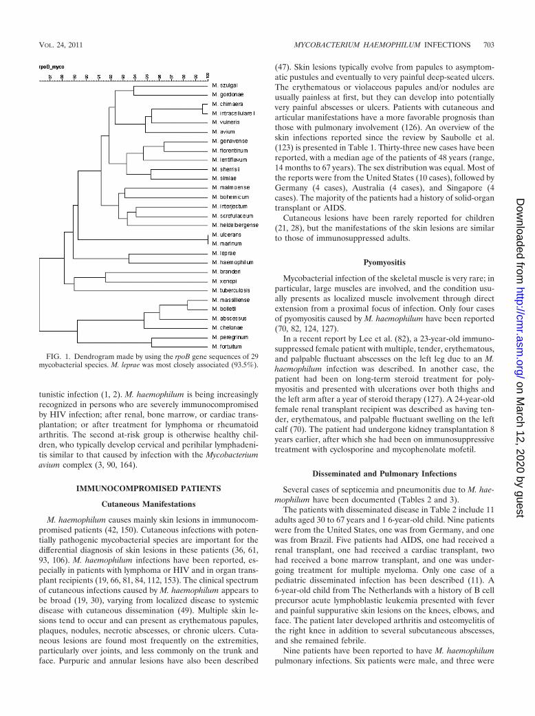
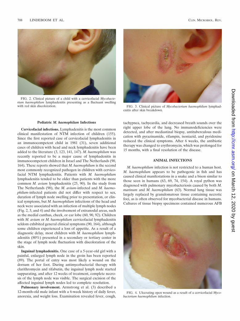
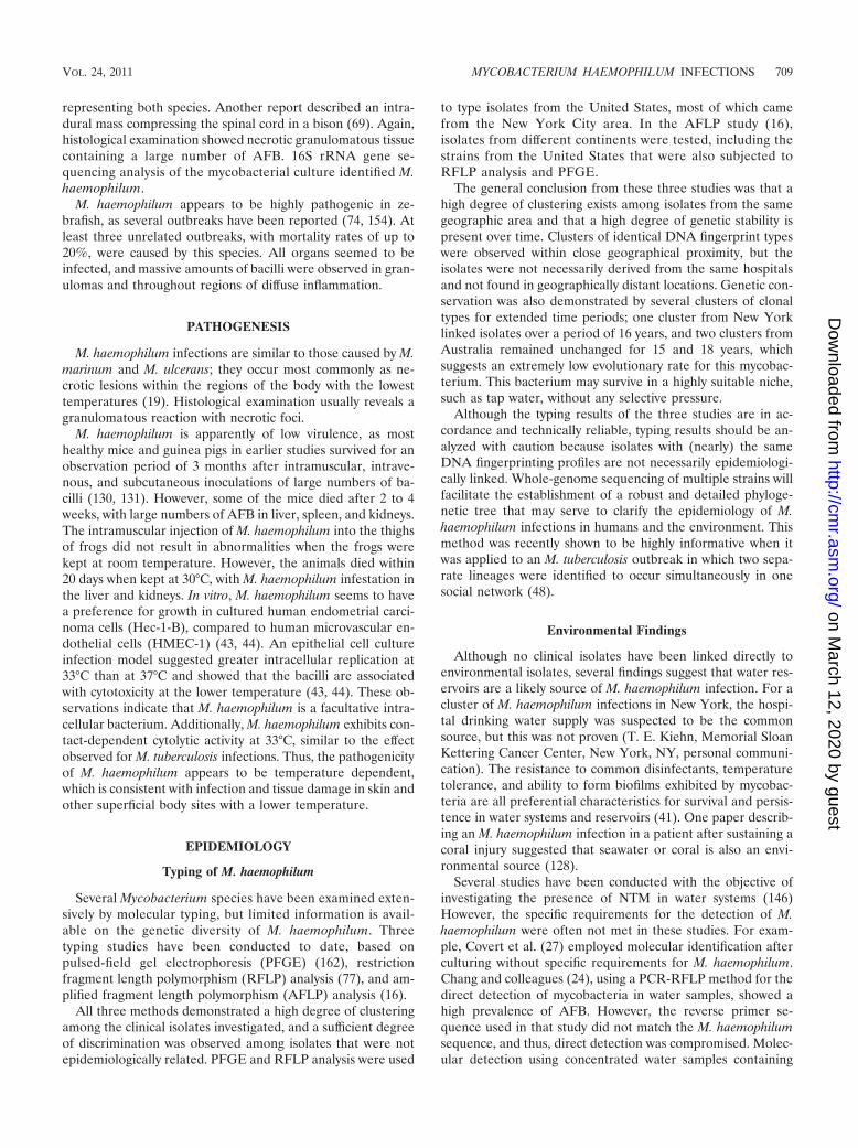
Cervicofacial infections. Lymphadenitis is the most commonclinical manifestation of NTM infection of children (155).Since the first reported case of cervicofacial lymphadenitis inan immunocompetent child in 1981 (31), seven additionalcases of children with head and neck lymphadenitis have beenadded to the literature (3, 123, 141, 147). M. haemophilum wasrecently reported to be a major cause of lymphadenitis inimmunocompetent children in Israel and The Netherlands (90,164). These reports showed that M. haemophilum is the secondmost commonly recognized pathogen in children with cervico-facial NTM lymphadenitis. Patients with M. haemophilumlymphadenitis tended to be older than patients with the morecommon M. avium lymphadenitis (25, 90). In the study fromThe Netherlands (90), the M. avium-infected and M. haemo-philum-infected patients did not differ with respect to sex,duration of lymph node swelling prior to presentation, or clin-ical symptoms, but M. haemophilum infections of the head andneck were associated with an infection of multiple lymph nodes(Fig. 2, 3, and 4) and the involvement of extranodal areas, suchas the medial canthus, cheek, or ear lobe (60, 90, 92). Childrenwith M. avium or M. haemophilum cervicofacial lymphadenitisseldom exhibited general clinical symptoms (90, 164), althoughsome children experienced a loss of appetite. As a result of adiagnostic delay, most children with M. haemophilum lymph-adenitis (80%) presented in a secondary or tertiary center inthe stage of lymph node fluctuation with discoloration of theskin.
Inguinal lymphadenitis. One case of a 5-year-old girl with apainful, enlarged lymph node in the groin has been reported(89). The portal of entry was most likely a wound on thedorsum of her foot. During antimycobacterial therapy withclarithromycin and rifabutin, the inguinal lymph node startedsuppurating, and after 12 weeks of treatment, complete necro-sis of the lymph node was visible. The surgical excision of theaffected inguinal lymph nodes led to complete resolution.
Pulmonary involvement. Armstrong et al. (3) described a12-month-old male infant with a 6-week history of daily fever,anorexia, and weight loss. Examination revealed fever, cough,
tachypnea, tachycardia, and decreased breath sounds over theright upper lobe of the lung. No immunodeficiencies weredetected, and after mediastinal biopsy, antituberculous medi-cation with pyrazinamide, rifampin, isoniazid, and pyridoxinereduced the clinical symptoms. After 6 weeks, the antibiotictherapy was changed to erythromycin, which was prolonged for15 months, with a final resolution of the disease.
ANIMAL INFECTIONS
M. haemophilum infection is not restricted to a human host.M. haemophilum appears to be pathogenic in fish and hascaused clinical manifestations in a snake and a bison similar tothose seen in humans (63, 69, 74, 154). A royal python wasdiagnosed with pulmonary mycobacteriosis caused by both M.marinum and M. haemophilum (63). Normal lung tissue waslargely replaced by granulomatous tissue containing necroticfoci, as is often observed for mycobacterial disease in humans.Cultures of tissue biopsy specimens contained numerous AFB
FIG. 2. Clinical picture of a child with a cervicofacial Mycobacte-rium haemophilum lymphadenitis presenting as a fluctuant swellingwith red skin discoloration. FIG. 3. Clinical picture of Mycobacterium haemophilum lymphad-
enitis after skin breakdown.
FIG. 4. Ulcerating open wound as a result of a cervicofacial Myco-bacterium haemophilum infection.
708 LINDEBOOM ET AL. CLIN. MICROBIOL. REV.
on March 12, 2020 by guest
http://cmr.asm
.org/D
ownloaded from
representing both species. Another report described an intra-dural mass compressing the spinal cord in a bison (69). Again,histological examination showed necrotic granulomatous tissuecontaining a large number of AFB. 16S rRNA gene se-quencing analysis of the mycobacterial culture identified M.haemophilum.
M. haemophilum appears to be highly pathogenic in ze-brafish, as several outbreaks have been reported (74, 154). Atleast three unrelated outbreaks, with mortality rates of up to20%, were caused by this species. All organs seemed to beinfected, and massive amounts of bacilli were observed in gran-ulomas and throughout regions of diffuse inflammation.
PATHOGENESIS
M. haemophilum infections are similar to those caused by M.marinum and M. ulcerans; they occur most commonly as ne-crotic lesions within the regions of the body with the lowesttemperatures (19). Histological examination usually reveals agranulomatous reaction with necrotic foci.
M. haemophilum is apparently of low virulence, as mosthealthy mice and guinea pigs in earlier studies survived for anobservation period of 3 months after intramuscular, intrave-nous, and subcutaneous inoculations of large numbers of ba-cilli (130, 131). However, some of the mice died after 2 to 4weeks, with large numbers of AFB in liver, spleen, and kidneys.The intramuscular injection of M. haemophilum into the thighsof frogs did not result in abnormalities when the frogs werekept at room temperature. However, the animals died within20 days when kept at 30°C, with M. haemophilum infestation inthe liver and kidneys. In vitro, M. haemophilum seems to havea preference for growth in cultured human endometrial carci-noma cells (Hec-1-B), compared to human microvascular en-dothelial cells (HMEC-1) (43, 44). An epithelial cell cultureinfection model suggested greater intracellular replication at33°C than at 37°C and showed that the bacilli are associatedwith cytotoxicity at the lower temperature (43, 44). These ob-servations indicate that M. haemophilum is a facultative intra-cellular bacterium. Additionally, M. haemophilum exhibits con-tact-dependent cytolytic activity at 33°C, similar to the effectobserved for M. tuberculosis infections. Thus, the pathogenicityof M. haemophilum appears to be temperature dependent,which is consistent with infection and tissue damage in skin andother superficial body sites with a lower temperature.
EPIDEMIOLOGY
Typing of M. haemophilum
Several Mycobacterium species have been examined exten-sively by molecular typing, but limited information is avail-able on the genetic diversity of M. haemophilum. Threetyping studies have been conducted to date, based onpulsed-field gel electrophoresis (PFGE) (162), restrictionfragment length polymorphism (RFLP) analysis (77), and am-plified fragment length polymorphism (AFLP) analysis (16).
All three methods demonstrated a high degree of clusteringamong the clinical isolates investigated, and a sufficient degreeof discrimination was observed among isolates that were notepidemiologically related. PFGE and RFLP analysis were used
to type isolates from the United States, most of which camefrom the New York City area. In the AFLP study (16),isolates from different continents were tested, including thestrains from the United States that were also subjected toRFLP analysis and PFGE.
The general conclusion from these three studies was that ahigh degree of clustering exists among isolates from the samegeographic area and that a high degree of genetic stability ispresent over time. Clusters of identical DNA fingerprint typeswere observed within close geographical proximity, but theisolates were not necessarily derived from the same hospitalsand not found in geographically distant locations. Genetic con-servation was also demonstrated by several clusters of clonaltypes for extended time periods; one cluster from New Yorklinked isolates over a period of 16 years, and two clusters fromAustralia remained unchanged for 15 and 18 years, whichsuggests an extremely low evolutionary rate for this mycobac-terium. This bacterium may survive in a highly suitable niche,such as tap water, without any selective pressure.
Although the typing results of the three studies are in ac-cordance and technically reliable, typing results should be an-alyzed with caution because isolates with (nearly) the sameDNA fingerprinting profiles are not necessarily epidemiologi-cally linked. Whole-genome sequencing of multiple strains willfacilitate the establishment of a robust and detailed phyloge-netic tree that may serve to clarify the epidemiology of M.haemophilum infections in humans and the environment. Thismethod was recently shown to be highly informative when itwas applied to an M. tuberculosis outbreak in which two sepa-rate lineages were identified to occur simultaneously in onesocial network (48).
Environmental Findings
Although no clinical isolates have been linked directly toenvironmental isolates, several findings suggest that water res-ervoirs are a likely source of M. haemophilum infection. For acluster of M. haemophilum infections in New York, the hospi-tal drinking water supply was suspected to be the commonsource, but this was not proven (T. E. Kiehn, Memorial SloanKettering Cancer Center, New York, NY, personal communi-cation). The resistance to common disinfectants, temperaturetolerance, and ability to form biofilms exhibited by mycobac-teria are all preferential characteristics for survival and persis-tence in water systems and reservoirs (41). One paper describ-ing an M. haemophilum infection in a patient after sustaining acoral injury suggested that seawater or coral is also an envi-ronmental source (128).
Several studies have been conducted with the objective ofinvestigating the presence of NTM in water systems (146)However, the specific requirements for the detection of M.haemophilum were often not met in these studies. For exam-ple, Covert et al. (27) employed molecular identification afterculturing without specific requirements for M. haemophilum.Chang and colleagues (24), using a PCR-RFLP method for thedirect detection of mycobacteria in water samples, showed ahigh prevalence of AFB. However, the reverse primer se-quence used in that study did not match the M. haemophilumsequence, and thus, direct detection was compromised. Molec-ular detection using concentrated water samples containing
VOL. 24, 2011 MYCOBACTERIUM HAEMOPHILUM INFECTIONS 709
on March 12, 2020 by guest
http://cmr.asm
.org/D
ownloaded from
AFB was unsuccessful overall, and the method was eventuallyapplied to the identification of isolates cultured without theculturing requirements necessary for M. haemophilum. Bothstudies showed that a variety of mycobacterial species werepresent in chlorine-treated water supplies and were thorough,but M. haemophilum might have been overlooked.
Only a few studies allowed the detection of M. haemophilumby molecular methods or specific culturing methods (40, 68,113, 154). Hussein and colleagues (68) did include speciesdetection, but they encountered only other NTM. Three stud-ies detected M. haemophilum. Falkinham et al. (40) found it inthree samples, comprising one water sample and two biofilmsamples, all from different water distribution systems in theUnited States. Whipps et al. (154) detected M. haemophilum inbiofilms from four zebrafish tank meniscuses and one tankdrain, all from a zebrafish research center in which M. haemo-philum caused significant mortality among the fish population.Pryor et al. (113) cultured M. haemophilum from a water dis-tribution system (unknown sample type) as one of many otherMycobacterium species.
In one publication, an environmental M. haemophilum iso-late not directly associated with water was described. Myco-bacterial isolates were cultured from the intestines and surfaceof hospital cockroaches in Taiwan, and M. haemophilum wasfound on the surface of one cockroach (109).
DIAGNOSTICS
Skin Testing
No specific antigen test is available for M. haemophiluminfections, although in the past, purified protein derivatives(PPDs) of M. avium, M. kansasii, M. scrofulaceum and M.marinum, M. intracellulare, M. gordonae, and M. fortuitum havebeen used for the diagnosis of NTM infections. Unfortunately,a few years ago the production of NTM-PPD (Statens SerumInstitute, Denmark) was terminated, although skin testing ap-peared to be useful for the diagnosis of NTM infections inchildren. Because of cross-reactivity between the immune re-actions to PPDs of different species, the tuberculin-PPD testoften shows false-positive reactions due to previous encounterswith NTM (91). The problem with previous NTM encountersis not expected in young children; therefore, a positive tuber-culin test can be indicative of NTM disease in this patientgroup, except for children living in a country where tubercu-losis is highly endemic. For the initial diagnosis of NTM lymph-adenitis, the tuberculin test has an optimal cutoff value of 5mm for a positive skin induration (91). Using a 5-mm cutoff,the tuberculin PPD has 71% sensitivity for M. haemophilumand a 98% positive predictive value (PPV). Using a 10-mmcutoff (the induration cutoff considered positive for M. tuber-culosis reactivity), 57% of all confirmed M. haemophilum in-fections yielded positive skin indurations.
Histopathology
Tissues infected with M. haemophilum show, almost withoutexception, granulomatous infiltrates with necrosis (19, 35). Thegranulomas comprise variable forms of granulocytes, lympho-cytes, monocytes, and multinucleated giant cells. Bacilli can be
observed both extracellularly and intracellularly, and they canbe abundant or scarce in affected tissue (19, 35, 126). Nospecific clinical and histological manifestations can be attrib-uted to M. haemophilum. M. haemophilum skin infection oftenmimics M. marinum infection: it forms erythematous papulesor nodules, often overlying or above the joints, and in laterstages, it becomes suppurative/ulcerative. However, in contrastto M. marinum infections, the nodules are painful, and sporo-trichoid spread is seldom seen in M. haemophilum infections(19). Skin manifestations sporadically include lichenoid der-matitis, panniculitis, vasculitis, or annular plaques.
Histological findings for 16 skin biopsy specimens from 11immunocompromised patients with culture-proven M. haemo-philum infections revealed most commonly (7 of 16 biopsyspecimens) a mixed histopathological pattern of suppurativeand granulomatous reactions (19). Four biopsy specimensshowed well-formed epithelioid granulomas. The authors ofthat study noted that infections by M. haemophilum can alsopresent with nongranulomatous or paucigranulomatous reac-tions without necrosis, probably due to the immunocompro-mised state of the patients.
Microscopy
M. haemophilum is a strongly acid-fast bacterium and can bestained with Ziehl-Neelsen, modified Kinyoun, or auraminedye. The bacilli appear as short, and often curved, rods (1.2 �mto 2.5 �m in length) and can be pleomorphic. No specificgrowth or morphological differences exist between this andother species. Because M. haemophilum has the tendency toclump, a stain from a cultured isolate can exhibit strings ofAFB, as is sometimes attributed exclusively to M. tubercu-losis. Cord formation or cording should no longer be attrib-uted exclusively to isolates of M. tuberculosis, as has recentlyalso been demonstrated for nonpathogenic mycobacteria(71).
Culture
Like most of the pathogenic Mycobacterium species, M. hae-mophilum is slowly growing. Visible growth can take as long as8 weeks. The normal growth temperature for mycobacteria is35°C to 37°C. M. haemophilum, however, prefers a lowergrowth temperature of 30°C to 32°C and requires iron sup-plements such as hemin or ferric ammonium citrate, whichcan be added to both liquid and solid media (32, 122).Culturing of mycobacteria is most frequently applied to asystem measuring the assimilation of bacteria in broth me-dium such as the BBL Mycobacteria Growth Indicator Tube(MGIT) containing Middlebrook 7H9 medium. A combina-tion of a liquid culture medium with a solid medium isrecommended. Solid egg-based media such as Lowenstein-Jensen (LJ), Coletsos, Stonebrink, Herrold’s, or Dubos me-dium and solid agar-based media such as Middlebrook 7H10and 7H11 agars are commercially available but must besupplemented with iron or hemin to allow the growth of M.haemophilum, as previously described (4).
Growth enhancers, such as mycobactin and OADC (contain-ing oleic acid, albumin, dextrose, catalase, and NaCl), andantibiotics to inhibit the growth of contaminants are often
710 LINDEBOOM ET AL. CLIN. MICROBIOL. REV.
on March 12, 2020 by guest
http://cmr.asm
.org/D
ownloaded from

added: PANTA (containing polymyxin, amphotericin B, na-lidixic acid, trimethoprim, and azlocillin) and/or PACT(containing polymyxin B, amphotericin B, carbenicillin, andtrimethoprim) (129, 158). The effect of these growth enhancersor antibiotic supplements on M. haemophilum has not beenexamined. The application of a decontamination protocolprior to culture helps to further decrease contamination withcommensals and to release culturable bacilli from tissue (17).Several decontamination protocols are available, but it shouldbe considered that most of them also decrease to some extentthe recovery of mycobacteria. In our institute, we follow aNALC (N-acetyl-L-cysteine)-NaOH procedure for those sam-ples that are contaminated and culture positive for rapidlygrowing bacteria on a standard blood agar medium (13).
Molecular Identification Methods
M. haemophilum can easily be differentiated from other spe-cies by sequencing. The representation of the species in thepublicly available GenBank databases is sufficient for identifi-cation. Complete or partial ITSs and 16S rRNA, rpoB, andhsp65 genes represent 28 of 48 M. haemophilum sequencessubmitted to the database to date (January 2011). For mostother housekeeping genes, only one sequence is available. Al-though the genetic marker most suitable for species identifi-cation is still unclear, all sequence targets in the databaseenable the identification of M. haemophilum.
A few commercial assays are available for the identificationof cultured NTM isolates. Two reverse line probe assays in-clude M. haemophilum: the GenoType Mycobacterium AS(Hain Lifescience GmbH, Nehren, Germany) (115) and theInno-LiPA-Mycobacteria V2 (Innogenetics, Ghent, Belgium)(143, 144) assays. Other assays do not include the species, suchas the AccuProbe assay, a chemiluminescence assay (Gen-ProbeInc./bioMerieux, Marcy l’Etoile, France), and the Speed-OligoMycobacteria assay, a hybridization dipstick test (Vircell,Spain). The newest software and database versions of Micro-seq 500 ID (Microseq ID 16S rDNA Full Gene Library v2.0,Applied Biosystems, Foster City, CA), a sequencing system,include a database with 86 mycobacterial species, including M.haemophilum (Applied Biosystems).
Also, several noncommercial molecular assays have beendeveloped to differentiate between Mycobacterium species andinclude M. haemophilum. High-performance liquid chromatog-raphy (HPLC) has also been successfully applied (140) Thenew assays either employ species-specific probe hybridization,such as array probes (142, 161), or use restriction patterns todifferentiate between species (118).
Newly developed methods that are currently being evaluatedfor application as tools to identify bacterial isolates might beapplicable for the identification of species of Mycobacteriumisolates. Matrix-assisted laser desorption ionization–time offlight mass spectrometry (MALDI-TOF MS) and Raman spec-trometry (18, 95, 120) as well as the new-generation sequenc-ing method pyrosequencing (145) have been described for thedifferentiation of NTM species. Although M. haemophilum hasbeen included in the NIH database, clinical isolates have notyet been tested (120).
Direct Detection Methods
The direct detection of NTM as a group is still being chal-lenged, as only a few molecular assays have been described andvalidated for direct application to clinical materials (5, 13, 67,116, 134).
Only two of these assays have been applied to the detectionof M. haemophilum in clinical materials. Conventional PCRand subsequent restriction analysis (PRA) of hsp65 in all My-cobacterium species were applied successfully to biopsy speci-mens from four patients with M. haemophilum skin infections(28, 151). A 439-bp fragment was amplified and digested intospecies-specific band patterns by two restriction enzymes.However, the assay includes the handling of PCR products andtherefore poses a contamination risk.
The second assay is a real-time PCR assay targeting the ITSbetween the 16S rRNA and 23S genes of all slowly growingMycobacterium species (13). Mismatches in the forward primerand genus-specific probe have been encountered in severalrapid-growing mycobacteria; therefore, for the detection ofthis group of species, this assay is less proficient. A species-specific probe subsequently enables the recognition of M. hae-mophilum. M. haemophilum-specific culture was found to beless sensitive than the real-time PCR assay when applied di-rectly to biopsy specimens from children with cervicofaciallymphadenitis (14). Of 16 patients with evidence of M. haemo-philum infection, 9 (56%) were positive by auramine staining,and 9 (56%) were positive by M. haemophilum-specific cul-tures. Thirteen specimens (81%) were positive by genus-spe-cific detection, 11 of which were also positive by M. haemophi-lum-specific detection.
This assay was also applied to formalin-fixed/paraffin-em-bedded biopsy specimens from patients with granulomatousinflammation of the skin, which were stored between 1984 and2004 (15). Of 30 patient materials tested, 13 (43%) were foundto contain mycobacterial DNA. Only 5 of the patients had beenpreviously diagnosed with a mycobacterial disease. M. haemo-philum was identified as the most common species (n � 7). Inthis study, PCR was not compared with conventional tech-niques.
Another possible approach for direct detection is the appli-cation of generic PCR targeting a Mycobacterium-specific frag-ment that is subsequently sequenced to identify the involvedspecies. This approach was applied in several reported M.haemophilum cases (52, 70, 114). The method can be per-formed by using a number of gene fragments (see “MolecularIdentification Methods” above).
Diagnostic Approach
M. haemophilum infection should be considered for immu-nocompetent patients with nonpyogenic cervicofacial lymph-adenitis. M. haemophilum can induce reactions in the tubercu-lin PPD skin test similar to those induced by M. tuberculosisand could be misdiagnosed when positive culture results arelacking (3, 59, 91). In general, a 10-mm tuberculin PPD cutoffpoint is recommended for the identification of latent M. tuber-culosis infections, whereas a reaction of 5 to 9 mm is morelikely to indicate NTM infection (45, 46). Therefore, althoughit is not decisive, the tuberculin PPD test can be helpful as a
VOL. 24, 2011 MYCOBACTERIUM HAEMOPHILUM INFECTIONS 711
on March 12, 2020 by guest
http://cmr.asm
.org/D
ownloaded from

diagnostic tool with an induration cutoff of �5 mm as anindication of NTM infection in children.
M. haemophilum involvement should also be suspected forimmunocompromised patients with typical NTM manifesta-tions combined with skin lesions. Specific M. haemophilumdetection should be carried out concurrently with standardmycobacterial detection for clinical samples obtained from su-perficial body sites, such as skin biopsy specimens and super-ficial lymph node biopsy specimens.
Overall, the failure to isolate a pathogen from clinical spec-imens with positive acid-fast stains should prompt a targetedsearch for M. haemophilum using appropriate culture condi-tions and molecular techniques.
A full diagnostic regimen for the optimal detection of M.haemophilum in biopsy specimens includes acid-fast staining,mycobacterial culturing at two temperatures using media withand without iron additives, and molecular detection. The di-agnosis of mycobacterial infection by the direct detection ofthe pathogen is achieved by use of fine-needle aspiration bi-opsy (13), excision of the affected tissue, or respiratory speci-mens. After decontamination using, for example, the NALC-NaOH decontamination protocol, biopsy specimens should bestained with auramine and investigated microscopically, fol-lowed by standard mycobacterial culturing at 35°C in liquidMGIT medium and on solid LJ medium. In addition to thisgeneric protocol, M. haemophilum-specific culturing should beperformed at 30°C on LJ medium supplemented with ironcitrate (preferably combined with a liquid medium using heminsupplementation). Because culture for M. haemophilum is lesssensitive than the real-time PCR assay described above (14),molecular diagnosis should also be attempted, preferably usinggenus-specific detection and M. haemophilum-specific detec-tion. Molecular detection also enables biopsy specimens andother histopathological materials to be examined for the pres-ence of mycobacterial DNA when culturing is not possible dueto tissue fixation (15). This approach offers an excellent op-portunity to investigate the presence of newly identified Myco-bacterium species in stored patient materials.
However, positive PCR results need to be interpreted withcaution. The widespread presence of NTM in the environmentmay result in the contamination of patient samples with bacillior DNA fragments during processing. Thus, the application ofa highly sensitive NTM DNA detection method can result infalse-positive results.
ANTIMICROBIAL SUSCEPTIBILITY
No standardized procedure is available for the susceptibilitytesting of M. haemophilum, although a recent CLSI documentincludes recommendations for a disk agar elution method forM. haemophilum (24a).
The application of different culture media can result in vari-ations in the MIC values obtained for the same isolate. More-over, European and U.S. guidelines do not always fully agreeon the critical concentrations and protocols for susceptibilitytesting (160). Therefore, the in vitro susceptibilities presentedin Table 5 are approximations. M. haemophilum appears to besusceptible to ciprofloxacin, clarithromycin, rifabutin, and clo-fazimine but resistant to isoniazid and ethambutol (96, 105,126, 141). Discrepant results have been observed for amikacin
and streptomycin; our results demonstrate high MIC values,allowing us not to consider aminoglycosides for the treatmentof M. haemophilum infections. Isoniazid may be more activethan indicated by the in vitro test results, since hemin, used asa broth supplement, can antagonize the in vitro activity ofisoniazid (9). Interesting results were obtained for cycloserine,with an MIC50 of 50 �g/ml. While macrolides and rifamycinappear to be highly active against M. haemophilum, resistanceis readily acquired by a single mutation in the 23S gene and therpoB gene, respectively (78, 110). Therefore, dual or tripletherapy is advised over monotherapy.
TREATMENT
No standard guidelines are available for the treatment of M.haemophilum infection. Although no optimal therapeutic reg-imen and treatment duration for M. haemophilum have beenestablished, experts generally agree that patients should beplaced on multiple antibiotics that include some combinationof clarithromycin, ciprofloxacin, and one of the rifamycins(123, 126) for a duration of 12 to 24 months (126). Therapyshould be tailored to the individual patient based on his or herdisease presentation and underlying degree of immune sup-pression. The contribution of antibiotics to the healing of M.haemophilum lesions is difficult to evaluate. Recovery maydepend mostly on an improved immunologic state (108). Evenif M. haemophilum infections are diagnosed early, adequatetreatment may be complicated by an inability to reduce im-mune suppression, adverse reactions to the antibiotics, patientintolerance of the antibiotics, the antimicrobial resistance ofM. haemophilum isolates, interactions between antimicrobialsand immunosuppressive agents, and superinfection of cutane-ous lesions with, for example, Staphylococcus aureus (39).
TABLE 5. Resistance of clinical isolates to antimicrobial agentsd
Antimicrobialagent
1993 study(n � 12)a
2001 study(n � 16)b
CHIMEDstudy,
2003–2004(n � 18)c
MIC50 MIC90Disk
(�g/ml) % sensitivity MIC50 MIC90
Ciprofloxacin 2 8 2 100 �1 4Clarithromycin �0.25 �0.25 3 100 �2 �2Rifabutin �0.03 �0.03 NT NT �0.2 �0.2Rifampin 0.5 1 1 94 0.2 1Amikacin 4 8 2 100 10 20Ethionamide R* R* 5 0 NT NTStreptomycin NT NT 10 100 10 20Ethambutol R* R* 5 0 �20 �20Isoniazid 8 �32 0.2 0 �20 �20Clofazimine 2 2 NT NT �0.5 �0.5Prothionamide NT NT NT NT 5 20Cycloserine NT NT NT NT 50 �50
a Data from reference 9. The method applied was a microtiter array withMiddlebrook 7H9 broth plus hemin. MICs are in �g/ml.
b Data from reference 126. The method applied was a disk elution method onMiddlebrook 7H10 agar with hemin.
c The method applied was an agar dilution method on Middlebrook 7H10medium with a hemin source. MICs are in �g/ml.
d R*, tested but not active; NT, not tested.
712 LINDEBOOM ET AL. CLIN. MICROBIOL. REV.
on March 12, 2020 by guest
http://cmr.asm
.org/D
ownloaded from
Immunocompromised Patients
Skin lesions. Only a few M. haemophilum infections in im-munocompromised children have been described (11, 21, 28).One patient was cured after antibiotics for 6 months (28),whereas the other two patients failed treatment (11, 21). Oneof these failing patients was cured after immune restoration,surgical drainage, and additional antibiotic treatment (11).
Numerous cases of skin infections in immunocompromisedadults have been reported (Table 1). In all patients for whomtherapy was reported, treatment consisted of antituberculousdrugs guided by the susceptibility pattern of the cultured mi-croorganism. The regimen usually consisted of at least threedrugs: almost always clarithromycin (29) plus ciprofloxacin,ethambutol, and/or rifabutin-rifampin. The treatment durationvaried between 3 and 42 months, with a median of approxi-mately 6 months. For AIDS patients, highly active antiretrovi-ral therapy (HAART) was usually also started. With one ex-ception (47), all patients were cured.
To summarize, antibiotic treatment is indicated for patientswith M. haemophilum skin infections. Curative surgical exci-sion is possible in rare cases with few infected sites (100). Theduration of antibiotic therapy is not well defined and dependson the clinical presentation, degree of immune suppression,and clinical course. In an earlier review the minimum recom-mended duration of antibiotic therapy was 12 months, buttreatment may need to be extended for up to 24 months (123,126). The exacerbation of the skin lesions shortly after theinitiation of treatment, however, is not uncommon. These ex-acerbations most likely occur as a result of a paradoxical re-action: an immune response to the local release of products ofmycobacterial cell death and lysis. These reactions tend toimprove within 2 to 3 weeks (85, 104, 126). In general, patientoutcomes tend to be satisfactory for M. haemophilum skininfections (126).
Disseminated infection/pulmonary infection. Tables 2 and 3give an overview of the reported cases of disseminated andpulmonary infections and the subsequent treatment. Fiveout of the 10 patients reported with disseminated diseaseresponded to treatment. For disseminated M. haemophiluminfections, a multidrug regimen combining clarithromycin,ciprofloxacin, and rifampin-rifabutin is recommended (157).
For the reported pulmonary infections (Table 3), the level ofresponse to treatment is lower. Only three patients from thenine reported cases responded permanently to the therapeuticregimen. Although no studies of the duration of treatment forM. haemophilum infections have been conducted, AmericanThoracic Society guidelines recommend treatment until cul-tures taken during therapy are negative for 1 year (55).Whether tumor necrosis factor alpha (TNF-�) treatment canbe continued during antimycobacterial treatment is a matter ofdebate (148). In active tuberculosis infections, treatment withTNF-� is contraindicated until patients complete a standardregimen of antituberculosis therapy. No information is avail-able for NTM disease (148).
Pyomyositis. The majority of the described cases of pyomyo-sitis were successfully treated with a combination of surgeryand antibiotic therapy. Surgical debridement of necrotic tissueis required when extensive inflammation is present (124).Based on limited data from individual cases (70, 82, 124), a
combination of surgery and antibiotic therapy with clarithro-mycin, ciprofloxacin, and one of the rifamycins appears to beeffective. However, the wounds did not resolve in a case re-ported by Shih et al. (127). The therapeutic regimen consistedof ethambutol, rifampin, clarithromycin, ciprofloxacin, andamikacin. Repeated debridement of the right thigh was per-formed, but the patient died of fungemia due to Candidaglabrata 3 months after admission.
The duration of therapy should depend on the patient’sunderlying disease presentation, degree of immunosuppres-sion, and response to therapy. Treatment should generally becontinued for at least 1 year and perhaps for as long as 2 years(123, 126).
Skeletal infections/osteomyelitis. Data from the publicationson skeletal M. haemophilum infections are presented in Table4. The clinical response to treatment varies, even when theabove-mentioned antibiotics are used. Prolonged maintenancetherapy lasting months, or even years, with several drugs isgenerally necessary, particularly for patients with sustainedimmunosuppression (83).
Immunocompetent Patients
Immunocompetent adults. In adults, the most frequentlyreported manifestations are skin lesions with or without lymph-adenitis. Success has been reported with antibiotic treatmentfor 4 to 6 months (8, 124, 128). Treatment in these patientsconsisted of clarithromycin, rifabutin, and ethambutol or cip-rofloxacin. In a recent case series of 12 patients with eyebrowlesions and cervicofacial lymphadenitis, surgical excision wascurative, and in the majority of the cases antibiotics were notsuccessful (52).
Immunocompetent children. The most common manifesta-tion of M. haemophilum infection in immunocompetent chil-dren is cervicofacial lymphadenitis. Excisional surgery leads toa quick resolution and the best esthetic outcome (87, 88). Inmore advanced stages with extensive necrosis and skin discol-oration, excisional surgery can be technically difficult. Whetherantibiotic treatment offers benefits over observation alone inthese cases is not clear. Both success and failure have beenreported for antibiotic treatment (60, 88, 92, 147). In the larg-est reported case series, 32 children in Israel were treated byobservation alone (164). Total resolution was achieved for71% of patients within 6 months and for the remaining patientswithin 9 to 12 months. For children with an advanced stage ofnontuberculous mycobacterial cervicofacial lymphadenitis, nosignificant difference in the median healing times between anobservational approach and antibiotic therapy with clarithro-mycin and rifabutin was found (86).
Treatment Outcome
In the cases reported after 1996, almost all immunocompro-mised patients were cured, with the few exceptions describedabove (47, 104, 126, 127, 149). Thus, in summary, most immu-nocompromised patients will recover after prolonged antimy-cobacterial treatment, especially those with skin infections, butmortality can occur in patients with deep-seated infections. Inthese circumstances, surgery is usually not an option, leaving
VOL. 24, 2011 MYCOBACTERIUM HAEMOPHILUM INFECTIONS 713
on March 12, 2020 by guest
http://cmr.asm
.org/D
ownloaded from

only an alleviation of immunosuppressive medication, if pos-sible, or, in AIDS patients, antiretroviral treatment.
RECOMMENDATIONS AND CONCLUSION
In conclusion, M. haemophilum infection should be consid-ered in the differential diagnosis of chronic cervicofaciallymphadenitis in young immunocompetent children and ulcer-ating skin lesions and/or arthritis in immunocompromised pa-tients, especially when AFB are seen by direct microscopy andwhen routine mycobacterial cultures remain sterile. Detailedclinical information, adjusted mycobacterial culture proce-dures, and molecular techniques all contribute to the adequateand rapid diagnosis of M. haemophilum infections. The out-come of M. haemophilum lymphadenitis in immunocompetentpatients favors surgical intervention rather than antibiotictreatment.
REFERENCES
1. Abbott, M. R., and D. D. Smith. 1981. Mycobacterial infections in immu-nosuppressed patients. Med. J. Aust. 1:351–353.
2. Abell, F., P. B. Harrison, and M. Seldon. 1994. Mycobacterium haemophi-lum infection in an elderly patient. Aust. N. Z. J. Med. 24:404.
3. Armstrong, K. L., R. W. James, D. J. Dawson, P. W. Francis, and B.Masters. 1992. Mycobacterium haemophilum causing perihilar or cervicallymphadenitis in healthy children. J. Pediatr. 121:202–205.
4. Atlas, R. M., and J. W. Snyder. 2006. Handbook of media for clinicalmicrobiology, 2nd ed., p. 307. CRC Press, Boca Raton, FL.
5. Azov, A. G., J. Koch, and S. J. Hamilton-Dutoit. 2005. Improved diagnosisof mycobacterial infections in formalin-fixed and paraffin-embedded sec-tions with nested polymerase chain reaction. APMIS 113:586–593.
6. Bachmann, S., U. Schnyder, G. E. Pfyffer, R. Luthy, and R. Weber. 1996.Mycobacterium haemophilum infection in a patient with AIDS. Dtsch. Med.Wochenschr. 121:1189–1192.
7. Reference deleted.8. Bekou, V., A. Buchau, M. J. Flaig, T. Ruzicka, and M. Hogardt. 2011.
Cutaneous infection by Mycobacterium haemophilum and kansasii in anIgA-deficient man. BMC Dermatol. 11:3.
9. Bernard, E. M., F. F. Edwards, T. E. Kiehn, S. T. Brown, and D. Armstrong.1993. Activities of antimicrobial agents against clinical isolates of Mycobac-terium haemophilum. Antimicrob. Agents Chemother. 37:2323–2326.
10. Besra, G. S., et al. 1991. Structural elucidation and antigenicity of a novelglycolipid antigen from Mycobacterium haemophilum. Biochemistry 30:7772–7777.
11. Bosma, F., et al. 2004. Mycobacterium reverse hybridization line-probe assayused to diagnose disseminated Mycobacterium haemophilum infection in achild with acute lymphoblastic leukemia. Eur. J. Clin. Microbiol. Infect. Dis.23:345–347.
12. Branger, B., et al. 1985. Mycobacterium haemophilum and Mycobacteriumxenopi associated infection in a renal transplant patient. Clin. Nephrol.23:46–49.
13. Bruijnesteijn van Coppenraet, E. S., et al. 2004. Real-time PCR assay usingfine-needle aspirates and tissue biopsy specimens for rapid diagnosis ofmycobacterial lymphadenitis in children. J. Clin. Microbiol. 42:2644–2650.
14. Bruijnesteijn van Coppenraet, L. E. S., E. J. Kuijper, J. A. Lindeboom,J. M. Prins, and E. C. Claas. 2005. Mycobacterium haemophilum and lymph-adenitis in children. Emerg. Infect. Dis. 11:62–68.
15. Bruijnesteijn van Coppenraet, L. E. S., V. T. H. B. M. Smit, K. E. Temple-ton, E. C. J. Claas, and E. J. Kuijper. 2007. Application of real-time PCRto recognize atypical mycobacteria in archival skin biopsies: high prevalenceof Mycobacterium haemophilum. Diagn. Mol. Pathol. 16:81–86.
16. Bruijnesteijn van Coppenraet, L. E. S., et al. 2009. Amplified fragmentlength polymorphism analysis of human clinical isolates of Mycobacteriumhaemophilum from different continents. Clin. Microbiol. Infect. 15:924–930.
17. Buijtels, P. C., and P. L. Petit. 2005. Comparison of NaOH-N-acetyl cys-teine and sulfuric acid decontamination methods for recovery of mycobac-teria from clinical specimens. J. Microbiol. Methods 62:83–88.
18. Buijtels, P. C., et al. 2008. Rapid identification of mycobacteria by Ramanspectroscopy. J. Clin. Microbiol. 46:961–965.
19. Busam, K. J., T. E. Kiehn, S. P. Salob, and P. L. Myskowski. 1999. Histo-logic reactions to cutaneous infections by Mycobacterium haemophilum.Am. J. Surg. Pathol. 23:1379–1385.
20. Cameselle, D., et al. 2007. Sporotrichoid cutaneous infection by Mycobac-terium haemophilum in an AIDS patient. Actas Dermosifiliogr. 98:188–193.(In Spanish.)
21. Campbell, L. B., M. Maroon, H. Pride, D. C. Adams, and W. B. Tyler. 2006.
Mycobacterium haemophilum in an immunosuppressed child. Pediatr. Der-matol. 23:481–483.
22. Castro-Silva, A. N., et al. 2011. Cutaneous Mycobacterium haemophiluminfection in a kidney transplant recipient after acupuncture treatment.Transpl. Infect. Dis. 13:33–37.
23. Centers for Disease Control. 1991. Mycobacterium haemophilum infec-tions—New York City metropolitan area, 1990–1991. MMWR Morb. Mor-tal. Wkly. Rep. 40:636–637, 643.
24. Chang, C. T., L. Y. Wang, C. Y. Liao, and S. P. Huang. 2002. Identificationof nontuberculous mycobacteria existing in tap water by PCR-restrictionfragment length polymorphism. Appl. Environ. Microbiol. 68:3159–3161.
24a.CLSI. 2005. Susceptibility testing of mycobacteria, nocardiae, and other aero-bic actinomycetes; approved standard, 2nd ed. M24-A2. CLSI, Wayne, PA.
25. Cohen, Y. H., et al. 2008. Mycobacterium haemophilum and lymphadenitis inimmunocompetent children, Israel. Emerg. Infect. Dis. 14:1437–1439.
26. Cooper, D. K. C., et al. 1983. Infectious complications after heart trans-plantation. Thorax 38:822–828.
27. Covert, T. C., M. R. Rodgers, A. L. Reyes, and G. N. Stelma, Jr. 1999.Occurrence of nontuberculous mycobacteria in environmental samples.Appl. Environ. Microbiol. 65:2492–2496.
28. Da Mata, O., et al. 2008. The diagnosis of two cases of cutaneous ulcercaused by infection with Mycobacterium haemophilum: direct identificationin a clinical sample by polymerase chain reaction-restriction endonucleaseanalysis. Int. J. Dermatol. 47:820–823.
29. Darling, T. N., et al. 1994. Treatment of Mycobacterium haemophiluminfection with an antibiotic regimen including clarithromycin. Br. J. Der-matol. 131:376–379.
30. Davis, B. R., J. Brumbach, W. J. Sanders, and E. Wolinsky. 1982. Skinlesions caused by Mycobacterium haemophilum. Ann. Intern. Med. 97:723–724.
31. Dawson, D. J., Z. M. Blacklock, and D. W. Kane. 1981. Mycobacteriumhaemophilum causing lymphadenitis in an otherwise healthy child. Med. J.Aust. 2:289–290.
32. Dawson, D. J., and F. F. Jennis. 1980. Mycobacteria with a growth require-ment for ferric ammonium citrate, identified as Mycobacterium haemophi-lum. J. Clin. Microbiol. 11:190–192.
33. Dever, L. L., J. W. Martin, B. Seaworth, and J. H. Jorgensen. 1992. Variedpresentations and responses to treatment of infections caused by Mycobac-terium haemophilum in patients with AIDS. Clin. Infect. Dis. 14:1195–1200.
34. Devulder, G., M. Perouse de Montclos, and J. P. Flandrois. 2005. A mul-tigene approach to phylogenetic analysis using the genus Mycobacterium asa model. Int. J. Syst. Evol. Microbiol. 55:293–302.
35. Dobos, K. M., F. D. Quinn, D. A. Ashford, C. R. Horsburgh, and C. H. King.1999. Emergence of a unique group of necrotizing mycobacterial diseases.Emerg. Infect. Dis. 5:367–378.
36. Dore, N., J. P. Collins, and E. Mankiewicz. 1979. A sporotrichoid-likeMycobacterium kansasii infection of the skin treated with minocycline hy-drochloride. Br. J. Dermatol. 101:75–79.
37. Elsayed, S., and R. Read. 2006. Mycobacterium haemophilum osteomyelitis:case report and review of the literature. BMC Infect. Dis. 6:70.
38. Endo, T., et al. 2001. Mycobacterium haemophilum infection in a Japanesepatient with AIDS. J. Infect. Chemother. 7:186–190.
39. Fairhurst, R. M., et al. 2002. Mycobacterium haemophilum infections inheart transplant recipients: case report and review of the literature. Am. J.Transplant. 2:476–479.
40. Falkinham, J. O., III, C. D. Norton, and M. W. LeChevallier. 2001. Factorsinfluencing numbers of Mycobacterium avium, Mycobacterium intracellulare,and other mycobacteria in drinking water distribution systems. Appl. En-viron. Microbiol. 67:1225–1231.
41. Falkinham, J. O., III. 2009. Surrounded by mycobacteria: nontuberculousmycobacteria in the human environment. J. Appl. Microbiol. 107:356–367.
42. Feldman, R. A., and E. Hershfield. 1974. Mycobacterial skin infection by anunidentified species: a report of 29 patients. Ann. Intern. Med. 80:445–452.
43. Fischer, L. J., F. D. Quinn, E. H. White, and C. H. King. 1996. Intracellulargrowth and cytotoxicity of Mycobacterium haemophilum in a human epithe-lial cell line (Hec-1-B). Infect. Immun. 64:269–276.
44. Fischer, L. J., F. D. Quinn, L. Kikuta-Oshima, E. M. Ribot, and C. H. King.1996. Identification of genes specifically expressed by Mycobacterium hae-mophilum in association with human epithelial cells. Ann. N. Y. Acad. Sci.797:277–279.
45. Fordham von Reyn, C., et al. 1994. Dual skin testing with Mycobacteriumavium sensitin and purified protein derivate: an open study of patients withM. avium complex infection or tuberculosis. Clin. Infect. Dis. 19:15–20.
46. Fordham von Reyn, C., et al. 1998. Dual skin testing with Mycobacteriumavium sensitin and purified protein derivate to discriminate pulmonarydisease due to M. avium complex from pulmonary disease due to Mycobac-terium tuberculosis. J. Infect. Dis. 177:730–736.
47. Friedli, A., J. Krischer, B. Hirschel, J. H. Saurat, and M. Pechere. 2000. Anannular plaque due to Mycobacterium haemophilum infection in a patientwith AIDS. J. Am. Acad. Dermatol. 43:913–915.
48. Gardy, J. L., et al. 2011. Whole-genome sequencing and social-networkanalysis of a tuberculosis outbreak. N. Engl. J. Med. 364:730–739.
714 LINDEBOOM ET AL. CLIN. MICROBIOL. REV.
on March 12, 2020 by guest
http://cmr.asm
.org/D
ownloaded from
49. Geisler, W. M., R. D. Harrington, C. K. Wallis, J. P. Harnisch, and W. C.Liles. 2002. Broad spectrum of dermatologic manifestations caused byMycobacterium haemophilum infection. Arch. Dermatol. 138:229–230.
50. Gey van Pittius, N. C., et al. 2006. Evolution and expansion of the Myco-bacterium tuberculosis PE and PPE multigene families and their associationwith the duplication of the ESAT-6 (esx) gene cluster regions. BMC Evol.Biol. 6:95.
51. Gibson, M. K., S. A. Villano, C. A. Huff, and J. Cofrancesco. 1999. Recru-descence of cutaneous Mycobacterium haemophilum lesions following tet-anus immunization. Clin. Infect. Dis. 28:919–920.
52. Giulieri, S., et al. 2011. Outbreak of Mycobacterium haemophilum infectionsafter permanent makeup of the eyebrows. Clin. Infect. Dis. 52:488–491.
53. Goodman, M. D., G. N. Elsasser, and J. Biehle. 2004. Cutaneous ulcerationin a patient with HIV. Am. Fam. Physician 70:1537–1538.
54. Gouby, A., B. Branger, R. Oules, and M. Ramuz. 1988. Two cases ofMycobacterium haemophilum infection in a renal-dialysis unit. J. Med. Mi-crobiol. 25:299–300.
55. Griffith, D. E., et al. 2007. An official ATS/IDSA statement: diagnosis,treatment, and prevention of nontuberculous mycobacterial diseases.Am. J. Respir. Crit. Care Med. 175:367–416.
56. Gruschke, A., R. Enzensberger, and V. Brade. 2002. Osteomyelitis of thetibial head caused by Mycobacterium haemophilium in a patient with AIDS.Dtsch. Med. Wochenschr. 127:1947–1950.
57. Gupta, I., et al. 1992. Mycobacterium haemophilum osteomyelitis in anAIDS patient. N. J. Med. 89:201–202.
58. Gutierrez, M. C., et al. 2005. Ancient origin and gene mosaicism of theprogenitor of Mycobacterium tuberculosis. PLoS Pathog. 1:e5.
59. Haimi-Cohen, Y., A. Zeharia, M. Mimouni, M. Soukhaman, and J. Amir.2001. Skin indurations in response to tuberculin testing in patients withnontuberculous mycobacterial lymphadenitis. Clin. Infect. Dis. 33:1786–1788.
60. Haimi-Cohen, Y., et al. 2009. Chronic cheek lesions: an unusual manifes-tation of nontuberculous mycobacterial cervicofacial infection. J. Pediatr.155:746–748.
61. Hanau, L. H., A. Leaf, R. Soeiro, L. M. Weiss, and S. S. Pollack. 1994.Mycobacterium marinum infection in a patient with the acquired immuno-deficiency syndrome. Cutis 54:103–105.
62. Harmsen, D., et al. 2003. RIDOM: comprehensive and public sequencedatabase for identification of Mycobacterium species. BMC Infect. Dis. 3:26.
63. Hernandez-Divers, S. J., and D. Shearer. 2002. Pulmonary mycobacteriosiscaused by Mycobacterium haemophilum and M. marinum in a royal python.J. Am. Vet. Med. Assoc. 220:1661–1663.
64. Hirsch, R., S. M. Miller, S. Kazi, T. R. Cate, and J. D. Reveille. 1996.Human immunodeficiency virus-associated atypical mycobacterial skeletalinfections. Semin. Arthritis Rheum. 25:347–356.
65. Holladay, K. L., and J. K. Carmichael. 1996. Mycobacterium haemophilumcellulitis and osteomyelitis in a man with AIDS. J. Am. Board Fam. Pract.9:122–124.
66. Holton, J., P. Nye, and R. Miller. 1991. Mycobacterium haemophilum infec-tion in a patient with AIDS. J. Infect. 23:303–306.
67. Hsiao, C. H., Y. T. Lin, C. C. Lai, C. H. Chou, and P. R. Hsueh. 2010.Identification of nontuberculous mycobacterial infection by IS6110 andhsp65 gene analysis on lung tissues. Diagn. Microbiol. Infect. Dis. 68:241–246.
68. Hussein, Z., O. Landt, B. Wirths, and N. Wellinghausen. 2009. Detection ofnon-tuberculous mycobacteria in hospital water by culture and molecularmethods. Int. J. Med. Microbiol. 299:281–290.
69. Jacob, B., B. M. Debey, and D. Bradway. 2006. Spinal intradural Mycobac-terium haemophilum granuloma in an American bison (Bison bison). Vet.Pathol. 43:998–1000.
70. Jang, E. Y., et al. 2007. Case of pyomyositis due to Mycobacterium haemo-philum in a renal transplant recipient. J. Clin. Microbiol. 45:3847–3849.
71. Julian, E., et al. 2010. Microscopic cords, a virulence-related characteristicof Mycobacterium tuberculosis, are also present in nonpathogenic mycobac-teria. J. Bacteriol. 192:1751–1760.
72. Kamboj, M., et al. 2008. Mycobacterium haemophilum infection after alem-tuzumab treatment. Emerg. Infect. Dis. 14:1821–1823.
73. Keller, M., A. Mak, L. Thibert, P. Rene, and M. B. Klein. 2008. Mycobac-terium haemophilum epididymal abscess in a renal transplant patient.J. Clin. Microbiol. 46:2459–2460.
74. Kent, M. L., et al. 2004. Mycobacteriosis in zebrafish (Danio rerio) researchfacilities. Comp. Biochem. Physiol. C Toxicol. Pharmacol. 138:383–390.
75. Kiehn, T. E., and M. White. 1994. Mycobacterium haemophilum: an emerg-ing pathogen. Eur. J. Clin. Microbiol. Infect. Dis. 13:925–931.
76. Kiehn, T. E., et al. 1993. A cluster of four cases of Mycobacterium haemo-philum infection. Eur. J. Clin. Microbiol. Infect. Dis. 12:114–118.
77. Kikuchi, K., E. M. Bernard, T. E. Kiehn, D. Armstrong, and L. W. Riley.1994. Restriction fragment length polymorphism analysis of clinical isolatesof Mycobacterium haemophilum. J. Clin. Microbiol. 32:1763–1767.
78. Klein, J. L., T. J. Brown, and G. L. French. 2001. Rifampin resistance inMycobacterium kansasii is associated with rpoB mutations. Antimicrob.Agents Chemother. 45:3056–3058.
79. Kristjansson, M., V. M. Bieluch, and P. D. Byeff. 1991. Mycobacteriumhaemophilum infection in immunocompromised patients: case report andreview of the literature. Rev. Infect. Dis. 13:906–910.
80. Kuijper, E. J., F. W. Wit, J. Veenstra, and E. C. Bottger. 1997. Recovery ofMycobacterium haemophilum skin infection in an HIV-1-infected patientafter the start of antiretroviral triple therapy. Clin. Microbiol. Infect. 3:584–585.
81. Lederman, C., et al. 1994. Mycobacterium haemophilum cellulites in a hearttransplant recipient. J. Am. Acad. Dermatol. 30:804–805.
82. Lee, W. J., et al. 2010. Non-tuberculous mycobacterial infections of the skin:a retrospective study of 29 cases. J. Dermatol. 37:965–972.
83. Lefkowitz, R. A., and R. D. Singson. 1998. Considering Mycobacteriumhaemophilum in the differential diagnosis for lytic bone lesions in AIDSpatients who present with ulcerating skin lesions. Skeletal Radiol. 27:334–336.
84. Lerner, G. W., A. Safdar, and S. Coppel. 1995. Mycobacterium haemophiluminfection in AIDS. Infect. Dis. Clin. Pract. 4:233–236.
85. Lin, J. H., W. Chen, J. Y. Lee, J. J. Yan, and J. J. Huang. 2003. Dissemi-nated cutaneous Mycobacterium haemophilum infection with severe hyper-calcaemia in a failed renal transplant recipient. Br. J. Dermatol. 149:200–202.
86. Lindeboom, J. A. 2011. Conservative wait-and-see therapy versus antibiotictreatment for nontuberculous mycobacterial cervicofacial lymphadenitis inchildren. Clin. Infect. Dis. 52:180–184.
87. Lindeboom, J. A., et al. 2009. Esthetic outcome of surgical excision versusantibiotic therapy for nontuberculous mycobacterial cervicofacial lymphad-enitis in children. Pediatr. Infect. Dis. J. 28:1028–1030.
88. Lindeboom, J. A., E. J. Kuijper, E. S. Bruijnesteijn van Coppenraet, R.Lindeboom, and J. M. Prins. 2007. Surgical excision versus antibiotic treat-ment for nontuberculous mycobacterial cervicofacial lymphadenitis in chil-dren: a multicenter, randomized, controlled trial. Clin. Infect. Dis. 44:1057–1064.
89. Lindeboom, J. A., C. F. Kuijper, and M. van Furth. 2007. Inguinal lymph-adenitis caused by Mycobacterium haemophilum in an immunocompetentchild. Pediatr. Infect. Dis. J. 26:84–86.
90. Lindeboom, J. A., J. M. Prins, E. S. Bruijnesteijn van Coppenraet, R.Lindeboom, and E. J. Kuijper. 2005. Cervicofacial lymphadenitis in chil-dren caused by Mycobacterium haemophilum. Clin. Infect. Dis. 41:1569–1575.
91. Lindeboom, J. A., E. J. Kuijper, J. M. Prins, L. E. S. Bruijnesteijn vanCoppenraet, and R. Lindeboom. 2006. Tuberculin skin testing is useful inthe screening for nontuberculous mycobacterial cervicofacial lymphadenitisin children. Clin. Infect. Dis. 43:1547–1551.
92. Lindeboom, J. A., E. J. Kuijper, E. S. Bruijnesteijn van Coppenraet, andJ. M. Prins. 2006. First case of an oculofacial lesion due to Mycobacteriumhaemophilum infection in an immunocompetent child. Oral Surg. OralMed. Oral Pathol. Oral Radiol. Endod. 101:774–776.
93. Lomvardias, S., and G. E. Madge. 1972. Chaetoconidium and atypical acidfast bacilli in skin ulcers. Arch. Dermatol. 106:875–876.
94. Lott, J. P., V. P. Werth, and C. L. Kovarik. 2008. Cutaneous Mycobacteriumhaemophilum infection in iatrogenically immunocompromised patientswithout transplantation. J. Am. Acad. Dermatol. 59:139–142.
95. Lotz, A., et al. 2010. Rapid identification of mycobacterial whole cells insolid and liquid culture media by matrix-assisted laser desorption ioniza-tion–time of flight mass spectrometry. J. Clin. Microbiol. 48:4481–4486.
96. Males, B. M., T. E. West, and W. R. Bartholomew. 1987. Mycobacteriumhaemophilum infection in a patient with acquired immune deficiency syn-drome. J. Clin. Microbiol. 25:186–190.
97. Malouf, M. A., and A. R. Glanville. 1999. The spectrum of mycobacterialinfection after lung transplantation. Am. J. Respir. Crit. Care Med. 160:1611–1616.
98. Martinelli, C., et al. 2004. First case of Mycobacterium haemophilum in-fection in an AIDS patient in Italy. J. Eur. Acad. Dermatol. Venereol.18:83–85.
99. McBride, M. E., et al. 1991. Diagnostic and therapeutic considerations forcutaneous Mycobacterium haemophilum infections. Arch. Dermatol. 127:276–277.
100. McGovern, J., B. C. Bix, and G. W. Webster. 1994. Mycobacterium haemo-philum skin disease successfully treated with excision. J. Am. Acad. Der-matol. 30:269–270.
101. Mezo, A., et al. 1979. Unusual mycobacteria in 5 cases of opportunisticinfections. Pathology 11:377–384.
102. Millar, M. J., C. Bulliard, C. Balachandran, and A. J. Maloof. 2007. My-cobacterium haemophilum infection presenting as filamentary keratopathyin an immunocompromised adult. Cornea 26:764–766.
103. Minani, T. J., M. A. Saubolle, E. Yu, and Z. Sussland. 2010. Mycobacteriumhaemophilum as a novel etiology of cervical lymphadenitis in an otherwisehealthy adult patient. J. Clin. Microbiol. 48:2636–2639.
104. Modi, D., et al. 2007. Mycobacterium haemophilum: a rare cause of endo-phthalmitis. Retina 27:1148–1151.
105. Moulsdale, M. T., J. M. Harper, G. N. Thatcher, and B. L. Dunn. 1983.
VOL. 24, 2011 MYCOBACTERIUM HAEMOPHILUM INFECTIONS 715
on March 12, 2020 by guest
http://cmr.asm
.org/D
ownloaded from

Infection by Mycobacterium haemophilum, a metabolically fastidious acid-fast bacillus. Tubercle 64:29–36.
106. Murdoch, M. E., and I. M. Leigh. 1989. Sporotrichoid spread of cutaneousMycobacterium chelonei infection. Clin. Exp. Dermatol. 14:309–312.
107. Olsen, R. J., P. L. Cernoch, and G. A. Land. 2006. Mycobacterial synovitiscaused by slow-growing nonchromogenic species: eighteen cases and areview of the literature. Arch. Pathol. Lab. Med. 130:783–791.
108. Paech, V., et al. 2002. Remission of cutaneous Mycobacterium haemophiluminfection as a result of antiretroviral therapy in a human immunodeficiencyvirus-infected patient. Clin. Infect. Dis. 34:1017–1019.
109. Pai, H. H., W. C. Chen, and C. F. Peng. 2003. Isolation of non-tuberculousmycobacteria from hospital cockroaches (Periplaneta americana). J. Hosp.Infect. 53:224–228.
110. Pfister, P., et al. 2004. The structural basis of macrolide-ribosome bindingassessed using mutagenesis of 23S rRNA positions 2058 and 2059. J. Mol.Biol. 342:1569–1581.
111. Phowthongkum, P., A. Puengchitprapai, N. Udomsantisook, S. Tumwa-sorn, and C. Suankratay. 2008. Spindle cell pseudotumor of the brainassociated with Mycobacterium haemophilum and Mycobacterium simiaemixed infection in a patient with AIDS: the first case report. Int. J. Infect.Dis. 12:421–424.
112. Plemmons, R. M., C. K. McAllister, M. C. Garces, and R. L. Ward. 1997.Osteomyelitis due to Mycobacterium haemophilum in a cardiac transplantpatient: case report and analysis of interactions among clarithromycin,rifampin, and cyclosporine. Clin. Infect. Dis. 24:995–997.
113. Pryor, M., et al. 2004. Investigation of opportunistic pathogens in municipaldrinking water under different supply and treatment regimes. Water. Sci.Technol. 50(1):83–90.
114. Rajpara, A., J. Sri, and M. Driscoll. 2010. Mycobacterium haemophilum:cutaneous nodules in a renal transplant patient. Dermatol. Online J.16(7):3.
115. Richter, E., S. Rusch-Gerdes, and D. Hillemann. 2006. Evaluation of theGenoType Mycobacterium Assay for identification of mycobacterial speciesfrom cultures. J. Clin. Microbiol. 44:1769–1775.
116. Ringuet, H., et al. 1999. hsp65 sequencing for identification of rapidlygrowing mycobacteria. J. Clin. Microbiol. 37:852–857.
117. Rogers, P. L., et al. 1988. Disseminated Mycobacterium haemophilum in-fection in two patients with the acquired immunodeficiency syndrome.Am. J. Med. 84:640–642.
118. Roth, A., et al. 2000. Novel diagnostic algorithm for identification ofmycobacteria using genus-specific amplification of the 16S-23S rRNA genespacer and restriction endonucleases. J. Clin. Microbiol. 38:1094–1104.
119. Sagi, L., et al. 2010. Mycobacterium haemophilum infection presenting asbilateral cellulitis and annular lesion in a heart transplant recipient. Isr.Med. Assoc. J. 12:57–58.
120. Saleeb, P. G., S. K. Drake, P. R. Murray, and A. M. Zelazny. 2011. Iden-tification of mycobacteria in solid-culture media by matrix-assisted laserdesorption ionization–time of flight mass spectrometry. J. Clin. Microbiol.49:1790–1794.
121. Sampaio, J. L., et al. 2002. Mycobacterium haemophilum: emerging orunderdiagnosed in Brazil? Emerg. Infect. Dis. 8:1359–1360.
122. Samra, Z., et al. 1999. Optimal detection and identification of Mycobacte-rium haemophilum in specimens from pediatric patients with cervicallymphadenopathy. J. Clin. Microbiol. 37:832–834.
123. Saubolle, M. A., T. E. Kiehn, M. H. White, M. F. Rudinsky, and D. Arm-strong. 1996. Mycobacterium haemophilum: microbiology and expandingclinical and geographic spectra of disease in humans. Clin. Microbiol. Rev.9:435–447.
124. Schumacher, O., J. Dabernig, I. Nenadic, G. Ingianni, and C. Cedidi. 2008.Severe course of a rare non-tuberculous mycobacteriosis (M. haemophilum)of the hand—case report and strategic comments. Handchir. Mikrochir.Plast. Chir. 40:342–347.
125. Seitz, C. S., A. Trautemann, E. B. Brocker, M. Abele-Horn, and M. Goe-beler. 2008. Skin nodules in rheumatoid arthritis due to infection withMycobacterium haemophilum. Acta Derm. Venereol. 88:428–429.
126. Shah, M. K., A. Sebti, T. E. Kiehn, S. A. Massarella, and K. A. Sepkowitz.2001. Mycobacterium haemophilum in immunocompromised patients. Clin.Infect. Dis. 33:330–337.
127. Shih, J. Y., et al. 1998. Pyomyositis due to Mycobacterium haemophilum ina patient with polymyositis and long-term steroid use. Clin. Infect. Dis.26:505–507.
128. Smith, S., G. D. Taylor, and E. A. Fanning. 2003. Chronic cutaneousMycobacterium haemophilum infection acquired from coral injury. Clin.Infect. Dis. 37:100–101.
129. Somoskovi, A., and P. Magyar. 1999. Comparison of the MycobacteriaGrowth Indicator Tube with MB Redox, Lowenstein-Jensen, and Middle-brook 7H11 media for recovery of mycobacteria in clinical specimens.J. Clin. Microbiol. 37:1366–1369.
130. Sompolinsky, D., A. Lagziel, D. Naveh, and T. Yankilevitz. 1978. Mycobac-terium haemophilum sp. nov., a new pathogen of humans. Int. J. Syst.Bacteriol. 28:67–75.
131. Sompolinsky, D., A. Lagziel, and I. Rosenberg. 1979. Further studies of a
new pathogenic mycobacterium (M. haemophilum sp. nov.). Can. J. Micro-biol. 25:217–226.
132. Soubani, A. O., A. Mohammed, and S. Forlenza. 1994. Successful treatmentof disseminated Mycobacterium haemophilum infection in a patient withAIDS. Clin. Infect. Dis. 18:475.
133. Sowden, D., R. Kemp, and D. Dawson. 1993. Osteomyelitis due to Myco-bacterium haemophilum in a patient with AIDS. Pathology 25:308–309.
134. Stauffer, F., et al. 1998. Genus level identification of mycobacteria fromclinical specimens by using an easy-to-handle Mycobacterium-specific PCRassay. J. Clin. Microbiol. 36:614–617.
135. Straus, W. L., et al. 1994. Clinical and epidemiologic characteristics ofMycobacterium haemophilum, an emerging pathogen in immunocompro-mised patients. Ann. Intern. Med. 120:118–125.
136. Sturenburg, E. E., et al. 2004. Disseminated Mycobacterium haemophiluminfection as initial manifestation of AIDS. Tuberculosis 84:341–345.
137. Swart, R. M., et al. 2009. Nontuberculous mycobacteria infection and tumornecrosis factor-alpha antagonists. Emerg. Infect. Dis. 15:1700–1701.
138. Tan, H. H., A. Tan, C. Theng, and S. K. Ng. 2004. Cutaneous Mycobacteriumhaemophilum infections in immunocompromised patients in a dermatologyclinic in Singapore. Ann. Acad. Med. Singapore 33:532–536.
139. Teh, C. L., K. O. Kong, A. P. Chong, and H. Badsha. 2002. Mycobacteriumhaemophilum infection in an SLE patient on mycophenolate mofetil. Lupus11:249–252.
140. Thibert, L., and S. Lapierre. 1993. Routine application of high-perfor-mance liquid chromatography for identification of mycobacteria. J. Clin.Microbiol. 31:1759–1763.
141. Thibert, L., F. Lebel, and B. Martineau. 1990. Two cases of Mycobacteriumhaemophilum infection in Canada. J. Clin. Microbiol. 28:621–623.
142. Tobler, N. E., M. Pfunder, K. Herzog, J. E. Frey, and M. Altwegg. 2006.Rapid detection and species identification of Mycobacterium spp. usingreal-time PCR and DNA-microarray. J. Microbiol. Methods 66:116–124.
143. Tortoli, E., et al. 2001. Performance assessment of new multiplex probeassay for identification of mycobacteria. J. Clin. Microbiol. 39:1079–1084.
144. Tortoli, E., A. Mariottini, and G. Mazzarelli. 2003. Evaluation of INNO-LiPA MYCOBACTERIA v2: improved reverse hybridization multipleDNA probe assay for mycobacterial identification. J. Clin. Microbiol. 41:4418–4420.
145. Tuohy, M. J., G. S. Hall, M. Sholtis, and G. W. Procop. 2005. Pyrosequenc-ing as a tool for the identification of common isolates of Mycobacterium sp.Diagn. Microbiol. Infect. Dis. 51:245–250.
146. Vaerewijck, M. J., G. Huys, J. C. Palomino, J. Swings, and F. Portaels.2005. Mycobacteria in drinking water distribution systems: ecology andsignificance for human health. FEMS Microbiol. Rev. 29:911–934.
147. Van de Griendt, E. J., P. J. Rietra, and R. N. van Andel. 2003. Mycobacte-rium haemophilum as the cause of lymphadenitis in the neck in an otherwisehealthy boy. Ned. Tijdschr. Geneeskd. 147:1367–1369.
148. Van Ingen, J., M. J. Boeree, P. N. Dekhuijzen, and D. van Soolingen. 2008.Mycobacterial disease in patients with rheumatic disease. Nat. Clin. Pract.Rheumatol. 4:649–656.
149. Von Stebut, E., K. Wiest, and W. Braeuninger. 2005. Chronic infiltrates andpersisting ulcerations on the arms and legs. Arch. Dermatol. 141:897–902.
150. Walder, B. K., et al. 1976. The skin and immunosuppression. Aust. J.Dermatol. 17:94–97.
151. Wang, S. X., L. H. Sng, H. N. Leong, and B. H. Tan. 2004. Direct identi-fication of Mycobacterium haemophilum in skin lesions of immunocompro-mised patients by PCR-restriction endonuclease analysis. J. Clin. Microbiol.42:3336–3338.
152. Ward, M. S., K. V. Lam, P. K. Cannell, and R. P. Herrmann. 1999. Myco-bacterial central venous catheter tunnel infection: a difficult problem. BoneMarrow Transplant. 24:325–329.
153. Weinstock, D. M., M. B. Feinstein, K. A. Sepkowitz, and A. Jakubowski.2003. High rates of infection and colonization by nontuberculous mycobac-teria after allogeneic hematopoietic stem cell transplantation. Bone Mar-row Transplant. 31:1015–1021.
154. Whipps, C. M., S. T. Dougan, and M. L. Kent. 2007. Mycobacterium hae-mophilum infections of zebrafish (Danio rerio) in research facilities. FEMSMicrobiol. Lett. 270:21–26.
155. White, M. P., H. Bangash, K. M. Goel, and P. A. Jenkins. 1986. Nontuber-culous mycobacterial lymphadenitis. Arch. Dis. Child. 61:368–371.
156. White, M. H., E. Papadopoulos, T. N. Small, T. E. Kiehn, and D. Arm-strong. 1995. Mycobacterium haemophilum infections in bone marrow trans-plant recipients. Transplantation 60:957–960.
157. White, D. A., T. E. Kiehn, A. Y. Bondoc, and S. A. Massarella. 1999.Pulmonary nodule due to Mycobacterium haemophilum in an immunocom-petent host. Am. J. Respir. Crit. Care Med. 160:1366–1368.
158. Whittier, S., R. L. Hopfer, M. R. Knowles, and P. H. Gilligan. 1993. Im-proved recovery of mycobacteria from respiratory secretions of patientswith cystic fibrosis. J. Clin. Microbiol. 31:861–864.
159. Winter, N., et al. 1995. Characterization of the gene encoding the immu-nodominant 35 kDa protein of Mycobacterium leprae. Mol. Microbiol. 16:865–876.
716 LINDEBOOM ET AL. CLIN. MICROBIOL. REV.
on March 12, 2020 by guest
http://cmr.asm
.org/D
ownloaded from

160. Woods, G. L., N. G. Warren, and C. B. Inderlied. 2007. Susceptibility testmethods: mycobacteria, nocardia, and other actinomycetes, p. 1223–1248.In P. R. Murray, E. J. Baron, J. H. Jorgensen, M. L. Landry, and M. A.Pfaller (ed.), Manual of clinical microbiology, 9th ed. ASM Press, Wash-ington, DC.
161. Xiong, L., F. Kong, Y. Yang, J. Cheng, and G. L. Gilbert. 2006. Use of PCRand reverse line blot hybridization macroarray based on 16S-23S rRNAgene internal transcribed spacer sequences for rapid identification of 34Mycobacterium species. J. Clin. Microbiol. 44:3544–3550.
162. Yakrus, M. A., and W. L. Straus. 1994. DNA polymorphisms detected inMycobacterium haemophilum by pulsed-field gel electrophoresis. J. Clin.Microbiol. 32:1083–1084.
163. Yarrish, R. L., et al. 1992. Osteomyelitis caused by Mycobacterium hae-mophilum: successful therapy in two patients with AIDS. AIDS 6:557–561.
164. Zeharia, A., et al. 2008. Management of nontuberculous mycobacteria-induced cervical lymphadenitis with observation alone. Pediatr. Infect. Dis.J. 27:920–922.
Jerome A. Lindeboom, M.D., D.D.S., Ph.D.,is an Oral and Maxillofacial surgeon at theDepartment of Oral and Maxillofacial Sur-gery at the Academic Medical Center(AMC) in Amsterdam and the AcademicCentre for Dentistry Amsterdam (ACTA),The Netherlands. He also works as an Oraland Maxillofacial surgeon at the Depart-ment of Oral and Maxillofacial Surgery atthe Amstelland Hospital, Amstelveen, TheNetherlands, and he is editor of the EuropeanJournal of Oral Implantology.
Lesla E. S. Bruijnesteijn van Coppenraet isa clinical molecular microbiologist at theDepartment of Medical Microbiology andInfectious Diseases in the Isala Clinics,Zwolle, The Netherlands. She conducted aPh.D. study with a special interest in Myco-bacterium haemophilum at the Leiden Uni-versity Medical Center, The Netherlands,and wrote her thesis about diagnostics ofnontuberculous mycobacteria in 2009.
Jan M. Prins is an infectious disease (ID)specialist and Professor of Infectious Dis-eases at the Department of Infectious Dis-eases, Tropical Medicine and AIDS, at theAcademic Medical Center (AMC) in Am-sterdam, The Netherlands. He is head of theInfectious Diseases Fellowship TrainingProgram at the AMC in Amsterdam, and heis chairman of the Dutch Working Party onAntibiotic Policy (SWAB) and chairs theSWAB guideline development committee.
Dick van Soolingen, Ph.D., is the head ofthe Mycobacteria Reference Laboratoryat the National Institute for Public Healthand the Environment (RIVM) in Bilthoven,The Netherlands. In addition, he is a pro-fessor at the Department of Microbiologyand of Pulmonary Diseases, Radboud Uni-versity Nijmegen Medical Centre/UniversityLung Centre Dekkerswald, Nijmegen, TheNetherlands. He produced multiple articleson the clinical relevance of nontuberculousmycobacteria in recent years.
Eduard J. Kuijper, M.D., Ph.D., medicalmicrobiologist, is the head of the Depart-ment of Experimental Microbiology at theLeiden University Medical Center. He in-troduced molecular biology and mass spec-trometry in diagnostics of bacterial diseasesand initiated research projects on Mycobac-terium haemophilum and Clostridium diffi-cile, with interest in epidemiology andpathogenesis. He is currently also leading anational workgroup on microbiological di-agnostics of mycobacterial diseases.
VOL. 24, 2011 MYCOBACTERIUM HAEMOPHILUM INFECTIONS 717
on March 12, 2020 by guest
http://cmr.asm
.org/D
ownloaded from