clinical and surgical correlation with mri findings in
TRANSCRIPT

32 Egy Spine J - Volume 14 - April 2015
Online ISSN : 2314-8969Print ISSN: 2314-8950
www.esa.org.eg
Clinical ArticleEgy Spine J 14:32-45, 2015
CLIN
ICAL
AN
D S
URG
ICAL
CO
RREL
ATIO
N W
ITH
MRI
FI
NDIN
GS
IN H
ERNI
ATED
LUM
BAR
DIS
C: T
OW
ARDS
CL
INIC
AL AN
D SU
RGIC
AL AP
PLIC
ATIO
N OF T
HE CU
RREN
T CL
ASSI
FICA
TIO
N O
RIEN
TED
UNDE
RSTA
NDIN
G O
F TH
E M
OR
PHO
LOG
Y O
F TH
E H
ERN
IATE
D L
UM
BAR
DIS
C Clinical and Surgical Correlation with MRI Findings in Herniated Lumbar Disc: Towards Clinical and Surgical Application of the Current Classification Oriented Understanding of the Morphology of the Herniated Lumbar Disc
Salah M Hamada MD, Ahmed H Abou-Zeid MD, FRCS (NS).Department of Neurosurgery, Ain Shams University, Cairo, Egypt.
AbstractBackground Data: Herniated lumbar discs requiring surgery are evaluated preoperatively by magnetic resonance imaging. This helps the surgeon to appreciate the size, direction and morphology of the herniated disc material and aids planning the surgical procedure. The currently available MRI based classifications and terminology do correlate clinical, surgical and prognostic information.Purpose: Our study aims to find clinical and/or surgical correlation between the morphology of different disc herniations based on MRI findings with correlation to the clinical and surgical findings.Study Design: Retrospective analysis of 117 patients who had lumbar microdiscectomy for single level herniated lumbar discs.Patients and Methods: Preoperative MRI was thoroughly examined and the level, laterality, the presence of High Intensity Zone (HIZ) on T2 MRI and Modic changes were recorded. Furthermore, all disc levels were analyzed using the Michigan University Grading System (MSU), the Pfirmann grading for degree of disc degeneration. We subdivided the fragment according to its base diameter on sagittal MRI into: uniform, protruded, extruded and sequestrated. Noted was the fragment direction. The signal intensity of the herniated material in T2 weighted images was reported. We then correlated using statistical analysis each of the MSU Grade, Pfirmann Grade, fragment morphology, fragment signal and fragment migration with the preoperative duration of symptoms, self-reported Visual Analogue Score for leg pain (VAS), neurological deficit, sphincters dysfunction and straight leg raising, blood loss, incision length, bony work, the amount of disc material removed and the shape of the fragment, length of hospital stay, early postoperative sciatica,
Received at:January 2nd, 2015Accepted at:March 12th, 2015

33Egy Spine J - Volume 14 - April 2015
unintended durotomy, recurrent sciatica, redo surgery within the first year.Results: The mean age was 39.3 years, 70% were males, 47% were L4-5, and 47.9% were L5-S1. Unilateral left herniation was reported in 52.1%, right in 31.6%, central in 12%, and bilateral in 4.3%. Patients were classified as IIAB in 36, IIB in 28, III AB in 24, HIZ in 7. Caudal migration was reported in 28.2%, straight posterior in 64.1%, and cranial migration in 7.7%. The fragments were dark gray on T2WI in 66 (56.4%) patients. Fragment base was uniform in 53 (45.3%) patients. Most of our surgically treated disc prolapses were Pfirmann, Grade 3 and 4. There was no statistical significance between any of the preoperative clinical or the surgical or the postoperative parameters and Pfrimann grade, MSU grade, fragment base, fragment signal, sagittal extent (P>0.05). Except for a statistically significance between the disc size according to MSU classification and the severity of the preoperative leg pain (VAS) (P=0.01) and the preoperative SLR (P=0.005). There was also a statistically significant correlation between the operative time and the fragment base (P=0.006)Conclusion: Not all disc herniations are similar. On our first attempt to clinically and surgically correlate some of these classifications, we found few clinical and surgical correlations with herniated fragment morphology. A more surgically oriented classification scheme would be useful and applicable for surgeons to anticipate the degree of difficulty of surgery and the plan required for adequate nerve root decompression. (2015ESJ079)Key words: Lumbar disc herniation, sequestration, protrusion, extrusion, prolapse, microdisectomy.
IntroductionLumbar microdiscectomy is the standard
treatment for herniated lumbar disc and has become an easy straightforward practice that has gained wide acceptance.1,6,7,17,21 MRI is the gold standard for evaluating the relationship of disc material to soft tissue and neural structures. Terminology used to describe lumbar disc herniation and nerve root compression has always been a source of confusion between healthcare providers.11 The currently favored terminology to describe lumbar disc abnormalities on MRI reports is identical with that used for CT and consists of classifying discs according to the morphology of their contour using the terms: Normal, Bulge, Protrusion and Extrusion.3,5,9,12 The distinction of herniation is made by the observation of displacement of disc material beyond the edges of the ring apophysis and does not designate etiology, relation to symptoms or treatment indications. Disc “protrusion” has a broad base, “extrusion” has a narrow base, and separated fragments are referred to as “sequestration”. The term “migration” may be used to signify displacement of disc material away from the site of extrusion regardless of the continuity or discontinuity with the parent disc “sequestered migration versus extruded migration”5. Further distinctions can often be made regarding containment, continuity, volume, composition, and location of the displaced disc material.5
All these descriptive terms and previous studies fail to give clinical or surgical significance of these
descriptions. Herniations with similar MRI features may vary in clinical pictures and moreover some cases of prolapsed disc might be challenging during surgery regarding adequate disc excision and root decompression.2 In our study we attempted to analyze the herniated fragment and correlate our description with clinical and surgical perspectives. Our study aims to find clinical and/or surgical correlation between the morphology of different disc herniations based on MRI findings with clinical and surgical findings.
Patients and MethodsThis is a descriptive cohort of 117 lumbar
discectomies operated under our care in Ain Shams University Hospitals, with radiological assessment done by the same radiologist during the period between 2010-2014. One hundred and seventeen patients with severe intractable sciatica who failed proper attempts of medical therapy, rest, physical therapy and life style modification were enrolled in our study. The inclusion criteria were the presence of a herniated lumbar disc observed on Magnetic Resonance Imaging (MRI) scans and the persistence of sciatica after 4 to 8 weeks. Patients with severe unbearable sciatica with no response to treatment or showing progressive neurological deficit or with cauda equina didn’t comply with the 4-8 weeks of initial conservative management. Only those patients with a final postoperative follow-up period of at least 1 year were included in this study.

34 Egy Spine J - Volume 14 - April 2015
Excluded from this study patients with; associated significant canal stenosis/ lateral recess stenosis, spinal instability, age older than 65 years, recurrent disc herniations, workers’ compensation payments. After the inclusion criteria were met and informed consent was obtained, all patients had a thorough history and clinical examination. Their Preoperative MRI was thoroughly examined and the level, laterality, the presence of High Intensity Zone (HIZ) on T2 MRI and Modic changes were recorded. Furthermore, all disc levels were analyzed using the Michigan University Grading System,14 the Pfirmann grading for degree of disc degeneration.16
In a further attempt to examine and study the fragments, we subdivided the fragment according to its base diameter on sagittal MRI into:5 Uniform; the base diameter is equal to that of the maximum diameter of the herniated fragment, Protruded: sessile wide base that is more than the largest diameter of the herniated material and this is further subdivided into spondylosis osteochondrans and protruded disc material, Extruded: (Pediculated) where the base is narrower than the maximum diameter of the extruded material yet there is still continuity between the fragment and the parent disc, and Sequestration: there is no continuity between the fragment and the parent disc.5
Another recorded note was the presence or absence of cranial or caudal migration. The signal intensity of the herniated material in T2 weighted images was reported. We then correlated using statistical analysis each of the MSU grade, Pfirmann Grade, fragment morphology, fragment signal and fragment migration with; the preoperative duration of symptoms, VAS leg pain, neurological deficit, sphincters dysfunction and straight leg raising (SLR), the operative data: blood loss, incision length, bony work, the amount of disc material removed and the shape of the fragment, and these were also correlated with the length of hospital stay, early postoperative sciatica, unintended durotomy, recurrent sciatica and redo surgery within the first year.Surgical Procedure:The surgical procedures were performed under general anesthesia with the patient in the prone position. Prophylactic IV antibiotics were given in 3 doses; first with induction of anesthesia and the two following doses. All patients were operated upon via
microdisectomy using Caspar’s microsurgical lumbar discectomy retractor and using the operating microscope or Keeler loupe 2.5X magnification with LED headlight. In this technique the paravertebral muscles are swept laterally from the laminae in a subperiosteal plane, small unilateral laminotomy, bilateral spinous process preserving laminotomies or spinous process removal and a laminectomy were done according to the surgeon’s decision according to the size and laterality of the herniated material. The surgical wound was closed with an absorbable subcuticular 3-0 suture and was assessed and redressed on the following morning of surgery and on the first postoperative visit 10-15 days after surgery. Postoperative braces were not used, and the patients were kept in the hospital until adequate pain control was achieved. The patients were re-evaluated 10-15 days after surgery, 1, 3, 6, and 12 months after surgery.
ResultsThe mean age was 39.3±9.9years (Range: 22-64).
Eighty two (70%) were males and 35 (30%) were females. Most of the operated levels were L4-5 (47%) and L5-S1 (47.9%) (Table 1). With regard to Laterality: (52.1%) had left herniations, 31.6 % right herniation, 12% were central disc herniations, and 4.1% were bilaterally herniated (Table 1). All of our patients were pre-operatively analyzed using the MSU grading system (Table 2). Most of the patient (N=36) were IIAB meaning not extending beyond the facet joint line and being central and paracentral in location, followed by IIB (N=28) being same degree of prolapse with only being paracentral with no central orientation. III AB (N=24) meaning that the disc is huge and extending beyond the facet joint line and AB meaning it is central and paracentral in location (Table 2).
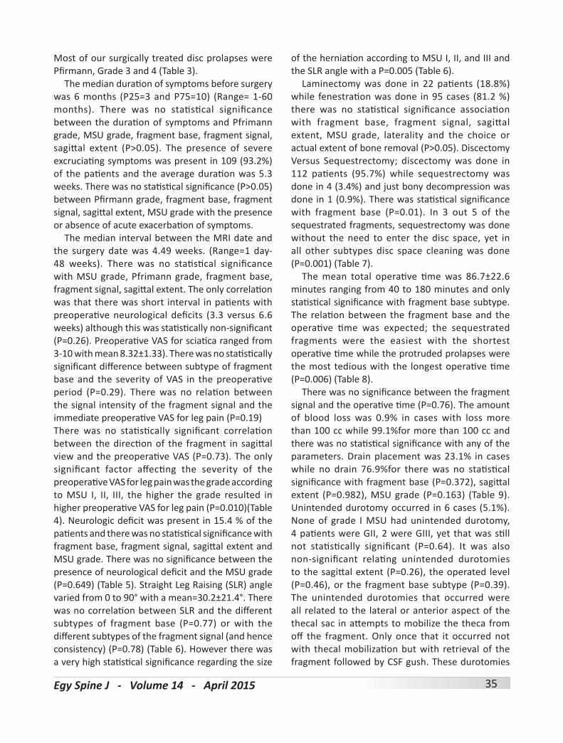
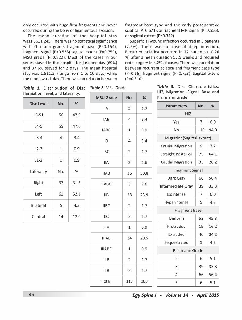
The HIZ denoting annular fissure was present in 7 (6 %) cases (Table 3). We divided the disc prolapses into 3 groups according to the direction of the prolapse, Caudal migration (28.2%) are occasionally impacted within the axilla of the traversing root. Straight posterior discs were most common (64.1%) (Table 3, Figure 1). We divided the fragment signal into four types (Figure 2).Most of the fragments were dark gray (66 patients (56.4%) (Table 3, Figure 2). Fragment base was uniform in 53 patients (45.3%)(Table 3, Figure 3).
35Egy Spine J - Volume 14 - April 2015
Most of our surgically treated disc prolapses were Pfirmann, Grade 3 and 4 (Table 3).
The median duration of symptoms before surgery was 6 months (P25=3 and P75=10) (Range= 1-60 months). There was no statistical significance between the duration of symptoms and Pfrimann grade, MSU grade, fragment base, fragment signal, sagittal extent (P>0.05). The presence of severe excruciating symptoms was present in 109 (93.2%) of the patients and the average duration was 5.3 weeks. There was no statistical significance (P>0.05) between Pfirmann grade, fragment base, fragment signal, sagittal extent, MSU grade with the presence or absence of acute exacerbation of symptoms.
The median interval between the MRI date and the surgery date was 4.49 weeks. (Range=1 day-48 weeks). There was no statistical significance with MSU grade, Pfrimann grade, fragment base, fragment signal, sagittal extent. The only correlation was that there was short interval in patients with preoperative neurological deficits (3.3 versus 6.6 weeks) although this was statistically non-significant (P=0.26). Preoperative VAS for sciatica ranged from 3-10 with mean 8.32±1.33). There was no statistically significant difference between subtype of fragment base and the severity of VAS in the preoperative period (P=0.29). There was no relation between the signal intensity of the fragment signal and the immediate preoperative VAS for leg pain (P=0.19)There was no statistically significant correlation between the direction of the fragment in sagittal view and the preoperative VAS (P=0.73). The only significant factor affecting the severity of the preoperative VAS for leg pain was the grade according to MSU I, II, III, the higher the grade resulted in higher preoperative VAS for leg pain (P=0.010)(Table 4). Neurologic deficit was present in 15.4 % of the patients and there was no statistical significance with fragment base, fragment signal, sagittal extent and MSU grade. There was no significance between the presence of neurological deficit and the MSU grade (P=0.649) (Table 5). Straight Leg Raising (SLR) angle varied from 0 to 90° with a mean=30.2±21.4°. There was no correlation between SLR and the different subtypes of fragment base (P=0.77) or with the different subtypes of the fragment signal (and hence consistency) (P=0.78) (Table 6). However there was a very high statistical significance regarding the size
of the herniation according to MSU I, II, and III and the SLR angle with a P=0.005 (Table 6).
Laminectomy was done in 22 patients (18.8%) while fenestration was done in 95 cases (81.2 %) there was no statistical significance association with fragment base, fragment signal, sagittal extent, MSU grade, laterality and the choice or actual extent of bone removal (P>0.05). Discectomy Versus Sequestrectomy; discectomy was done in 112 patients (95.7%) while sequestrectomy was done in 4 (3.4%) and just bony decompression was done in 1 (0.9%). There was statistical significance with fragment base (P=0.01). In 3 out 5 of the sequestrated fragments, sequestrectomy was done without the need to enter the disc space, yet in all other subtypes disc space cleaning was done (P=0.001) (Table 7).
The mean total operative time was 86.7±22.6 minutes ranging from 40 to 180 minutes and only statistical significance with fragment base subtype. The relation between the fragment base and the operative time was expected; the sequestrated fragments were the easiest with the shortest operative time while the protruded prolapses were the most tedious with the longest operative time (P=0.006) (Table 8).
There was no significance between the fragment signal and the operative time (P=0.76). The amount of blood loss was 0.9% in cases with loss more than 100 cc while 99.1%for more than 100 cc and there was no statistical significance with any of the parameters. Drain placement was 23.1% in cases while no drain 76.9%for there was no statistical significance with fragment base (P=0.372), sagittal extent (P=0.982), MSU grade (P=0.163) (Table 9). Unintended durotomy occurred in 6 cases (5.1%). None of grade I MSU had unintended durotomy, 4 patients were GII, 2 were GIII, yet that was still not statistically significant (P=0.64). It was also non-significant relating unintended durotomies to the sagittal extent (P=0.26), the operated level (P=0.46), or the fragment base subtype (P=0.39). The unintended durotomies that occurred were all related to the lateral or anterior aspect of the thecal sac in attempts to mobilize the theca from off the fragment. Only once that it occurred not with thecal mobilization but with retrieval of the fragment followed by CSF gush. These durotomies
36 Egy Spine J - Volume 14 - April 2015
only occurred with huge firm fragments and never occurred during the bony or ligamentous excision.
The mean duration of the hospital stay was1.56±1.245. There was no statistical significance with Pfirmann grade, fragment base (P=0.164), fragment signal (P=0.533) sagittal extent (P=0.759), MSU grade (P=0.822). Most of the cases in our series stayed in the hospital for just one day (69%) and 37.6% stayed for 2 days. The mean hospital stay was 1.5±1.2, (range from 1 to 10 days) while the mode was 1 day. There was no relation between
fragment base type and the early postoperative sciatica (P=0.671), or fragment MRI signal (P=0.556), or sagittal extent (P=0.352)
Superficial wound infection occurred in 3 patients (2.6%). There was no case of deep infection. Recurrent sciatica occurred in 12 patients (10.26 %) after a mean duration 57.5 weeks and required redo surgery in 4.2% of cases. There was no relation between recurrent sciatica and fragment base type (P=0.66), fragment signal (P=0.723), Sagittal extent (P=0.310).
Table 1. Distribution of Disc Herniation: level, and laterality.
Disc Level No. %
L5-S1 56 47.9
L4-5 55 47.0
L3-4 4 3.4
L2-3 1 0.9
L1-2 1 0.9
Laterality No. %
Right 37 31.6
Left 61 52.1
Bilateral 5 4.3
Central 14 12.0
Table 2. MSU Grade.
MSU Grade No. %
IA 2 1.7
IAB 4 3.4
IABC 1 0.9
IB 4 3.4
IBC 2 1.7
IIA 3 2.6
IIAB 36 30.8
IIABC 3 2.6
IIB 28 23.9
IIBC 2 1.7
IIC 2 1.7
IIIA 1 0.9
IIIAB 24 20.5
IIIABC 1 0.9
IIIB 2 1.7
IIIB 2 1.7
Total 117 100
Table 3. Disc Characteristics: HIZ, Migration, Signal, Base and Pfirrmann Grade.
Parameters No. %
HIZ
Yes 7 6.0
No 110 94.0
Migration(Sagittal extent)
Cranial Migration 9 7.7
Straight Posterior 75 64.1
Caudal Migration 33 28.2
Fragment Signal
Dark Gray 66 56.4
Intermediate Gray 39 33.3
Isointense 7 6.0
Hyperintense 5 4.3
Fragment Base
Uniform 53 45.3
Protruded 19 16.2
Extruded 40 34.2
Sequestrated 5 4.3
Pfirrmann Grade
2 6 5.1
3 39 33.3
4 66 56.4
5 6 5.1

37Egy Spine J - Volume 14 - April 2015
Table 4. Relationship between Severity of Sciatica with Fragment Type, Fragment Signal, Sagittal Extent & MSU Grade
Variable No. Mean±SD 95% CI(Lower-Upper) Min-Max P-Value
Fragment Type
Uniform 53 8.2±1.3 7.9-8.6 5-10
0.29Protruded 19 8.4±1.1 7.9-9.0 7-10
Extruded 40 8.3±1.5 7.8-8.8 3-10
Sequestrated 5 9.4±0.9 8.3-10.5 8-10
Fragment signal
Dark Gray 66 8.4±1.4 8.0-8.7 3-10
0.185Intermediate Gray 39 8.1±1.3 7.7-8.5 6-10
Isointense 7 9.3±0.8 8.6-10 8-10
Hyperintense 5 8.2±0.8 7.2-9.2 7-9
Sagittal Extent
Posterior-Cranial Migration 9 8.0±2.1 6.4-9.6 3-10
0.728Straight Posterior 75 8.4±1.2 8.1-8.7 5-10
Posterior-Cranial Migration 33 8.3±1.3 7.8-8.8 6-10
MSU Grade
I 13 7.3±1.3 6.5-8.1 5-10
0.01II 75 8.4±1.2 8.1-8.7 6-10
III 29 8.6±1.5 8.0-9.2 3-10
Table 5. Relationship between Neurologic Deficit and MSU Grade
MSU GradeNeurological Deficit
P-ValueYes no
I 1 12
0.649II 13 62
III 4 25

38 Egy Spine J - Volume 14 - April 2015
Table 6. Relationship between Straight Leg Raising Angle with fragment type, fragment signal, sagittal extent & MSU Grade
Variable No. Mean±SD 95% CI (Lower-Upper) Min-Max P-Value
Fragment Type
Uniform 53 31.9±21.6 24.0-35.0 0-90
0.770Protruded 19 31.1±17.4 26.1-39.1 5-70
Extruded 40 28.3±22.7 4.3-54.3 5-80
Sequestrated 5 24±20 9.4-36.6 5-30
Fragment Signal
Dark Gray 66 29.5±22.3 8.0-8.7 3-10
0.774Intermediate Gray 39 32.6±20.1 7.7-8.5 5-10
Isointense 7 29.3±27.0 8.6-10 8-10
Hyperintense 5 23±11.0 7.2-9.2 7-9
Sagittal Extent
Posterior-Cranial Migration 9 31.7±27.4 10-52.7 10-80
0.564Straight Posterior 75 31.5±21.0 26-36.4 0-90
Posterior-Cranial Migration 33 26.8±20.0 19.7-33.9 5-70
MSU Grade
I 13 43.5±13.8 35.2-51.8 30-60
0.005II 75 31.3±20.8 26.6-36.1 0-90
III 29 21.4±22.6 812.8-30.0 0-80
Table 7. Discectomy versus Sequestrectomy and Fragment Base
Fragment Base Bony Decompression Discectomy Sequestrectomy
Uniform 0 53 0
Protrusion 1 18 0
Extrusion 0 39 1
Sequestration 0 2 3

39Egy Spine J - Volume 14 - April 2015
Table 8. Relationship between Operative Time with fragment type & fragment signal.
No. Mean±SD Minimum Maximum P-Value
Fragment Base
Uniform 53 80.0±15.3 40.0 120.0
Protrusion 19 95.3±30.7 60.0 180.0 0.006
Extrusion 40 93.0±24.2 60.0 180.0
Sequestration 5 74.0±15.2 60.0 90.0
Fragment Intensity
Dark Gray 66 88.3±26.0 40.0 180.0
Intermediate Gray 39 83.5±17.0 60.0 120.0
Isointense 7 88.6±19.3 70.0 120.0 0.762
Hyperintense 5 88.0±18.9 75.0 120.0
Table 9. Drain Placement, Unintended Durotomy, and Reoperations
No. %
Placement Of Drain
Yes 27 23.1
No 90 76.9
Unintended Durotomy
Yes 6 5.1
No 111 94.9
Reoperation
Yes 5 4.3
No 112 95.7
Figure 1. Migration (Sagittal Extent): Caudal (A), Straight Posterior (B) & Cranial (C).
A CB
40 Egy Spine J - Volume 14 - April 2015
Figure 2. Fragment Signal: Dark Gray (A), Intermediate Gray (B), Isointense (C) and Hyperintense (D)
Figure 3. Fragment Base: Sequestrated (A), Extruded (B), Protruded (C) & Uniform (D)
Figure 4. A:raised cranially and caudally creeping edges of the offending herniation/ B: After annulotomy the hard raised edges are still protruding and offending the nerve root ( red) C: The raised edges are removed ( green circles) D: The nerve root ( red ) is no more offended by disc material in its path to the foramen.
A
A
A
C
C
C
D
D
D
B
B
B
41Egy Spine J - Volume 14 - April 2015
DiscussionThe effect of disc prolapse depends on the location
and extent of the herniation relative to the diameter of the spinal canal.5,18 The duration of the symptoms with long standing prolapse offers more difficult fragment excision due to the adhesions between the fragment and the overlying dura and due to the change in the nature of prolapsed fragment edges from soft consistency to osteo-cartilagenous nature. In our cohort, there was no statistical association between the duration of symptoms and the difficulty in surgery reflected as operative time, bleeding, occurrence of operative complications as incidental durotomy (P=0.75); refuting the misconception that the prolonged duration of symptoms prior to surgery would increase the adhesions between the thecal sac and the herniated fragment and increase the incidence of dural injury during mobilization of the root. This suggests that the severity and early clinical course of the leg symptoms may be correlated with the local condition of the herniated nucleus pulposus (HNP) and the nerve root, such as the relative volume of the HNP in the spinal canal, the location of the HNP relative to the nerve root, or the shape of the lateral recess.10
In some selected cases where the patients were not responding to medical treatment with severe unbearable symptoms with a huge extruded disc, it would be justifiable to operate early and not subject the patient to a deemed failed medical attempt. This would definitely not be the case in smaller GII or GI discs were a trial of full medical treatment was warranted.
Direction of the fragment: Bonneville and Wiltse proposed different methods to classify, according to location, the position of disc fragments that have migrated in the horizontal or sagittal plane.8,22 Caudally migrated fragments seem to be more impacted within the root axilla, might be more difficult to remove as the root is tented and fixed on the fragment. Thus, the initial root mobilization is not only difficult but extremely hazardous, and therefore initial generous foraminotomy should be completed before attempting to mobilize the root or address the fragment. After the fragment is removed, completion of the foraminotomy might be needed as initially the compressed root within the foramen might not allow safe and easy introduction of the
Kerrison rongeurs. Posteriorly directed fragments: with no cranio-caudal inclination seem to be the easiest as they are directly within the field especially in L5-S1 discs due to the wide interlaminar space, long root and the anterior orientation of the S1 foramen rather than a laterally placed L5 and above foramina. That is why a cranial migrated fragment might need extension of the fenestration especially in L4-5 discs and above as the interlaminar space is at a level below the disc space. Therefore, if the cranially migrating fragment is hard or adherent and can’t be squeezed out then a more generous fenestration should be done.
Regarding medio-lateral position of the fragment: “B” position MSU14 have been the easiest immediately under the dural edge without the need for theca retraction, squeezing of the posterior longitudinal ligament or subannular decompression of the disc material or even some times extended medial annulutomy in hard raised annuli in central discs(A Position). The real problem is with Position C discs (foraminal and far lateral where the decompression of the exiting root is important but with preservation of facet integrity with no extension of the fenestration either laterally or superiorly. So it is very important to preoperatively estimate the fragment consistency and ease of excision because we can only hope for a soft loose fragment that will be easily delivered otherwise it might be mandatory to sacrifice the facet.
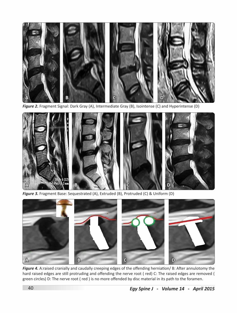
The configuration of the fragment differed in difficulty ranging from “easy” to “difficult”. The “sequestrated” fragment is easiest followed by the pediculated narrow based extrusion” fragment, then by the “uniform based” fragment, and finally the most difficult is the “sessile broad based” prolapse, where the base of the fragment is spreading over the upper and lower posterior vertebral bodies (Spondylosis Osteochondrans) (Figure 4). The point that is of surgical consideration is that performing a discectomy by a simple annulotomy and evacuating the fragment and the loose disc material leaves the described hard raised edges of the annulus in place. This keeps the offending margins raising, tenting and stretching the nerve root regardless of the evacuated central content and therefore requires further excision of these edges using the hard sharp-biting disc rongeurs in a direction 90 perpendicular to the disc space, providing a corridor in the path

42 Egy Spine J - Volume 14 - April 2015
of the nerve root with a few millimeters wider than the diameter of the nerve root to allow space for mobility.
The size of the fragment: A scheme to define the degree of canal compromise produced by disc displacement should be practical, objective, reasonably precise, and clinically relevant as suggested by Fardon et al,4 In 2004 Pfirmann16 graded the herniated discs according to the degree of root compression in “no compromise”, “just contact”, “root deviation” and “root compression” and correlated this with the intraoperative findings and found good correlation between the MRI grading and the intraoperative findings. In our study according to this grading all but 2 of our patients were GIII (root compression).
In our attempt to categorize the lumbar disc prolapse we didn’t address the small confusing “bulges” or annular relaxations” nor did we operate on small herniations that were just touching or mildly displacing the roots according to Pfirmann16 but we subdivided the more significant herniations, uniform protrusions, broad base pointing protrusions, extrusions and sequestrations , extrusions according to the morphology of the prolapsed portion in attempt to find if there is any preoperative clinical, surgical or postoperative significance.
The Michigan State University (MSU) classification14 separated lumbar disc disease into different zones based purely on location. The authors theorized that the location of lumbar discs had a significant effect on symptomatology. Prospectively they applied their classification scheme to 100 patients and performed microdiscectomies on only those in Zones 2 and 3 (larger and more extensive). Their surgical success correlated with patient selection based solely on the grading scale (all patients with Size 1 lesions were excluded from surgical consideration) and their surgical results showed 90% to 96% and 80% to 84% good to excellent outcomes on Oswestry Disability Index at 1- and 5-year follow-up, respectively. MSU classification is a reliable method to objectively classify significant lumbar disc disease and can serve as an adjunct to patient selection for single-level discectomy.14 In fact, larger disc fragments with more pronounced compres¬sion of the thecal sac are another predictor of failure to respond to conservative management.13
In our cohort, higher grades of prolapses
according to MSU (i.e. the size of the fragment) correlated with a higher preoperative VAS and smaller angle for straight leg raising. According to MSU grading for degree of prolapse we attempted to correlated between the size of the fragment and the ease of surgery, we got the impression that the larger the fragmented the initial difficult is its release from the root but once delivered (if possible) the room and displacement caused by the size of the fragment gives space for navigating around the root. In very large fragment where there is no space for delivery with severe root compression, more lateral annulotomy with subannular decompression and possible fragment debulking may provide some cleavage between the fragment and the dura and then would allow easier delivery.
Composition of the fragment: Composition of the displaced material may be characterized by terms such as nuclear, cartilaginous, bony, calcified, ossified, collagenous, scarred, desiccated, gaseous, or liquefied. Clinical significance related to the observation of volume and composition depends on the correlation with clinical data and cannot be inferred from morphologic data alone.5 MRI signal characteristics may, on rare occasion, allow differentiation of acute and chronic disc herniations.4,12 In such cases, acutely herniated disc material may appear brighter than the disc of origin on T2-weighted sequences. 4,15 The signal intensity of the prolapsed fragment in T2 gives an idea about the consistency of the fragment with the least water content being the hardest, yet carries no extra difficult unless being in the “mushroom” prolapse. A hyperintense on T2 MRI sequestrated fragment can easily be squeezed from under the annulus and the posterior longitudinal ligament, with a relatively significantly shorter duration of surgery while hypointense hard fragment especially with raised osteo-cartilagenous edges needs more tedious decompression.
Is there a need for laminectomy rather than a fenestration in large fragments? If huge massive fragments that are located unilaterally; we believe that it can be adequately done through the fenestration without the need for further bone removal. Sublaminar flavectomy might be done to allow retraction and better visualization; however, in large fragments the retraction is even more limited than the smaller fragments. A laminectomy might

43Egy Spine J - Volume 14 - April 2015
be needed in huge disc that is spreading bilaterally even if the symptoms are unilateral in attempt to probe the contralateral hump and feel its relation to the root.
ConclusionFrom a surgical perspective, not all herniations
are similar. Many different radiological classifications are present to describe the herniated lumbar disc, the degree of spinal canal and neural structure violation, but there are still no comprehensive studies on the clinical and surgical significance of these classifications and nomenclatures. During our first attempt to clinically correlate some of these classifications we found that GII and GIII herniations on the MSU grading correlated with the preoperative severity of radicular pain and with the SLR angle, but had no intraoperative or post-operative significance.
We also found a surgical significance between different types of shape and signal intensity of the herniated portion with the broad based osteocartilageous hypointense herniation being the most difficult and time consuming during surgery. We believe that our study would lead to the start of a formulation of a more surgical significant classification scheme based on the fragment morphology on the MRI combined different already present classifications. It would be useful and applicable for surgeons to anticipate the degree of difficulty of surgery and the plan required for adequate nerve root decompression.
We believe that more prospective studies need to be done preferably complemented by 3- 6 months post-operative MRI scanning with post-operative clinical correlation aided by functional outcome indices to get a better understanding of the impact of different nomenclatures and classification on clinical and surgical course.
References1. Alexander Aichmair, Jerry Y Du, Jennifer
Shue, Gisberto Evangelisti, Andrew A Sama, Alexander P Hughes, et al. “Microdiscectomy for the Treatment of Lumbar Disc Herniation: An Evaluation of Reoperations and Long-Term Outcomes.” Evidence-based spine-care journal 5(2):77–86, 2014
2. Boos N, Dreier D, Hilfiker E, Schade V, Kreis R, Hora J, Aebi M, BoeschC “Tissue Characterization of Symptomatic and Asymptomatic Disc
Herniations by Quantitative Magnetic Resonance Imaging.” J Orthop Res 15(1):141–149, 1997
3. Brant-Zawadzki MN, Jensen MC, Obuchowski N, Ross JS, Modic MT. Interobserver and intraobserver variability in interpretation of lumbar disc abnormalities. A comparison of two nomenclatures (discussion 1264) Spine 1995:20(11):1257–1263
4. Brock M, S Patt, H-M Mayer: “The Form and Structure of the Extruded Disc.” Spine 17(12):1457–1461, 1992
5. David F Fardon, Alan L Williams, Edward J Dohring, F Reed Murtagh, Stephen L Gabriel Rothman, Gordon K Sze: “Lumbar Disc Nomenclature: Version 2.0.” The Spine Journal 14(11):2525–2545, 2014
6. German JW, Adamo MA, Hoppenot RG Blossom JH, Nagle HA: “Perioperative Results Following Lumbar Discectomy: Comparison of Minimally Invasive Discectomy and Standard Microdiscectomy.” Neurosurgical focus 25(2):E20, (2008)
7. Goald HJ: Surgical Technique. “Microlumbar Discectomy: Follow-up of 477 Patients.” Journal of microsurgery 2(2):95–100, 1980
8. Jean-François Bonneville: “Plaidoyer Pour Une Classification Par L’image Des Hernies Discales Lombaires: La Carte-Image.” Rev Im Med 2:557–60, 1990
9. Kieffer SA, Stadlan EM, Mohandas A, Peterson HO: “Discographic-Anatomical Correlation of Developmental Changes with Age in the Intervertebral Disc.” Acta Radiologica: Diagnosis 9:733–9, 1969
10. Komori H: “Factors Predicting the Prognosis of Lumbar Radiculopathy due to Disc Herniation.” Journal of orthopaedic science : official journal of the Japanese Orthopaedic Association 7(1):56–61, 2002
11. Li Y, Vance F, Daniel KR: “How Should We Grade Lumbar Disc Herniation and Nerve Root Compression? A Systematic Review.” Clinical Orthopaedics and Related Research® 473(6):1896–1902, 2015
12. Milette PC, Fontaine S, Lepanto L, Cardinal E, Breton G: “Differentiating Lumbar Disc Protrusions, Disc Bulges, and Discs with Normal Contour but Abnormal Signal Intensity. Magnetic Resonance Imaging with Discographic
44 Egy Spine J - Volume 14 - April 2015
Correlations.” Spine 24(1):44–53, 199913. Motiei-Langroudi R, Homa S, Amir SS: “Clinical
and Magnetic Resonance Imaging Factors Which May Predict the Need for Surgery in Lumbar Disc Herniation.” Asian spine journal 8(4):446–52, 2014
14. Mysliwiec LW, Cholewicki J, Winkelpleck MD, Eis GP: “MSU Classification for Herniated Lumbar Discs on MRI: Toward Developing Objective Criteria for Surgical Selection.” European spine journal 19(7):1087–93, 2010
15. Pfirrmann CW, Metzdorf A, Zanetti M, Hodler J, Boos N: “Magnetic Resonance Classification of Lumbar Intervertebral Disc Degeneration.” Spine 26(17):1873–8, 2001
16. Pfirrmann CWA, Dora C, Schmid MR, Zanetti M, Hodler J, Boos M: “MR Image-Based Grading of Lumbar Nerve Root Compromise due to Disk Herniation: Reliability Study with Surgical Correlation.” Radiology 230(2):583–8, 2004
17. Porchet F, Bartanusz V, Kleinstueck FS, Lattig F, Jeszenszky D, Grob D, Mannion AF: “Microdiscectomy Compared with Standard
Discectomy: An Old Problem Revisited with New Outcome Measures within the Framework of a Spine Surgical Registry.” European Spine Journal 18(3):360–366, 2009
18. Porter RW, Hibbert CS, Wicks M: “The Spinal Canal in Symptomatic Lumbar Disc Lesions.” J Bone Joint Surg Br 60(4): 485–487, (1978)
19. Pritzker KP: “Aging and Degeneration in the Lumbar Intervertebral Disc.” Orthop Clin North Am 8(1):66–77, 1977
20. Quencer RM: “ The Abnormal Annulus Fibrosus: Can We Infer the Acuteness of an Annular Injury?” AJNR. American journal of neuroradiology 23(7):1069, 2002
21. Soliman J, Harvey A, Howes G, Seibly J, Dossey J, Nardone E: “Limited Microdiscectomy for Lumbar Disk Herniation: A Retrospective Long-Term Outcome Analysis.” Journal of spinal disorders & techniques 27(1):E8–E13, 2014
22. Wiltse LL, Berger PE, McCulloch JA: “A System for Reporting the Size and Location of Lesions in the Spine.” Spine 22(13):1534–7, 1997
Ahmed Abou-Zeid, MDDepartment of Neurosurgery, Ain Shams University, Cairo, EgyptEmail: [email protected]
Address reprintrequest to:
45Egy Spine J - Volume 14 - April 2015
العلاقـة بيـن الصـورة الأكلينيكيـة و المشـاهدة الجراحيـة وبيـن صـورة الرنيـن المغناطيسـي فـي حـالات الغضـروف القطني المنزلق
البيانـات الخلفيـة: تعـد جراحـة اسـتئصال الغضـروف القطنـي المنزلـق مـن أكثـر جراحـات العمـود الفقـري شـيوعا و يمثـل الرنين المغناطيسي حجر الزاوية في تقييم درجة و شدة الانزلاق الغضروفي كما يوضح مكان واتجاه ودرجة الانزلاق مع كثرة التقسيمات والتسميات المختلفة لشكل الانزلاق الغضروفي في صورة أشعة الرنين المغناطيسي والتي تعبر عن توصيف دقيق للصورة لا يزال الربط ضعيفا بين صورة الأشـعة والفحص الأكلينيكي والمشـاهدات أثناء الجراحة وبالتالي يصعـب التنبـؤ بصعوبـة الجراحـة أثنـاء اسـتئصال الغضـروف لرفـع الضغـط عـن جذور الأعصـاب، إضافة إلى ذلـك يصعب جدا توقع حالة المريض في فترة النقاهة بعد الجراحة من حيث الألم والقدرة على الحركة والمضاعفات بناءا على التسميات
والتقسيمات المتاحة لأشعة الرنين المغناطيسي.
الغرض: دراسة العلاقة بين البيانات السريرية والجراحية والأشعية في حالات الغضروف القطني.
تصميم الدراسة: دراسه سريريه تحليليه بأثر رجعي.
الطـرق و المرضـي: قمنـا فـي هـذا البحـث بتطبيـق قواعد التقسـيمات والتوصيفات المعتمدة عالميا على مائة وتسـعة وتسعين مريضا أجري لهم جراحة ميكروسكوبية لاستئصال الغضروف القطني المنزلق وحاولنا إيجاد العلاقة المفتقدة بيـن شـكل ودرجـة الانـزلاق فـي صـورة الأشـعة وبيـن الحالـة الأكلينيكيـة قبل وبعـد الجراحة و أيضا حاولنـا إيجاد علاقة بين الصورة الإشعاعية و مدى سلاسة أو صعوبة الجراحة. استخدمنا في البحث الأشعات التي أجريت للمرضى قبل الجراحة إضافـة إلـى الفحـص الأكلينيكـي ممثـلا فـي درجـة الألـم و وجـود )أو عـدم وجـود( ضعـف فـي أحـد الطرفييـن السـفليين أو كليهما أو صعوبة في التحكم في الإخراج واستخدمنا أيضا بيانات الجراحة ممثلة في طول الجرح و كمية النزيف و كمية الغضـروف المسـتأصل و مـدة البقـاء فـي المستشـفى بالإضافـة لدرجـة التحسـن والمضاعفـات بمـا فـي ذلـك قطـع الأم
الجافية و ارتجاع الغضروف والحاجة لإجراء جراحات أخرى خلال العام الأول بعد الجراحة.
النتائـج: وجدنـا فـي البحـث قصـور التوصيفات والتسـميات المتاحة وعدم ارتباطها الوثيـق بالملاحظات أثناء الجراحة فيما عدا العلاقة الدالة إحصائيا بين حجم الغضروف في مقياس )MSU( و درجة الألم التي يشعر بها المريض وأيضا محدودية
رفع الساق بشكل مستقيم
الاستنتاج: نستنتج من البحث الحاجة لإيجاد توصيف و تقسيم جديد للغضروف القطني المنزلق ينصب اهتمامه بشكل أكبـر علـى الجانـب الجراحـي مـن المـرض و يسـاعد جـراح العمـود الفقـري علـى التخطيـط الجيـد للجراحـة و التنبـؤ بالنتيجـة
الجراحية
الملخص العربي