mri of the psoas major muscle: origin, attachments ... psoas major arrs 2016.pdf · mri of the...
TRANSCRIPT
MRI OF THE PSOAS MAJOR MUSCLE: ORIGIN, ATTACHMENTS, ANATOMICAL VARIANTS AND CORRELATION WITH THE LUMBAR DISC EXTRUSION
APARNA KAKARALA (1), ARNE S BORTHNE (2), CLAUDE PIERRE-JEROME (2)
EMORY UNIVERSITY SCHOOL OF MEDICINE, ATLANTA GA USA (1)
AKERSHUS UNIVERSITY HOSPITAL, OSLO NORWAY (2)

DISCLOSURE OF COMMERCIAL INTEREST
• Neither I nor the co-authors have a financial relationship with a commercial organization
that may have a direct or indirect interest in the content of this presentation
INTRODUCTION: ANATOMY AND BIOMECHANICS
The Psoas Major muscle is a flexor and a stabilizer
It plays an important role in:
Motion and stabilization of the lumbar spine
Flexion of the Hip
INTRODUCTION: ANATOMY
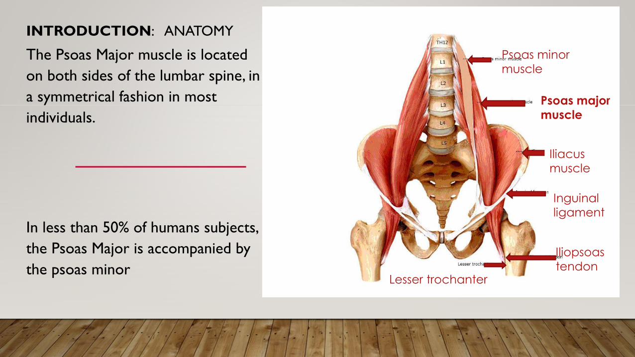
The Psoas Major muscle is located
on both sides of the lumbar spine, in
a symmetrical fashion in most
individuals.
In less than 50% of humans subjects,
the Psoas Major is accompanied by
the psoas minor
Psoas minor
muscle
Psoas major
muscle
Iliacus
muscle
Inguinal
ligament
Iliopsoas
tendonLesser trochanter
INTRODUCTION: ANATOMY
The infrastructure of the Psoas Major muscle is made of a series of
overlapping segmental fascicles.
Each fascicle consists of bundles of fibers. These fascicles connect bilaterally
with:
- the lumbar intervertebral discs
- the transverse processes
- and the vertebral bodies
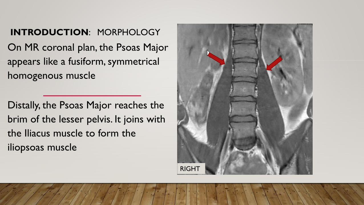
INTRODUCTION: MORPHOLOGY
On MR coronal plan, the Psoas Major
appears like a fusiform, symmetrical
homogenous muscle
Distally, the Psoas Major reaches the
brim of the lesser pelvis. It joins with
the Iliacus muscle to form the
iliopsoas muscle
RIGHT
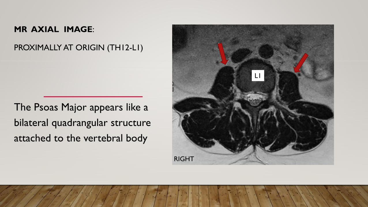
MR AXIAL IMAGE:
PROXIMALLY AT ORIGIN (TH12-L1)
The Psoas Major appears like a
bilateral quadrangular structure
attached to the vertebral body
L1
RIGHT
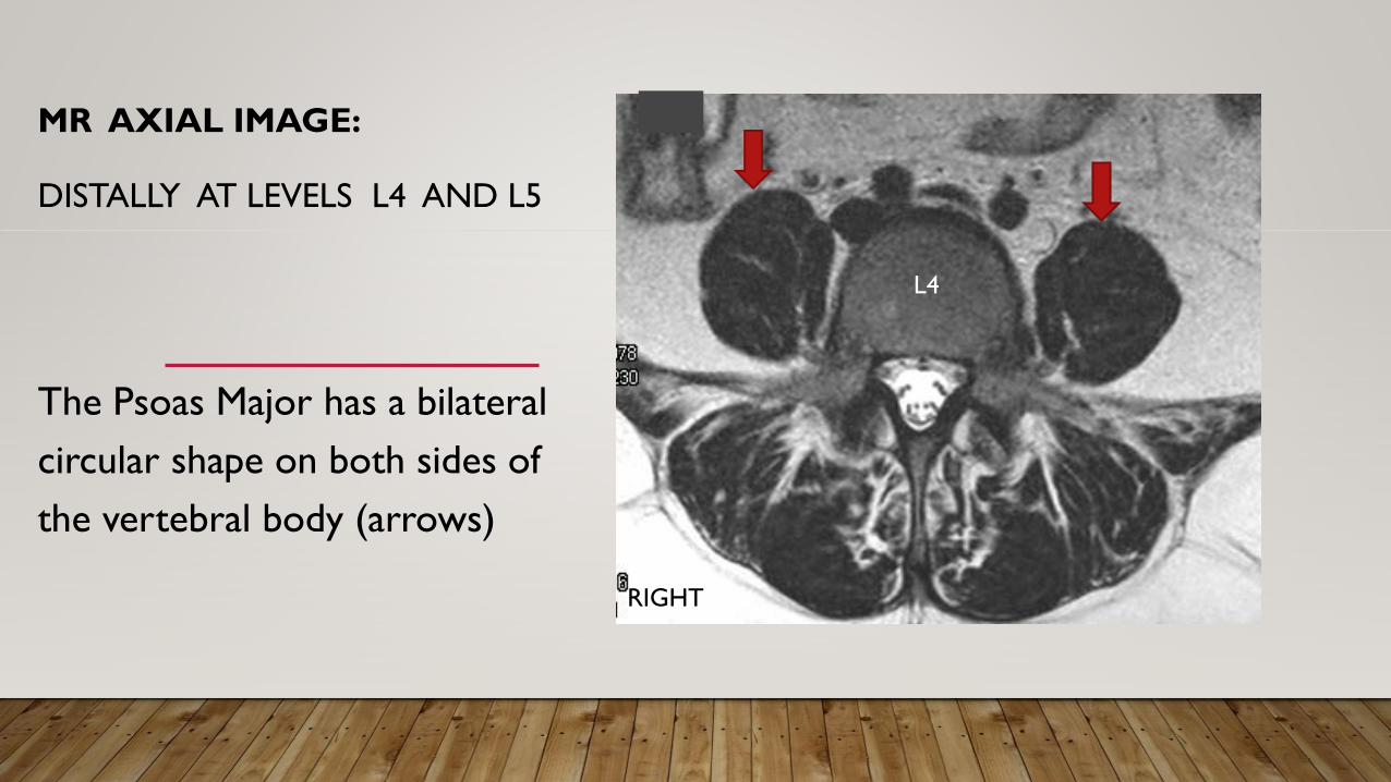
MR AXIAL IMAGE:
DISTALLY AT LEVELS L4 AND L5
The Psoas Major has a bilateral
circular shape on both sides of
the vertebral body (arrows)
L4
RIGHT
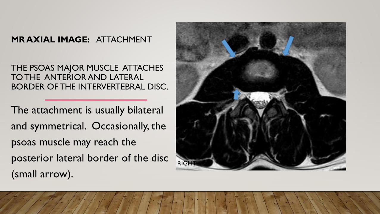
MR AXIAL IMAGE: ATTACHMENT
THE PSOAS MAJOR MUSCLE ATTACHES TO THE ANTERIOR AND LATERAL BORDER OF THE INTERVERTEBRAL DISC.
The attachment is usually bilateral
and symmetrical. Occasionally, the
psoas muscle may reach the
posterior lateral border of the disc
(small arrow).
RIGHTRIGHT
PURPOSES OF THE STUDY
1)To verify the anatomical origin of the psoas major muscle and its anatomical variants
2) To evaluate the attachments to the lumbar discs and the variants
3) To assess the prevalence of disc extrusion in individuals with absent of partial
attachment of the psoas to the lumbar discs.
MATERIALS
From two Institutions: Emory University Atlanta, GA – USA / Oslo University,
Norway
MR images of the Lumbar spine from the last 5 years were retrieved
Exclusion criteria:
acute trauma
acute inflammatory diseases
and neoplasia and metastases
Incomplete examination of the spine
MATERIALS
Population (after exclusion): 383 subjects
Gender: 213 females / 170 males
Age: mean age 60,3 years (range15 to 85years)
All had MR examinations of the lumbar spine in sagittal, axial and coronal
planes
METHODS - ANALYSIS OF IMAGES
1. The origin of the psoas major was assessed on the coronal and axial images
2. The attachments of the psoas major muscle to the:
vertebral bodies
transverse processes
and intervertebral discs
were evaluated on both axial and coronal images
METHODS –ANALYSIS OF IMAGES
We searched for:
a) The exact level of the origin of the psoas muscle
on both sides of the spine, above or below Th12-L1 level.
b) the symmetry of the origin of the muscle
METHODS –ANALYSIS OF IMAGES
c) Attachments of the psoas to the lumbar discs
at all levels until L5-S1
d) Symmetrical (bilateral) attachment of the Psoas
to the lumbar discs
METHODS –ANALYSIS OF IMAGES
e) Search for the presence of lumbar disc herniation
f) Co-existence of disc herniation and partial or total absence
of psoas attachment to the disc at each level
METHODS – DEFINITION OF VARIANTS
Variant of origin:
We considered as anatomical variants of the psoas major muscle origin,
when the muscle originated above or below the level of Th12-L1 disc
Variant of attachment:
When there is partial (unilateral) or total absent attachment of the psoas
muscle to the intervertebral disc.
METHODS - ANALYSIS OF IMAGES
The analysis of images was done independently by four readers:
two readers in Atlanta
and two readers in Oslo
Agreement was reached by consensus
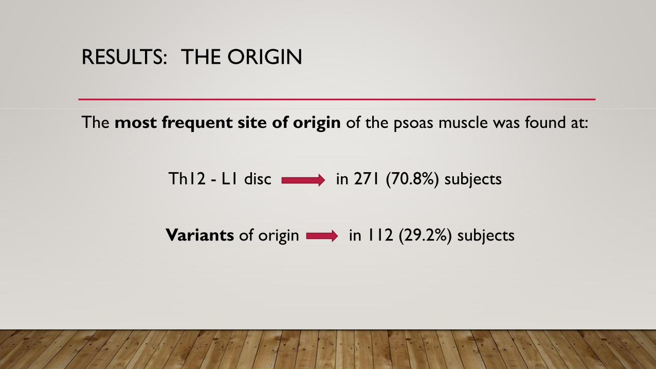
RESULTS: THE ORIGIN
The most frequent site of origin of the psoas muscle was found at:
Th12 - L1 disc in 271 (70.8%) subjects
Variants of origin in 112 (29.2%) subjects
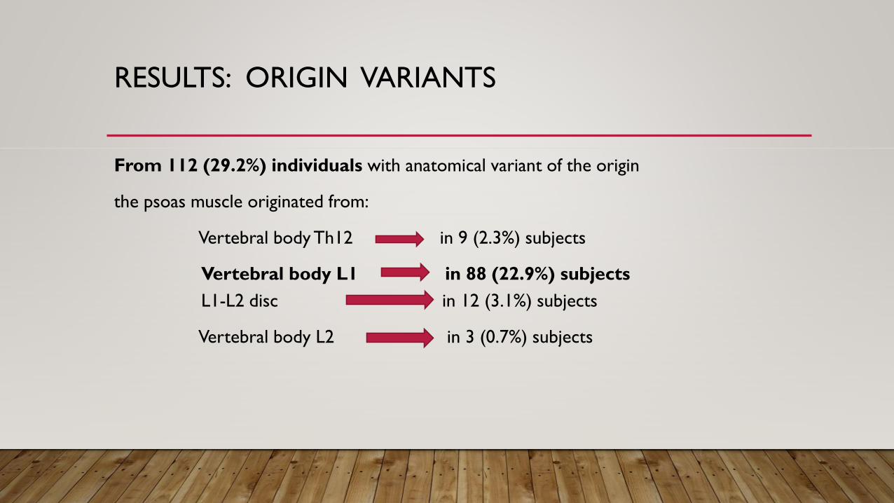
RESULTS: ORIGIN VARIANTS
From 112 (29.2%) individuals with anatomical variant of the origin
the psoas muscle originated from:
Vertebral body Th12 in 9 (2.3%) subjects
Vertebral body L1 in 88 (22.9%) subjects
L1-L2 disc in 12 (3.1%) subjects
Vertebral body L2 in 3 (0.7%) subjects
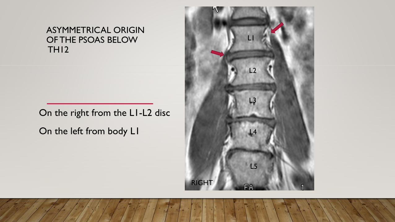
ASYMMETRICAL ORIGIN OF THE PSOAS BELOWTH12
On the right from the L1-L2 disc
On the left from body L1
RIGHT
L5
L4
L3
L2
L1
RESULTS: ORIGIN VARIANTS
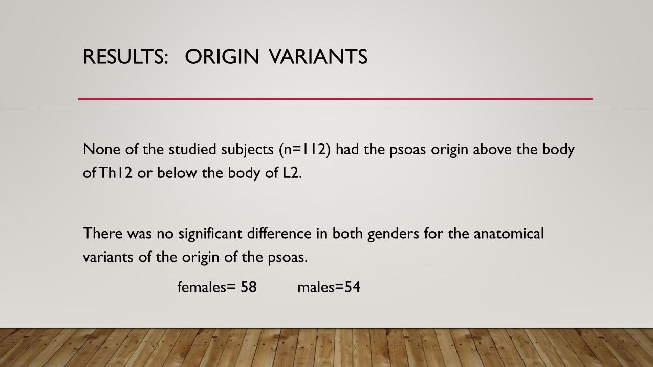
None of the studied subjects (n=112) had the psoas origin above the body
of Th12 or below the body of L2.
There was no significant difference in both genders for the anatomical
variants of the origin of the psoas.
females= 58 males=54
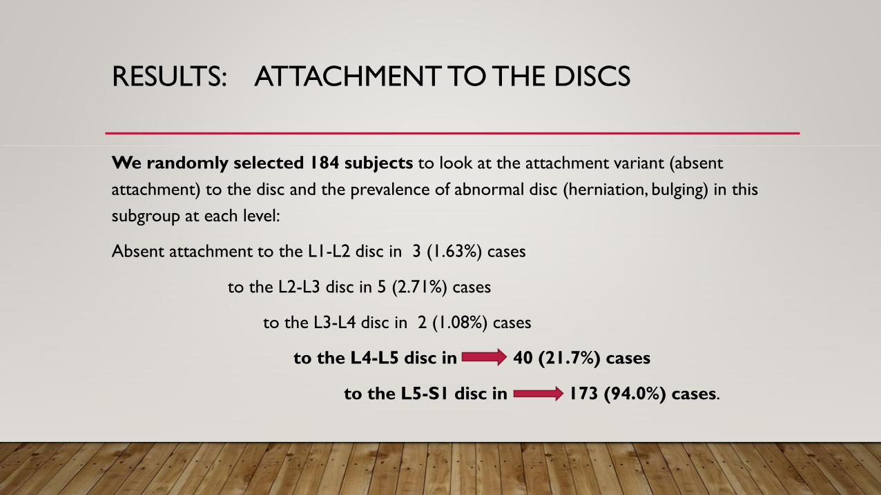
RESULTS: ATTACHMENT TO THE DISCS
We randomly selected 184 subjects to look at the attachment variant (absent
attachment) to the disc and the prevalence of abnormal disc (herniation, bulging) in this
subgroup at each level:
Absent attachment to the L1-L2 disc in 3 (1.63%) cases
to the L2-L3 disc in 5 (2.71%) cases
to the L3-L4 disc in 2 (1.08%) cases
to the L4-L5 disc in 40 (21.7%) cases
to the L5-S1 disc in 173 (94.0%) cases.
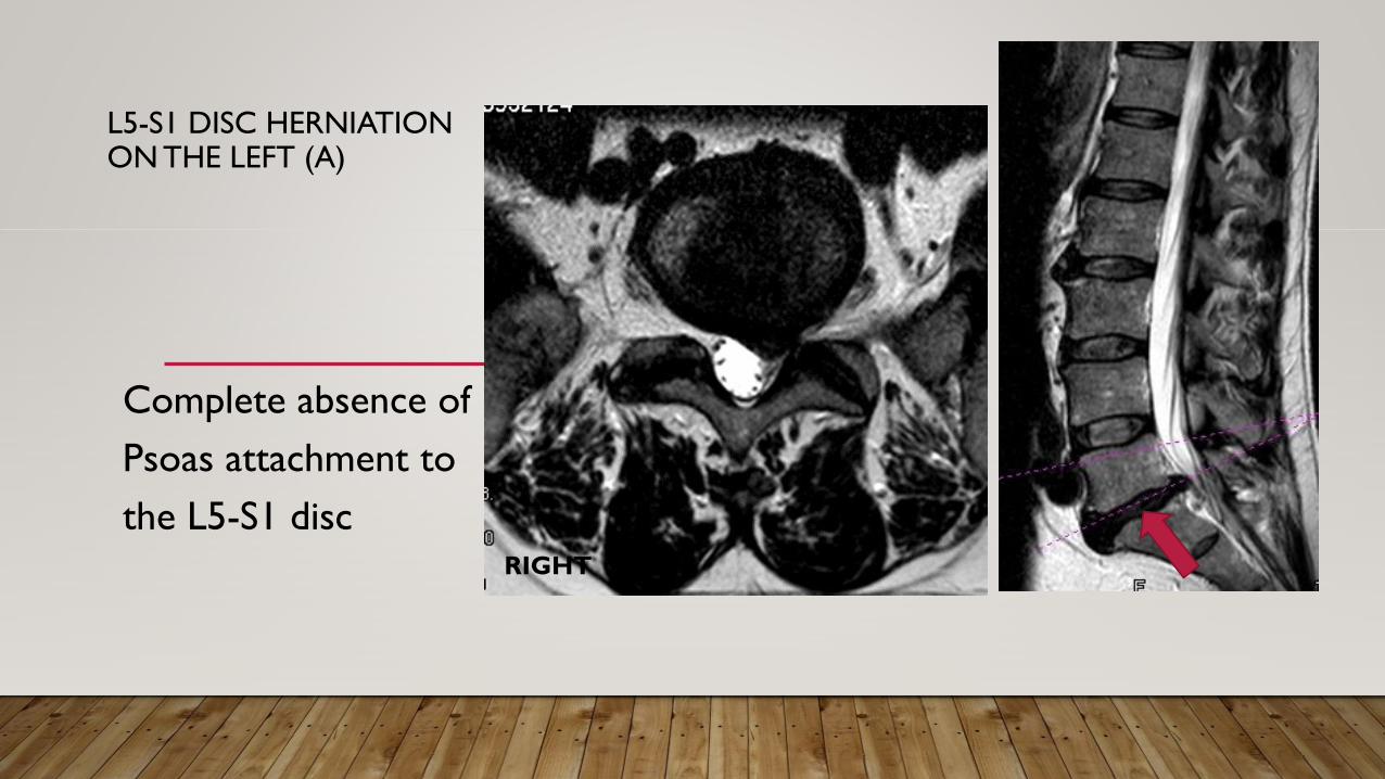
L5-S1 DISC HERNIATION ON THE LEFT (A)
Complete absence of
Psoas attachment to
the L5-S1 discRIGHT
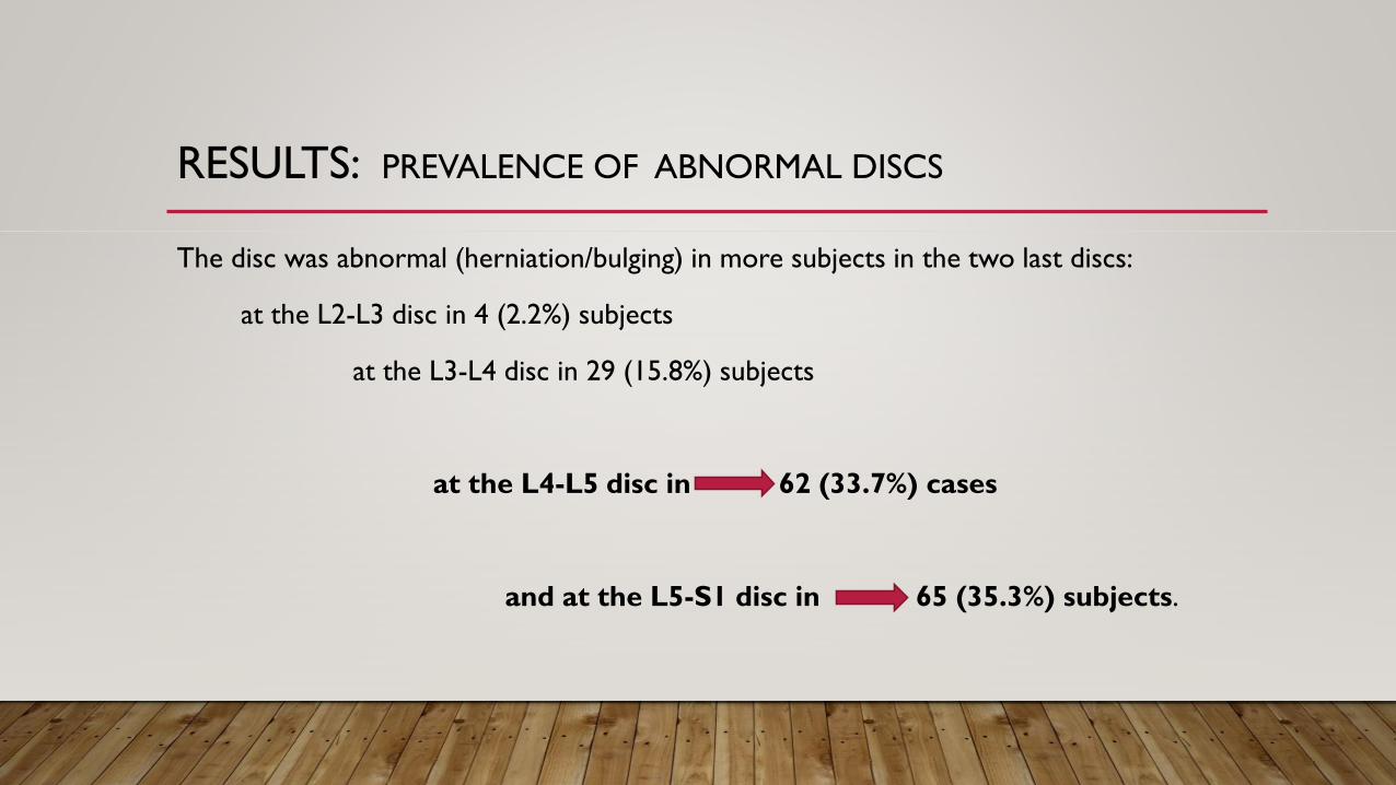
RESULTS: PREVALENCE OF ABNORMAL DISCS
The disc was abnormal (herniation/bulging) in more subjects in the two last discs:
at the L2-L3 disc in 4 (2.2%) subjects
at the L3-L4 disc in 29 (15.8%) subjects
at the L4-L5 disc in 62 (33.7%) cases
and at the L5-S1 disc in 65 (35.3%) subjects.
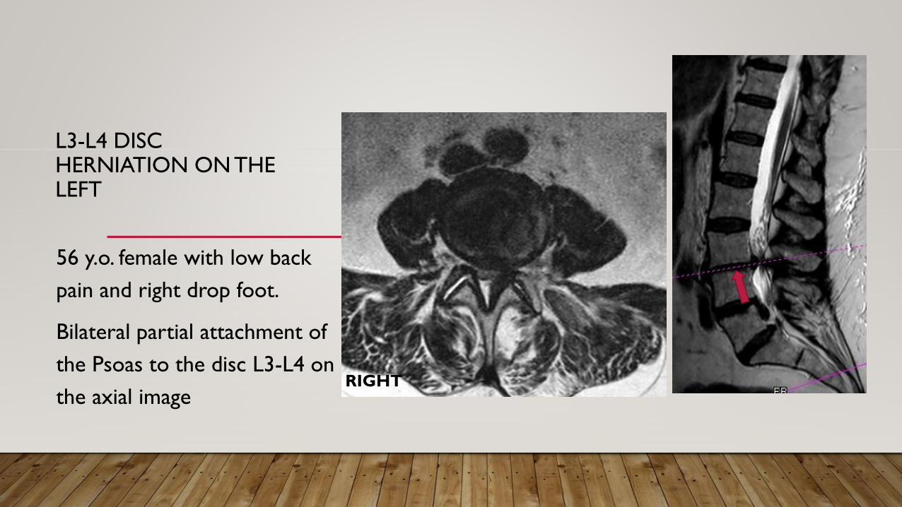
L3-L4 DISC HERNIATION ON THE LEFT
56 y.o. female with low back
pain and right drop foot.
Bilateral partial attachment of
the Psoas to the disc L3-L4 on
the axial image RIGHT
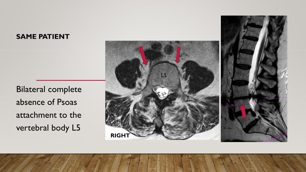
SAME PATIENT
Bilateral complete
absence of Psoas
attachment to the
vertebral body L5
L5
RIGHT
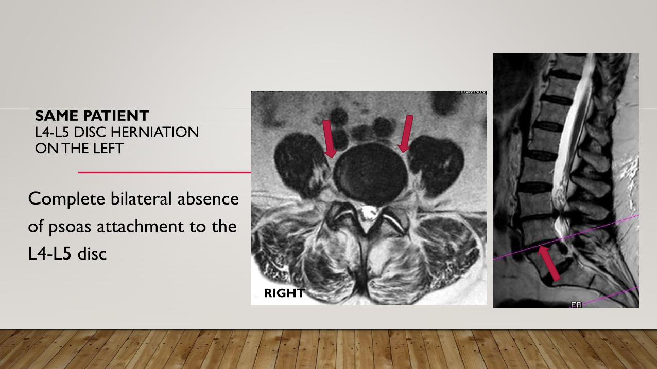
SAME PATIENTL4-L5 DISC HERNIATION ON THE LEFT
Complete bilateral absence
of psoas attachment to the
L4-L5 disc
RIGHT
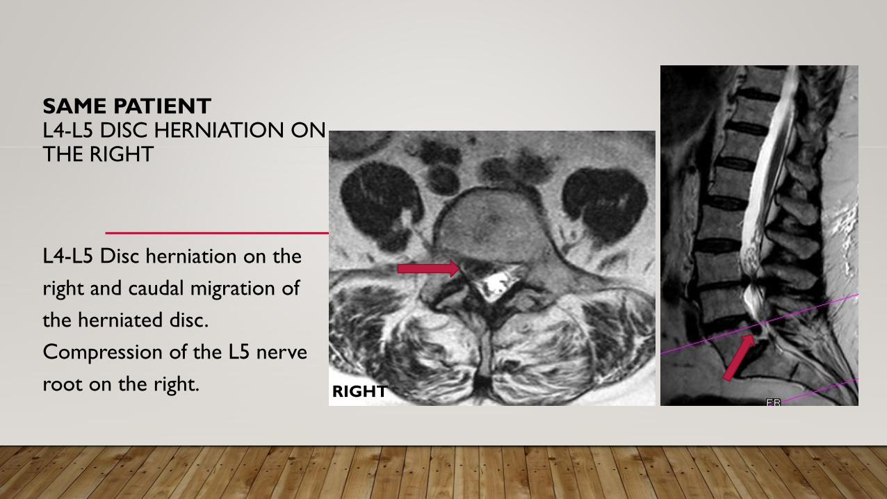
SAME PATIENTL4-L5 DISC HERNIATION ON THE RIGHT
L4-L5 Disc herniation on the
right and caudal migration of
the herniated disc.
Compression of the L5 nerve
root on the right. RIGHT
CONCLUSIONS
1. The psoas major muscle originates most frequently from the Th12-L1 disc
It does not originate from above the vertebral body Th12 or from below L2
2. The psoas major muscle rarely attaches to the L5-S1 disc
3. We found a higher prevalence of abnormal disc at L4-L5 and L5-S1 where partial or total
absence of attachment of the psoas muscle to the disc occurred.
CONCLUSIONS
Weakness of the study:
1. This is a retrospective study
2. The results are preliminary. And the results from the subgroups based on age and
genders and attachments to the vertebral body and transverse processes
are not available
3. Further investigations needed:
Based on the results, further investigations are necessary to determine whether the
absence of attachment of the psoas major muscle to the lower lumbar discs can relate
to the higher occurrence of disc herniation at those two levels.

THANK YOU