cleavage of tau by asparagine endopeptidase mediates the
TRANSCRIPT

Cleavage of tau by asparagine endopeptidasemediates the neurofibrillary pathology inAlzheimer's diseaseZhentao Zhang, Emory UniversityMingke Song, Emory UniversityXia Liu, Emory UniversitySeong Su Kang, Emory UniversityIl-Sun Kwon, Emory UniversityDuc M. Duong, Emory UniversityNicholas Seyfried, Emory UniversityWilliam Hu, Emory UniversityZhixue Liu, Chinese Academy of SciencesJian-zhi Wang, Huazhong University of Science & Technology
Only first 10 authors above; see publication for full author list.
Journal Title: Nature MedicineVolume: Volume 20, Number 11Publisher: Nature Publishing Group | 2014-11-01, Pages 1254-1262Type of Work: Article | Post-print: After Peer ReviewPublisher DOI: 10.1038/nm.3700Permanent URL: https://pid.emory.edu/ark:/25593/rwm9n
Final published version: http://dx.doi.org/10.1038/nm.3700
Copyright information:© 2014, Rights Managed by Nature Publishing Group
Accessed February 24, 2022 9:23 AM EST

Cleavage of tau by asparagine endopeptidase mediates the neurofibrillary pathology in Alzheimer’s disease
Zhentao Zhang1,2, Mingke Song3, Xia Liu1, Seong Su Kang1, Il-Sun Kwon1, Duc M. Duong4,5, Nicholas T. Seyfried4,5, William T. Hu4, Zhixue Liu6, Jian-zhi Wang7, Liming Cheng8,9, Yi E. Sun8, Shan Ping Yu3, Allan I. Levey4, and Keqiang Ye1,9
1Department of Pathology and Laboratory Medicine, Center for Neurodegenerative Diseases, Emory University School of Medicine, Atlanta, GA 30322, USA
2Department of Neurology, Renmin Hospital of Wuhan University, Wuhan 430060, China
3Department of Anesthesiology, Center for Neurodegenerative Diseases, Emory University School of Medicine, Atlanta, GA 30322, USA
4Department of Neurology, Center for Neurodegenerative Diseases, Emory University School of Medicine, Atlanta, GA 30322, USA
5Department of Biochemistry, Center for Neurodegenerative Diseases, Emory University School of Medicine, Atlanta, GA 30322, USA
6Key Laboratory of Nutrition and Metabolism, Institute for Nutritional Sciences, Shanghai Institutes for Biological Sciences, Chinese Academy of Sciences, Graduate School of the Chinese Academy of Sciences, Shanghai 200031, China
7Department of Pathophysiology, Key Laboratory of Ministry of Education of Neurological Diseases, Tongji Medical College, Huazhong University of Science and Technology, Wuhan, China
8Translational Center for Stem Cell Research, Tongji Hospital, Department of Regenerative Medicine, Tongji University School of Medicine, Shanghai 200065, China
Abstract
Neurofibrillary tangles (NFTs), composed of truncated and hyperphosphorylated tau, are a
common feature of numerous aging-related neurodegenerative diseases including Alzheimer’s
disease (AD). However, the molecular mechanisms mediating tau truncation and aggregation
Users may view, print, copy, and download text and data-mine the content in such documents, for the purposes of academic research, subject always to the full Conditions of use:http://www.nature.com/authors/editorial_policies/license.html#terms9To whom correspondence should be addressed ([email protected]; [email protected]).
AUTHOR CONTRIBUTIONSK.Y. conceived the project, designed the experiments, and wrote the manuscript. Z.Z. designed and performed most of the experiments. M. S. and S.P.Y. performed the electrophysiological experiments. X.L. prepared primary neurons and assessed with animal experiments. S.S.K performed the stereotaxic injection of virus. D.M.D. and N.T.S performed the mass spectrometry analysis. I.K. assisted with the molecular biology experiments. L.C., W.T.H. Z.L., J. W., Y.E.S., and A.I.L. designed the experiments, assisted with data analysis and interpretation and critically read the manuscript.
COMPETING FINANCIAL INTERESTRSThe authors declare no competing financial interests.
HHS Public AccessAuthor manuscriptNat Med. Author manuscript; available in PMC 2015 May 01.
Published in final edited form as:Nat Med. 2014 November ; 20(11): 1254–1262. doi:10.1038/nm.3700.
Author M
anuscriptA
uthor Manuscript
Author M
anuscriptA
uthor Manuscript
during aging remain elusive. Here we show that asparagine endopeptidase (AEP), a lysosomal
cysteine proteinase, is activated during aging and proteolytically degrades tau, abolishes its
microtubule assembly function, induces tau aggregation, and triggers neurodegeneration. AEP is
upregulated and active during aging, and is activated in tau P301S transgenic mice and human AD
brain, leading to tau truncation in NFTs. Deletion of AEP from tau P301S transgenic mice
substantially reduces tau hyperphosphorylation, alleviates the synapse loss and rescues impaired
hippocampal synaptic function and the cognitive deficits. Infection of uncleavable tau
N255AN368A mutant rescues tau P301S-induced pathological and behavioral defects. Together,
these observations indicate that AEP acts as a crucial mediator of tau-related clinical and
neuropathological changes in neurodegenerative diseases. Inhibition of AEP may be
therapeutically useful for treating tau-mediated neurodegenerative diseases.
INTRODUCTION
Alzheimer’s disease (AD) is a progressive neurodegenerative disease characterized by two
neuropathological hallmarks: extracellular senile plaque deposits, composed of amyloid beta
(Aβ), and intracellular neurofibrillary tangles (NFTs), made of truncated and
hyperphosphorylated tau. Tau-mediated neurodegeneration may result from the combination
of toxic gains-of-function acquired by the aggregates and the detrimental effects that arise
from the loss of the normal function(s) 1. Tau is mainly expressed in neurons and abundant
in the neuronal axons, regulating microtubule (MT) polymerization and stabilizing MT.
Through alternative splicing, the MAPT gene yields six major isoforms 2. While normal
phosphorylation of tau controls the dynamics of MT, establishing neuronal polarity, axonal
outgrowth and axonal transport 3–6, pathological hyperphosphorylation in disease severely
interferes with tau’s ability to regulate MT dynamics 7–9. Hyperphosphorylated tau displays
an increased propensity to form paired helical filaments (PHFs) in vitro and sequesters full-
length tau and other microtubule associated proteins 10–12, indicating that
hyperphosphorylation is a potent inducer of tau pathology. In AD, tau undergoes a number
of other posttranslational modifications in addition to phosphorylation that contribute to the
tau aggregation and disease pathology.
Tau is a substrate for various proteases. Tau can be cleaved by several caspases at Asp421.
In AD brain, tau truncated at Asp421 is a component of NFTs, and Aβ induces this cleavage
in cultured neurons 13–15. In AD brain, calpain 1 and calpain 2 are abnormally activated 16.
Aβ treatment leads to activation of calpains and production of a 17 kDa fragment in neurons
(tau45–230). Overexpression of tau45–230 induces neuronal apoptosis 17. In addition to
caspases and calpains, thrombin and cathepsins have also been implicated in processing
tau 18–20. However, many tau fragments found in AD are not well characterized and the
proteases responsible for their generation have not all been identified. For instance, a 25–35
kDa tau fragment in the cerebrospinal fluid (CSF) has been used as an early marker of
AD 21,22, but the proteases responsible for this cleavage event are unknown.
Mammalian asparagine endopeptidase (AEP), also known as legumain (LGMN), is a
lysosomal cysteine protease that cleaves protein substrates on the C-terminal side of
asparagine 23,24. AEP activation is autocatalytic and requires sequential removal of C- and
Zhang et al. Page 2
Nat Med. Author manuscript; available in PMC 2015 May 01.
Author M
anuscriptA
uthor Manuscript
Author M
anuscriptA
uthor Manuscript

N-terminal propeptides at different pH thresholds 25. Recently, we showed that neuronal
AEP is involved in neuronal apoptosis by degrading DNase inhibitor SET during excito-
neurotoxicity 26 and that AEP cleaves TDP-43 in post-mortem brain from humans with
frontotemporal lobar degeneration 27. In this report, we show that AEP cleaves tau at both
N255 and N368 residues, induces tau aggregation and attenuates its MT stabilizing activity.
Furthermore, AEP is highly activated in tau P301S transgenic mice and human AD brains.
Consistently, an AEP-cleaved tau fragment at N368 is detected in human AD brains.
Knockout of AEP in tau P301S mice leads to the reduction of tau hyperphosphorylation,
protecting against memory loss. Blockade of tau cleavage by AEP rescues tau P301S-
triggered pathological and behavioral defects. Hence, our results support that tau is a
physiological substrate of AEP, and that AEP is a mechanism based therapeutic target for
treating tauopathies including AD.
RESULTS
AEP directly cleaves tau
To explore whether AEP proteolytically processes tau, we conducted an in vitro cleavage
assay with kidney lysates prepared from wild-type (+/+) versus Lgmn knockout (−/−) mice
under pH 7.4 or 6.0, respectively. Active AEP cleaved tau into two fragments at pH 6.0,
whereas inactive AEP at pH 7.4 failed (Fig. 1a, left panel). AEP enzymatic activity in
kidney lysates was confirmed by the activity assay (Fig. 1a, right panel). When
cotransfected with GST-tau, wild-type AEP strongly triggered tau fragmentation, while AEP
mutants that abolish the cysteine protease activity of AEP (C189S) and the zymogen
autocleavage required for its activation (N323A) 25 were unable to provoke tau cleavage,
indicating AEP is responsible for eliciting tau proteolytic cleavage (Fig. 1b). Furthermore,
the peptide inhibitor AENK completely suppressed tau cleavage by AEP, whereas the
inactive control AEQK had no effect (Fig. 1c). The selective anti-AEP antibody also
attenuated tau fragmentation (Fig. 1d), suggesting that tau can be specifically cleaved in an
AEP-dependent manner. Biochemical assay with purified active AEP and recombinant GST-
tau revealed that purified AEP indeed potently and directly cleaved tau (Fig. 1e). Moreover,
tau was degraded in both Lgmn+/+ and Lgmn+/− mice at pH 6.0, but this effect was
substantially attenuated in Lgmn−/− mice. By contrast, at pH 7.4, where AEP is inactive, tau
remained intact regardless of AEP genotypes (Fig. 1f). AEP enzymatic activities were also
validated by fluorescent substrate cleavage assay (Fig. 1g). Together, these results strongly
support that tau is a direct substrate of the cysteine protease AEP.
AEP cleaves endogenous tau at N255 and N368
To identify the cleavage sites on tau by AEP, we purified the fragmented GST-tau proteins
with molecular weights of approximately 50 and 60 kDa, respectively, and performed mass
spectrometry analysis (Fig. 2a). Two partial-tryptic peptides ending with N255 and N368 for
the cleaved recombinant proteins were identified from the LC-MS/MS (Fig. 2b). The amino
acid number is based on the longest tau isoform (isoform 2, a.a. 1–441). To avoid
unnecessary confusion, we adopted isoform 2’s numbering system, though we employed
isoform 4 (a.a. 1–352) in all of our in vitro experiments. Mutation of either N368A or
N255A blocked the appearance of the band at 60kDa or 50kDa, respectively. Tau double
Zhang et al. Page 3
Nat Med. Author manuscript; available in PMC 2015 May 01.
Author M
anuscriptA
uthor Manuscript
Author M
anuscriptA
uthor Manuscript

mutant (N255A/N368A) remained completely intact in the presence of active AEP (Fig. 2c),
suggesting that N255 and N368 are the two major AEP cleavage sites on tau. Moreover,
peptide N368 was identified by MS in human AD brain samples by mass spectrometry (Fig.
2d), supporting that tau cleavage at N368 by AEP occurs in human AD brain. Finally, both
N255 and N368 peptides were identified in whole mouse brain extracts and comparative
label-free proteomic analysis against Lgmn−/− mice revealed greater than 20 folds
enrichment for both N368 and N255 in Lgmn+/+ versus Lgmn−/− brain extracts (Fig. 2e).
Hence, tau is a physiological substrate of AEP, which cleaves tau at both N255 and N368
sites.
AEP cleaves tau independent of caspases or calpains
Tau can be proteolytically processed by numerous proteinases including caspases, calpains,
cathepsins, thrombin, puromycin-sensitive aminopeptidase (PSA) 28. We assessed the effect
of their specific inhibitors on the processing of tau by AEP, and found that tau cleavage was
selectively reduced by the specific AEP inhibitor AENK but not by any of the other protease
inhibitors (Supplementary Fig. 1a). Remarkably, point mutation of the cleavage sites by
various proteases (R155A and K257A for thrombin; M419A for cathepsin; D421E for
caspase-3; L43A/V229A for calpain) were prominently fragmented by active AEP, whereas
AEP mutant (N255A/N368A) remained intact (Supplementary Fig. 1b). These results
indicate that cleavage by other proteases is not necessary for AEP processing of tau. On the
other hand, calpain cleavage analysis revealed that tau wild-type, N255A/N368A mutant and
tau fragment (1–255) were cleaved in a time-dependent manner with consistent rates
(Supplementary Fig. 1c), indicating that AEP cleavage of tau does not affect proteolytic
processing by calpains. We made the similar observation with both wild-type and tau
N255A/N368A mutant in caspase-3 cleavage assay (Supplementary Fig. 1d). Thus, AEP and
other proteases can independently fragment tau. To test whether tau hyperphosphorylation
influences tau cleavage by AEP, we pretreated GST-tau transfected HEK293 cells with
protein phosphatase PP2 inhibitor okadaic acid (OA), and monitored tau fragmentation.
GST-tau was cleaved in a time-dependent manner regardless of OA treatment
(Supplementary Fig. 1e,f), indicating that hyperphosphorylation of tau does not interfere
with AEP cleavage.
AEP is activated during aging process and in human AD brains
Since age is the major risk factor for AD, and the pH in the brain gradually decreases during
aging 29,30, we sought to investigate tau degradation by AEP in the brain at different ages.
Tau was fragmented progressively with degradation products detectable as early as 8 months
of age (Fig. 3a). To further investigate tau fragmentation by AEP, we developed an anti-tau
N368 antibody, which specifically recognized the cleaved tau 1–368 band but not full-length
tau or tau 1–255 band (Supplementary Fig. 2a,b). The immunoprecipitated bands by anti-tau
N368 antibody was recognized by two different tau antibodies (tau1 and tau5), suggesting
indeed AEP-cleaved tau but not other proteins was selectively immunoprecipitated
(Supplementary Fig. 2c). We detected tau immunoreactive fragments at 8 and 13 months old
mouse brains with anti-tau N368 (Fig. 3a, 2nd panel). In agreement with this finding, we
found that AEP activity also increased with aging, correlating with tau cleavage pattern (Fig.
3b). The tau fragmentation was completely abolished in Lgmn−/− brain, supporting that tau
Zhang et al. Page 4
Nat Med. Author manuscript; available in PMC 2015 May 01.
Author M
anuscriptA
uthor Manuscript
Author M
anuscriptA
uthor Manuscript

degradation during aging is mediated by AEP (Fig. 3c). The anti-tau N368 antibody also
robustly labeled tau N368 immunoreactivity in AD brain sections, whereas the signals were
markedly attenuated in age-matched control brains (Fig. 3d). Pre-incubation with the antigen
peptide (tau 360–368) abolished immunoreactivity (Supplementary Fig. 2d), supporting the
specificity of the anti-tau N368 antibody. Furthermore, we found that tau N368
immunoreactive fragments were abundant in human AD brains but barely detectable in
controls (Fig. 3e). Moreover, immunofluorescent staining revealed that tau N368 colocalized
with thioflavin S-positive NFTs in human AD brains (Fig. 3f), and tau N368 also
colocalized with phosphorylated tau (Supplementary Fig. 2e), indicating that AEP induced
fragments of tau are constituents of NFTs in vivo. AEP enzymatic activities in human AD
brains were also higher than controls (Fig. 3g), fitting with the marked tau N368
fragmentation in human AD samples. Further, AEP activity was elevated in tau P301S
transgenic mice versus the age-matched control non-transgenic mice (Fig. 3h). As expected,
the pH in the transgenic mice brain cortex and hippocampus was decreased compared to
non-transgenic control (Fig. 3i). Hence, AEP is activated in aging brain, and it is also
activated in both human AD brains and tau P301S transgenic mice.
Noticeably, Aβ oligomers elicited both AEP and tau fragmentation in a dose-dependent
manner in primary neuronal cultures (Supplementary Fig. 3a,b). AEP was activated by Aβ
dose-dependently, whereas cathepsin B, an AEP-related cysteine protease, was not
activated. As expected, caspase-3 activity increased with Aβ in a concentration-dependent
manner (Supplementary Fig. 3c–e). Moreover, overexpression of AEP in primary neurons
elicited demonstrable tau N368 degradation (Supplementary Fig. 3f). Thus, Aβ may provoke
AEP activation, resulting in tau degradation at N368.
AEP cleavage of tau generates neurotoxic fragments
To assess whether AEP cleavage may affect the functions of tau in promoting MT
polymerization, we conducted an in vitro MT polymerization assay with purified tubulin in
the presence of His-tagged tau fragments. As expected, full-length tau strongly augmented
MT polymerization; in contrast, the stimulatory effects of truncated tau fragments 1–368 and
256–441 were greatly reduced. Although tau fragment 256–368 moderately increased MT
polymerization in vitro, tau fragments 1–255 and tau 369–441 failed to induce
polymerization (Fig. 4a–c). Accordingly, overexpression of full-length tau in primary
neurons notably increased axon length; in contrast, axon elongation was not enhanced in
neurons expressing any one of the tau fragments (Fig. 4d).
In neurons transfected with a mixture of the tau fragments, apoptosis was increased more
than 60% compared to neurons transfected with full-length tau (Fig. 4e). TUNEL staining
showed that the tau truncates, 1–368 and 256–368, triggered substantial apoptosis compared
to wild-type or other tau fragments (Fig. 4f). These results were confirmed in neurons
infected with adeno-associated virus (AAVs) encoding tau full-length or tau 1–368
(Supplementary Fig. 4a,b). Hence, tau fragments that lack the C-terminal tail after N368
residue cut by AEP are neurotoxic. Furthermore, none of the CDK5, GSK3, and PKA
inhibitors (roscovitine, SB216763, and H89, respectively) attenuated the toxic effect of tau
Zhang et al. Page 5
Nat Med. Author manuscript; available in PMC 2015 May 01.
Author M
anuscriptA
uthor Manuscript
Author M
anuscriptA
uthor Manuscript

1–368, indicating the neurotoxic effect of tau 1–368 is not dependent on its phosphorylation
(Fig. 4g).
AEP-generated tau fragments form insoluble fibrils in vitro
To investigate the effect of AEP cleavage on tau fragment propensity for filament formation,
we monitored accumulation of PHFs using purified His-tagged tau recombinant proteins.
The tau truncate 256–368 displayed the strongest effect on self-assembly into filamentous
structures, followed by the tau 1–368 and tau 256–441. These fragments all exhibited a
greater ability to form PHFs than full-length tau. Notably, tau 1–255 and tau 369–441
fragments were unable to aggregate into PHFs, whereas the mixture of these aforementioned
fragments demonstrated an increased propensity to form PHFs (Supplementary Fig. 5a).
Consistent with the Thioflavine S assay, PHFs were observed under negative stain electron
microscopy in samples prepared with tau truncates of 1–368, 256–368, 256–441 and full-
length tau. Again, a mixture of all of these fragments also formed filamentous structures
(Supplementary Fig. 5b). Fractionation of heparin-induced insoluble tau fibrils via
ultracentrifugation revealed similar results, with tau fibrils of 256–368, 256–441 and 1–368
displaying the most robust aggregates, followed by full-length tau (Supplementary Fig. 5c).
Hence, AEP processing of tau promotes its assembly into filaments.
To explore whether AEP-cleaved tau fragments induce tau phosphorylation, we transfected
primary neurons with HA-tagged tau 1–255 or tau 1–368 and performed immunofluorescent
staining with anti-AT8. Overexpression of either tau fragment from AEP fragmentation
elicited robust AT8 immunoreactivity, whereas full-length tau displayed no signal above
endogenous levels (Supplementary Fig. 6). Conceivably, AEP cleaves tau on both N255 and
N368 sites, and the resultant fragments elicit tau hyperphosphorylation and aggregation.
Knockout of AEP prevents cognitive deficits in tau P301S mice
To assess the physiological role of AEP in synaptic function and behavior, we bred Lgmn−/−
mice with tau P301S transgenic mice to knock out AEP in tau P301S mice. The genotypes
of transgenic mice were validated (Supplementary Fig. 7). Immunoreactivity for the tau
N368 fragment was evident in tau P301S mice, but was not detectable in tau P301S/Lgmn−/−
mice (Fig. 5a). The absence of tau N368 in tau P301S/Lgmn−/− mice was also confirmed by
immunohistochemistry (Fig. 5b). Approximately 30% reduction of AT8- and AT100-
positive neurons was found in tau P301S/Lgmn−/− mice compared to tau P301S transgenic
mice both in the hippocampus and cortex (Fig. 5c and Supplementary Fig. 8). Electron
microscope analysis of brain sections showed that at 6 months of age, tau P301S mice
exhibited overt reduction in synapses compared to wild-type and Lgmn−/− mice. Notably,
deletion of AEP greatly ameliorated synapse loss in tau P301S mice (Fig. 5d and
Supplementary Fig. 9a). Golgi staining revealed that AEP gene deletion prevented the loss
of denritic spines in tau P301S mice (Supplementary Fig. 9b,c). Electrophysiological
analysis found that the input/output (I/O) relation was suppressed and the averaged field
excitatory post-synaptic potentials (fEPSPs) slope was substantially reduced in tau P301S
mice compared to wild-type and tau P301S/Lgmn−/− mice (Supplementary Fig. 10a,b),
indicating that synaptic transmission is impaired in 6 months old tau P301S mice but
rescued by AEP gene deletion. The ratios of paired pulse (induced by 100- and 200-ms
Zhang et al. Page 6
Nat Med. Author manuscript; available in PMC 2015 May 01.
Author M
anuscriptA
uthor Manuscript
Author M
anuscriptA
uthor Manuscript

intervals) were lower in tau P301S mice but higher in WT and tau P301S mice with AEP
knockout (Fig. 5e), suggesting an amelioration of presynaptic dysfunction due to AEP gene
deletion. The long-term potentiation (LTP) of fEPSPs in the hippocampal CA1 region,
which represents the molecular basis of learning and memory, was also diminished in tau
P301S mice compared with the control wild-type and tau P301S/Lgmn−/− mice. Hence,
knockout of AEP gene in tau P301S mice substantially prevents the impairment in synaptic
function caused by the tau mutation (Fig. 5f).
Next, we assessed the effect of deleting Lgmn gene on memory functions in tau P301S
transgenic mice by the Morris Water Maze. During the training phase, the swim distance
and latency to find the platform were progressively decreased, demonstrating a learning
effect in the tau P301S transgenic mice, albeit substantially impaired compared to wild-type
control or Lgmn−/− mice. However, knockout of AEP gene from tau P301S mice greatly
reduced the learning deficits (Fig. 5g,h). Probe trail found that deletion of AEP in tau P301S
mice improved memory retention as illustrated by the higher percentage of time spent in the
target quadrant. All groups of animals displayed comparable swim speed (Fig. 5i), indicating
that the genetic manipulations did not have any deleterious effects on motor function.
Next, we also conducted the contextual and cued fear conditioning tests with these animals.
Strikingly, tau P301S mice displayed substantially lower levels of freezing compared to the
wild-type or Lgmn−/− mice. Lgmn gene knockout from tau P301S mice increased memory in
both the cued fear test and the contextual fear assay (Fig. 5j). Therefore, deletion of AEP
greatly reverses the spatial and associative learning deficits in tau P301S transgenic mice.
Preservation of cognition in mice expressing uncleavable tau P301S
To confirm the effects of AEP are mediated via cleavage of tau, we injected AAVs encoding
human tau P301S or AEP non-cleavable tau P301S (tau P301SN255AN368A) into the
hippocampus of wild-type mice. Two months later, similar levels of tau P301S and tau
P301SN255AN368A were detected in the hippocampus by immunohistochemistry and
Western blot using the human tau specific antibody HT-7 (Fig. 6a,b). The density of synapse
in the hippocampus of mice injected with AAV-tau P301S was substantially less than AAV-
GFP control. However, the synaptic density was preserved when the mice were injected with
AAV-tau P301SN255AN368A (Fig. 6c,d). Electrophysiological analysis found that the
ratios of paired pulse were lower in mice expressing tau P301S compared with mice injected
with control virus, indicating evident presynaptic dysfunction. N255AN368A mutation
reversed the reduction of paired pulse ratios (Fig. 6e). Notably, the N255AN368A mutation
also attenuated the impairment of LTP induced by injection of AAV-tau P301S (Fig. 6f).
The slope of I/O curve was suppressed by tau P301S, but not by tau P301SN255AN368A
(Fig. 6g,h). These results indicate that the synaptic function is preserved in mice expressing
non-cleavable tau P301S. In the Morris Water Maze test, tau P301SN255AN368A mice
showed decreased latency to find the platform during the training phase, and increased
percentage of time in the target quadrant during the probe test compared to mice injected
with AAV-tau P301S, indicating preserved cognitive function (Fig. 6i,j). The preservation of
synaptic and cognitive function by the mutation of AEP cleavage site indicates that the
cleavage of tau is required for the effects of AEP in the current paradigm.
Zhang et al. Page 7
Nat Med. Author manuscript; available in PMC 2015 May 01.
Author M
anuscriptA
uthor Manuscript
Author M
anuscriptA
uthor Manuscript

DISSCUSION
In the present study, we have identified that AEP acts as a physiological protease that
cleaves tau in an age-dependent manner in mouse and human brain, including in AD. AEP
processes tau at both N255 and N368 sites, inhibiting tau-mediated effects on MT assembly
and axon elongation. The resultant C-terminus-deleted tau fragments are prone to aggregate
and strongly trigger neurodegeneration, indicating that AEP activation may induce neuronal
cell death through proteolytic degradation of tau. These findings fit with the most recent
report that AEP is activated in human AD brains, and it translocates from the lysosomes into
the cytoplasm 31. Presumably, the cytoplasmic translocated AEP may cleave tau, leading to
disruption of microtubule dynamics, NFT formation, and neurodegeneration. We noticed
that the different pathogenic processes are not necessarily associated with the same AEP
derived fragment. However, the one AEP derived fragment that does show some of the
common toxic effects is tau 1–368. Our data also support that deletion of AEP from tau
P301S transgenic mice substantially decreases tau hyperphosphorylation and alleviates
synaptic loss and memory deficit in the mice.
Previous studies in a transgenic (tau P301S) tauopathy mouse model revealed that synapse
loss and microglial activation precede the appearance of NFTs, presumably due to the
impaired transport that results from tau hyperphosphorylation 32. Here, we provide
convergent biochemical and cellular evidence that suggests AEP cleavage of tau also
contributes to synaptic loss in tau P301S mice (Fig. 5). Interestingly, tau
hyperphosphorylation does not affect tau proteolytic degradation by AEP (Supplementary
Fig. 1). Thus, while tau phosphorylation may prevent its cleavage by caspase-3 33, AEP
cleavage of tau appears phosphorylation independent. However, we found that truncation of
tau by AEP facilitates tau hyperphosphorylation (Supplementary Fig. 6). Consistent with our
findings, the expression of the truncated tau 151–391 in the brain of transgenic animals
induces the complete tau cascade of neurofibrillary degeneration as found in humans 34.
Therefore, all these observations strongly support truncation of tau as a key posttranslational
modification provoking neurofibrillary degeneration with the pathological features similar to
those found in human AD.
AEP levels increase in an age-dependent manner (Fig. 3a). As expected, tau is fragmented
by AEP at the age of 8 months, when AEP is augmented. NFTs are the first pathology that
occurs during aging and AD and their burden correlates with the degree of cognitive
impairment 35–37, providing the initial circumstantial evidence to suggest that toxic gains-of-
function by NFTs might play an important part in the progression of the disease. The
reduction of hyperphosphorylated tau in tau P301S/Lgmn−/− mice compared to age-matched
tau P301S mice (Fig. 5) indicates AEP might act as a crucial protease triggering NFT
pathology in AD during aging. Moreover, AEP knockout ameliorates synaptic loss, LTP
impairment and memory loss in tau P301S transgenic mice, supporting that AEP might be
an upstream trigger in AD onset and progression. The preservation of synaptic and cognitive
function in mice expressing AEP non-cleavable tau P301S further confirms that the effects
of AEP are mediated by the cleavage of tau. Conceivably, blockade of this devastating
protease may provide an innovative therapeutic intervention for treating neurodegenerative
diseases including AD.
Zhang et al. Page 8
Nat Med. Author manuscript; available in PMC 2015 May 01.
Author M
anuscriptA
uthor Manuscript
Author M
anuscriptA
uthor Manuscript

ONLINE METHODS
Mice
Tau P301S mice on a C57BL/6J background (line PS19) and wild-type C57BL/6J mice were
products of the Jackson Laboratory (stock number: 008169 and 000664, respectively). The
AEP knockout mice on a mixed 129/Ola and C57BL/6 background were generated as
reported 38. Animal care and handling was performed according to the Declaration of
Helsinki and Emory Medical School guidelines. Tau P301S mice were crossed with
Lgmn−/− mice to generate tau P301S/Lgmn−/− mice. The following animal groups were
analyzed: WT, Lgmn−/−, tau P301S, tau P301S/Lgmn−/−. 6-month-old male mice were used
for experiments unless otherwise mentioned (8 mice per group). Sample size was
determined by Power and Precision (Biostat). Investigators were blinded to the group
allocation during the animal experiments. The protocol was reviewed and approved by the
Emory Institutional Animal Care and Use Committee.
Human tissue samples
Post-mortem brain samples were dissected from frozen brains of 8 AD cases (age 74.5 ±
11.2 years, mean ± SD) and 8 nondemented controls (age 73.9 ± 12.7 years) from the Emory
Alzheimer’s Disease Research Center. The study was approved by the Biospecimen
Committee. AD was diagnosed according to the criteria of the Consortium to Establish a
Registry for AD and the National Institute on Aging. Diagnoses were confirmed by the
presence of amyloid plaques and neurofibrillary tangles in formalin-fixed tissue. Informed
consent was obtained from the subjects.
Transfection and infection of the cells
HEK293 cells were transfected with plasmids encoding wild-type or point mutant mGST-tau
isoform 4 (a.a. 1–352), myc-AEP, myc-AEP C189S, or myc-AEP N323A by the calcium
phosphate precipitation method. To express myc-AEP, tau full-length or tau fragments in
primary neurons, Lipofectamine 2000 (Invitrogen) was used as described previously 39. To
express tau full-length and tau 1–368 in primary neurons, 1 μl AAV-tau or AAV-tau 1–368
(1X10¹⁴ vg ml min−1) was added to 1 ml culture medium. The expression of tau full-length
and tau 1–368 was assessed 96 h after infection.
In vitro tau cleavage assay
To assess the cleavage of tau by AEP in vitro, HEK293 cells were transfected with GST-tau
DNA by the calcium phosphate precipitation method. 48 h after transfection, the cells were
collected, washed once in PBS, lysed in buffer (50 mM sodium citrate, 5 mM DTT, 0.1%
CHAPS and pH 5.5, 0.5% Triton X-100), and centrifuged for 10 min at 14,000 g at 4°C. The
supernatant were then incubated with mouse kidney lysate at pH 7.4 or 6.0 at 37°C for 30
min. To test the effect of caspase, cathepsin, calpain, thrombin, PSA and AEP inhibitors on
the cleavage of tau by AEP, inhibitors were used against: caspase (ZVAD-fmk,
Calbiochem), cathepsin (E64, Sigma-Aldrich), calpains (ALLN, Sigma-Aldrich), thrombin
(PMSF, Sigma-Aldrich), PSA (purimycin, Sigma-Aldrich), and AEP (AENK peptide
inhibitor and inactive control AEQK). To measure the cleavage of purified tau fragments by
Zhang et al. Page 9
Nat Med. Author manuscript; available in PMC 2015 May 01.
Author M
anuscriptA
uthor Manuscript
Author M
anuscriptA
uthor Manuscript

AEP, caspase-3 or calpain, GST-tagged tau full-length or fragments were purified with
Glutathione beads. The purified tau protein was incubated with recombinant AEP protein
(Novoprotein, 5 μg ml−1) in AEP buffer (50 mM sodium citrate, 5 mM DTT, 0.1% CHAPS,
and 0.5% Triton X-100, pH 6.0), recombinant caspase-3 (Calbiochem, 5 ug ml−1) in caspase
buffer (100 mM NaCl, 50 mM HEPES, 10 mM DTT, 1 mM EDTA, 10% glycerol, 0.1%
CHAPS, pH 7.4), or recombinant calpain (Sigma-Aldrich, 50 ug ml−1) in calpain buffer (50
mM Tris-HCl, 100 mM NaCl, 2 mM DTT, 1mM EDTA, 3 mM CaCl2, pH 7.5). The
samples were then boiled in 1XSDS loading buffer and analyzed by immunoblotting.
AEP activity assay and measurement of pH in the brain lysates
Tissue homogenates or cell lysates (10 μg) were incubated in 200 μl assay buffer (20 mM
citric acid, 60 mM Na2HPO4, 1 mM EDTA, 0.1% CHAPS, and 1 mM DTT, pH 6.0)
containing 20 μM AEP substrate Z-Ala-Ala-Asn-AMC (Bachem). AMC released by
substrate cleavage was quantified by measuring at 460 nm in a fluorescence plate reader at
37°C for 1 h in kinetic mode for 5 min. The pH of hippocampus and cortex of tau P301S and
control mice was read at 37°C using 2% (wt/vol) brain homogenates in distilled water as
described previously 29.
Mass spectrometry analysis
Protein samples were in-gel digested with trypsin. Peptide samples were resuspended in
loading buffer (0.1% formic acid, 0.03% trifluoroacetic acid, 1% acetonitrile) and loaded
onto a 20 cm nano-HPLC column (internal diameter 100 μm) packed with Reprosil-Pur 120
C18-AQ 1.9 μm beads (Dr. Maisch) and eluted over a 2 h 4–80% buffer B reverse phase
gradient (Buffer A: 0.1% formic acid, 1% acetonitrile in water; Buffer B: 0.1% formic acid
in acetonitrile) generated by a NanoAcquity UPLC system (Waters Corporation). Peptides
were ionized with 2.0 kV electrospray ionization voltage from a nano-ESI source (Thermo)
on a hybrid LTQ XL Orbitrap mass spectrometer (Thermo). Data dependent acquisition of
centroid MS spectra at 30,000 resolution and MS/MS spectra were obtained in the LTQ
following collision induced dissociation (collision energy 35%, activation Q 0.25, activation
time 30 ms) for the top 10 precursor ions with charge determined by the acquisition software
to be z ≥ 2. Dynamic exclusion of peaks already sequenced was for 20 s with early
expiration for 2 count events with signal-to-noise > 2. Automatic gating control was set to
150 ms maximum injection time or 106 counts. To identify AEP cleavage sites on human
tau, the SageN Sorcerer SEQUEST 3.5 algorithm was used to search and match MS/MS
spectra to a complete semi-tryptic human proteome database (NCBI reference sequence
revision 50, with 66,652 entries) plus pseudo-reversed decoys sequences 40,41 with a 20 ppm
mass accuracy threshold. Only b and y ions were considered for scoring (Xcorr) and Xcorr
along with ΔCn were dynamically increased for groups of peptides organized by a
combination of trypticity (fully or partial) and precursor ion charge state to remove false
positive hits along with decoys until achieving a false discovery rate (FDR) of < 5% (<
0.25% for proteins identified by more than one peptide). The FDR was estimated by the
number of decoy matches (nd) and total number of assigned matches (nt). FDR = 2*nd/nt,
assuming mismatches in the original database were the same as in the decoy database. All
semi-tryptic MS/MS spectra for putative AEP generated APP cleavage sites were manually
Zhang et al. Page 10
Nat Med. Author manuscript; available in PMC 2015 May 01.
Author M
anuscriptA
uthor Manuscript
Author M
anuscriptA
uthor Manuscript

inspected. A user defined precursor mass tolerance of ± 20 ppm was employed for extracted
ion chromatogram (XIC) based quantification as previously described 42.
Western blot analysis
The mouse brain tissue or human tissue samples was lysed in lysis buffer (50 mM Tris, pH
7.4, 40 mM NaCl, 1 mM EDTA, 0.5% Triton X-100, 1.5 mM Na3VO4, 50 mM NaF, 10 mM
sodium pyrophosphate, 10 mM sodium β-glycerophosphate, supplemented with protease
inhibitors cocktail), and centrifuged for 15 min at 16,000 g. The supernatant was boiled in
SDS loading buffer. After SDS-PAGE, the samples were transferred to a nitrocellulose
membrane. Primary antibodies to the following targets were used: GST-HRP (Sigma-
Aldrich), tubulin (Sigma-Aldrich), HA and myc (both from Santa Cruz), His (GE
healthcare), HT7, tau5, AT8 and AT100 (Thermo), and tau-1 (Calbiochem).
Immunostaining
For visualization of PHF-tau, Free-floating 30 μm brain sections the brain sections were
treated with 0.3% H2O2 for 10 min. Then, sections were washed three times in PBS and
blocked in 1% BSA, 0.3% Triton X-100, for 30 min followed by overnight incubation with
anti-tau N368, anti-AT8 or anti-AT100 antibody (1: 500) at 4°C. The signal was developed
using Histostain-SP kit (Invitrogen). To detect the localization of AEP-cleaved tau and PHF
in human AD brain sections, the sections were incubated with mouse anti-tau N368 primary
antibody overnight at 4°C. The sections were washed three times in PBS, and incubated with
Texas Red conjugated anti-mouse IgG for 1 h at room temperature. After brief rinse in PBS,
the sections were stained for 5 min with 0.0125% Thioflavin-S in 50% ethanol. The sections
were washed with 50% ethanol and placed in distilled water. Then the sections were covered
with a glass cover using mounting solution and examined under a fluorescence microscope.
Purification of recombinant tau protein and preparation of PHF
His-tagged full-length tau, tau 1–255, 1–368, 256–368, 256–441, and 368–441 was purified
from E. coli using His bind purification kit (Calbiochem). The purified tau fragments were
induced to aggregate as described previously 43. Briefly, purified tau fragments (50 μM)
were incubated in PBS (pH 7.4) containing 12.5 μM heparin, 2 mM DTT and a protease
inhibitor cocktail. The samples were incubated with 50 μM thioflavin S for 45 min in the
dark at room temperature. PHF formation was quantified by measuring the fluorescence
with an excitation at 440 nm and an emission wavelength of 510 nm. The overall appearance
of the PHFs was visualized by negative stain electron microscopy. Briefly, the reaction
samples was adsorbed onto carbon/formvar-coated 400 mesh copper grids (EM Sciences)
for 30 s, and stained with 2% uranyl acetate for 30 s. Excess liquid in the sample was
wicked using filter paper. The grids were examined with a Philips 208S electron microscope
(Philips, Hillsboro, OR). The remaining solutions of aggregated tau were centrifuged at
100,000 g for 30 min to separate aggregated tau pallet and non-aggregated tau supernatant,
and analyzed by Western blot.
Zhang et al. Page 11
Nat Med. Author manuscript; available in PMC 2015 May 01.
Author M
anuscriptA
uthor Manuscript
Author M
anuscriptA
uthor Manuscript

Microtubule assembly assay
His-tagged full-length tau and tau fragments were purified from E. coli. 1.5 μM tau
fragments were incubated with 20 μM tubulin (Cytoskeleton) at 37°C, and polymerization of
microtubules was monitored by measuring the absorbance at 350 nm over 20 min as
described previously 44.
AAV vector packaging
pAAV vectors carrying the longest human tau isoform (Tau4R2N) or GFP use the human
synapsin I promoter to drive neuron-specific gene expression 45. All of the mutations were
introduced using site-directed mutagenesis kit (Agilent Technologies). The AAV particles
were prepared by Viral Vector Core at Emory University.
Stereotaxic injection of the virus
3-month-old wild-type C57BL/6J mice were anesthetized with phenobarbital (75 mg kg−1).
Bilateral intracerebral injection of AAV-GFP, AAV-tau P301S, and AAV-tau
P301SN255AN368A was performed stereotactically at coordinates posterior 1.94 mm,
lateral 1.4 mm, ventral 2.2 mm relative to bregma. 2 μl of viral suspension containing
2X10¹¹ vector genome (vg) was injected in to each point using 10 μl glass syringes with a
fixed needle at a rate of 0.5 μl min−1. The needle was remained in place for 5 min before it
was removed slowly (throughout 2 min). The mice were placed on heating pad until it began
to recover from the surgery.
Electron microscopy of synapse
Synaptic density was determined by electron microscopy. Briefly, after deep anesthesia,
mice were perfused transcardially with 2% glutaraldehyde and 3% paraformaldehyde in
PBS. Hippocampal slices were postfixed in cold 1% OsO4 for 1 h. Samples were prepared
and examined using standard procedures. Ultrathin sections (90 nm) were stained with
uranyl acetate and lead acetate and viewed at 100 kV in a JEOL 200CX electron
microscope. Synapses were identified by the presence of synaptic vesicles and postsynaptic
densities. Synapse density in area CA1 of the hippocampus was calculated.
Golgi stain
Mouse brains were fixed in 10% formalin for 24 h, and then immersed in 3% potassium
bichromate for 3 d in the dark. The solution was changed each day. Then the brains were
transferred into 2% silver nitrate solution and incubated for 24 h in the dark. Vibratome
sections were cut at 60 μm, air dried for 10 min, dehydrated through 95% and 100% ethanol,
cleared in xylene and assembled on coverslips. Bright-field images of pyramidal neurons in
the hippocampus and cortex were taken at 100X magnification using a Zeiss Axioplan
(Zeiss, Decatur, GA, USA) microscope. To measure the spine density, all clearly evaluable
areas of 50–100 μm of secondary dendrites from each imaged neuron were used.
Electrophysiology
Acute hippocampal transversal slices were prepared form 6-month-old WT, Lgmn−/−, tau
P301S, tau P301S/Lgmn−/− mice as previously described 44. Briefly, mice hippocampi were
Zhang et al. Page 12
Nat Med. Author manuscript; available in PMC 2015 May 01.
Author M
anuscriptA
uthor Manuscript
Author M
anuscriptA
uthor Manuscript

dissected and cut into 400-μm thick transverse slices. A 0.1 M tungsten monopolar electrode
was used to stimulate the Schaffer collaterals. The field excitatory post-synaptic potentials
(fEPSPs) were recorded in CA1 stratum radiatum by a glass microelectrode filled with
artificial CSF with resistance of 3–4 MΩ. Field potential input-output curves were
constructed by measuring fEPSP slopes responding to the stimulus intensity increasing from
0.5 to 10 V, with an 0.5 V increment. Paired-pulse facilitation (PPF) was examined by
applying pairs of pulses which were separated by 20–500 ms intervals. LTP of fEPSPs was
induced by 3 theta-burst-stimulation (TBS); it is 4 pulses at 100 Hz, repeated 3 times with a
200-ms interval). The magnitudes of LTP are expressed as the mean percentage of baseline
fEPSP initial slope.
Generation of antibodies that specifically recognize the AEP-generated tau fragment (anti-tau N368)
Three Balb/c mice were immunized with the peptide Ac-CITHVPGGGN-OH that includes
the 9 amino acids in tau that precede the AEP cleavage site at N368 as well as an amino-
terminal cysteine residue to allow coupling to KLH. The mice were boosted 4 times with the
immunizing peptide with 3-week intervals between injections. The antiserum was pooled
and the titers against the immunizing peptide were determined by ELISA. The maximal
dilution giving a positive response using chromogenic substrate for horseradish peroxidase
was 1: 30000. The immunoactivity of the antiserum was further confirmed by Western blot
and immunohistochemistry.
Morris water maze
6-month-old WT, Lgmn−/−, tau P301S, tau P301S/Lgmn−/− mice were trained in a round,
water-filled tub (52 inch diameter) in an environment rich with extra maze cues as described
previously 44. Each subject was given 4 trials/day for 5 consecutive days with a 15-min
inter-trial interval. The maximum trial length was 60 s and if subjects did not reach the
platform in the allotted time, they were manually guided to it. Following the 5 d of task
acquisition, a probe trial was presented during which time the platform was removed and the
percentage of time spent in the quadrant which previously contained the escape platform
during task acquisition was measured over 60 s. All trials were analysed for latency and
swim speed by means of MazeScan (Clever Sys, Inc.).
Fear conditioning test
The ability to form and retain an association between an aversive experience and
environmental cues was tested with a standard fear conditioning paradigm that occurs over a
period of three days. Mice were placed in the fear conditioning apparatus (7″ W, 7″ D X 12″
H, Coulbourn) composed of plexiglass with a metal shock grid floor and allowed to explore
the enclosure for 3 min. Following this habituation period, 3 conditioned stimulus (CS)-
unconditioned stimulus (US) pairings was presented with a 1 min intertrial interval. The CS
was composed of a 20 second 85 db tone and US was composed of 2 s of a 0.5 mA
footshock, which was co-terminate with each CS presentation. One minute following the last
CS-US presentation, mice were returned to their home cage. On day 2 the mice were
presented with a context test, during which subjects were placed in the same chamber used
Zhang et al. Page 13
Nat Med. Author manuscript; available in PMC 2015 May 01.
Author M
anuscriptA
uthor Manuscript
Author M
anuscriptA
uthor Manuscript

during conditioning on Day 1, and the amount of freezing was recorded via a camera and the
software provided by Colbourn. No shocks were given during the context test. On day 3, a
tone test was presented, during which time subjects were exposed to the CS in a novel
compartment. Initially, animals were allowed to explore the novel context for 2 min. Then
the 85 db tone was presented for 6-min, and the amount of freezing behaviour was recorded.
Primary neuron cultures
Primary rat cortical neurons were cultured as previously described 26. To measure the effect
of tau fragments on neurons, neurons cultured 7 days in vitro (DIV 7) were transfected with
myc-AEP, HA-tau full-length, or HA-tau fragments using Lipofectamine 2000 (Invitrogen).
72 h later, the neurons were fixed in 4% formaldehyde, permeabilized and immunostained
with anti-MAP2, anti-myc, anti-HA, anti-AT8, anti-AT100, or anti-tau N368 antibody. The
toxic effect of tau fragments was detected with the In situ cell death detection kit (Roche).
The apoptotic index was expressed as the percentage of TUNEL-positive neurons out of the
total number of MAP2-positive neurons.
Caspase and cathepsin activity assay
Cell lysates (10 μg) were incubated in 200 μl caspase assay buffer (100 mM HEPES 0.1%
CHAPS, 10% sucrose, pH 7.4) containing 25 μM caspase substrate Ac-Asp-Glu-Val-Asp-
AMC (Bachem), or in 200 μl cathepsin assay buffer (100 mM sodium acetate, 1 mM EDTA,
pH 5.5) containing 25 μM cathepsin substrate D-Val-Leu-Lys-AMC (Bachem) for 1 h.
AMC released by substrate cleavage was quantified by measuring at 460 nm in a
fluorescence plate reader.
Statistical analysis
Statistical analysis were performed using either Student’s t-test (two-group comparison) or
one-way ANOVA followed by LSD post hoc test (more than two groups), and differences
with P values less than 0.05 were considered significant.
Supplementary Material
Refer to Web version on PubMed Central for supplementary material.
Acknowledgments
This work was supported by grants from RO1 (NS045627 and NS060680) from NIH to K. Y., the NIH/NIAP50 ADRC center grant to A.I.L., grant from National Natural Science Foundation of China (No. 81100958) to Z. Z., National Key Basic Research Program of China Grant (2010CB945202) to Y.E.S and National Science Foundation of China Grant (81330030) to L.C. and Y.E.S. We thank D. Weinshenker (Emory University) for tau P301S transgenic mice, S. Kuegler (Max Planck Institute of Psychiatry, Germany) for pAAV vectors carrying tau GFP, and C. Hales (Emory University) for technical support.
References
1. Ballatore C, Lee VM, Trojanowski JQ. Tau-mediated neurodegeneration in Alzheimer’s disease and related disorders. Nat Rev Neurosci. 2007; 8:663–672. [PubMed: 17684513]
2. Binder LI, Frankfurter A, Rebhun LI. The distribution of tau in the mammalian central nervous system. J Cell Biol. 2565; 101:1371–1378. [PubMed: 3930508]
Zhang et al. Page 14
Nat Med. Author manuscript; available in PMC 2015 May 01.
Author M
anuscriptA
uthor Manuscript
Author M
anuscriptA
uthor Manuscript
3. Harada A, et al. Altered microtubule organization in small-calibre axons of mice lacking tau protein. Nature. 1994; 369:488–491. [PubMed: 8202139]
4. Esmaeli-Azad B, McCarty JH, Feinstein SC. Sense and antisense transfection analysis of tau function: tau influences net microtubule assembly, neurite outgrowth and neuritic stability. J Cell Sci. 1994; 107 (Pt 4):869–879. [PubMed: 8056843]
5. Caceres A, Kosik KS. Inhibition of neurite polarity by tau antisense oligonucleotides in primary cerebellar neurons. Nature. 1990; 343:461–463. [PubMed: 2105469]
6. Dixit R, Ross JL, Goldman YE, Holzbaur EL. Differential regulation of dynein and kinesin motor proteins by tau. Science. 2008; 319:1086–1089. [PubMed: 18202255]
7. Lindwall G, Cole RD. Phosphorylation affects the ability of tau protein to promote microtubule assembly. J Biol Chem. 2564; 259:5301–5305. [PubMed: 6425287]
8. Iqbal K, Zaidi T, Bancher C, Grundke-Iqbal I. Alzheimer paired helical filaments. Restoration of the biological activity by dephosphorylation. FEBS Lett. 1994; 349:104–108. [PubMed: 8045285]
9. Kopke E, et al. Microtubule-associated protein tau. Abnormal phosphorylation of a non-paired helical filament pool in Alzheimer disease. J Biol Chem. 1993; 268:24374–24384. [PubMed: 8226987]
10. Alonso AC, Grundke-Iqbal I, Iqbal K. Alzheimer’s disease hyperphosphorylated tau sequesters normal tau into tangles of filaments and disassembles microtubules. Nat Med. 1996; 2:783–787. [PubMed: 8673924]
11. Alonso AC, Zaidi T, Grundke-Iqbal I, Iqbal K. Role of abnormally phosphorylated tau in the breakdown of microtubules in Alzheimer disease. Proc Natl Acad Sci USA. 1994; 91:5562–5566. [PubMed: 8202528]
12. Alonso A, Zaidi T, Novak M, Grundke-Iqbal I, Iqbal K. Hyperphosphorylation induces self-assembly of tau into tangles of paired helical filaments/straight filaments. Proc Natl Acad Sci USA. 2001; 98:6923–6928. [PubMed: 11381127]
13. Gamblin TC, et al. Caspase cleavage of tau: linking amyloid and neurofibrillary tangles in Alzheimer’s disease. Proc Natl Acad Sci USA. 2003; 100:10032–10037. [PubMed: 12888622]
14. Rissman RA, et al. Caspase-cleavage of tau is an early event in Alzheimer disease tangle pathology. J Clin Invest. 2004; 114:121–130. [PubMed: 15232619]
15. Basurto-Islas G, et al. Accumulation of aspartic acid421- and glutamic acid391-cleaved tau in neurofibrillary tangles correlates with progression in Alzheimer disease. J Neuropathol Exp Neurol. 2008; 67:470–483. [PubMed: 18431250]
16. Saito K, Elce JS, Hamos JE, Nixon RA. Widespread activation of calcium-activated neutral proteinase (calpain) in the brain in Alzheimer disease: a potential molecular basis for neuronal degeneration. Proc Natl Acad Sci USA. 1993; 90:2628–2632. [PubMed: 8464868]
17. Park SY, Ferreira A. The generation of a 17 kDa neurotoxic fragment: an alternative mechanism by which tau mediates beta-amyloid-induced neurodegeneration. J Neurosci. 2005; 25:5365–5375. [PubMed: 15930385]
18. Arai T, Guo JP, McGeer PL. Proteolysis of non-phosphorylated and phosphorylated tau by thrombin. J Biol Chem. 2005; 369:5145–5153. [PubMed: 15542598]
19. Yang AJ, Chandswangbhuvana D, Margol L, Glabe CG. Loss of endosomal/lysosomal membrane impermeability is an early event in amyloid Abeta1–42 pathogenesis. J Neurosci Res. 1998; 52:691–698. [PubMed: 9669318]
20. Kenessey A, Nacharaju P, Ko LW, Yen SH. Degradation of tau by lysosomal enzyme cathepsin D: implication for Alzheimer neurofibrillary degeneration. J Neurochem. 1997; 69:2026–2038. [PubMed: 9349548]
21. Johnson GV, et al. The tau protein in human cerebrospinal fluid in Alzheimer’s disease consists of proteolytically derived fragments. J Neurochem. 1997; 68:430–433. [PubMed: 8978756]
22. Portelius E, et al. Characterization of tau in cerebrospinal fluid using mass spectrometry. J Proteome Res. 2008; 7:2114–2120. [PubMed: 18351740]
23. Chen JM, Dando PM, Stevens RA, Fortunato M, Barrett AJ. Cloning and expression of mouse legumain, a lysosomal endopeptidase. Biochem J. 1998; 335 (Pt 1):111–117. [PubMed: 9742219]
Zhang et al. Page 15
Nat Med. Author manuscript; available in PMC 2015 May 01.
Author M
anuscriptA
uthor Manuscript
Author M
anuscriptA
uthor Manuscript

24. Chen JM, Rawlings ND, Stevens RA, Barrett AJ. Identification of the active site of legumain links it to caspases, clostripain and gingipains in a new clan of cysteine endopeptidases. FEBS Lett. 1998; 441:361–365. [PubMed: 9891971]
25. Li DN, Matthews SP, Antoniou AN, Mazzeo D, Watts C. Multistep autoactivation of asparaginyl endopeptidase in vitro and in vivo. J Biol Chem. 2003; 278:38980–38990. [PubMed: 12860980]
26. Liu Z, et al. Neuroprotective actions of PIKE-L by inhibition of SET proteolytic degradation by asparagine endopeptidase. Mol Cell. 2008; 29:665–678. [PubMed: 18374643]
27. Herskowitz JH, et al. Asparaginyl endopeptidase cleaves TDP-43 in brain. Proteomics. 2012; 12:2455–2463. [PubMed: 22718532]
28. Wang Y, Garg S, Mandelkow EM, Mandelkow E. Proteolytic processing of tau. Biochem Soc Trans. 2010; 38:955–961. [PubMed: 20658984]
29. Yates CM, Butterworth J, Tennant MC, Gordon A. Enzyme activities in relation to pH and lactate in postmortem brain in Alzheimer-type and other dementias. J Neurochem. 1990; 55:1624–1630. [PubMed: 2213015]
30. Pirchl M, Humpel C. Does acidosis in brain play a role in Alzheimer’s disease? Neuropsychiatr. 2009; 23:187–192. [PubMed: 19703385]
31. Basurto-Islas G, Grundke-Iqbal I, Tung YC, Liu F, Iqbal K. Activation of asparaginyl endopeptidase leads to tau hyperphosphorylation in Alzheimer disease. J Biol Chem. 2013; 288:17495–17507. [PubMed: 23640887]
32. Yoshiyama Y, et al. Synapse loss and microglial activation precede tangles in a P301S tauopathy mouse model. Neuron. 2007; 53:337–351. [PubMed: 17270732]
33. Guillozet-Bongaarts AL, et al. Pseudophosphorylation of tau at serine 422 inhibits caspase cleavage: in vitro evidence and implications for tangle formation in vivo. J Neurochem. 2006; 97:1005–1014. [PubMed: 16606369]
34. Zilka N, et al. Truncated tau from sporadic Alzheimer’s disease suffices to drive neurofibrillary degeneration in vivo. FEBS Lett. 2006; 580:3582–3588. [PubMed: 16753151]
35. Arriagada PV, Growdon JH, Hedley-Whyte ET, Hyman BT. Neurofibrillary tangles but not senile plaques parallel duration and severity of Alzheimer’s disease. Neurology. 1992; 42:631–639. [PubMed: 1549228]
36. Arriagada PV, Marzloff K, Hyman BT. Distribution of Alzheimer-type pathologic changes in nondemented elderly individuals matches the pattern in Alzheimer’s disease. Neurology. 1992; 42:1681–1688. [PubMed: 1307688]
37. Braak H, Braak E. Staging of Alzheimer’s disease-related neurofibrillary changes. Neurobiol Aging. 1995; 16:271–278. discussion 278–284. [PubMed: 7566337]
38. Shirahama-Noda K, et al. Biosynthetic processing of cathepsins and lysosomal degradation are abolished in asparaginyl endopeptidase-deficient mice. J Biol Chem. 2003; 278:33194–33199. [PubMed: 12775715]
39. Kaech S, Banker G. Culturing hippocampal neurons. Nat Protoc. 2006; 1:2406–2415. [PubMed: 17406484]
40. Elias JE, Gygi SP. Target-decoy search strategy for increased confidence in large-scale protein identifications by mass spectrometry. Nat Methods. 2007; 4:207–214. [PubMed: 17327847]
41. Xu P, Duong DM, Peng J. Systematical Optimization of Reverse-Phase Chromatography for Shotgun Proteomics. J Proteome Res. 2009; 8:3944–3950. [PubMed: 19566079]
42. Herskowitz JH, et al. Phosphoproteomic analysis reveals site-specific changes in GFAP and NDRG2 phosphorylation in frontotemporal lobar degeneration. J Proteome Res. 2010; 9:6368–6379. [PubMed: 20886841]
43. Barghorn S, Biernat J, Mandelkow E. Purification of recombinant tau protein and preparation of Alzheimer-paired helical filaments in vitro. Methods Mol Biol. 2005; 299:35–51. [PubMed: 15980594]
44. Hong Y, et al. SRPK2 phosphorylates tau and mediates the cognitive defects in Alzheimer’s disease. J Neurosci. 2012; 32:17262–17272. [PubMed: 23197718]
45. Jaworski T, et al. AAV-tau mediates pyramidal neurodegeneration by cell-cycle re-entry without neurofibrillary tangle formation in wild-type mice. PLoS One. 2009; 4:e7280. [PubMed: 19794916]
Zhang et al. Page 16
Nat Med. Author manuscript; available in PMC 2015 May 01.
Author M
anuscriptA
uthor Manuscript
Author M
anuscriptA
uthor Manuscript
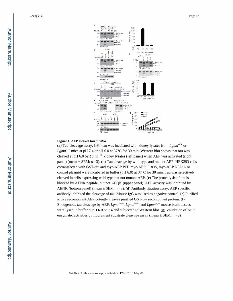
Figure 1. AEP cleaves tau in vitro(a) Tau cleavage assay. GST-tau was incubated with kidney lysates from Lgmn+/+ or
Lgmn−/− mice at pH 7.4 or pH 6.0 at 37°C for 30 min. Western blot shows that tau was
cleaved at pH 6.0 by Lgmn+/+ kidney lysates (left panel) when AEP was activated (right
panel) (mean ± SEM; n =3). (b) Tau cleavage by wild-type and mutant AEP. HEK293 cells
cotransfected with GST-tau and myc-AEP WT, myc-AEP C189S, myc-AEP N323A or
control plasmid were incubated in buffer (pH 6.0) at 37°C for 30 min. Tau was selectively
cleaved in cells expressing wild-type but not mutant AEP. (c) The proteolysis of tau is
blocked by AENK peptide, but not AEQK (upper panel). AEP activity was inhibited by
AENK (bottom panel) (mean ± SEM; n =3). (d) Antibody titration assay. AEP specific
antibody inhibited the cleavage of tau. Mouse IgG was used as negative control. (e) Purified
active recombinant AEP potently cleaves purified GST-tau recombinant protein. (f) Endogenous tau cleavage by AEP. Lgmn+/+, Lgmn+/−, and Lgmn−/− mouse brain tissues
were lysed in buffer at pH 6.0 or 7.4 and subjected to Western blot. (g) Validation of AEP
enzymatic activities by fluorescent substrate cleavage assay (mean ± SEM; n =3).
Zhang et al. Page 17
Nat Med. Author manuscript; available in PMC 2015 May 01.
Author M
anuscriptA
uthor Manuscript
Author M
anuscriptA
uthor Manuscript

Figure 2. AEP cleaves tau at N255 and N368 residues(a) Cleavage of purified GST-tau analyzed by immunoblotting (upper panel) or Coomassie
blue staining (bottom panel). (b) Mass spectrometry analysis of recombinant tau fragmented
by AEP. The detected MS/MS peptide spectra are listed. (c) Cleavage of mutant tau by AEP.
Tau cleavage was analyzed by Western blot after GST-tau wide-type, N255A, N368A, or
N255A/N368A mutant were incubated with active mouse kidney lysates. (d) MS/MS
spectrum showing the cleavage of tau after N368 in brain samples from subjects with AD.
(e) Representative extracted ion chromatograms for tau N255 and N368 peptide from wild-
type and Lgmn−/− mouse brain samples. Signal intensities were then normalized to wild-type
samples, setting the maximum signal intensity to 100%. Values are represented as raw
peptide extracted ion intensity.
Zhang et al. Page 18
Nat Med. Author manuscript; available in PMC 2015 May 01.
Author M
anuscriptA
uthor Manuscript
Author M
anuscriptA
uthor Manuscript

Figure 3. AEP is upregulated and cleaves tau during aging and in Alzheimer’s disease(a) Western blot analysis of tau and AEP in mouse brain during aging process. (b) AEP
activity assay (mean ± SEM; n = 6; *P < 0.05 compared with 1- and 2-months old mouse
brain, **P < 0.01 compared with 1-, 2-, and 4-months old mouse brain, one-way ANOVA).
(c) Western blot detection of tau fragments in the Lgmn−/− and Lgmn+/+ mice brain. (d)
Immunostaining of tau N368 fragments in brain sections of subjects with AD (mean ± SEM,
*P < 0.01, Student’s t-test). Scale bar, 50 μm. (e) Western blot detection of tau fragments in
human brain samples from subjects with AD and age-matched controls. (f) Immunostaining
showing colocalization of tau N368 fragment with PHFs. Brain sections from subjects with
AD were immunostained with anti-tau N368 antibody, and then stained with Thioflavin S,
which labels both the senile plaque (arrow head) and PHFs (arrow). Scale bar, 50 μm. (g)
AEP activity assay in brain samples from subjects with AD and age-matched controls (mean
± SEM; *P < 0.05 compared with control group, one-way ANOVA). (h) AEP activity assay
in 6-month-old tau P301S mice and non-transgenic controls (mean ± SEM; n = 6; *P < 0.01,
Zhang et al. Page 19
Nat Med. Author manuscript; available in PMC 2015 May 01.
Author M
anuscriptA
uthor Manuscript
Author M
anuscriptA
uthor Manuscript

Student’s t-test). (i) pH in the brain cortex and hippocampus of control and tau P301S
transgenic mice (n = 6, *P < 0.01, Student’s t-test).
Zhang et al. Page 20
Nat Med. Author manuscript; available in PMC 2015 May 01.
Author M
anuscriptA
uthor Manuscript
Author M
anuscriptA
uthor Manuscript

Figure 4. Tau cleavage by AEP disrupts its microtubule assembly activity and is toxic to neurons(a) Schematic diagram of tau isoform 4 domains and its cleavage by AEP. I, Inserts; P,
proline rich; R, repeats; R′, pseudo-repeat; C, C-terminal tail. (b) Western blot analysis of
purified His-tagged tau fragments. (c) Microtubule assembly assay. (d)Axon elongation in
primary neurons transfected with control plasmid, HA-tau, or HA-tau fragments (mean ±
SEM; n = 5; *P < 0.05, one-way ANOVA). (e, f) TUNEL assay showing the neurotoxicity
of AEP-derived tau fragments. Mixed tau fragments (e), tau 1–368 and tau 256–368 (f) induced significant neuronal apoptosis (mean ± SEM; n = 3; *P < 0.01, one-way ANOV).
Scale bar, 10 μm. (g) Effect of kinase inhibitors on the neurotoxic effect of tau 1–368.
Primary neurons infected with AAV-tau 1–368 were treated with 10 μM roscovitine (CDK5
inhibitor), SB216763 (GSK3 inhibitor), or H89 (PKA inhibitor) for 12 h. Cell apoptosis was
detected by TUNEL staining (n = 4). Scale bar, 20 μm.
Zhang et al. Page 21
Nat Med. Author manuscript; available in PMC 2015 May 01.
Author M
anuscriptA
uthor Manuscript
Author M
anuscriptA
uthor Manuscript

Figure 5. AEP gene deficiency prevents tau phosphorylation, synaptic dysfunction, and memory deficits in the tau P301S mouse(a) The processing of tau in WT, Lgmn−/−, tau P301S, and tau P301S/Lgmn−/− mice. (b)
Immunostaining of AEP-derived tau fragments in brain sections of tau P301S and tau
P301S/Lgmn−/− mice. Scale bar, 20 μm. (c) AT8 immunostaining of hippocampal and cortex
neurons (mean ± SEM; *P < 0.05, **P < 0.01, Student’s t-test). Scale bar, 50 μm. (d)
Synaptic density analysis (mean ± SEM; n = 6; **P < 0.01, one-way ANOVA). (e) The ratio
of paired pulses (mean ± SD; n = 6 in each group; *P < 0.05, one-way ANOVA). (f) LTP of
fEPSPs (mean ± SD; n = 6 in each group; *P < 0.05, one-way ANOVA). Shown traces are
representative fEPSPs recorded at the time point 1 and 2 (tau P301S/Lgmn−/−), 3 and 4 (tau
Zhang et al. Page 22
Nat Med. Author manuscript; available in PMC 2015 May 01.
Author M
anuscriptA
uthor Manuscript
Author M
anuscriptA
uthor Manuscript

P301S). (g, h) Morris water maze analysis as distance traveled (millimeters) and integrated
distance (AUC) for WT, Lgmn−/−, tau P301S and tau P301S/Lgmn−/− mice (mean ± SEM; n
= 8; *P < 0.05, one-way ANOVA). (i) Probe trail of Morris water maze test (left panel,
mean ± SEM; n = 8; *P < 0.05, **P < 0.01, one-way ANOVA). AEP gene deletion did not
interfere with the swim speed (right panel, mean ± SEM; n = 8; P = 0.786, one-way
ANOVA). (j) Fear conditioning test. Tau P301S/Lgmn−/− mice show more freezing time
than tau P301S littermates both in cued fear conditioning test (left panel), and contextual
fear conditioning test (right panel) (mean ± SEM; n = 8; *P < 0.05, **P < 0.01, one-way
ANOVA).
Zhang et al. Page 23
Nat Med. Author manuscript; available in PMC 2015 May 01.
Author M
anuscriptA
uthor Manuscript
Author M
anuscriptA
uthor Manuscript

Figure 6. Blocking of tau cleavage by AEP prevents tau P301S-induced synaptic dysfunction and cognitive impairment(a, b) Immunostaining and Western blot showing the expression of tau P301S and tau
P301SN255AN368A in mice brain. Scale bar, 50 μm. (c) Electron microscopy of the
synapses. Arrows indicate the synapses. Scale bar, 1 μm. (d) Quantification of synaptic
density (mean ± SEM; n = 6; *P < 0.05, ** P < 0.01, one-way ANOVA). (e) The ratio of
paired pulses is greater in tau P301SN255AN368A mice than in tau P301S mice (mean ±
SD; n = 6 in each group; *P < 0.05, one-way ANOVA). (f) LTP of fEPSPs (mean ± SD; n =
6 in each group; *P < 0.05, one-way ANOVA). Shown traces are representative fEPSPs
recorded at the time point 1 and 2 (tau P301S), 3 and 4 (tau P301SN255AN368A). (g) I/O
curves obtained in hippocampal slices prepared from mice injected with AAV-GFP control,
AAV-tau P301S, and AAV-tau P301SN255AN368A. (h) Averaged slope of I/O curves
(mean ± SD; n = 6; *P < 0.05, one-way ANOVA). (i) Morris water maze analysis as time to
platform (sec) and integrated latency (AUC) for mice injected with AAV-GFP control,
AAV-tau P301S, or AAV-tau P301SN255AN368A (mean ± SEM; n = 10; **P < 0.01, one-
way ANOVA). (j) Probe trail result (left panel, mean ± SEM; n = 10; *P < 0.05, **P < 0.05,
one-way ANOVA). The swim speed of the mice was not affect by the expression of tau
P301S or tau P301SN255N368 (right panel, mean ± SEM; n = 10; P = 0.512, one-way
ANOVA).
Zhang et al. Page 24
Nat Med. Author manuscript; available in PMC 2015 May 01.
Author M
anuscriptA
uthor Manuscript
Author M
anuscriptA
uthor Manuscript