three-dimensional structure ofescherichia coli asparagine
TRANSCRIPT

Three-Dimensional Structure ofEscherichia coliAsparagine Synthetase B: A ShortJourney from Substrate to Product†,‡
Todd M. Larsen,§ Susan K. Boehlein,| Sheldon M. Schuster,*,| Nigel G. J. Richards,*,| James B. Thoden,§
Hazel M. Holden,§ and Ivan Rayment*,§
Department of Biochemistry, UniVersity of Wisconsin, Madison, Wisconsin 53706, Department of Biochemistry and MolecularBiology, College of Medicine, UniVersity of Florida, GainesVille, Florida 32611, and Department of Chemistry and
Biotechnology Program, UniVersity of Florida, GainesVille, Florida 32611
ReceiVed July 8, 1999; ReVised Manuscript ReceiVed August 23, 1999
ABSTRACT: Asparagine synthetase B catalyzes the assembly of asparagine from aspartate, Mg2+ATP, andglutamine. Here, we describe the three-dimensional structure of the enzyme fromEscherichia colidetermined and refined to 2.0 Å resolution. Protein employed for this study was that of a site-directedmutant protein, Cys1Ala. Large crystals were grown in the presence of both glutamine and AMP. Eachsubunit of the dimeric protein folds into two distinct domains. The N-terminal region contains two layersof antiparallelâ-sheet with each layer containing six strands. Wedged between these layers of sheet is theactive site responsible for the hydrolysis of glutamine. Key side chains employed for positioning theglutamine substrate within the binding pocket include Arg 49, Asn 74, Glu 76, and Asp 98. The C-terminaldomain, responsible for the binding of both Mg2+ATP and aspartate, is dominated by a five-strandedparallel â-sheet flanked on either side byR-helices. The AMP moiety is anchored to the protein viahydrogen bonds with Oγ of Ser 346 and the backbone carbonyl and amide groups of Val 272, Leu 232,and Gly 347. As observed for other amidotransferases, the two active sites are connected by a tunnellined primarily with backbone atoms and hydrophobic and nonpolar amino acid residues. Strikingly, thethree-dimensional architecture of the N-terminal domain of asparagine synthetase B is similar to thatobserved for glutamine phosphoribosylpyrophosphate amidotransferase while the molecular motif of theC-domain is reminiscent to that observed for GMP synthetase.
Asparagine synthetase, isolated from both prokaryotes andeukaryotes, catalyzes the ATP-dependent conversion ofaspartic acid to asparagine, employing either glutamine orammonia as the nitrogen source (1). In Escherichia coli, thereare two unlinked genes,asnA and asnB, that code formechanistically distinct enzymes(2-4). Asparagine syn-thetase A is strictly ammonia-dependent, whereas the pre-ferred source of nitrogen for asparagine synthetase B isglutamine. According to the presently available experimentaldata, the overall transformation of aspartic acid to asparagineoccurs via three separate reactions as indicated in Scheme1: the activation of aspartate by the formation of aâ-aspartyl-AMP intermediate, the hydrolysis of glutamine to glutamateand ammonia, and the final breakdown of theâ-aspartyl-AMP intermediate via a nucleophilic attack by the ammonia(5, and references therein).
On the basis of amino acid sequence analyses, asparaginesynthetase B has been shown to belong to a larger family of
enzymes referred to as the glutamine amidotransferases andelegantly reviewed in refs6 and7. In all of these enzymes,glutamine serves as the preferred source of nitrogen wherebyan acceptor molecule is subsequently aminated. Two differentcategories of glutamine amidotransferases have been identi-fied thus far, namely the Triad (formerly referred to asTrpGor class I) and the Ntn (formerly referred to as thePurF orclass II) types. The Triad enzymes, such as GMP synthetaseand carbamoyl phosphate synthetase, employ a histidine-cysteine couple for catalytic activity (8-10). In the Ntnenzymes, of which asparagine synthetase is a member, theactive-site cysteine is invariably located at the N-terminus.Proteins belonging to the Ntn family do not contain acatalytic triad(6, 11).
The glutamine amidotransferases are of significant researchinterest, in part, because their biochemical transformationsoccur via two or more distinct reactions. Strikingly, thesereactions have been shown to take place at separate and quitespatially distinct active sites. Indeed, in glutamine phospho-ribosylpyrophosphate amidotransferase (GPATase),11 an Ntnclass enzyme, the two active sites are separated by 20 Å(12), while in carbamoyl phosphate synthetase, a Triad class
† This research was supported in part by NIH grants (AR35186 toI.R., GM55513 to H.M.H., CA28725 to S.M.S., and GM39082 toG.H.R.).
‡ The X-ray coordinates have been deposited in the Protein DataBank (1CT9).
* To whom correspondence should be addressed. Phone: (608) 262-0529. Fax: (608) 265-2904. E-mail: [email protected].
§ University of Wisconsin.| Department of Biochemistry and Molecular Biology and Depart-
ment of Chemistry and Biotechnology Program, University of Florida.
1 Abbreviations: GPATase, glutamine phosphoribosylpyrophosphateamidotransferase; DTT, dithiothreitol; EDTA, ethylenediaminetetraace-tic acid; Hepes,N-(2-hydroxyethyl)piperazine-N′-(ethanesulfonic acid);MAD, multiple wavelength anomalous dispersion; DON, 6-diazo-5-oxonorleucine; PPi, pyrophosphate.
16146 Biochemistry1999,38, 16146-16157
10.1021/bi9915768 CCC: $18.00 © 1999 American Chemical SocietyPublished on Web 11/12/1999
protein, the three active sites are separated by nearly 100 Å(13). Molecular tunnels connecting the various active siteshave been observed in both of the above-mentioned enzymes(12, 13). The manner in which the active sites in theseproteins are coordinated and regulated remains an area ofintense investigation. Recent results have shown that, forGPATase, the binding of phosphoribosylpyrophosphate tothe C-terminal domain results in three structural changesoccurring in the protein: a kinking of the C-terminal helix(Asp 471-Gln 492), an ordering of a flexible loop from Val325-Arg 354, and a restructuring of the loop defined byresidues Arg 73 to Ser 79, which allows the guanidiniumgroup of Arg 73 to interact with the glutamine substrate inthe N-terminal domain (12). These structural changes clearlyplay a pivotal role in signaling between the two active sitesof GPATase.
In an effort to both complement and extend our currentunderstanding of asparagine synthetase, and of the Ntn classamidotransferases in general, an X-ray crystallographicanalysis of asparagine synthetase B fromE. coli was initiated.Here we report the molecular architecture of the catalyticallyinactive Cys1Ala mutant of the enzyme complexed withL-glutamine and AMP and determined to a nominal resolu-tion of 2.0 Å. This study was conducted in order to addressseveral key structural issues concerning asparagine syn-thetase. For example, what is the spatial relationship betweenthe active sites utilized for the hydrolysis of glutamine andfor the formation of theâ-aspartyl AMP intermediate? Howsimilar is the molecular motif of asparagine synthetase tothe structures of other Ntn class enzymes? What residuesare important for the binding of glutamine? How is ATPaccommodated within the active site and what amino acidsare critical for the production and stabilization of theâ-aspartyl AMP intermediate? How is the ammonia gener-ated from the hydrolysis of glutamine transported to theactive site responsible for the final production of asparagine?Does a molecular tunnel exist in asparagine synthetase, andif so, how similar is it to that observed in GPATase? Themodel described here begins to address several of these keystructural/functional questions.
MATERIALS AND METHODS
Purification and Crystallization Procedures.Protein em-ployed for this investigation was purified as described
elsewhere (14) and stored in 2.0 M ammonium sulfate. Forcrystallization experiments, the protein was dialyzed against20 mM Bis-Tris (pH 6.5), 150 mM NaCl, 0.5 mM EDTA,and 2 mM DTT and either used immediately or frozen inliquid nitrogen in 30µL droplets. Crystals were grown bythe hanging drop method of vapor diffusion at 4°C. Forthese experiments, the protein concentration was adjustedto 6.5 mg/mL and contained 10 mM MgCl2, 5 mMglutamine, and 10 mM AMP. Typically 10µL of the proteinsolution was mixed with 10µL of a precipitant solutioncontaining 14% (w/v) poly(ethylene glycol) 8000 and 50 mMHepes (pH 7.0). These droplets were then suspended againstwells containing 15% poly(ethylene glycol) 8000 and 50 mMHepes (pH 7.0). Small crystals appeared within approxi-mately 3 days and grew as rectangles with well-defined faces.Large crystals were subsequently obtained by the techniqueof macroseeding (15, 16). For these experiments, smallcrystals were washed in 8% poly(ethylene glycol) 8000 and50 mM Hepes (pH 7.0) and placed in a fresh 20µL dropletof the protein solution as described before but with a slightlylower poly(ethylene glycol) 8000 concentration. These 20µL droplets were subsequently equilibrated against 15% poly-(ethylene glycol) 8000 and 50 mM Hepes (pH 7.0). Thisprocedure was repeated an additional time to produce crystalswith overall dimensions of 0.3 mm× 0.5 mm× 1.0 mm.The crystals belonged to the space groupP212121 with unitcell dimensions ofa ) 101.0 Å,b ) 127.8 Å, andc ) 205.6Å and contained two dimers per asymmetric unit.
X-ray Data Collection and Processing.Initial X-ray datawere recorded at-3 °C with a Bruker HiSTAR area detectorpositioned at a crystal-to-detector distance of 25 cm. Forheavy-atom derivative searches, crystals were transferred toa synthetic mother liquor containing 14% poly(ethyleneglycol) 8000, 50 mM Hepes (pH 7.0), 100 mM NaCl, 100mM tetramethylammonium sulfate, 10 mM MgCl2, 8 mMglutamine, and 10 mM AMP and allowed to equilibrate forat least 24 h. Over 40 heavy metal salts were surveyed. Onlytwo compounds, namely K2PtCl4 and uranyl oxalate, pro-duced interpretable isomorphous heavy-atom derivatives. Theuranyl oxalate derivative was subsequently utilized todetermine the structure of asparagine synthetase by thetechnique of multiple wavelength anomalous dispersion(MAD).
Scheme 1
Structure of Asparagine Synthetase Biochemistry, Vol. 38, No. 49, 199916147

For MAD X-ray data collection, the crystals were soakedin 2 mM uranyl oxalate for 45 h and then serially transferredin six steps to 14% poly(ethylene glycol) 8000, 50 mM Hepes(pH 7.0), 280 mM NaCl, 100 mM tetramethylammoniumsulfate, 10 mM MgCl2, 8 mM glutamine, and 10 mM AMPcontaining 24% glycerol and 2 mM uranyl oxalate over aperiod of 1 day. The crystals were captured in a loop ofsurgical suture and flash frozen in a stream of nitrogen gasat -160 °C.
X-ray data were collected on beam line 19ID of theStructural Biology Center at the Advanced Photon Source(Argonne National Laboratory, Argonne, IL). All X-ray datawere recorded from a single frozen crystal. Diffractionpatterns were recorded with a 3× 3 mosaic CCD areadetector (17) and reduced with the HKL2000 package (18).The MAD data were collected at four wavelengths with 1°frames at a crystal-to-detector distance of 30 cm and anexposure time of 2 s. Although the crystal exhibiteddiffraction to a nominal resolution of 1.9 Å resolution,radiation damage limited the MAD X-ray data to 2.6 Å. Uponcompletion of the MAD X-ray data collection runs, thecrystal was translated and a high-resolution X-ray data setto 1.9 Å was collected with 0.25° frames, 4 s per exposure,and a crystal-to-detector distance of 23 cm. X-ray datacollection statistics are summarized in Table 1.
Structural Determination.The initial heavy-atom positionswere determined by visual inspection of Patterson mapscalculated to 4.0 Å resolution. Originally, four heavy-atombinding sites were located. Upon closer examination ofdifference Fourier maps, however, it became apparent thatthese major sites were actually composed of three “subsites”each and separated from one another by∼4 Å. Thisarrangement led to a total of 12 uranium atoms perasymmetric unit. The heavy-atom positions were refined andprotein phases calculated to 2.6 Å resolution with thesoftware package SOLVE (19, 20). Since there were fourcopies of asparagine synthetase in the asymmetric unit, itwas possible to improve the quality of the electron densitymap by the techniques of molecular averaging and solvent-flattening as implemented in the software package DM (21).The relationships between the four subunits in the asym-metric unit were determined with the program MUNCHKINS(22). Residues Ala 1-Phe 196, Glu 210-Lys 247, Leu 267-Tyr 357, His 370-Ile 415, and Val 456-Val 514 were builtinto the averaged electron density map. This model wassubsequently expanded into the unit cell and subjected toleast squares refinement with the program TNT (23) at 2.0Å. The X-ray data from 2.0- 1.9 Å were weak and werenot included in the refinement.
Within the unit cell, it was possible to locate many of themissing residues. In each subunit, however, approximately40 of the C-terminal residues were disordered. During theearly stages of least-squares refinement, 10% of the X-raydata were excluded for the required calculation ofRfree. Inthat all X-ray data are important for the Fourier synthesis,however, these data were ultimately included in the finalstages of the refinement and model building. Alternate cyclesof refinement and model building reducedRfree to 29.7% andRoverall to 19.7% for all measured X-ray data from 30 to 2.0Å. Note that the high-resolution native X-ray set employedfor the least-squares refinement was collected from a crystalsoaked in uranyl oxalate. Consequently, in addition to watermolecules, four uranium sites per subunit were included inthe refinement. Each subunit in the asymmetric unit alsocontained a bound glutamine and an AMP moiety. Thefollowing regions were disordered in the electron densitymap: Ala 251 to Gln 266, Gly 423 to Gly 425, and Gly 517to Lys 553 (subunit I), Ala 251 to Trp 263, Gly 423 to Gly425, Lys 449 to Ser 453, and Gly 517 to Lys 553 (subunitII), Ala 251 to Trp 263, Gly 423 to Gly 425, Lys 449 to Ser453, and Gly 517 to Lys 553 (subunit III), and Arg 252 toPro 265, Asn 424 to Gly 425, Lys 449 to Ser 453, and Gly517 to Lys 553 (subunit IV). Relevant refinement statisticscan be found in Table 2. Portions of the electron densitymap corresponding to the bound ligands are displayed inFigure 1. For the sake of simplicity, the monomer describedin the Results refers to subunit I in the X-ray coordinate setwhile the dimer presented refers to subunits I and IV.
Table 1: MAD X-ray Data Collection Statistics
resolution edge peak high low high-resolution data set
wavelength (Å) 0.7219 0.7216 0.7012 0.7433 0.7433resolution (Å) 2.4 2.4 2.4 2.4 1.9total observations 2 339 392 2 341 855 2 303 772 2 321 221 1 085 969unique reflections 105 296 105 284 104 341 104 251 200 469redundancya 11.1 (4.1) 11.1 (4.2) 11.0 (3.7) 11.1 (4.0) 5.4 (3.2)completeness 99.9 (99.8) 99.9 (99.8) 99.8 (99.1) 99.9 (99.8) 98.0 (93.6)Rmerge(%) 8.8 (31.4) 8.7 (29.7) 9.4 (33.1) 8.6 (31.8) 6.3 (33.1)R 5.0 6.96 7.1
a Numbers in parentheses are related to the highest resolution shell, which is 2.49-2.40 Å for the MAD X-ray data and 1.97-1.90 Å formonochromatic X-ray data.
Table 2: Least-Squares Refinement Statistics
resolution limits (Å) 30.0-2.0Roverall (%)a 19.7Rfree(%) 29.7no. of reflections used 172 857no. of protein atoms 15 780no. of water moleculesb 1048weighted root-mean-square deviations from idealitybond length (Å) 0.012bond angle (deg) 2.4planarity (trigonal) (Å) 0.006planarity (other planes) (Å) 0.012torsional angle (deg)c 18.3
a R-factor ) Σ|Fo - Fc|/Σ|Fo| whereFo is the observed structure-factor amplitude andFc is the calculated structure-factor amplitude.Roverall includes all measured X-ray data.Rfree is based on 10% of theX-ray data.b In addition to the water molecules, the X-ray model alsoincluded 16 uranium ions, four chloride ions, four AMP moieties, andfour glutamines.c The torsional angles were not restrained during therefinement.
16148 Biochemistry, Vol. 38, No. 49, 1999 Larsen et al.
RESULTS
Tertiary and Quaternary Structure.Asparagine synthetasecontains 553 amino acid residues (excluding the N-terminalmethionine) and functions as a dimer (4). For the numberingemployed here, residue 1 corresponds to the N-terminalalanine which in the native protein is a cysteine residue. Aribbon representation of one asparagine synthetase subunitis displayed in Figure 2a. As can be seen, the monomerconsists of two structural motifs formed by Ala 1 to Asp194 and Trp 195 to Gly 516 and referred to as the N- andC-terminal domains, respectively. In addition to the surfaceloops between Ala 250-Leu 267 and Cys 422-Ala 426,the last 37 residues of the C-terminus are also disordered inthe present structure (subunit I). Most likely these C-terminal
residues play a role in binding the aspartate substrate.
The N-terminal domain, responsible for the hydrolysis ofglutamine, consists of fourR-helical regions that flank theouter surfaces of two layers of antiparallelâ-sheet. Each layerof sheet contains sixâ-strands that range in length from threeto nine residues. The helical regions are formed from fourto 12 residues with theR-helix defined by Met 161 to Pro166 being decidedly distorted. These secondary structuralelements are connected to one another by ten type I, onetype II, one type II′, and four type III turns. Nearly 80% ofthe amino acid residues in the N-terminal domain occur inwell-defined secondary structural elements. In addition, Pro61 adopts the cis-conformation. As expected from the knownbiochemical features of asparagine synthetase, the glutamine
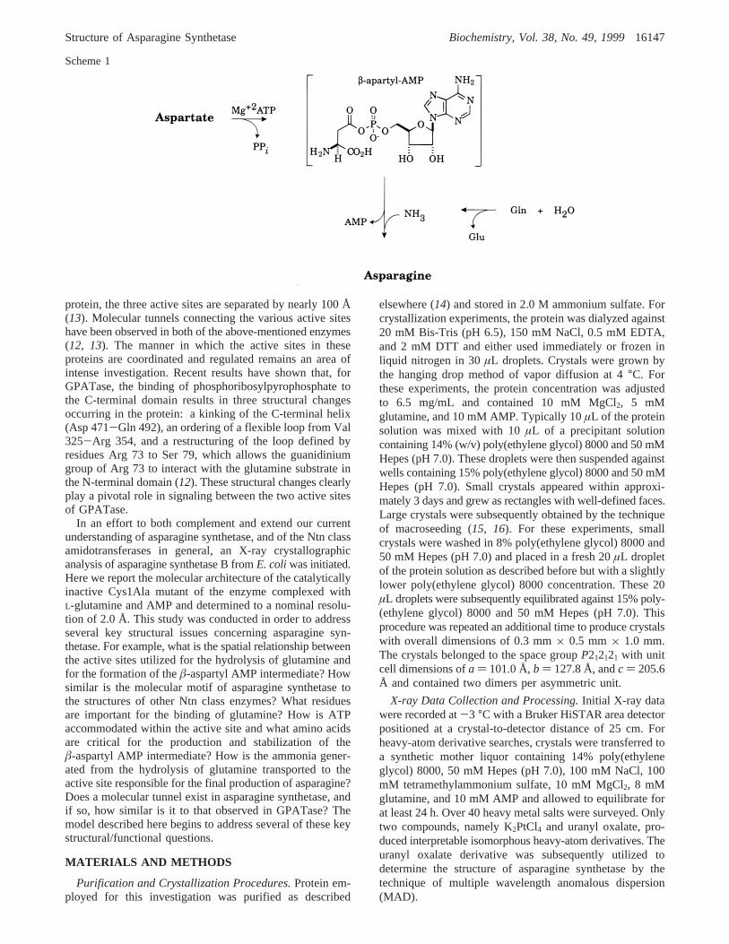
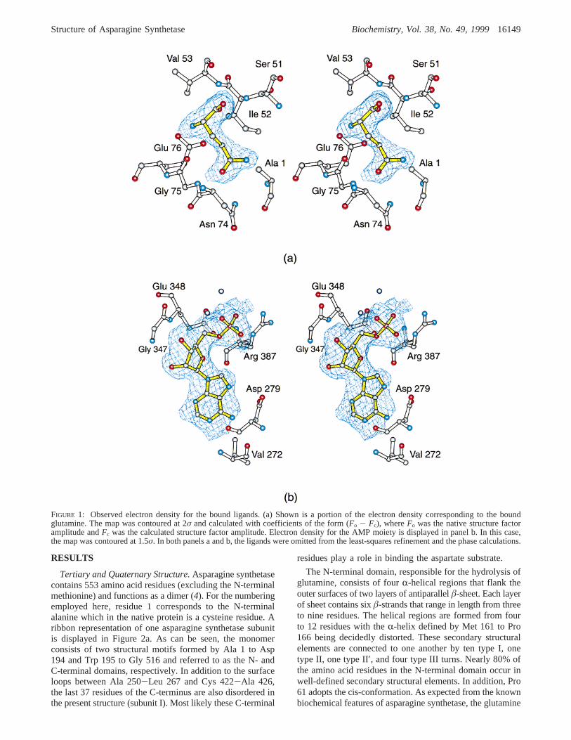
FIGURE 1: Observed electron density for the bound ligands. (a) Shown is a portion of the electron density corresponding to the boundglutamine. The map was contoured at 2σ and calculated with coefficients of the form (Fo - Fc), whereFo was the native structure factoramplitude andFc was the calculated structure factor amplitude. Electron density for the AMP moiety is displayed in panel b. In this case,the map was contoured at 1.5σ. In both panels a and b, the ligands were omitted from the least-squares refinement and the phase calculations.
Structure of Asparagine Synthetase Biochemistry, Vol. 38, No. 49, 199916149
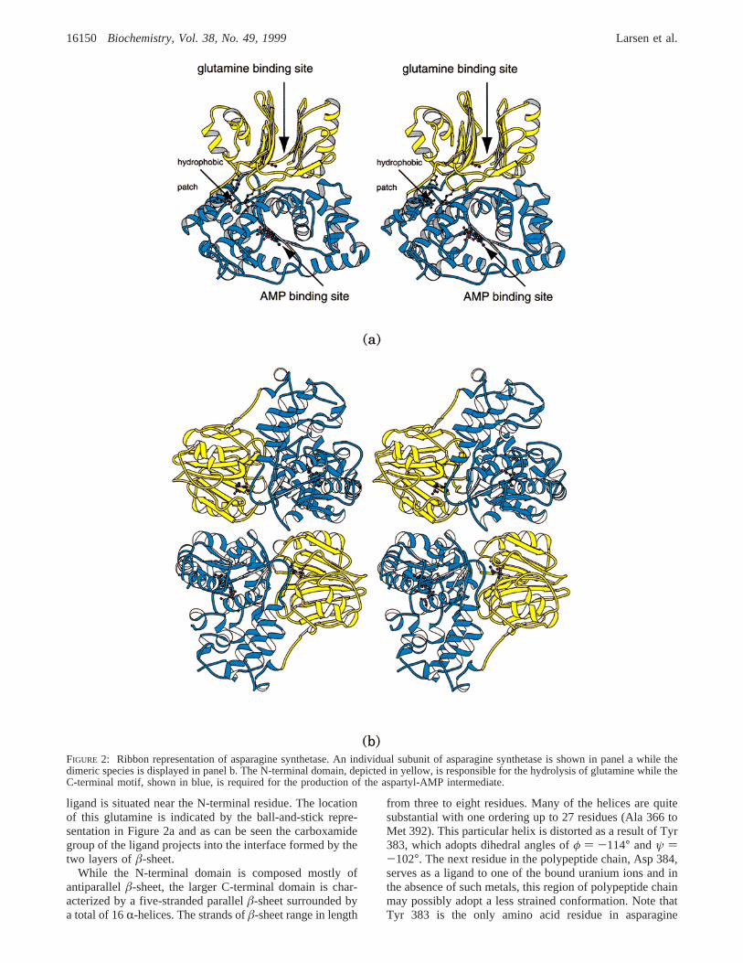
ligand is situated near the N-terminal residue. The locationof this glutamine is indicated by the ball-and-stick repre-sentation in Figure 2a and as can be seen the carboxamidegroup of the ligand projects into the interface formed by thetwo layers ofâ-sheet.
While the N-terminal domain is composed mostly ofantiparallelâ-sheet, the larger C-terminal domain is char-acterized by a five-stranded parallelâ-sheet surrounded bya total of 16R-helices. The strands ofâ-sheet range in length
from three to eight residues. Many of the helices are quitesubstantial with one ordering up to 27 residues (Ala 366 toMet 392). This particular helix is distorted as a result of Tyr383, which adopts dihedral angles ofφ ) -114° andψ )-102°. The next residue in the polypeptide chain, Asp 384,serves as a ligand to one of the bound uranium ions and inthe absence of such metals, this region of polypeptide chainmay possibly adopt a less strained conformation. Note thatTyr 383 is the only amino acid residue in asparagine
FIGURE 2: Ribbon representation of asparagine synthetase. An individual subunit of asparagine synthetase is shown in panel a while thedimeric species is displayed in panel b. The N-terminal domain, depicted in yellow, is responsible for the hydrolysis of glutamine while theC-terminal motif, shown in blue, is required for the production of the aspartyl-AMP intermediate.
16150 Biochemistry, Vol. 38, No. 49, 1999 Larsen et al.
synthetase whose dihedral angles lie well outside of theallowed regions of the Ramachandran plot. In addition totheR-helices andâ-strands, there are seven type I, two typeII, and two type III turns also present in the C-terminaldomain. Again, most of the residues forming the C-terminaldomain are situated in well-defined secondary structuralelements with only∼24% lying in random coil regions. TheAMP moiety observed in the electron density map lies acrossthe C-terminal edge of theâ-sheet as indicated by the ball-and-stick representation in Figure 2a. The glutamine andAMP ligands are separated by approximately 19 Å.
From Figure 2a, it is immediately obvious that the N- andC-domains of asparagine synthetase are quite distinct bio-chemical units with no commonly shared secondary structuralelements. The molecular interface between these motifs isextensive with a buried surface area of∼2500 Å2 ascalculated according to the algorithm of GRASP (24).Specifically, this domain:domain interface is built primarilyfrom the regions of polypeptide delineated by His 29 to Asp33, Leu 50 to Asp 54, Glu 76 to Tyr 78, Asp 115 to Gln118, His 139 to Tyr 146, and Glu 160 to Glu 173 in theN-terminal domain and by Ser 221 to Asp 226, Tyr 313 toGlu 316, Lys 342, Lys 367 to His 381, and Asn 389 to Arg414 in the C-terminal domain. While the intramolecularinterface contributed by the N-terminal domain is formedprimarily by reverse turns and random coil regions, themolecular surface donated by the C-terminal domain iscomposed mostly ofR-helices and oneâ-strand. Bothhydrophobic and electrostatic interactions occur within theseregions. As an example of the former, the molecular interfaceformed by Leu 50 to Asp 54 is reasonably hydrophobic withthe side-chain groups of Leu 50 and Ile 52 pointing towardthe interior. As examples of the latter, there are salt bridgesformed between the functional groups of Asp 33 and Lys342, Asp 115 and Lys 406, and Lys 163 and Glu 316. Astriking hydrophobic patch exists at the transitional regionbetween the two domains as indicated in Figure 2a. In thisarea there is a crown of leucine residues (140, 377, 378,380, 404, 409) surrounded by Tyr 191, Trp 195, and Phe196. Interestingly, the distortedR-helix that includes Tyr 383discussed above forms part of the intramolecular interfacethus providing a direct link between the active site of theC-terminal domain and the surface of the N-terminal motif.
As depicted in Figure 2b, the asparagine synthetase dimeris fairly symmetrical with overall dimensions of approxi-mately 98 Å× 103 Å × 74 Å and a buried surface area of∼2220 Å2. The two bound glutamine ligands within thedimer are 27 Å apart while the two nucleotide monophos-phate moieties are separated by 39 Å. A distance of 25 Åseparates the glutamine/nucleotide pairs in the neighboringsubunits of the physiological dimer. The dimeric interfaceis formed predominately by four regions of polypeptide chaincontributed by each subunit and delineated by Arg 17 to Glu48 (anR-helix-turn-â-sheet-turn-â-sheet motif), Gln 302 toHis 314 (anR-helix), Leu 331 to Lys 342 (anR-helix), andSer 393 to Gly 396 (a type I turn). Along with the numerouswater molecules that line the subunit:subunit interface, thereare various salt bridges that occur including those betweenArg 17 and Asp 306*, Glu 48 and Arg 334*, Asp 306 andArg 17*, and Arg 334 and Glu 48* (where those residuesindicated by the asterisks belong to subunit II). Additionally,the side-chain groups of Arg 25 and Arg 28 (subunit I)
project into the dimeric interface such that both of theirguanidinium groups lie within∼3.3 Å of the carboxylategroup of Asp 310 (subunit II). The symmetry-related ionpairs between Arg 25 and Arg 28 of subunit II and Asp 310(subunit I) are also present in the dimeric species. In additionto numerous electrostatic interactions, there are severalhydrophobic patches that contribute to the subunit:subunitinterface of asparagine synthetase, including the regionsurrounding the side chain of Trp 395, which is located inthe ∼Type I turn listed above.
ActiVe Site of the N-Terminal Domain.Since the structureof asparagine synthetase reported here is that of a mutantprotein whereby the active nucleophile Cys 1 was changedto an alanine residue, it was possible to crystallize the enzymein the presence of its natural glutamine substrate. A close-up view of the glutaminase active site in the N-terminaldomain is displayed in Figure 3a, and a cartoon of potentialhydrogen bonding interactions between the ligand and theprotein is given in Figure 3b. For the sake of clarity, onlythose residues that lie within approximately 4 Å of theligandare shown. There are only two bound water moleculesobserved in the active site. One of these solvents interactswith the R-carboxylate group of the substrate while thesecond forms a hydrogen bond with the carbonyl oxygen ofside-chain carboxamide group. As indicated by the dashedlines in Figure 3b, the side-chain functional groups of Arg49, Asn 74, Glu 76, and Asp 98 form hydrogen bonds withthe glutamine ligand. These hydrogen bonds range in lengthfrom 2.6 to 3.1 Å. In addition to these side chains, thebackbone amide groups of Ile 52, Val 53, and Gly 75 andthe carbonyl oxygens of Leu 50 and Gly 75 also play keyroles in the proper positioning of the ligand within the active-site pocket. The observed interactions between the ligandand Arg 49, Asn 74, and Asp 98 were, in fact, previouslypredicted from model building studies of asparagine syn-thetase (5) based on the structural analyses of theE. coliGPATase (25) and an N-terminal proteolytic fragment oftheE. coli glutamine fructose-6-phosphate amidotransferase(26).
Note that Câ of Ala 1 is situated at 3.9 Å from the carbonylcarbon of the substrate that would be attacked by theN-terminal cysteine residue in the native protein. With theassumption that the glutamine ligand binds to this Cys1Alamutant protein in an identical manner to that expected forthe native protein, it can be speculated that the nucleophilicattack by the thiolate of Cys 1 occurs at thesi-face of theglutamine substrate, yielding a tetrahedral intermediate withan S-configuration. The oxyanion of the tetrahedral inter-mediate presumably is stabilized via hydrogen bonds donatedby the backbone amide group of Gly 75 and Nδ2 of Asn 74.In the structure reported here, the distance between the side-chain carbonyl oxygen of the ligand and the backbone amidegroup of Gly 75 is 2.5 Å. Similarly, Nδ2 of Asn 74 ispositioned within 3.0 Å to this oxygen atom.
ActiVe Site of the C-Terminal Domain.Unlike the activesite in the glutaminase portion of asparagine synthetase, theC-terminal active site is less well-defined in the presentcomplex. From Figure 2a, it is immediately apparent thatthe binding pocket for the observed AMP moiety is quiteopen and most likely the disordered regions in the presentmodel play critical roles in the binding of Mg2+ATP andaspartate. Even in light of this caveat, the general region for
Structure of Asparagine Synthetase Biochemistry, Vol. 38, No. 49, 199916151

the C-terminal domain active site has been partially definedin the present structural analysis and is shown in Figure 4a.In addition to the bound nucleotide, three uranium ions wereobserved in the active site. Most likely some of these metalsare mimicking the binding of magnesium ions. The riboseof the nucleotide adopts the C3′-endo pucker while theadenine ring lies in the anti-conformation. Two specificelectrostatic interactions exist between the purine ring of thenucleotide and the protein: one of these occurs between thebackbone carbonyl group of Val 272 and the amino groupat position 6 of the adenine ring while the second occursbetween the amide group of Val 272 and N1 of the purinemoiety. In both cases, the distances between the oxygensand nitrogens in these hydrogen bonds are∼3.1 Å. The 2′-hydroxyl group of the nucleotide ribose is linked to theprotein via O of Leu 232, Oγ of Ser 346, and N of Glu 347.The only other interactions between the nucleotide and theprotein occur via the bound uranium ions. One of the uraniumions sits at 2.6 Å from the 3′-hydroxyl group of the ribose.
The other two uranium sites are coordinated by phosphoryloxygens of the AMP moiety. One of these ions is furtherligated to the protein via the carboxylate side chains of Asp238 and Asp 351 while the second metal is surrounded bythe carboxylate side chains of Glu 352 and Asp 384 and Oη
of Tyr 357. The metal:ligand bond distances range in lengthfrom 2.3 to 2.8 Å. The coordination geometry for the metalsand the observed protein:nucleotide interactions will mostlikely change when the structure of asparagine synthetase issolved in the presence ofâ-aspartyl-AMP mimics andmagnesium ions.
DISCUSSION
Significant progress was made on our understanding ofthe amidotransferases by the informative X-ray crystal-lographic studies of GMP synthetase fromE. coli byDavisson and Smith (9) and of GPATase, also fromE. coli,by Zalkin and Smith (12, 25, 27). These specific enzymesbelong to the Triad and Ntn classes, respectively. Subsequent
FIGURE 3: Close-up view of the N-terminal domain active site. Those protein atoms located within approximately 4 Å of the glutamineligand are shown in panel a. A cartoon of the possible electrostatic interactions between the ligand and the protein, within 3.2 Å, is givenin panel b.
16152 Biochemistry, Vol. 38, No. 49, 1999 Larsen et al.

structural analyses of two additional enzymes, namely theN-terminal domain of glucosamine 6-phosphate synthase (26)and carbamoyl phosphate synthetase (10, 13, 28, 29, 30)added important new information regarding the Ntn and Triadfamilies, respectively. From a combination of both biochemi-cal and structural data, it is now known that the Ntnamidotransferases, as the name implies, employ an N-terminal cysteine residue as the active-site nucleophile forthe hydrolysis of glutamine to glutamate and ammonia. Asa consequence of the location of the nucleophile, theglutaminase domains of the Ntn class proteins are alwayslocated at the N-terminal portions of the molecules. In thecase of the Triad family, however, this restriction does notapply. These enzymes contain a conserved Cys-His couplethat is located in the N-terminal domain of GMP synthetase,but in the C-terminal domain of the carbamoyl phosphatesynthetase small subunit. While these enzymes are referredto as the Triad family, this may, in fact, be a misnomer.Presently, there is some discrepancy in the literature regard-ing the third residue in the Triad, namely a conservedglutamate. In GMP synthetase, it has been speculated thatthis glutamate is part of a catalytic triad (9). However, astudy on the glutaminase subunit ofp-aminobenzoate syn-
thetase has demonstrated that replacement of this residue withan alanine or aspartate results in little loss of catalytic activity(31). Regardless of the actual role of the conserved glutamate,these enzymes will most likely continue to be referred to asthe Triad class in the literature, and, as such, this nomen-clature will be retained here.
Prior to the structural analysis of asparagine synthetasedescribed in this report, it had already been predicted thatits N-terminal domain would have a similar molecular foldto that observed in GPATase and that its C-terminal domainwould be homologous to the C-terminal domain of GMPsynthetase (9). These predictions of similar structures werebased on both amino acid sequence homologies and con-sideration of common catalytic mechanisms and wereexceedingly accurate (32). Indeed, asparagine synthetase canbe envisioned simply as a chimera of GPATase and GMPsynthetase whereby an Ntn class glutaminase domain hasbeen attached to a synthetase domain belonging to a Triadfamily. This mixing and matching of donor (glutaminase)and acceptor (synthetase) domains is a common themeamong the amidotransferases.
Shown in Figure 5a is a superposition of the N-terminaldomain of GPATase onto the N-terminal domain of aspar-
FIGURE 4: Close-up view of the AMP binding region in the C-terminal domain. Those protein atoms located within approximately 4 Å ofthe nucleotide monophosphate are shown in panel a. A cartoon of the possible electrostatic interactions between the protein and the AMP,within 3.2 Å, is displayed in panel b. The bound uranium ions are indicated by the yellow/gray spheres.
Structure of Asparagine Synthetase Biochemistry, Vol. 38, No. 49, 199916153

agine synthetase. TheR-carbons for these two modelssuperimpose with a root-mean-square deviation of 1.4 Å for176 structurally equivalent residues. It is immediatelyobvious that the two structures differ only in a few regions.The largest difference occurs at Ser 40 (asparagine syn-thetase) or Ile 36 (GPATase). Here, there is an insertion inthe GPATase model whereby the polypeptide chain wrapsaround to form an additionalâ-strand-helix motif. As aconsequence, the firstâ-sheet in GPATase contains sevenrather than sixâ-strands. The two polypeptide chainssuperimpose again at Ile 44 (asparagine synthetase) or Gly68 (GPATase). There is a second, rather large insertion, thatoccurs in the region defined by Glu 108 to Phe 113 inasparagine synthetase or Ser 137 to Thr 159 in GPATase.Finally, there are two smaller insertions that occur betweenGlu 151 to Gly 153 and Ser 182 to Gly 185 in asparaginesynthetase and correspond to Asp 196 to Thr 202 and Tyr231 to Phe 239 in GPATase.
The structure of GPATase was solved in the presence ofa glutamine analogue, namely 6-diazo-5-oxonorleucine andreferred to as DON (12). Shown in Figure 5b is a superposi-tion of the active sites for the glutaminase domains ofGPATase and asparagine synthetase. The chemical environ-ments of the active sites observed in asparagine synthetase
and GPATase are somewhat different. For example, Tyr 74,Pro 75, Thr 76, Ala 77, Asn 103, and Ser 128 in GPATaseare replaced by Leu 50, Ser 51, Ile 52, Val 53, Glu 76, andCys 99 in asparagine synthetase. As can be seen, there is aclose three-dimensional correspondence between the DONinhibitor bound to GPATase and the actual substrate,glutamine, as observed in asparagine synthetase. In GPATase,the side-chain carbonyl group of the inhibitor is hydrogenbonded to the backbone amide group of Gly 102 and Nδ2 ofAsn 101. These interactions are identical to those depictedfor asparagine synthetase in Figure 3b. Likewise, as observedin asparagine synthetase, the carboxylate group of the DONinhibitor forms a salt bridge with an arginine residue inGPATase, namely Arg 73. The amino group of the DONinhibitor interacts with GPATase in a slightly differentmanner to that observed for glutamine bound to asparaginesynthetase. Specifically, Oγ of Thr 76 and Oδ1 of Asp 127lie within hydrogen-bonding distance to the amino group.These residues are replaced by Glu 76 and Asp 98 inasparagine synthetase. The structure of asparagine synthetasereported here is that of a site-directed mutant, Cys1Ala. It isimportant to note the close correspondence between theN-terminal alanine of asparagine synthetase and the N-terminal cysteine of GPATase. This similarity indicates that
FIGURE 5: Comparison of the glutaminase domains in asparagine synthetase and GPATase. X-ray coordinates for GPATase (12) wereobtained from the Brookhaven Protein Data Bank (1ECC). A superposition of theR-carbons for the two N-terminal domains in asparaginesynthetase (blue) and GPATase (red) is depicted in panel a. A close-up view of the active site regions is shown in panel b. Note that thestructure of GPATase was solved in the presence of a glutamine affinity analogue covalently attached to the N-terminal cysteine residue.
16154 Biochemistry, Vol. 38, No. 49, 1999 Larsen et al.
the structural results presented here, albeit based on a mutantprotein, represent a good mimic for the native enzyme.
The synthetase domain of asparagine synthetase is respon-sible for the binding of Mg2+ATP and aspartate and thesubsequent production of theâ-aspartyl intermediate and PPi.These functional properties place asparagine synthetase intothe category of ATP pyrophosphatases (33). Likewise, theC-terminal domain of GMP synthetase catalyzes the produc-tion of the O2-adenyl-XMP intermediate and PPi from XMP
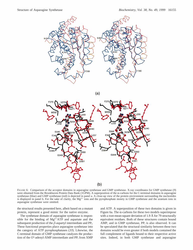
and ATP. A superposition of these two domains is given inFigure 6a. TheR-carbons for these two models superimposewith a root-mean-square deviation of 1.9 Å for 79 structurallyequivalent residues. Both of these structures contain boundAMP, and in GMP synthetase, PPi is also observed. It canbe speculated that the structural similarity between these twodomains would be even greater if both models contained thefull complement of ligands bound to their respective activesites. Indeed, in both GMP synthetase and asparagine
FIGURE 6: Comparison of the acceptor domains in asparagine synthetase and GMP synthetase. X-ray coordinates for GMP synthetase (9)were obtained from the Brookhaven Protein Data Bank (1GPM). A superposition of theR-carbons for the C-terminal domains in asparaginesynthetase (blue) and GMP synthetase (red) is depicted in panel a. A close-up view of the protein environment surrounding the nucleotidesis displayed in panel b. For the sake of clarity, the Mg2+ ions and the pyrophosphate moiety in GMP synthetase and the uranium ions inasparagine synthetase were omitted.
Structure of Asparagine Synthetase Biochemistry, Vol. 38, No. 49, 199916155
synthetase, there are residues missing from the presentmodels: the loop between Ala 346 and Lys 369 in GMPsynthetase and the loops between Ala 250 to Leu 267 andCys 422 to Ala 426 in asparagine synthetase. Interestingly,the structural correspondence observed between the C-terminal domains of asparagine synthetase B and GMPsynthetase does not extend to the molecular motif recentlydetermined by Nakatsu et al., (34) for theE. coli asparaginesynthetase A. Rather, this enzyme closely resembles thecatalytic domain of yeast aspartyl-tRNA synthetase (35).
There are four major regions where the two polypeptidechains differ significantly between GMP synthetase andasparagine synthetase: (i) a 13 residue insertion in asparaginesynthetase beginning at Ala 250, (ii) a four residue insertionin asparagine synthetase beginning at Thr 300 and resultingin a completely different disposition of the surface loopsdefined by Thr 300 to Ala 325 in asparagine synthetase andGlu 291 to Ile 311 in GMP synthetase, (iii) an extended helix-turn-helix motif delineated by Val 353 to Gly 396 inasparagine synthetase which corresponds to the disorderedregion in GMP synthetase, and (iv) a 21 residue insertion inasparagine synthetase beginning at Lys 406.
A close-up view of the protein environment surroundingthe bound nucleotides in asparagine synthetase and GMPsynthetase is given in Figure 6b. Like that observed inasparagine synthetase, both N1 of the adenine ring and theN6 amino group of the base are hydrogen bonded to GMPsynthetase via the backbone nitrogen and carbonyl oxygenof Val 260, respectively. The “P-loops”, delineated by Ser234 to Ser 240 in asparagine synthetase and Ser 235 to Ser241 in GMP synthetase, are virtually identical with the onlyexception being Leu 237 in asparagine synthetase beingreplaced with a valine residue in GMP synthetase. As canbe seen in Figure 6b, there are significant differences between
the two models near the phosphate binding region which,most likely, are a result of the different constituents boundwithin the active sites (i.e., uranium ions in asparaginesynthetase and PPi in GMP synthetase).
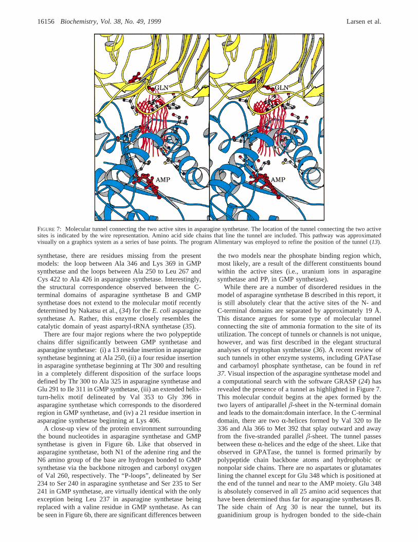
While there are a number of disordered residues in themodel of asparagine synthetase B described in this report, itis still absolutely clear that the active sites of the N- andC-terminal domains are separated by approximately 19 Å.This distance argues for some type of molecular tunnelconnecting the site of ammonia formation to the site of itsutilization. The concept of tunnels or channels is not unique,however, and was first described in the elegant structuralanalyses of tryptophan synthetase (36). A recent review ofsuch tunnels in other enzyme systems, including GPATaseand carbamoyl phosphate synthetase, can be found in ref37. Visual inspection of the asparagine synthetase model anda computational search with the software GRASP (24) hasrevealed the presence of a tunnel as highlighted in Figure 7.This molecular conduit begins at the apex formed by thetwo layers of antiparallelâ-sheet in the N-terminal domainand leads to the domain:domain interface. In the C-terminaldomain, there are twoR-helices formed by Val 320 to Ile336 and Ala 366 to Met 392 that splay outward and awayfrom the five-stranded parallelâ-sheet. The tunnel passesbetween theseR-helices and the edge of the sheet. Like thatobserved in GPATase, the tunnel is formed primarily bypolypeptide chain backbone atoms and hydrophobic ornonpolar side chains. There are no aspartates or glutamateslining the channel except for Glu 348 which is positioned atthe end of the tunnel and near to the AMP moiety. Glu 348is absolutely conserved in all 25 amino acid sequences thathave been determined thus far for asparagine synthetases B.The side chain of Arg 30 is near the tunnel, but itsguanidinium group is hydrogen bonded to the side-chain
FIGURE 7: Molecular tunnel connecting the two active sites in asparagine synthetase. The location of the tunnel connecting the two activesites is indicated by the wire representation. Amino acid side chains that line the tunnel are included. This pathway was approximatedvisually on a graphics system as a series of base points. The program Alimentary was employed to refine the position of the tunnel (13).
16156 Biochemistry, Vol. 38, No. 49, 1999 Larsen et al.
carboxamide groups of Asn 74 and Asn 389. Both Arg 30and Asn 74 are absolutely conserved while Asn 389 isreplaced with an aspartate residue in some asparaginesynthetases. Those residues with side chains pointing intothe tunnel include Met 120, Ile 142, Ile 143, Leu 232, Met329, Ser 346, Ala 388, Met 392, Ser 393, Ala 399, and Val401. Leu 232, Met 329, Ser 346, and Ala 388 form a clusternear the C-terminal portion of the tunnel and are absolutelyconserved among all 25 asparagine synthetase B amino acidsequences. Most of the other residues listed above are locatedmore toward the N-terminal portion of the tunnel and arenot strictly conserved. Both Met 120 and Ile 142, however,are replaced with side chains of comparable chemicalreactivities. In the present model of asparagine synthetasewith bound glutamine and AMP, there are several watermolecules located within the tunnel. The positions of thesesolvents most likely change upon catalysis.
In summary, the structure of asparagine synthetase pre-sented here provides another entry into the growing databaseconcerning the Ntn-class amidotransferases. From this in-vestigation, the spatial relationships between the two activesites have been determined, and the manners in whichglutamine and AMP are accommodated within their respec-tive binding pockets have been defined. In addition, atentative molecular tunnel connecting the two active siteshas been identified. Most likely the amino acid residues thatare disordered in the current model play a key role in bindingboth the aspartate substrate and theâ-aspartyl AMP inter-mediate. Further definition of the C-terminal domain activesite and the molecular tunnel must await additional structuralanalyses of the enzyme in the presence of aspartate analoguesand â-aspartyl AMP mimics; however, the current studyestablishes a solid foundation for these efforts. This work isin progress.
ACKNOWLEDGMENT
We thank Dr. W. W. Cleland for critically reading thismanuscript and Dr. G. H. Reed for the financial support ofT. M. L. In addition, we thank Dr. Gary Wesenberg forhelpful discussions regarding the molecular tunnel. Use ofthe Argonne National Laboratory Structural Biology Centerbeamlines at the Advanced Photon Source was supportedby the U.S. Department of Energy, Office of EnergyResearch, under Contract W-31-109-ENG-38. We thank Drs.Frank Rotella and Norma Duke for their assistance in settingup the MAD experiment. Finally, we acknowledge thesupport of the Florida Space Grant Consortium for theirsummer fellowship to Stephen Goble who was involved inthe initial crystallization experiments.
REFERENCES
1. Milman, H. A., and Cooney, D. A. (1979)Biochem. J. 181,51-59.
2. Zalkin, H., and Truit, C. D. (1977)J. Biol. Chem. 252, 5341-5346.
3. Nakamura, M., Yamada, M., Hirota, Y., Sugimoto, K., Oka,A., and Takanami, M. (1981)Nucleic Acids Res. 9, 4669-4676.
4. Scofield, M. A., Lewis, W. S., and Schuster, S. M. (1990)J.Biol. Chem. 265, 12895-12902.
5. Richards, N. G., and Schuster, S. M. (1998)AdV. Enzymol.72, 145-198.
6. Zalkin, H. (1993)AdV. Enzymol. Relat. Areas Mol. Biol. 66,203-309.
7. Zalkin, H., and Smith, J. L. (1998)AdV. Enzymol. Relat. AreasMol. Biol. 72, 87-144.
8. Amuro, N., Paluh, J. L., and Zalkin, H. (1985)J. Biol. Chem.260, 14844-14849.
9. Tesmer, J. J. G., Klem, T. J., Deras, M. L., Davisson, V. J.,and Smith, J. L. (1996)Nat. Struct. Biol. 3, 74-86.
10. Thoden, J. B., Miran, S. G., Phillips, J. C., Howard, A. J.,Raushel, F. M., and Holden, H. M. (1998)Biochemistry 37,8825-8831.
11. Smith, J. L., Zaluzec, E. J., Wery, J. P., Niu, L., Switzer, R.L., Zalkin, H., and Satow, Y. (1994)Science 264, 1427-1433.
12. Krahn, J. M., Kim, J. H., Burns, M. R., Parry, R. J., Zalkin,H., and Smith, J. L. (1997)Biochemistry 36, 11061-11068.
13. Thoden, J. B., Holden, H. M., Wesenberg, G., Raushel, F. M.,and Rayment, I. (1997)Biochemistry 36, 6305-6316.
14. Boehlein, S. K., Richards, J. G. J., and Schuster, S M. (1994)J. Biol. Chem. 269, 7450-7457.
15. Thaller, C., Weaver, L. H., Eichele, G., Wilson, E., Karlsson,R., and Jansonius, J. N. (1981)J. Mol. Biol. 147, 465-469.
16. Thaller, C., Eichele, G., Weaver, L. H., Wilson, E., Karlsson,R., and Jansonius, J. N. (1985)Methods Enzymol. 114, 132-135.
17. Westbrook, E. M., and Naday, I. (1997)Methods Enzymol.276, 244-268.
18. Otwinowski, Z., and Minor, W. (1997)Methods Enzymol. 276,307-326.
19. Terwilliger, T. C., and Eisenberg, D. (1983)Acta Crystallogr.,Sect. A 39, 813-817.
20. Terwilliger, T. C. (1997)Methods Enzymol. 276, 530-537.21. Cowtan, K., and Main, P. (1998)Acta Crystallogr., Sect. D
54, 487-493.22. Rypniewski, W. R., Breiter, D. R., Benning, M. M., Wesen-
berg, G., Oh, B.-H., Markley, J. L., Rayment, I., and Holden,H. M. (1991)Biochemistry 30, 4126-4131.
23. Tronrud, D. E., Ten Eyck, L. F., and Matthews, B. W. (1987)Acta Crystallogr., Sect. A. 43, 489-501.
24. Nicholls, A., Sharp, K. A., and Honig, B. (1991)Proteins:Struct., Funct., Genet. 11, 281-296.
25. Kim, J. H., Krahn, J. M., Tomchick, D. R., Smith, J. L., andZalkin, H. (1996)J. Biol. Chem. 271, 15549-15557.
26. Isupov, M. N., Obmolova, G., Butterworth, S., Badet-Denisot,M. A., Badet, B., Polikarpov, I., Littlechild, J. A., andTeplyakov, A. (1996)Structure 4, 801-810.
27. Muchmore, C. R., Krahn, J. M., Kim, J. H., Zalkin, H., andSmith, J. L. (1998)Protein Sci. 7, 39-51.
28. Thoden, J. B., Raushel, F. M., Benning, M. M., Rayment, I.,and Holden, H. M. (1999)Acta Crystallogr., Sect. D 55, 8-24.
29. Thoden, J. B., Wesenberg, G., Raushel, F. M., and Holden,H. M. (1999)Biochemistry 38, 2347-2357.
30. Thoden, J. B., Raushel, F. M., Wesenberg, G., and Holden,H. M. (1999)J. Biol. Chem(in press).
31. Roux, B., and Walsh, C. T. (1993)Biochemistry 32, 3763-32. 3768.33. Boehlein, S. K., Walworth, E. S., Schuster, S. M., and
Richards, N. G. J. (1999) Manuscript in preparation.34. Bork, P., and Koonin, E. V. (1994)Proteins: Struct., Funct.,
Genet. 20, 347-355.35. Nakatsu, T., Kato, H., and Oda, J. (1998)Nat. Struct. Biol. 5,
15-19.36. Ruff, M., Krishnaswamy, S., Boeglin, M., Poterszman, A.,
Mitschler, A., Podjarny, A., Rees, B., Thierry, J. C., and Moras,D. (1991)Science 252, 1682-1689.
37. Hyde, C. C., Ahmed, S. A., Padlan, E. A., Miles, E. W., andDavies, D. R. (1988)J. Biol. Chem. 263, 17857-17871.
38. Miles, E. W., Rhee, S., and Davies, D. R. (1999)J. Biol. Chem.274, 12193-12196.
BI9915768
Structure of Asparagine Synthetase Biochemistry, Vol. 38, No. 49, 199916157