classification pathogenesis and treatment of benign ... · vascular anomalies in children with a...
TRANSCRIPT

9/2/2014
1
Classification, Pathogenesis Classification, Pathogenesis and Treatment of Benign and Treatment of Benign
Vascular Anomalies in ChildrenVascular Anomalies in ChildrenWith a review of the pathology
Martin C. Mihm Jr., MDMartin C. Mihm Jr., MDDirector, Melanoma ProgramDirector, Melanoma Program
Brigham and Women’s HospitalBrigham and Women’s HospitalHarvard Medical SchoolHarvard Medical School
Conflict of Interest
Chairman Scientific Advisory Board –Caliber I.D. Inc.
Member Scientific Advisory Board – MELA Sciences IncSciences Inc.
Vascular anomalies are either Vascular anomalies are either hemangiomas or malformationshemangiomas or malformations
10% of all children are born 10% of all children are born with a vascular birthmarkwith a vascular birthmark
90% resolve by age 2; the90% resolve by age 2; the 90% resolve by age 2; the 90% resolve by age 2; the remaining are either a remaining are either a problematic problematic hemangiomahemangioma(infantile), or type of (infantile), or type of hemangiomahemangioma (NICH/RICH) or a (NICH/RICH) or a vascular malformation (the vascular malformation (the most common is a port wine most common is a port wine stain)stain)
Facial hemangioma
Facial Port Wine Stain
Mulliken & Young (1988)

9/2/2014
2
HemangiomasHemangiomas--Clinical PresentationClinical Presentation Typically Typically infantile infantile hemangiomashemangiomas appearappear
3 to 4 weeks after birth3 to 4 weeks after birth
Start as a flat Start as a flat blanched lesion blanched lesion
Begin proliferation Begin proliferation at 4 to 6 weeksat 4 to 6 weeks
Can be both Can be both superficial/deep superficial/deep
Waner and Suen (1999)Waner and Suen (1999)
Hemangioma “before” signs of proliferation
Hemangioma during proliferation

9/2/2014
3
Infantile Hemangioma, Proliferative phase
Infantile Hemangioma, Proliferative phase

9/2/2014
4
Infantile Hemangioma:
Involution
Infantile Hemangioma, Mid-Involution

9/2/2014
5
Infantile Hemangioma, End-stage

9/2/2014
6
One prominent One prominent histological feature histological feature of infantile of infantile hemangiomas, the hemangiomas, the presence of presence of endoneurial endoneurial pseudoinvasion, pseudoinvasion, led us to led us to investigate bloodinvestigate blood--nerve barrier nerve barrier competency in competency in these lesions.these lesions.
GLUT1GLUT11.1. One of a family of facilitative glucose One of a family of facilitative glucose
transporter protein isoforms, each with a transporter protein isoforms, each with a limited tissue distribution in vivo.limited tissue distribution in vivo.
2. Expression found in normal tissues highly2. Expression found in normal tissues highly2. Expression found in normal tissues highly 2. Expression found in normal tissues highly restricted to erythrocytes, perineural cells, restricted to erythrocytes, perineural cells, endothelial cells at bloodendothelial cells at blood--tissue barriers as tissue barriers as brain, nerve and placenta, and some brain, nerve and placenta, and some epithelial barriers.epithelial barriers.
3. Up3. Up--regulation in many malignant cells, regulation in many malignant cells, but not in benign tumors.but not in benign tumors.
Pyogenic Granuloma
GLUT1

9/2/2014
7
Hemangioma
Lesion Gene Locus Pathway Therapy
IH VEGFR3; PDGFR-beta; FLT4;
5p31-33 VEGF receptor pathway; EC proliferation;
Propranolol; acebutolol; corticosteroids;
VEGFR2; TEM8
p ;tubular morphogenesis; sprouting integrin-like receptor
Uebelhoer M., Boon LM, ikkula M. CSH Perspectives; 2012
Preferred Treatment of Infantile Hemangiomas Early proliferating superficial lesions, especially segmental, should be treated with pulse dye laser or topicals
Steroids are still preferred for intralesional injection into small focal hemangiomas since propranolol has not been found to be effective when injected directly into a lesion
Propranolol is first line oral/systemic treatment for large, disfiguring and problematic segmental and focal hemangiomas
Surgery is considered for lesions that fail drug and/or laser therapy or when a vital structure is impaired and there is insufficient time to wait for drug therapy to take effect
Labreze, de la Roque, Hubiche, Boralevi, Bordeau Children’s Labreze, de la Roque, Hubiche, Boralevi, Bordeau Children’s Hospital, June 2008 (Hospital, June 2008 (New England Journal of MedicineNew England Journal of Medicine) 358) 358--26492649--26512651
C
GLUT1 Le Y
The Unique Vascular Phenotype of Infantile Hemangioma
FcgammaRIIFcgammaRII MEROSIN

9/2/2014
8
GLUT1 LeY
PLACENTA
FcγIlR MEROSIN
POSSIBLE MECHANISMS FOR SHARED POSSIBLE MECHANISMS FOR SHARED HEMANGIOMAHEMANGIOMA--PLACENTAL PHENOTYPEPLACENTAL PHENOTYPE
1. Embolization1. Embolization of placentallyof placentally--derived vascular cells or derived vascular cells or precursors to fetal tissues during gestation or birth (North precursors to fetal tissues during gestation or birth (North P. et al.; P. et al.; Hum PathHum Path; 2001; Mihm MC. Nelson S. ; 2001; Mihm MC. Nelson S. Cutaneous PatholCutaneous Pathol; 2010).; 2010).
2. 2. Colonization by angioblasts Colonization by angioblasts (Boye E. et al.; (Boye E. et al.; JCI; 2001)JCI; 2001)aberrantly “switched” to the placental phenotype by either:aberrantly “switched” to the placental phenotype by either:
a. Somatic mutation.a. Somatic mutation.b. Abnormal local inductive influences. b. Abnormal local inductive influences.
3. Infantile hemangioma 3. Infantile hemangioma stem cellsstem cells give rise to both give rise to both endothelial and pericytic cells. (Boscolo E. et al.; endothelial and pericytic cells. (Boscolo E. et al.; Arteriosclr Thromb Vasc BiolArteriosclr Thromb Vasc Biol; 2013); 2013)
Recent Investigations (Cont.)Recent Investigations (Cont.)
Striking similarities of transcriptomes between Striking similarities of transcriptomes between placenta and hemangioma when studying placenta and hemangioma when studying hierarchical and nonhierarchical clustering hierarchical and nonhierarchical clustering analysis of >7,800 genes from a variety of analysis of >7,800 genes from a variety of tissuestissues
Comparing the two studying arrays of 21 Comparing the two studying arrays of 21 endothelial cell genes in 1000 polymporphisms, endothelial cell genes in 1000 polymporphisms, great similarities were found. (Barnes et al. great similarities were found. (Barnes et al. PNAS, 2005)PNAS, 2005)

9/2/2014
9
Vascular MalformationsVascular MalformationsHigh FlowHigh Flow
Arteriovenous MalformationsArteriovenous Malformations Arteriovenous FistulasArteriovenous Fistulas
Low FlowLow Flow
LymphaticLymphatic CapillaryCapillary VenousVenous MixedMixed
Arteriovenous Malformation
HIGH FLOW
Lymphatic Malformation
LOW FLOW
Most cases occur sporadicallyMost cases occur sporadically Heritable AVMs have been associated with a Heritable AVMs have been associated with a
cutaneous capillary malformation and hereditary cutaneous capillary malformation and hereditary hemorrhagic telangiectasia (HHT)hemorrhagic telangiectasia (HHT)
Arteriovenous Malformations
g g ( )g g ( ) ArterioArterio--venous fistulae are commonly trauma venous fistulae are commonly trauma
associatedassociated Lesions present as often small pulsatile Lesions present as often small pulsatile
cutaneous plaques or nodules with overlying cutaneous plaques or nodules with overlying normal or Port Wine Stainnormal or Port Wine Stain--like skinlike skin
HHTHHT--associated AVMs involve endoglin associated AVMs involve endoglin and activin receptorand activin receptor--like kinase 1 geneslike kinase 1 genes
Loss of function results in impaired TGFLoss of function results in impaired TGF--beta signaling necessary for AVbeta signaling necessary for AV
Arteriovenous Malformations
beta signaling, necessary for AV beta signaling, necessary for AV differentiationdifferentiation
The cutaneous capillary malformation The cutaneous capillary malformation associated with AVM involve mutations in associated with AVM involve mutations in RASA 1 that affects the RAS/MAP Kinase RASA 1 that affects the RAS/MAP Kinase pathwaypathway
Whitehead KJ et al. 2013; CHS Perspectives on medicine

9/2/2014
10
Arteriovenous Malformations
Arteriovenous Malformations

9/2/2014
11
AVM

9/2/2014
12
AVM involving the skin
AVM HistopathologyAVM Histopathology

9/2/2014
13
NIDUS
artery
NIDUS
vein
TheoryTheory
Relative or absolute absence of preRelative or absolute absence of pre--capillary sphincters/sphincter control.capillary sphincters/sphincter control.
Results in continuous shunting of blood Results in continuous shunting of blood across the nidusacross the nidusacross the nidus.across the nidus.
This in turn results in expansion of the This in turn results in expansion of the nidus, venous dilatation and arterial nidus, venous dilatation and arterial hypertrophy.hypertrophy.

9/2/2014
14
Primary nidus Primary nidus –– capillary malformationcapillary malformation Arterial hypertrophy and venous Arterial hypertrophy and venous
dilatationdilatation--secondary changessecondary changesN f li i ll diff ti tiN f li i ll diff ti ti
TheoryTheory
No way of clinically differentiating No way of clinically differentiating between primary nidus and secondary between primary nidus and secondary changes.changes.
Difficulty in determining “tumor margins”, Difficulty in determining “tumor margins”, assuming that the nidus is fixed.assuming that the nidus is fixed.
Treatment of AVMsTreatment of AVMs Requires multidisciplinary Requires multidisciplinary
team approach with team approach with Interventional Radiologist and Interventional Radiologist and SurgeonSurgeon
Goal is to manage, attempt to Goal is to manage, attempt to curecure
Embollization/AngiographicEmbollization/Angiographic
SclerotherapySclerotherapy
Combination with surgery. Combination with surgery. Must remove the NIDUSMust remove the NIDUS
All tissue must be removedAll tissue must be removedWaner & Suen (1999)
ArteriovenousArteriovenous MalformationMalformation
Lesion Gene Locus Pathway Therapy
AVM RASA1 5q13-22 Ras/MAPK inhibiton; Cell motility; Survival
mTOR inhibitors? Ras inhibitors?Survival inhibitors?
Uebelhoer M., Boon LM, ikkula M. CSH Perspectives; 2012

9/2/2014
15
Glomulovenous Glomulovenous MalformationMalformation
“Glomangioma”“Glomangioma”GlomangiomaGlomangioma
Differential diagnosis in infancy:– Blue Rubber Bleb Nevus Syndrome– Leukemia Cutis (Blueberry Muffin Syndrome)
Venous Malformations
Glomulovenous MalformationGlomulovenous Malformation
– Venous Malformations

9/2/2014
16
Glomulovenous MalformationGlomulovenous Malformation
Lesion Gene Locus Pathway Therapy
Glomuvenous Malformation
GLMN 1p21-22 SMC differentiation
P t i
? mTOR inhibitors
; Protein synthesis/ degradation; TGF-beta, HGF pathways
Uebelhoer M., Boon LM, ikkula M. CSH Perspectives; 2012
Low Flow Low Flow –– Lymphatic Lymphatic MalformationMalformation
Lymphatic malformations (cystic Lymphatic malformations (cystic hygroma or lymphangioma are hygroma or lymphangioma are classified as microcystic, classified as microcystic, macrocystic, or mixed. macrocystic, or mixed.
Most lymphatic malformations Most lymphatic malformations (approximately 75%) occur in the (approximately 75%) occur in the cervicofacial region. cervicofacial region.
The overlying skin can be healthy, The overlying skin can be healthy, or it may have tiny characteristic or it may have tiny characteristic vesicles. vesicles.
Lymphatic MalformationsLymphatic Malformations
Lesions often first present or become Lesions often first present or become more extensive at times of hormonal more extensive at times of hormonal change, such as puberty, or associated change, such as puberty, or associated with infectionwith infectionwith infectionwith infection
Recurring infections lead to extensive Recurring infections lead to extensive growth and often require prophylactic growth and often require prophylactic antibioticsantibiotics

9/2/2014
17
LYMPHATIC MALFORMATIONS
Lymphatic MalformationLymphatic Malformation
Lymphatic MalformationLymphatic Malformation
9/2/2014
18
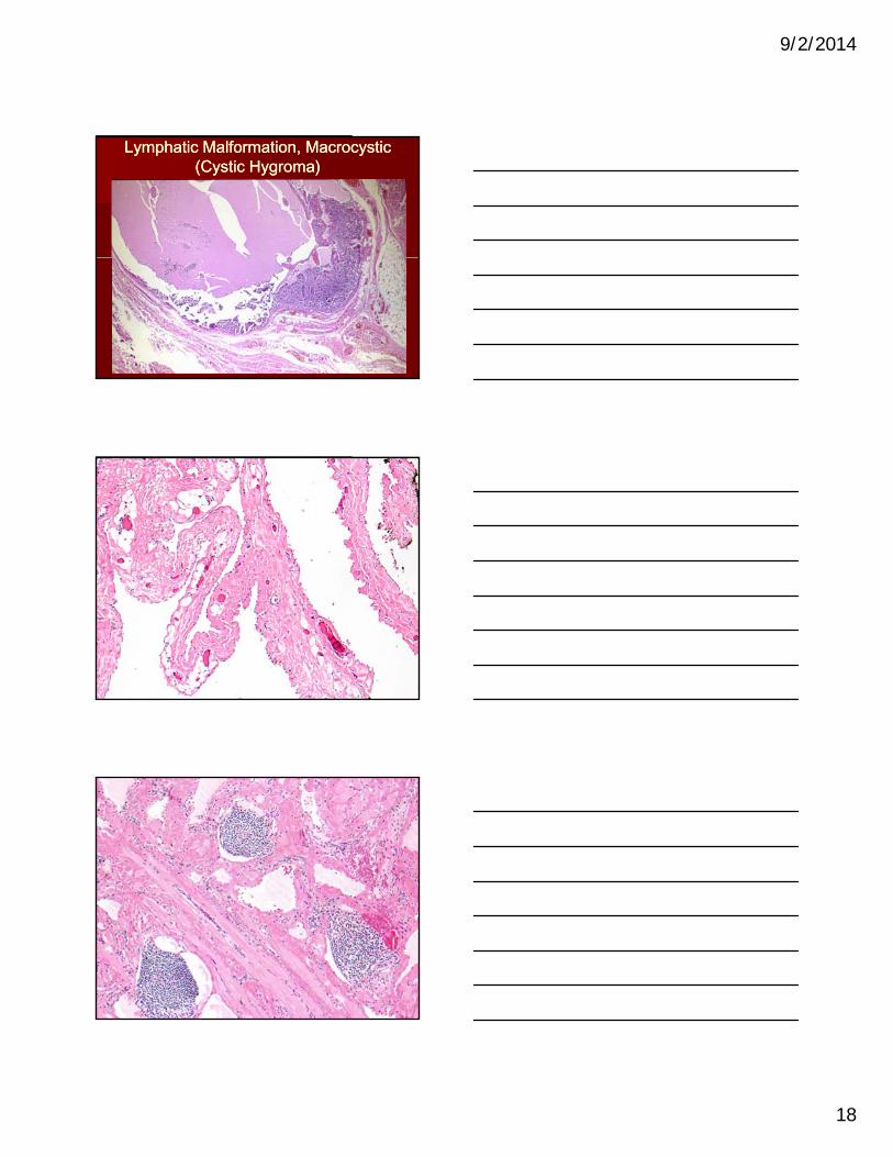
Lymphatic Malformation, Macrocystic Lymphatic Malformation, Macrocystic (Cystic Hygroma)(Cystic Hygroma)

9/2/2014
19
Management of Lymphatic Management of Lymphatic MalformationsMalformations
Surgical resectionSurgical resection
Laser therapyLaser therapy
OK 432OK 432 OK 432OK 432
Management with Management with antibioticsantibiotics
Studies currently Studies currently underway with Rapamycin underway with Rapamycin and Viagra (Sildenafil)and Viagra (Sildenafil)
Before
After numerous surgeries
Low Flow Low Flow –– Capillary MalformationCapillary MalformationPort Wine StainsPort Wine Stains
Also known as port wine Also known as port wine stainsstains
Sometimes referred to as Sometimes referred to as venular malformationsvenular malformations
Present at birth as a flat Present at birth as a flat red/purple birthmarkred/purple birthmark
Never regressNever regress
Some can thicken, Some can thicken, cobble, and cause tissue cobble, and cause tissue overgrowthovergrowth

9/2/2014
20
PORT WINE STAIN
Port Wine Stain Dermal Venulocapillary Malformation
Mature Port Wine Stain: Cobblestoning and Nodularity

9/2/2014
21
Port Wine Stain TreatmentPort Wine Stain Treatment
Laser treatmentLaser treatment Sometimes Sometimes
require tissue require tissue debulkingdebulkingdebulkingdebulking
Photos courtesy of www.birthmark.org

9/2/2014
22
Pulse Dye Laser (PDL) is current treatment Pulse Dye Laser (PDL) is current treatment of choiceof choice
Selectively destroys subsurface targets Selectively destroys subsurface targets without inducing thermal damage in without inducing thermal damage in
Port Wine Stain TreatmentPort Wine Stain Treatment
adjacent normal tissueadjacent normal tissue PDL first generation used 577 nm. PDL first generation used 577 nm.
wavelength and 300 us. pulse durationwavelength and 300 us. pulse duration Now 585 nm. wavelength available for Now 585 nm. wavelength available for
adult PWS treatmentadult PWS treatment
Angiogenesis inhibitor Rapamycin (RPM) Angiogenesis inhibitor Rapamycin (RPM) has been combined with PDL to potentially has been combined with PDL to potentially enhance PWS therapeutic outcomeenhance PWS therapeutic outcome
RPM can suppress theRPM can suppress the
Port Wine Stain TreatmentPort Wine Stain Treatment
RPM can suppress the RPM can suppress the VEGF/PI3K/AKT/mTOR pathway and VEGF/PI3K/AKT/mTOR pathway and inhibit reperfusion of blood vessels post inhibit reperfusion of blood vessels post PDL in PWS patientsPDL in PWS patients
Further study is needed for more efficient Further study is needed for more efficient therapeutic modalitiestherapeutic modalities
Venous MalformationVenous Malformation Clinical FeaturesClinical Features Incidence is 1 in 5,000 to 1 in 10,000 personsIncidence is 1 in 5,000 to 1 in 10,000 persons Thrombosis common and associated with pain Thrombosis common and associated with pain
as well as clinical nodularityas well as clinical nodularityO i l d i l di Kli lO i l d i l di Kli l Occur in complex syndromes including KlippelOccur in complex syndromes including Klippel--Trenaunay, Maffucci, and Blue Rubber Bleb Trenaunay, Maffucci, and Blue Rubber Bleb Nevus syndromeNevus syndrome

9/2/2014
23
Low Flow Low Flow -- Venous Venous MalformationsMalformations
Venous Malformations are Venous Malformations are usually soft and easily usually soft and easily compressible softcompressible soft--tissue mass tissue mass that is associated with bluish that is associated with bluish skin discoloration. skin discoloration.
Increasing engorgement with Increasing engorgement with dependency is typical.dependency is typical.
These birthmarks can be These birthmarks can be small and localized or small and localized or extensive and involve the extensive and involve the entire extremity or body entire extremity or body part.part.
Venous Malformation
GLUT1

9/2/2014
24

9/2/2014
25
Venous Malformation TreatmentVenous Malformation Treatment
Surgical resectionSurgical resection EmbollizationEmbollization SclerotherapySclerotherapy RapamycinRapamycin NdYagNdYag LaserLaser
Venous MalformationVenous MalformationLesion Gene Locus Pathway Therapy
VM TIE2/TEK 9p21 Tyrosine kinase receptor; EC migration, proliferation, survival; SMC recruitment; Vascular sprouting; Maturation, stability;
TIE2 inhibitors?
, y;Hematopoietic quiescence
Uebelhoer M., Boon LM, ikkula M. CSH Perspectives; 2012

9/2/2014
26
Low Flow Low Flow –– Mixed MalformationMixed Malformation LymphoLympho--venous malformations are often referred to as mixed venous malformations are often referred to as mixed
lesions. lesions.
They contain both abnormal lymphatic and venous channels.They contain both abnormal lymphatic and venous channels.
They may be scattered in one extremity or may be a focal They may be scattered in one extremity or may be a focal malformationmalformation
Treatment consists of embollization, sclerotherapy, Treatment consists of embollization, sclerotherapy, compression management and lasercompression management and laser
Orhan Konez (2008)
Malformation SyndromesMalformation Syndromes
Shortcut to Geoffforstudy010.lnk
Sturge-Weber SyndromeKlippel-Trenaunay Syndrome
KlippelKlippel--TrenaunayTrenaunay SyndromeSyndrome Affects one or more limbs or trunk Affects one or more limbs or trunk
region.region.
Triad of stain, tissue hypertrophy, and Triad of stain, tissue hypertrophy, and bone overgrowthbone overgrowth
Most cases girth of limb is larger but in Most cases girth of limb is larger but in some cases the nonsome cases the non--affected limb can be affected limb can be clinically smallerclinically smaller
Stain is different than typical port wine Stain is different than typical port wine stainstain
Lateral Marginal Vein varicosity Lateral Marginal Vein varicosity diagnosticdiagnostic
9/2/2014
27
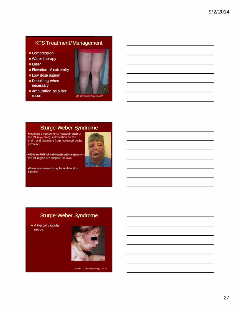
KTS Treatment/ManagementKTS Treatment/Management
CompressionCompressionWater therapyWater therapy LaserLaser
El ti f t itEl ti f t it Elevation of extremityElevation of extremity Low dose aspirinLow dose aspirin Debulking when Debulking when
necessarynecessary Amputation as a last Amputation as a last
resortresort VBF Birthmarks Fact Booklet
SturgeSturge--Weber SyndromeWeber SyndromeInvolves 3 components, vascular stain of the V1 (eye area), calcification on the brain, and glaucoma from increased ocular pressure
30% to 70% of individuals with a stain in30% to 70% of individuals with a stain in the V1 region are suspect for SWS
Brain involvement may be unilateral or bilateral
A typical vascular nevus
SturgeSturge--Weber SyndromeWeber Syndrome
Ellison D., Neuropathology; 3rd Ed.

9/2/2014
28
Bilateral meningeal angiomatosis.
SturgeSturge--Weber SyndromeWeber Syndrome
Ellison D., Neuropathology; 3rd Ed.
Coronal slices of a surgical specimen show the narrowed dark granular cortical
SturgeSturge--Weber SyndromeWeber Syndrome
ribbon.
Ellison D., Neuropathology; 3rd Ed.
Microscopy shows the abnormal leptomeningeal venous plexus and a linear array of
SturgeSturge--Weber SyndromeWeber Syndrome
linear array of superficial calcifications in the thin atrophic cortex.
Ellison D., Neuropathology; 3rd Ed.

9/2/2014
29
Severe astrogliosis with Rosenthal fiber formation and many calcospherites in the
SturgeSturge--Weber SyndromeWeber Syndrome
superficial cortex.
Ellison D., Neuropathology; 3rd Ed.
The leptomeningeal venous angioma lacks elastic fibers.
SturgeSturge--Weber SyndromeWeber Syndrome
Ellison D., Neuropathology; 3rd Ed.
Vascular Tumors and Vascular Tumors and Malformations associated with Malformations associated with
CoagulopathyCoagulopathy MildMild--toto--moderate chronic consumptive moderate chronic consumptive
coagulopathycoagulopathy –– large venous andlarge venous andcoagulopathycoagulopathy large venous and large venous and lymphatic malformationslymphatic malformations
Severe thrombocytopenia due to platelet Severe thrombocytopenia due to platelet trapping (Kasabachtrapping (Kasabach--Merritt phenomenon)Merritt phenomenon)–– kaposiform hemangioendothelioma and kaposiform hemangioendothelioma and tufted angiomatufted angioma

9/2/2014
30
Kaposiform hemangioendotheliomaKaposiform hemangioendothelioma
Infantile hemangioma TA KHE
Kasabach-Merritt Phenomenon
GLUT1 positive
No KMP
GLUT1 negative
+/- KMP
TA

9/2/2014
31
KHE
KHE
KHE

9/2/2014
32
Intramuscular “Hemangiomas”
Large vessel malformations (mostly venous)
Small vessel type (vascular malformations or tumors?)or tumors?)
Mixed small and large vessel type No infantile hemangiomas
Intramuscular “Hemangiomas”
Most are low-flow venous malformations.
Th ll l “ ll l” t i i i f til The cellular, “small-vessel” type mimic infantile hemangioma in histology somehat, but are negative for GLUT1, etc. These present as “masses” by MRI and typically show angiographic and/or clinical features of AV-shunting. They are clinically consistent with vascular malformations and do not regress.
Intramuscular venous malformation

9/2/2014
33
Intramuscular venous malformation
Intramuscular “hemangioma”
In SummaryIn Summary Vascular tumors of childhood represent a number Vascular tumors of childhood represent a number
of distinct entities with diverse etiologies of distinct entities with diverse etiologies –– many many with diagnostic histopathological features.with diagnostic histopathological features.
Some lesions continue to defy classification and Some lesions continue to defy classification and are best viewed as complex, dynamic processes are best viewed as complex, dynamic processes responding to as yet unidentified factors. For responding to as yet unidentified factors. For these, the object for the pathologist is not to these, the object for the pathologist is not to “pigeon hole”, but to describe as accurately as “pigeon hole”, but to describe as accurately as possible. possible.

9/2/2014
34
Everyone has the right to look Everyone has the right to look normalnormal
Photos courtesy of VBF
AcknowledgmentsAcknowledgments Paula North, MD, PhDPaula North, MD, PhD Milton Waner, MDMilton Waner, MD Teresa O, MDTeresa O, MD Adriano Piris, MDAdriano Piris, MD Ignacio Ignacio CarpinteroCarpintero, MD, MD Larry Larry EichenfieldEichenfield, MD, MD IlonaIlona FriedenFrieden, MD, MD Christine Lian, MDChristine Lian, MD Labib Zakka, MDLabib Zakka, MD Linda Linda RozellRozell--Shannon, PhDShannon, PhD
Thank you for your kind Thank you for your kind attentionattention