classification of mandibular fractures
TRANSCRIPT
13
MAJMAAH JOURNAL OF HEALTH SCIENCE, Vol 2, Issue 2, 2014
Kaban in their classification divided the mandibularfractures into five groups according to locationof the fracture[3]. Though it is most currentlyused classification, but it doesn’t fulfil clinicalrequirements. Yet, another classification was givenby Gratz. He used tumor classification (TNM) patternto classify the mandibular fractures[4]. It is anaccepted classification but it is missing informationabout presence or absence of teeth in fracturelines, or whether dislocation has occurred or not.Pankratov & Robustowa proposed a classificationwhich uses numericals for fracture[5] but doesn’tgive radiological findings. A classification approvedby WHO has divided the fractures into 10 groups[6].These groups give information about the locationof the fracture only. Clinicians prefer to use easyand ready-to-be used type of classification. Le forteclassification for maxillary fractures though not verycomprehensive but is used by most clinician becauseof its simplicity. Keeping the necessity of easy-to-use classification in mind, a retrospective study hasbeenperformedon1745patientstreatmentrecords.These patients attended Maxillofacial Departmentat Warsaw Medical University for treatment ofmandibular fractures.
Objectives1. To analyze mandibular fractures on clinical and
radiological bases in the patients treated formandibular fractures.
2. To classify mandibular fractures on the basis ofclinical and radiological evaluation.
Materials & MethodsThis is a retrospective study of patients treatmentrecords and radiographs who attended maxillofacialunit of Warsaw University in the year 1988-1992 and2001-2005. It was found that a single fracture waseasytomentionandanypreviousclassificationcouldservethepurpose.Difficultyariseswhenthefractureis multiple. Therefore, records of 1745 patients wereanalyzed including 1492 males and 253 females whowere treated for mandibular fractures. Based onclinical and radiological evaluation, fractures lineswere identified. The patients were divided into twogroups: 1st group included unilateral fractures, andthe 2nd group included multiple fractures. The firstgroup was further divided depending on the locationof the fracture to eight locations: 1- incisors, 2–canines and premolars, 3- molars, 4- mandibularangle, 5- ramus, 6- coronoid process, 7–condylar
A Proposed Easy-to-Use Classification of Mandibular Fractures, Mouetaz K
ORIGINAL ARTICLE
area and 8– alveolar area. The multiple fractures ofthe mandible were divided into five patterns:1. Bilateral in the body of the mandible (b-b).2. Bilateral in the body and in the condylar process
(b-c).3. Trilateral in the body and in the condylar process
(b-b-c).4. Trilateral in the body and in both condylar
processes (b-c-c).5. Numerous fractures (n).
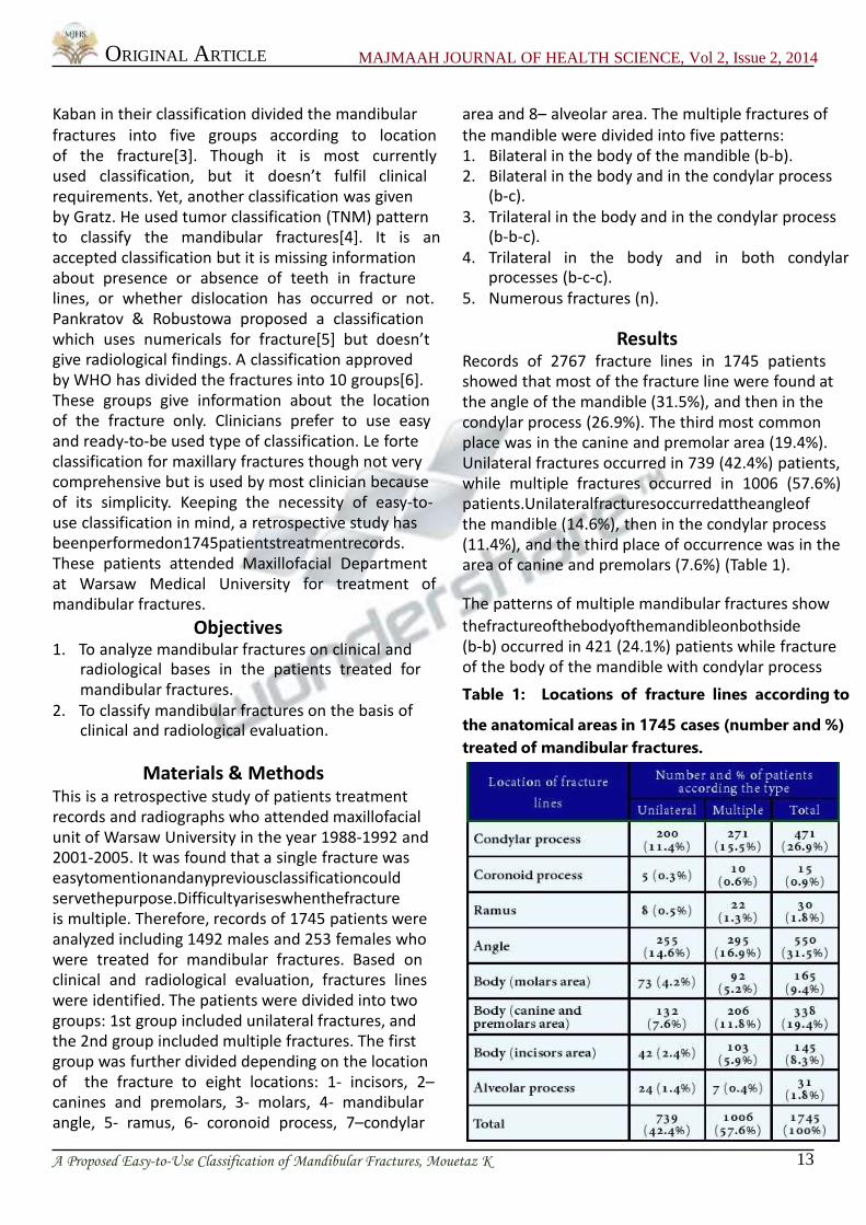
ResultsRecords of 2767 fracture lines in 1745 patientsshowed that most of the fracture line were found atthe angle of the mandible (31.5%), and then in thecondylar process (26.9%). The third most commonplace was in the canine and premolar area (19.4%).Unilateral fractures occurred in 739 (42.4%) patients,while multiple fractures occurred in 1006 (57.6%)patients.Unilateralfracturesoccurredattheangleofthe mandible (14.6%), then in the condylar process(11.4%), and the third place of occurrence was in thearea of canine and premolars (7.6%) (Table 1).
The patterns of multiple mandibular fractures show
thefractureofthebodyofthemandibleonbothside(b-b) occurred in 421 (24.1%) patients while fractureof the body of the mandible with condylar process
Table 1: Locations of fracture lines according to
the anatomical areas in 1745 cases (number and %)
treated of mandibular fractures.