cjo rco - cao · cjo rco vol 74 no 4 | 2012 improved ways to screen for patients with fabry...
TRANSCRIPT

C A N A D I A N J O U R N A L O F O P T O M E T R Y R E V U E C A N A D I E N N E D ’ O P T O M É T R I E
CJO RCOVO L 74 N O 4 | 2 012
Improved Ways to Screen for Patients with Fabry Disease, Involving Optometry in a Multidisciplinary Approach
Under Pressure: a Review of Normal-tension Glaucoma
The COETF Annual Awards Report

MOSTTRUSTEDUUUUUUUUUUUUUSSSSSSSSSSSSSTTTTTTTTTTTTRRRRRRRRRRRR TTTTTTTTTTTTEEEEEEEEEEEEEDDDDDDDDDDDDDCANADIANBRANDDDDDDDDDDDDDNNNNNNNNNNNNAAAAAAAAAAAAARRRRRRRRRRRRBBBBBBBBBBBBB
CCCCCCCCCCCCCAAAAAAAAAAAAANNNNNNNNNNNNAAAAAAAAAAAADDDDDDDDDDDDIIIIIIIIIIAAAAAAAAAAAANNNNNNNNNNNNN*
®
EYE CARE PROFESSIONALS
We look forward to continuing to support
you in providing your patients with an
exceptional vision care experience.
*Most trusted brand of contact lenses by Canadian Optometrists, Opticians and Ophthalmologists. Data on fi le. Johnson & Johnson Inc. 2011.ACUVUE® and INNOVATION FOR HEALTHY VISION™ are trademarks of Johnson & Johnson Inc. ©Johnson & Johnson Inc. 2012
*
LS
ort
an
ACU Trust_CND JRNL Optometry_EN_November.indd 1 12-10-12 11:22 AM ACU Oasys CND JRNL Optometry_EN_November.indd 1 12-10-12 11:22 AM
VOL 74 | NO 4 2012C A N A D I A N J O U R N A L O F O P T O M E T R Y | R E V U E C A N A D I E N N E D ’ O P T O M É T R I E 1
President’s Podium / Mot du président
By/par Dr. Lil Linton . . . . . . . . . . . . . . . . . . . . . . . . . . . . . . . . . . . n 3
Members' News / Nouvelles pour les membres . . . . . . . . . . . . . . . . . . . . . .s 6
Book Review
By Dr. Cheryl Zimmer . . . . . . . . . . . . . . . . . . . . . . . . . . . . . . . . . . . . . . .r 14
The COETF Annual Awards Program Report for 2012. . . . . . . . . . . . . . . . . . . . . . 19
Improved Ways To Screen For Patients With Fabry Disease, Involving Optometry in a Multidisciplinary Approach
By Dr. Langis Michaud & Dr. Christiane Auray-Blais. . . . . . . . . . . . . . . . . . . 25
Under pressure: a review of normal-tension glaucoma
By Dr. Derek MacDonald. . . . . . . . . . . . . . . . . . . . . . . . . . . . . . . . . . . . .dd 33
Uniform requirements for manuscripts: login to the member site at opto.caor contact CAO.
Exigences uniformes pour les manuscrits: voir sur le site des membres àopto.ca ou contacter l’ACO.
CANADIAN JOURNAL OF OPTOMETRY
REVUE CANADIENNE D’OPTOMÉTRIE
VOL 74, NO 42012(Date of issue: December 2012)(Date de parution : decembre 2012)ISSN 0045-5075
The Canadian Journal of Optometry is the official publication of theCanadian Association of Optometrists (CAO) / La Revue canadienne d’optométrie est la publication officielle del’Association canadienne des optométristes (ACO) :234 Argyle Avenue, Ottawa, ON, K2P 1B9. Phone 613 235-7924 / 888 263-4676, fax 613 235-2025, e-mail [email protected], website www.opto.ca. Publications Mail Registration No. 558206 / Envoi de publication – Enregistrement no. 558206.The Canadian Journal of Optometry / La Revue canadienne d’optométrie (USPS#0009-364) is published four times per year at CDN$55, and CDN$65 for subsriptions outside of Canada. Address changes should be sent toCAO, 234 Argyle Avenue, Ottawa, ON K2P 1B9.
•
The CJO*RCO is the official publication of the CAO. However, opinions andcommentaries published in the CJO*RCO are not necessarily either the official opinion or policy of CAO unless specifically identified as such.Because legislation varies from province to province, CAO advisesoptometrists to consult with their provincial licensing authority beforefollowing any of the practice management advice offered in CJO*RCO.
The CJO*RCO welcomes new advertisers. In keeping with our goal of advancing awareness, education and professionalism of members of theCAO, any and all advertising may be submitted, prior to its publication,for review by the National Publications Committee of the CAO. CAO reserves the right to accept or reject any advertisement submitted for placement in the CJO*RCO.
•
La CJO*RCO est la publication officielle de l’ACO. Les avis et lescommentaires publiés dans la CJO*RCO ne répresentent toutefois pasnécessairement la position ou la politique officielle de l’ACO, à moinsqu’il en soit précisé ainsi. Étant donné que les lois sont différentes d’une province à l’autre, l’ACO conseille aux optométristes de vérifier avec l’organisme provincial compétent qui les habilite avant de se conformer aux conseils de la CJO*RCO sur la gestion de leurs activités.
La CJO*RCO est prête à accueillir de nouveaux annonceurs. Dansl’esprit de l’objectif de la CJO*RCO visant à favoriser la sensibilisation, la formation et le professionnalisme des membres de l’ACO, on pourra soumettre tout matériel publicitaire avant publication pour examen parle Comité national des publications de l’ACO. L’ACO se réserve le droit d’accepter ou de refuser toute publicité dont on a demandé l’insertiondans la CJO*RCO.
•
Editor in Chief / l’éditeur en chef :Dr B. Ralph Chou
Chair, National Publications Committee / Président,Comité national des publications : Dr Paul Geneau
Academic Editors / Rédacteurs académiques :University of Waterloo, Dr B. Ralph ChouUniversité de Montréal, Dr Claude Giasson
Managing Editor / Rédactrice administrative ; Advertising Coordinator / Coordonnatrice des publicités :Design/LayoutLeslie Laskarin
Copy-editors:Tony Gibbs, Catherine Heinmiller
Printing Consultant / Impression : Vurtur Communications
Translation / Traduction:Tessier Translations / Les Traductions Tessier
Translation Editor / Réviseure des traductions :Claudette Gagnon
C A N A D I A N J O U R N A L O F O P T O M E T R Y R E V U E C A N A D I E N N E D ’ O P T O M É T R I E
CJO RCO

NEW!
New Advancements inNutrition for Eye Health.
For Adults With AMD AFor AFor A For Adults Over 50
The Global Leader in Ocular Vitamins.†
With 1000 mg of Omega-3

VOL 74 | NO 4 2012C A N A D I A N J O U R N A L O F O P T O M E T R Y | R E V U E C A N A D I E N N E D ’ O P T O M É T R I E 3
PRESIDENT'S PODIUM | MOT DU PRÉSIDENT
BY / PAR DR. LIL LINTON, OD
DECEMBER 2012
It has taken over a year and a half for thelatest iteration of our attempts to have
non-corrective (cosmetic) contact lensesclassifi ed as medical devices to make its way through parliament to the point of approval. When all is said and done you could have completed an MBA program in the time that it will have taken for the bill to come into force. By all accounts thishas been a fairly rapid process for sucha simple bill. Given a more complex orcontroversial bill, the length of time toprogress through each legislative step willincrease to the point where one couldlikely obtain a baccalaureate degree beforeits progress is complete. Suffi ce it to saythat changing legislation at any level is a time consuming and laborious process.
This year the CAO has worked with varying federal government agenciesincluding Health Canada, Canada RevenueAgency, Human Resources and Skills Development Canada, Industry Canada, Corporations Canada, Citizenship and Im-migration Canada, Canadian Institute forHealth Information and the Public HealthAgency of Canada. We have discussed is-sues around non-corrective contact lenses,non-insured health benefi ts, interim health benefi ts, grants and other issues important to eye health and the optometric profes-sion. We have developed relationships with MPs, senators, bureaucrats and staff assistants. It has been a very active year ingovernment relations for the CAO.
Government advocacy and relations is also a pressing need at the provincial level. Regardless of the diff erent regulations that exist provincially, one commonality existsin that every provincial optometry as-sociation in Canada has pursued advocacyon one issue or another in their province — government relations and the eff orts that go into trying to achieve change are substantial.
We know that changing legislationafter it has been instituted is an incrediblydiffi cult task. Every association’s eff orts arebest placed in preventing change that is not in the public interest before it takesplace. Being proactive requires eff ort that aff ects change when the policy makers are most receptive. Those eff orts should focus on building strategic relationships,advocating policy positions, and educatingpolicy infl uencers and decision makers.
There are many issues taking place provincially that should be on thenational radar. Governments take theircues for policy positions from the public, interest groups, industry, and other governments. Policies being discussed in Saskatchewan and Manitoba can eas-ily catch the attention of the Alberta orOntario governments. Optometry must be constantly aware of policies and issuesbeing discussed in other provinces, theUnited States, and other countries. In the information age, the world is too small for distance to be a barrier for domestic orforeign policies to set root.
Fortunately, there is a vehicle that existswithin CAO to be able to discuss opportu-nities, issues and needs at the policy level and strategize plans to advance them. That vehicle is the CAO Government Rela-tions Committee. It exists to bring provin-cial perspectives together so that we can
discuss issues and opportunities, unite andproactively move forward together. It isthe forum where we bring our collectiveknowledge, experience and GR resources together to aff ect change.
A primary role of the government rela-tions committee is to determine initiativesthat educate and infl uence government decision makers. CAO and the provincial associations need to take advantage of what the government relations committeeoff ers; to monitor information, educatedecision makers, advocate policy posi-tions and work for change. This requires dynamic initiatives to bring eye healthand optometry to the forefront through co-operative and strategic eff orts within our profession.
We know how long policy change or creation can take. If we want changes within the next fi ve years, eff orts need tobegin now.
I l a fallu plus d’un an et demi pour quela dernière édition des eff orts que nous
faisions pour que les lentilles cornéennesnon correctives (cosmétiques) soientreconnues comme dispositifs médicauxen arrivent au stade de l’approbation au Parlement. Tout compte fait, il aurait étépossible de terminer un programme de MBA pendant le temps qu’il aura fallu auprojet de loi pour devenir loi. L’exercice amalgré tout été relativement rapide pourun projet de loi aussi simple. Le tempsqu’il faut à une mesure plus complexe ou controversée pour franchir chaque étapelégislative augmente au point où il seraitpossible d’obtenir un baccalauréat avantque l’étude en soit terminée. Il suffi t dedire que le changement d’une mesurelégislative à n’importe quel niveau est chronophage et laborieux.
Cette année, l’ACO a collaboré avecdivers organismes du gouvernement fé-déral, y compris Santé Canada, l’Agence du

C a n a d i a n J o u r n a l o f o p t o m e t r y | r e v u e C a n a d i e n n e d ’ o p t o m é t r i evol 74 | no 4 20124
revenu du Canada, Ressources humaines et Développement des compétences Canada, Industrie Canada, Corporations Canada, Citoyenneté et Immigration Canada, l’Institut canadien d’information sur la santé et l’Agence de la santé pu-blique du Canada. Nous avons discuté des lentilles cornéennes non correctives, des services de santé non assurés, des services de santé intérimaires, des subventions et d’autres questions importantes pour la santé oculovisuelle et la profession opto-métrique. Nous avons établi des liens avec des députés, des sénateurs, des fonction-naires et des adjoints. L’année a été très active pour l’ACO sur le plan des relations gouvernementales.
La représentation et les relations gou-vernementales constituent aussi un besoin pressant à l’échelon provincial. Sans égard aux différents règlements en vigueur dans les provinces, il existe un aspect commun, soit que les associations d’optométrie de chaque province du Canada sont intervenues dans un grand dossier ou un autre dans leur province – les relations gouvernementales et les efforts déployés pour essayer d’instaurer le changement sont importants.
Nous savons qu’il est incroyablement difficile de modifier une loi en vigueur. Il est préférable que les efforts de chaque association visent à prévenir au préalable un changement qui n’est pas dans l’intérêt public. Pour être proactif, il faut faire des efforts qui ont une incidence sur les changements lorsque les responsables des politiques sont les plus réceptifs. Ces ef-forts doivent viser avant tout à établir des liens stratégiques, défendre des positions stratégiques et informer les stratèges et les décideurs.
Il y a beaucoup de questions à l’échelon provincial qui devraient être visibles sur la scène nationale. Lorsqu’il s’agit d’arrêter des positions stratégiques, les gouvernements sont à l’écoute du
public, des groupes spécialisés, de l’industrie et d’autres gouvernements. Des politiques qui font l’objet de discussions en Saskatchewan et au Manitoba peuvent facilement attirer l’attention des gou-vernements de l’Alberta ou de l’Ontario. L’optométrie doit être constamment à l’affût des politiques et des enjeux abordés dans d’autres provinces, aux États-Unis et ailleurs. À l’ère de l’information, le monde est trop petit pour que les distances empêchent les politiques nationales ou étrangères de s’implanter.
Il existe heureusement à l’ACO un organe qui permet de discuter de pos-sibilités, d’enjeux et de besoins à l’échelon stratégique et d’établir des plans straté-giques pour faire avancer ces dossiers. Cet organe est le Comité des relations avec les gouvernements de l’ACO qui doit unir les points de vue des provinces afin que nous puissions discuter d’enjeux et de possibilités, faire front commun et aller de l’avant ensemble de façon proactive. C’est la tribune où nous réunissons nos connais-sances, notre expérience et nos ressources collectives pour instaurer le changement.
Le comité des relations avec les gouvernements, a pour rôle premier de déterminer les initiatives qui informent et influencent les décideurs des gouverne-ments. L’ACO et les associations provinciales doivent profiter de ce que le comité des relations avec les gouvernements peut leur offrir, surveiller l’information, informer les décideurs, préconiser des positions stratégiques et chercher à instaurer le changement. Il faut à cette fin des initia-tives dynamiques pour placer la santé oculovisuelle et l’optométrie à l’avant-scène par des efforts stratégiques et basés sur la coopération à l’intérieur même de la profession.
Nous savons combien de temps il faut pour créer ou modifier une politique. Si nous voulons instaurer des changements au cours des cinq prochaines années, il faut nous mettre à l’œuvre maintenant.
CJO_Issue_4_Insides_2012.indd 4 12/12/12 3:04 PM

PR
OD
PR
OD
UC
ED
UC
ED
AN
DA
ND
AD
ISD
ISSD
TR
IBT
RIB
RU
TE
DU
TE
DT
BY
B
Y
YLU
XO
LUX
OX
OL
TTTT
ICA
IC
A
CG
RO
UG
RO
UG
RO
RPP
--MMM
OD
OD
OD
. P
H.
PHH
P2
08
20
82
08
0333
CO
LC
OL
CO
LC
. 5
0.
50
50
31
31
31
C A N A D I A N J O U R N A L O F O P T O M E T R Y | R E V U E C A N A D I E N N E D ’ O P T O M É T R I EVOL 74 | NO 4 20126
Blue Cross Announcement As of September 17, 2012, Medavie Blue Cross will begin introducing a new two-sided plastic identi� cation card, similar to those used for banking. The new card design will be provided to new Medavie Blue Cross members as well as to any existing members when and if they require a change in their current ID card informa-tion such as name or dependent status.
Medavie Blue Cross is taking a staggered approach to introducing this new ID card and will not be replacing all cards currently in use. Many Medavie Blue Cross members will continue to use a four-sided blue lami-nated card. In Quebec, a two-sided yellow plastic card also exists. Please note that federal program members (RCMP, Veterans A� airs, Canadian Forces and Citizenship and Immigration Canada) do not carry Medavie Blue Cross cards.
Providers may call the Customer Service Centre toll-free if they have further questions: 1-800-667-4511 (in Atlantic), 1-800-355-9133 (in Ontario).1-888-588-1212 (in Quebec). Providers in other provinces please contact your local Blue Cross.
Annonce sur la Croix-BleueLe 17 septembre 2012, Croix Bleue Medavie a lancé une nouvelle carte d'identité en plastique à deux côtés, semblable aux cartes bancaires. La nouvelle carte sera fournie aux nouveaux membres de Croix Bleue Medavie, ainsi qu'à tout membre actuel qui doit modi� er l'information contenue sur sa carte d'identité actuelle, comme son nom ou son statut de per-sonne à charge. Croix Bleue Medavie met cette nouvelle carte d'identité en service de façon décalée et ne remplacera pas toutes les cartes en vigueur. Beaucoup de membres de Croix Bleue Medavie continu-eront d'utiliser une carte laminée bleue à quatre côtés. Au Québec, une carte en plastique jaune à deux côtés existe aussi. Il faut noter que les membres du programme fédéral (GRC, Anciens Combattants, Forces canadiennes et Citoyenneté et Immigra-tion Canada) n'ont pas de carte Croix Bleue Medavie.
S'ils ont d'autres questions, les fournis-seurs peuvent appeler gratuitement le Centre des services à la clientèle : 1-800-667-4511 (Atlantique), 1-800-355-9133 (Ontario), 1-888-588-1212 (Québec). Les fournisseurs des autres provinces sont priés de communiquer avec leur Croix Bleue locale.
Eyefoods Book O� erThank your faithful patients, referring doctors, or sta� and show them your ap-preciation with a gift of good health this holiday season. The Eyefoods book makes a � tting present and an acknowledgment of their patronage and support throughout the year. Show your gratitude and take advantage of our special holiday o� er: or kick start January's "get healthy" resolutions and sell Eyefoods books in your practice
MEMBERS' NEWS | NOUVELLES POUR LES MEMBRES
PH
OTO
CO
URT
ESY
OF
JASO
N M
ARG
ARITA
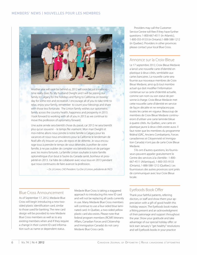
Another year will soon be behind us, 2012 will soon be just a memory –time really does � y. My husband Dwight and I will be joining our family in Calgary for the holidays and � ying to California on boxing day for some rest and recreation. I encourage all of you to take time to relax, enjoy your family, remember to count your blessings and share with those less fortunate. The Linton family wishes our optometric family across the country health, happiness and prosperity in 2013. I look forward to working with all of you in 2013 as we continue to move the profession of optometry forward.
Une autre année sera bientôt chose du passé, car 2012 ne sera bientôt plus qu’un souvenir – le temps � le vraiment. Mon mari Dwight et moi-même allons nous joindre à notre famille à Calgary pour les vacances et nous nous envolerons pour la Californie le lendemain de Noël a� n d’y trouver un peu de repos et de détente. Je vous encou-rage tous à prendre le temps de vous détendre, à pro� ter de votre famille, à ne pas oublier de compter vos bénédictions et de partager avec les moins fortunés. La famille Linton souhaite à notre famille optométrique d’un bout à l’autre du Canada santé, bonheur et pros-périté en 2013. J’ai hâte de collaborer avec vous tous en 2013 pendant que nous continuons de faire avancer la profession.
– Dr. Lil Linton, CAO President / La Dre Lil Linton, présidente de l’ACO
CJO_Issue_4_Insides_2012.indd 6 12/12/12 3:10 PM

TOPCON CANADA INC.
www.topcon.ca
e-mail: [email protected]
Exclusive Canadian Distributor for: Topcon, Amtek, Welch Allyn, Paradigm (Dicon), Gulden,
M&S Technologies, Tinsley (Selected Products), Icare, Mortan
Eastern Canada • 1-800-361-3515
Ontario • 1-800-387-6768
Western Canada • 1-800-661-8349
CONNECTING VISIONS
TRK-1P 4 in 1 Fully Automated• Easily interfaces with EMR systems for
improved patient flow
• Kerato-refractometer, Non-contact Tonometer and Pachymeter are now combined in one single unit
• The TRK-1P provides 4 beneficial automated functions for both kerato-ref and IOP measurement (Auto/Manual selectable)
• The TRK-1P has a 6.5” VGA large colour LCD screen with tilting mechanism for easier and smoother operations
Pre-testing just got efficientMORE

© Optos 2012. All rights reserved. Optos, optos and optomap are registered trademarks of Optos.plc. PN GA-00109 optos.com
optomap® Ultra-Widefield color Image optomap®af Ulf tra-Widefield Autofluorescence Image
A R E Y O U S E E I N GT H E F U L L P I C T U R E ?T H E F U L L P I C T U R E ?
Up to 200° of the retina in a single capture
Ultra-high resolution digital images
Non-mydriatic, through 2mm pupils
In less than a second
For more information
call 800-854-3039 or
email [email protected]
e
Building The Retina Company

VOL 74 | NO 4 2012C A N A D I A N J O U R N A L O F O P T O M E T R Y | R E V U E C A N A D I E N N E D ’ O P T O M É T R I E 9
for $24.95. Click the link to purchase booksfrom the Eyefoods online store. Purchase a box of 36 books for $13 a book ($468plus tax and shipping). That's a combinedtotal of $1,195 off the retail price! http://eyefoods.3dcartstores.com/36-Books-Eyefoods-A-Food-Plan-for-Healthy-Eyes-Holiday-Gift-Special_p_23.html
Off re EyefoodsRemerciez vos patients fi dèles, les méde-cins qui les réfèrent ou les membres du personnel en leur témoignant votre ap-préciation par un cadeau de bonne santéau cours de la saison des Fêtes. L'ouvrage Eyefoods constitue un cadeau bien adaptéet reconnaît leur clientèle ou leur soutientout au long de l'année. Montrez-leur votre gratitude et profi tez d'une off respéciale des Fêtes, ou lancez rapidement les résolutions « de bonne santé » dejanvier et vendez les ouvrages Eyefoodsdans votre cabinet au montant de 24,95 $.Cliquez sur le lien de l'ouvrage au magasinen ligne d'Eyefoods. Achetez un carton de36 ouvrages au prix de 13 $ l'unité (468 $plus taxes et frais de port). Ce montantreprésente au total 1 195 $ de moins que leprix de détail!http://eyefoods.3dcartstores.com/36-Books-Eyefoods-A-Food-Plan-for-Healthy-Eyes-Holiday-Gift-Special_p_23.html
New Canada Not-For-Profi t Corporations ActOn October 17, 2011 the Federal Govern-ment enacted the new Not-For-Profi t Cor-porations Act and all federally incorporatednot-for-profi t organizations have 3 yearsto transition to the new act. This compre-hensive act provides a foundation for theinternal management of not-for-profi t cor-porations. The transition process involves a review of the articles of incorporation andby-laws and to adopt changes to comply with the new act. The CAO studied the requirements to transition with the intentof presenting the necessary changes for
member adoption at the next businessmeeting of members in Edmonton in July2013. The timeline was recognized as verytight. The CAO was originally incorporatedunder a Special Act of Parliament. Special Act organizations are not required to transi-tion by the October 17, 2014 deadline. Therefore, the CAO will take the time it has available to fully investigate a transitionto the new act. CAO Council still expects to recommend several by-law changes at the general business meeting in Edmon-ton. Further information will be broughtforward in the near future.
Nouvelle loi canadienne sur les organisations à but non lucratifLe 17 octobre 2011, le gouvernementfédéral a adopté la nouvelle Loi canadiennesur les organisations à but non lucratif. Tous les organismes sans but lucratif quiont une charte fédérale ont trois ans pours'y conformer. Cette loi détaillée jette lesbases de la gestion interne des personnesmorales sans but lucratif. Le processus detransition comporte une revue des statuts constitutifs et des règlements et l'adoptionde modifi cations pour devenir conforme àla nouvelle loi. L'ACO a étudié les exigencesrelatives à la transition afi n de soumettre leschangements qui s'imposent aux membrespour qu'ils les adoptent au cours de leurprochaine séance de travail qui aura lieu à Edmonton en juillet 2013. Le calendrier est très serré. L'ACO a été constituée à l'origine en vertu d'une loi spéciale du Parlement. Les entités constituées en vertu d'une loi spéciale ne sont pas tenues d'eff ectuer latransition au plus tard à la date limite, fi xéeau 17 octobre 2014. L'ACO prendra donc le temps mis à sa disposition pour étudierà fond le virage vers la nouvelle loi. Le Conseil de l'ACO s'attend quand même àrecommander plusieurs modifi cations des règlements au cours de la séance de travail générale à Edmonton. D'autres renseigne-ments vous parviendront sous peu.
Director AnnouncementWaterloo's School of Optometry & Vision Science is pleased to announce that Profes-sor Paul Murphy, BSc, FCOptom, PhD, FAAO, FBCLA, FEAOO will be the school's next director. Dr. Murphy is an optometrist (Car-diff ), with a PhD (Glasgow) in ocular surfacesensation and a postgraduate certifi cate intertiary level teaching methods. He is cur-rently working toward acquiring his MBAfrom the University of Glamorgan. He was previously a lecturer in the Department of Vision Sciences (Glasgow) and is currently Reader and Director of Teaching at the School of Optometry and Vision Sciences at Cardiff University. The attached link to the school's web-site provides a brief descrip-tion of Dr. Murphy's career to date – uwaterloo.ca/optometry-vision-science/news/director-announcement. Theschool's interim administration will continue its work through to ProfessorMurphy's arrival.
Nomination d'un directeur annoncéeL'École d'optométrie et des sciences de la vision de Waterloo est heureuse d'annoncer que le Pr Paul Murphy, BS,FCOptom, PhD, FAAO, FBCLA, FEAOO,sera le prochain directeur de l'École. Le Dr Murphy est optométriste (Cardiff ) ettitulaire d'un doctorat (Glasgow) en sensa-tion des surfaces oculaires et d'un certifi catpostdoctoral en méthodes d'enseignement au niveau tertiaire. Il prépare actuellement son MBA de l'Université de Glamorgan.Auparavant chargé de cours au Départe-ment des sciences de la vision (Glasgow),il est actuellement maître de conférence et directeur de l'enseignement à l'École d'optométrie et des sciences de la visionde l'Université de Cardiff . Le lien ci-joint vers le site Web de l'école donne accès à une brève description de la carrière duDr Murphy jusqu'à maintenant - uwater-loo.ca/optometry-vision-science/news/director-announcement. L'administration intérimaire de l'école poursuivra son travailjusqu'à l'arrivée du Pr Murphy.

C A N A D I A N J O U R N A L O F O P T O M E T R Y | R E V U E C A N A D I E N N E D ’ O P T O M É T R I EVOL 74 | NO 4 201210
CCOHS PodcastDr. Cheryl Zimmer, Interim Director, Third Party Plans, CAO participated in an interview with the Canadian Centrefor Occupational Health and Safety for apodcast about Computer Vision Syndrome. CCOHS is promoting the podcast throughits Health and Safety Report newsletteremailed to 32,000 subscribers and LiaisonReport that goes to 11,000 subscribers. It is also being promoted through social media. You may listen to the podcast atthis link: ccohs.libsyn.com/shedding-light-on-computer-vision-syndrome.
Balado du CCHST La Dre Cheryl Zimmer, directrice intéri-maires, Régimes de tiers, ACO, a participé à une entrevue avec le Centre canadiend'hygiène et de sécurité au travail pour unbalado portant sur le syndrome de visioninformatique. Le CCHST fait la promotion du balado dans son bulletin Rapport surla santé et la sécurité envoyé par cour-riel à 32 000 abonnés et dans le bulletin Liaison, que reçoivent 11 000 abonnés. Onen fait aussi la promotion dans les médiassociaux. Vous pouvez écouter le balado en suivant ce lien : ccohs.libsyn.com/shed-ding-light-on-computer-vision-syndrome.
New Optometry Forum an email-based and commercially-inde-pendent forum restricted to Canadian op-tometrists and optometric educators (i.e.professors in a school of optometry). TheCOG delivers email posted to subscribers by other subscribers. The originators hope this simple resource will be helpful for colleagues across Canada to communicateanything relevant to optometric practice - clinical questions, eye-care news, practice management tips, etc. It is a forum for op-tometrists organized and operated by two Canadian optometrists, Peter Rozanec, OD& Glen Chiasson, OD. If you are interested in becoming a member, please let them
know and spread the word. To join, send an email to: [email protected].
Nouveau Forum de l'optométrieLe Canadian Optometry Group (COG) est un forum électronique et indépendantsur le plan commercial qui est réservé auxoptométristes et aux éducateurs en opto-métrie du Canada (c.-à-d. aux professeurs des écoles d'optométrie). Le COG livre des messages électroniques aux abonnés affi chés par d'autres abonnés. Les auteurs espèrent que cette ressource simple aiderades collègues de partout au Canada à diff user tout ce qui est important pour la pratique de l'optométrie - questions cli-niques, nouvelles sur les soins oculovisuels,conseils sur la gestion d'un cabinet, etc. Ce forum qui s'adresse aux optométristes est organisé et administré par deux optomé-tristes canadiens, Peter Rozanec, OD, etGlen Chiasson, OD. Si vous souhaitez de-venir membre, veuillez les en informer et faire passer le mot. Pour adhérer, envoyez un message électronique à : [email protected].
Website Updates Recent website changes of note includethe CAO history page, which now includesa copy of the wording of the Federal Act to Incorporate the Canadian Association of Optometrists, a gallery of CAO presidents,and dates/locations of CAO Congresses. In-formation about Occupational Vision Care programs has been moved from the Openyour eyes micro site to opto.ca/ovp. Thispage includes links to provincial OVC/OVPsand tips for completing forms. For those members who participate in the OntarioOVP, there is also additional content found when clicking on the 'Ontario' link.
Mises à jour de site Web Des modifi cations récentes et dignesde mention du site Web comprennent la page sur l'histoire de l'ACO, qui inclut maintenant une copie du texte de laLoi fédérale constitutive de l'Associationcanadienne des optométristes, une galeriede portraits des présidents de l'ACO etles dates et lieux des congrès de l'ACO. L'information sur les programmes de soins professionnels de la vue a été transféréedu microsite Ouvrez les yeux à opto.ca/rpv. Cette page comprend des liens versles SPV/RPSV des provinces, ainsi que des conseils sur la façon de remplir les for-mulaires. Les membres qui participent auprogramme RPSV de l'Ontario y trouverontdu contenu supplémentaire en cliquantsur le lien « Ontario ».
Incident ReportingCAO reminds members to reportpatient incidents on the nationalincident reporting site. Add to your provincial total by reporting asymp-tomatic patients, invalid prescrip-tions, online ordering, sight tests, and cosmetic contact lenses.
Please support this eff ort! To report an incident, visit: www.survey-monkey.com/s/ODincidentreport
Déclaration des incidents
L'ACO rappelle aux membres de déclarer les incidents liés à despatients sur le site national dedéclaration des incidents. Contribuez aux totaux de votre province endéclarant les patients asymptoma-tiques, les prescriptions non valides, les commandes en ligne, les tests de la vue et les lentilles cornéennes àbut esthétique.
Veuillez appuyer cet eff ort! Pour signaler un incident, rendez-vous à :http://www.surveymonkey.com/s/ODrapportincident


Health Care Banking222 Queen Street WestToronto, ON M5V1Z3Irv Handler, Sr. Manager647-286-5839
You define richness. With the Scotia Professional® Plan, customized for your uniquebanking needs, we can help with the money part.
To learn more about Scotia Professional Plan, visit your nearest Scotiabankbranch or visit scotiabank.com/professional today.

Wearers with aaststigmatismWWWearers llooking
for better vision
Astigmatic contact lenses –A huge unmet need
FiFirst-time wearers
F O R A S T I G M A T I S M
1 Young G, Sulley A, Hunt C. Prevalence of astigmatism in relation to soft contact lens fitting. Eye Contact Lens. 2011 Jan;37(1):20-5. 2Astigmatism: Incidence & Barriers. U.S. Market Research Report. Decision Analyst. December 2008. 3Needs, Symptoms, Incidence, Global Eye Health Trends (NSIGHT) Study. Market Probe Europe. December 2009. 4 ECP Toric Needs Study: US. Millward Brown. December 2010. 5Consumer Toric Needs Study: US. Millward Brown.
December 2010. © 2012 Bausch & Lomb Incorporated. ®/™ are trademarks of Bausch & Lomb Incorporated or its affiliates. All other brand/product names are trademarks of their respective owners . PNS06162 SL6881 HL5446-1
A D V E R T I S I N G F E A T U R E
from their doctor.2
Add this to the fact that globally, 43% of
spectacle-wearing and 38% of contact-lens
wearing patients with astigmatism reported
less-than-complete satisfaction with the
spectacles or contact lenses they wear most
often, as judged by a score of 7 or less on a scale
of 1 (very dissatisfied) to 10 (very satisfied).3
Such findings point to unmet needs that could,
in the case of astigmatic spectacle wearers,
potentially be addressed by consideration
of properly fitted toric contact lenses. The
dissatisfaction among astigmatic contact lens
wearers could similarly reflect a need for more
appropriately selected and fitted lenses.
What are eye care professionals looking
for in a toric lens to satisfy their astigmatic
patients? In a study identifying the most
important product attributes for eye care
You can be your patient’s hero when
they are in need of a new or updated
vision prescription. But are all the
options that might improve patient
satisfaction – including, possibly,
specialty contact lenses – receiving
adequate consideration?
A recent publication evaluated a database of
11,624 spectacle prescriptions to calculate the
prevalence of astigmatism of varying degrees
and found that the prevalence of patients with
astigmatism of 0.75 and 1.00 D or greater in
at least one eye was 47.4% and 31.8%.1 This
represents a wealth of patients who may be
coming to you for toric contact lenses. Yet,
about four out of 10 astigmats who have never
worn contacts have not tried them because of
information from family, friends or something
they read that people with astigmatism could
not wear lenses. Even more amazing is that
three out of 10 have not tried them due to advice
professionals when recommending soft toric
contact lenses, the top 3 toric lens benefits
related to vision, with the attribute of highest
relative importance being “delivers crisp, sharp
vision all day.”4 Likewise, when patients with
astigmatism were asked about the top product
attributes when selecting contact lenses, the
top 5 toric lens attributes were visual benefits,
with the benefit of highest relative importance
being “delivers consistently clear vision at all
times.”5
The opportunity to fit and satisfy astigmatic
patients is well within our reach; however
there is a genuine need and opportunity for
practitioners to proactively recommend and
prescribe a more satisfying solution for their
astigmatic patients. The good news is that eye care
professionals and patients do agree that when
it comes to selecting
toric soft lenses, the
importance of visual
b e n e f i t s r i s e s t o
the top. +
You can be your patient’s hero.

C A N A D I A N J O U R N A L O F O P T O M E T R Y | R E V U E C A N A D I E N N E D ’ O P T O M É T R I EVOL 74 | NO 4 201214
BY CHERYL ZIMMER, OD, BSc
Tallowed premature infants a 90%survival rate after only 27 weeks gestation, according to the March of Dimes.1
Developmentally and intellectuallydelayed children now live to adulthoodand seniors are living longer than ever imagined, often with multiple health is-sues. As optometrists, primary health care professionals, we have the responsibility to provide vision care to everyone, and we must be prepared for that endeavour.Visual Diagnosis and Care of the Patientwith Special Needs written by Marc B.Taub, Mary Bartuccio and Dominick M. Maino, all doctors of optometry, is the essential resource for taking care of specialpopulations.
The populations in this book include those that may be born with a disability orsyndrome as a result of problems duringgestation or genetic mutations, such asthose with cerebral palsy, Down syndrome,fragile X syndrome as well as intellectualdisabilities of unknown origins, and thosespecial populations that have acquiredillnesses such as brain injury from disease, accident or stroke, psychiatric disorders and neurodegenerative diseases. Eachcondition is discussed from a systemicstandpoint and oculovisual anomalies andtheir clinical implications are addressed.
The management and treatment of those with autism spectrum disorders,attention defi cit hyperactivity disorder andlearning disabilities are also discussed atlength. These conditions are more readily diagnosed than ever before and more prevalent in the class room and the prima-ry care optometrist's examination room. The Canadian Association of Optometrists, Eye Health Month's slogan for 2012 was Look, See, Learn. This applies to all children,and this book equips the optometrist tobetter deal with those for whom visionand learning are not straightforward.
Some visual issues aff ecting thesepopulations include refractive errors,strabismus, amblyopia and visual fi eld de-fects, as well as oculomotor dysfunctions.Computers have provided patients withbetter access to visual rehabilitation, treat-ment and enhancement. These techniques and procedures are addressed in detail for both in-offi ce assessment and home use.
One other area of interest discussedat length is the visual processing issuesthat special needs populations encounterthat cannot be treated with traditionalspectacle therapy. The book introducesadditional procedures not commonlyused during the conventional compre-hensive assessment of the visual system,but necessary for the assessment of thenon-verbal patient or those with multiple system delay.
One of the most intriguing chaptersoutlines the optometric managementof functional vision disorders. Treatment includes the use of spectacles and prismsto relieve stress on the visual system and improve binocular performance. In conjunc-tion, occlusion and vision therapy may alsobe implemented. Case examples are used to illustrate these management techniques.The neurological basis for vision therapyis also discussed in this chapter, stressing
the integration of vision with other sensoryinputs and how these interwoven systemsplay such a vital role in a person's localizationand orientation in the world.
The authors stress a multi-disciplinary approach where the patient's needs are assessed and treated by a variety of spe-cializations including, but not limited, to their physician, neurologist, ophthalmolo-gist and optometrist as well as adjunct health care workers such as chiropractors,occupational therapists, social workers anddieticians. The objective is to reduce thetotal load of systemic and visual assault onthe body experienced by special needspatients with many of these debilitatingsyndromes. Enhanced communicationbetween the disciplines is in the best interest of the patient.
The Visual Diagnosis and Care of the Patient with Special Needs may be read from cover to cover, providing the reader with a wealth of incredible and pertinentinformation, or used as a reference manual in the primary care optometry offi ce. The authors take into account that thereaders of this publication are well versed in the procedures used during a routineeye examination, but they expand thereader's knowledge base and apply these diagnostic and treatment techniques tothose with special needs. Each chapter hasa multitude of informed contributors and is eloquently and concisely written. There are extensive references at the end of each chapter providing resources for those who may want more insight into certaintopics. Congratulations to Marc B. Taub, Mary Bartuccio and Dominick M. Mainofor providing our profession with such anoutstanding and informative resource.
Endnotes1 http://www.marchofdimes.com/baby/
loss_neonataldeath.html
BOOK REVIEW
Visual Diagnosis and Care of the Patientwith Special Needs

Treat the Signs and Symptoms
• ALREX® for temporary relief of the signs and symptoms of seasonal allergic conjunctivitis1
• Proven efficacy with an excellent safety profile1
• Available in 5 mL bottles
ALREX® (loteprednol etabonate) Ophthalmic Solution 0.2% is indicated for temporary short-term relief of the signs and symptoms of seasonal
allergic conjunctivitis.
Alrex® is for ophthalmic, short-term use only (up to 14 days). If Alrex® is used for 10 days or longer, intraocular pressure should be monitored.
Alrex® is contraindicated in suspected or confirmed infections of the eye: viral diseases of the cornea and conjunctiva including epithelial herpes
simplex keratitis (dendritic keratitis), vaccinia, and varicella; untreated ocular infection of the eye; mycobacterial infection of the eye and fungal x
diseases of ocular structures; hypersensitivity to this drug or any ingredient in the formulation or container, or to other corticosteroids.
Reactions associated with ophthalmic steroids include elevated intraocular pressure, which may be associated with optic nerve damage, visual acuity
and field defects, posterior subcapsular cataract formation, secondary ocular infection from pathogens including herpes simplex, and perforation of x
the globe where there is thinning of the cornea or sclera.
In clinical studies, adverse events related to loteprednol etabonate were generally mild to moderate, non-serious and did not interrupt continuation in
the studies. The most frequent ocular event reported as related to therapy was increased IOP: 6% (77/1209) in patients receiving loteprednol etabonate,
as compared to 3% (25/806) in the placebo treated patients.
Bausch & Lomb Canada Inc., Vaughan, Ontario L4K 4B4© Bausch & Lomb Incorporated®/™ denotes trademark of Bausch & Lomb Incorporated
References: 1. ALREX Product Monograph, December 22, 2008
Target Seasonal Allergic Conjunctivitis with Alrex®
16

(loteprednol etabonate ophthalmic suspension 0.2% w/v)
THERAPEUTIC CLASSIFICATIONCorticosteroidINDICATIONS AND CLINICAL USEAlrex® (loteprednol etabonate) Ophthalmic Suspension is indicated for temporaryshort-term relief of the signs and symptoms of seasonal allergic conjunctivitisCONTRAINDICATIONSSuspected or confirmed infection of the eye: viral diseases of the cornea andconjunctiva including epithelial herpes simplex keratitis (dendritic keratitis), vaccinia,and varicella; untreated ocular infection of the eye; mycobacterial infection of theeye and fungal diseases of ocular structures; hypersensitivity to this drug or anyingredient in the formulation or container, or to other corticosteroids.SPECIAL POPULATIONSUse in Pediatrics (< 18 years of age):Alrex® should not be used in pediatric patients.Use in Geriatrics:Alrex® should not be used in geriatric patients. The safety and efficacy of Alrex®
have not been established in patients > 65 years of age.Pregnant Women:Alrex® should not be used in pregnant women, unless the benefit clearly outweighsthe risks. Studies in pregnant women have not been conducted.Nursing Women:Alrex® should not be used in lactating women, unless the benefit clearly outweighsthe risks.
WARNINGS AND PRECAUTIONSGeneralFor ophthalmic, short-term use only (up to 14 days).The initial prescription and renewal of Alrex® should be made by a physician onlyafter appropriate ophthalmologic examination is performed. If signs and symptomsfail to improve after two days, the patient should be re-evaluated. If Alrex® is usedfor 10 days or longer, intraocular pressure should be closely monitored. Prolonged use of corticosteroids may result in cataract and/or glaucoma formation.Alrex® should not be used in the presence of glaucoma or elevated intraocularpressure, unless absolutely necessary and close ophthalmologic monitoring isundertaken. Extreme caution should be exercised, and duration of treatment shouldbe kept as short as possible.Alrex® should not be used in cases of existing (suspected or confirmed) ocular viral,fungal, or mycobacterial infections. Alrex® may suppress the host response and thusincrease the hazard of secondary ocular infections. The use of Alrex® in patientswith a history of herpes simplex requires great caution and close monitoring.Alrex® contains benzalkonium chloride.Alrex® has not been studied in pregnant or nursing women, but has been found tobe teratogenic in animals. Alrex® should not be used in pregnant or nursing womenunless the benefits clearly outweigh the risks.Carcinogenesis and MutagenesisLong-term animal studies have not been conducted to evaluate the carcinogenicpotential of loteprednol etabonate. Loteprednol etabonate was not genotoxic in vitroin the Ames test, the mouse lymphoma tk assay, or in a chromosome aberrationtest in human lymphocytes, or in vivo in the single dose mouse micronucleus assay.OphthalmologicAlrex® should be used as a brief temporary treatment. If Alrex® is used for 10 daysor longer, intraocular pressure should be closely monitored. The initial prescriptionand renewal of Alrex® should be made by a physician only after appropriateophthalmologic examination is performed, ie. slit lamp biomicroscopy or fluoresceinstaining if appropriate. If signs and symptoms fail to improve after two days, the
patient should be re-evaluated.Prolonged use of corticosteroids may result in glaucoma with damage to the opticnerve, defects in visual acuity and fields of vision, and in posterior subcapsularcataract formation. Alrex® should not be used in the presence of glaucoma orelevated intraocular pressure, unless absolutely necessary and careful and closeappropriate ophthalmologic monitoring (including intraocular pressure and lensclarity) is undertaken.Corneal fungal infections are particularly prone to develop coincidentally withlong-term local steroid application. Fungus invasion must be considered in anypersistent corneal ulceration involving steroid use. Fungal cultures should be takenwhen appropriate.Prolonged use of corticosteroids may suppress the host response and thus increasethe hazard of secondary ocular infections. In those diseases causing thinning of thecornea or sclera, perforations have been known to occur with the use of topicalsteroids. In acute purulent conditions of the eye, steroids may mask infection orenhance existing infection.Use of ocular steroids may prolong the course and may exacerbate the severity ofmany viral infections of the eye (including herpes simplex). Employment of acorticosteroid medication in the treatment of patients with a history of herpessimplex requires great caution.Formulations with benzalkonium chloride should be used with caution in softcontact lens wearers.ADVERSE REACTIONSOverviewReactions associated with ophthalmic steroids include elevated intraocular pressure,which may be associated with optic nerve damage, visual acuity and field defects,posterior subcapsular cataract formation, secondary ocular infection from pathogensincluding herpes simplex, and perforation of the globe where there is thinning ofthe cornea or sclera.In nineteen clinical trials ranging from 1 to 42 days in length, 1,209 patientsreceived various concentrations of loteprednol etabonate in topical ocular drops(0.005%, 0.05%, 0.1%, 0.2%, 0.5%). Adverse events related to loteprednoletabonate were generally mild to moderate, non-serious and did not interruptcontinuation in the studies. The most frequent ocular event reported as related totherapy was increased IOP: 6% (77/1209) in patients receiving loteprednoletabonate, as compared to 3% (25/806) in the placebo treated patients.With the exception of elevations in IOP, the incidence of events in the LE group wassimilar to, or less than that of the placebo control groups. Itching was reported asrelated to therapy in 3% of the loteprednol treated eyes, injection, epiphora,burning/stinging other than at instillation, foreign body sensation, andburning/stinging at instillation were each reported for 2% of eyes. The mostfrequent non-ocular event reported as related to therapy was headache, reportedfor 1.2% of the loteprednol treated subjects and 0.6% of the placebo treatedsubjects.To report an adverse event, contact your Regional Adverse Reaction MonitoringOffice at 1-866-234-2345 or Bausch & Lomb at 1-888-459-5000
One drop instilled into the affected eye(s) four times daily for up to 14 days. Ifscheduled dose is missed, patient should be advised to wait until the next dose andthen continue as before.SHAKE VIGOROUSLY BEFORE USING. Alrex® should be stored upright between15°–25°C for up to 28 days after first opening.The preservative in Alrex®, benzalkonium chloride, may be absorbed by soft contactlenses, and can discolour soft contact lenses. Therefore, Alrex® should not be usedwhile the patient is wearing soft contact lenses. Patients who wear soft contactlenses and whose eyes are not red should wait ten to fifteen minutes after instillingAlrex® before they insert their contact lenses.Patients should be advised not to wear a contact lens if their eye is red. Alrex®
should not be used to treat contact lens related irritation.
SUPPLEMENTAL PRODUCT INFORMATION
WARNINGS AND PRECAUTIONSSexual Function/ReproductionThe effects of Alrex® on sexual function and reproduction have not been studied in humans. Treatment of male andfemale rats with up to 50 mg/kg/day and 25 mg/kg/day of loteprednol etabonate, respectively, (1000 and 500 timesthe Alrex® clinical dose) prior to and during mating, was clearly harmful to the rats, but did not impair their copulation
Prescribing Summary
Patient Selection Criteria
Safety Information
Administration

VOL 74 | NO 4 2012C A N A D I A N J O U R N A L O F O P T O M E T R Y | R E V U E C A N A D I E N N E D ’ O P T O M É T R I E 17
THANK YOU | MERCI
Alcon Johnson & JohnsonVision Care
Bausch & Lomb
CooperVision
Nikon Optical Canada
Scotiabank
Transitions
Carl Zeiss Canada
Centennial Optical
Essilor
Hoya Vision Care
Luxottica Group
performance and fertility (i.e., ability of female rats to become pregnant). However, these doses were highly toxic andhad significant toxic effects on the pregnancies, and the survival and development of the offspring. Maternal toxicity,possible occurrence of abnormalities and growth retardation started at 10 times the Alrex® clinical dose.
NeurologicDisturbances and suppression of the Hypothalamic-Pituitary-Adrenal (HPA) axis can occur with systemic exposure tocorticosteroids. However, given the very low systemic exposure to loteprednol etabonate when using Alrex® asdirected, these possible effects are not likely.
Endocrine and MetabolismGlucocorticoids, mostly when systemic exposure occurs, decrease the hypoglycemic activity of insulin and oralhypoglycemics, so that a change in dose of the antidiabetic drugs may be necessitated. In high doses, glucocorticoidsalso decrease the response to somatotropin. The usual doses of mineralocorticoids and large doses of someglucocorticoids cause hypokalemia and may exaggerate the hypokalemic effects of thiazides and high-ceiling diuretics.In combination with amphotericin-B, they also may cause hypokalemia. Glucocorticoids appear to enhance theulcerogenic effects of non-steroidal anti-inflammatory drugs. They decrease the plasma levels of salicylates, andsalicylism may occur on discontinuing steroids. Glucocorticoids may increase or decrease the effects of prothrombopenicanticoagulants. Estrogens, phenobarbital, phenytoin and rifampin increase the metabolic clearance of adrenal steroidsand hence necessitate dose adjustments.However, given the very low systemic exposure to loteprednol etabonate when using Alrex® as directed, these possibleeffects are not likely.
ImmuneCortisol and the synthetic analogs of cortisol have the capacity to prevent or suppress the development of the localheat, redness, swelling, and tenderness by which inflammation is recognized. At the microscopic level, they inhibit notonly the early phenomena of the inflammatory process (edema, fibrin deposition, capillary dilation, migration ofleukocytes into the inflamed area, and phagocytic activity) but also the later manifestations, such as capillaryproliferation, fibroblast proliferation, deposition of collagen, and, still later, cicatrisation.Clinical Trial Adverse Drug Rg eactionsPossibly or probably related adverse events from two Phase III studies are listed below:
One patient in the Alrex® group and one patient in the placebo group experienced increases in IOP of ≥10 mm Hg.Among these, one in each group had an IOP increase of ≥15 mm Hg, reaching IOP values over 30 mm Hg.In both studies, there were more patients with IOP increases of 6 to 9 mm Hg in the Alrex® group than in the placebogroup (see table below). In study A, among the patients with IOP increases of 6 to 9 mm Hg, four reached an IOPvalue of 22 to 23 mm Hg, and one patient reached 29 mm Hg and was discontinued (clinically significant increase inIOP). All these five patients were from the Alrex® groups.
Incidence of IOP increases of 6 to 9 mm Hg from baseline(number of patients and percentages)
Due to the sample size for each arm of the two phase III studies in SAC, all events captured are greater than 1% of n.
SYMPTOMS AND TREATMENT OF OVERDOSAGEFor management of suspected accidental oral ingestion or drug overdose, consult your regional poison control centre.No cases of overdose have been reported.Full Product Monograph available for health professionals at: http://www.bausch.ca
© 2008 Bausch & Lomb Canada Incorporated.Vaughan Ontario L4K 4B4
™/® Denote trademarks of Bausch & Lomb Incorporated or its affiliates.
Alrex® 0.2% PlaceboN = 133 N = 135
SPECIAL SENSES (EYE DISORDERS)( )Intraocular Pressure- elevation of 6 to 9mm Hg* 2% to 12%* 0% to 6%*- elevation of ≥10mm Hgg 1 (1%) 1 (1%)( ) ( )Chemosis 6 (5%) 7 (5%)( ) ( )Vision, Abnormal or Blurred 4 (3%) 5 (4%)( ) ( )Burning/Stinging, on instillationg/ g g 3 (2%) 6 (4%)( ) ( )Itching Eyeg y 3 (2%) 3 (2%)( ) ( )Dry Eyey y 2 (2%) 4 (3%)( ) ( )Burning/Stinging, not on instillationg/ g g 2 (2%) 2 (1%)( ) ( )Epiphorap p 1 (1%) 9 (7%)( ) ( )Dischargeg 1 (1%) 3 (2%)( ) ( )Foreign Body Sensationg y 1 (1%) 1 (1%)( ) ( )Discomfort Eye 1 (1%) 0 (0%)Injectionj 1 (1%) 0 (0%)( ) ( )Eye Pain 1 (1%) 0 (0%)
Sticky Eye 0 (0%) 7 (5%)
Erythema Eyelids 0 (0%) 2 (1%)
Eye Disorder 0 (0%) 2 (1%)BODY AS A WHOLE
Face Edema (Head)( ) 1 (1%) 0 (0%)( ) ( )Allergic Reaction 1 (1%) 0 (0%)
MUSCULOSKELETAL SYSTEM
Twitching 0 (0%) 1 (1%)* for IOP increase of 6 to 9 mm Hg, please see below * f f l b l
Duration of treatmentDay 7 Day 14 Day 28
Alrex®
Study-A 6 (9%) 6 (9%) 8 (12%)Study-B 3 (5%) 1 (2%) 4 (6%)
y ( ) ( ) ( )y ( ) ( ) (
PlaceboStudy-A 0 (0%) 4 (6%) 1 (2%)Study-B 0 (%) 0 (%) 0 (%)
y ( ) ( ) ( )y ( ) ( ) (
BRON
ZE
OPEN YOUR EYES
PART
NER 2
012
SILVE
ROPEN YOUR EYES
PART
NER 2
012
GOLD
OPEN YOUR EYES
PART
NER 2
012

C a n a d i a n J o u r n a l o f o p t o m e t r y | r e v u e C a n a d i e n n e d ’ o p t o m é t r i evol 74 | no 4 201218
EYE HEALTH COUNCIL OF C ANADA | LE CONSEIL C ANADIEN DE L A SANTÉ DE L'OEIL
Hoya Vision Care’s iQ Family of lenses, using TrueForm Lens Technology, offers the best of both worlds: the benefits of FreeForm design with an ideal solution to upgrade both progressive and single vision patients to an affordable cus-tom option.
With TrueForm Lens Technol-ogy, HOYA applies FreeForm design principles to a semi-finished front surfaced lens. By applying additional aber-ration correction along each line of sight, supported by an aspheric/atoric back surface, the visual performance of the lens can be optimized over the entire lens for each individual prescription.
The iQ Summit ecp and cd, as well as the iQ Amplitude and iQ Amplitude Mini lenses take an existing premium progres-sive design, then uses Free-Form back surfacing and pol-ishing techniques to optimize each prescription. Patients will experience noticeable improvements in peripheral vision at all distances, as well as easier adaptation and wider and clearer viewing ranges.
iQ Single Vision, the newest lens in the iQ TrueForm family, combines a spherical front surface single vision lens with HOYA FreeForm custom back surfacing for enhanced optical performance. iQ Single Vision lenses provide a better vision solution for patients, especially for those with higher prescrip-tions and/or astigmatism.
So the only question left is, what’s your iQ?
La famille de lentilles de Hoya Vision, utilisant la technologie TrueForm, offre le meilleur des deux mondes : les béné-fices d’un design FreeForm avec une solution idéale pour rehausser autant les patients porteurs de progressifs que de simple vision, et ce étant une option abordable.
Avec la technologie TrueForm, HOYA applique les principes de designs FreeForm à un semi-fini ayant un design de face avant. En appliquant une correction d’aberrations additionnelle à chaque ligne de vision, supportée par une surface interne asphérique/atorique, la performance
visuelle de la lentille peut être optimisée sur toute la surface de la lentille et ce, pour chaque prescription individuelle.
La lentille iQ Summit ECP et CD, et la iQ Amplitude et iQ Amplitude Mini prennent un design progressif primé, puis utilisent les techniques de sur-façage et polissage FreeForm pour optimiser chaque pres-cription. Les patients expéri-menteront des améliorations apparentes en vision périphéri-que à toutes les distances, ainsi qu’une adaptation plus facile avec une étendue plus large et claire de la vision.
La iQ Single Vision, la lentille la plus récente de la famille iQ TrueForm, combine une lentille sphérique simple vision sur la face avant à une face arrière sur mesure surfacée par la technologie HOYA FreeForm pour une performance op-tique rehaussée. Les lentilles iQ Single Vision offrent à vos patients une meilleure solution visuelle, spécifiquement pour les hautes prescriptions et/ou astigmatisme.
Alors, la question qui reste, quel est votre QI?
Introducing the iQ TrueForm FamilyIntroduisant la Famille iQ TrueForm
SILVE
ROPEN YOUR EYESPA
RTNE
R 201
2
CJO_Issue_4_Insides_2012.indd 18 12/12/12 3:10 PM
VOL 74 | NO 4 2012C A N A D I A N J O U R N A L O F O P T O M E T R Y | R E V U E C A N A D I E N N E D ’ O P T O M É T R I E 19
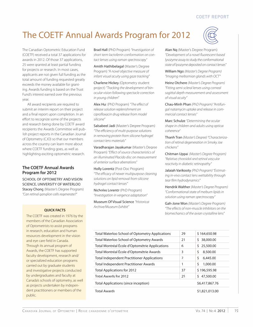
The Canadian Optometric Education Fund(COETF) received a total 37 applications for awards in 2012. Of those 37 applications,25 were granted at least partial funding for projects or research. In most cases, applicants are not given full funding as thetotal amount of funding requested greatlyexceeds the money available for grant-ing. Awards funding is based on the Trust Fund’s interest earned over the previousyear.
All award recipients are required tosubmit an interim report on their project and a fi nal report upon completion. In an eff ort to recognize some of the projectsand research being done by COETF award recipients the Awards Committee will pub-lish project reports in the Canadian Journalof Optometry (CJO) so that our membersacross the country can learn more aboutwhere COETF funding goes, as well ashighlighting exciting optometric research.
The COETF Annual Awards Program for 2012SCHOOL OF OPTOMETRY AND VISION SCIENCE, UNIVERSITY OF WATERLOOStacey Chong (Master’s Degree Program) “Can retinal ganglion cells regenerate?”
Brad Hall (PhD Program) “Investigation of short term lactoferrin conformation on con-tact lenses using raman spectroscopy”
Amith Hathibelagal (Master’s DegreeProgram) “A novel objective measure of infant visual acuity using gaze tracking”
Charlene Hickey (Optometry studentproject) “Tracking the development of bin-ocular vision following spectacle correctionin young children”
Alex Hui (PhD Program) “The eff ect of release solution replenishment on ciprofl oxacin drug release from model silicone”
Salsabeel Jadi (Master’s Degree Program) “The effi ciency of multi-purpose solutionsin removing protein from silicone hydrogel contact lens materials”
Varadharajan Jayakumar (Master’s DegreeProgram) “Eff ect of source characteristics of an illuminated Placido disc on measurement of anterior surface aberrations”
Holly Lorentz (Post-Doc Program) “The effi cacy of newer multipurpose cleaning solutions on lipid removal from silicone hydrogel contact lenses”
Nicholas Lorentz (PhD Program)“Investigation in vergence adaptation”
Museum Of Visual Science “Historical Archive/Museum Exhibit”
Alan Ng (Master’s Degree Program) “Development of a novel fl uorescent based lysozyme assay to study the conformational state of lysozyme deposited on contact lenses”
William Ngo (Master’s Degree Program) “Imaging meibomian glands with OCT”
Heinz Otchere (Master’s Degree Program) “Fitting semi-scleral lenses using corneal sagittal depth measurement and assessment of visual acuity”
Chau-Minh Phan (PhD Program) “Antifun-gal natamycin uptake and release in com-mercial contact lenses”
Marc Schulze “Determining the ocular shape in children and adults using optical coherence”
Thanh Tran (Master’s Degree) “Characteriza-tion of retinal degeneration in Smoky Joechickens”
Chitman Uppal (Master’s Degree Program)“Relative choroidal and retinal vascular reactivity in diabetic retinopathy”
Jalaiah Varikooty (PhD Program) “Estimat-ing in-vivo contact lens wettability throughtear fi lm hydrodynamics”
Hendrik Walther (Master’s Degree Program)“Conformational state of meibum lipids in solution using raman spectroscopy”
Gah-Jone Won (Master’s Degree Program)“The eff ects of non-muscle inhibitors on the biomechanics of the avian crystalline lens”
Total Waterloo School of Optometry Applications 29 $ 164,650.98
Total Waterloo School of Optometry Awards 21 $ 38,000.00
Total Montréal École d’Optométrie Applications 6 $ 25,500.00
Total Montréal École d’Optométrie Awards 3 $ 8,500.00
Total Independent Practitioner Applications 2 $ 6,445.00
Total Independent Practitioner Awards 1 $ 1,000.00
Total Applications for 2012 37 $ 196,595.98
Total Awards for 2012 25 $ 47,500.00
Total Applications (since inception) $6,417,867.76
Total Awards $1,821,013.00
The COETF Annual Awards Program for 2012
COETF REPORT
QUICK FACTS
The COETF was created in 1976 by the members of the Canadian Associationof Optometrists to assist programsin research, education and humanresources development in the vision and eye care fi eld in Canada.Through its annual program of Awards, the COETF has supported faculty development, research and/or specialized education programs carried out by graduate students and investigative projects conductedby undergraduates and faculty atCanada’s schools of optometry, as well as projects undertaken by indepen-dent practitioners or members of thepublic.
C A N A D I A N J O U R N A L O F O P T O M E T R Y | R E V U E C A N A D I E N N E D ’ O P T O M É T R I EVOL 74 | NO 4 201220
Witer Learning Resource Centre “Continuance of ‘Library InformationResources & Services for Canadian Optometrists’ program”
ÉCOLE D’OPTOMÉTRIE, UNIVERSITÉ DE MONTRÉAL
Estefania Chriqui (Master’s Degree Program) “Optimizing the determination of visual acuity in seniors who have consider-able diffi culties communicating or co-operating during the visual exam.”
Hélène Kergoat, Elizabeth Irving“Convergence insuffi ciency and Parkinson’sdisease”
Yves Momplaisir, Maxime Gosselin“Canadian optometrists’ contribution to themanagement of eye disease”
INDEPENDENT PRACTITIONER
Alissa Boroditsky “Oral History of Optometry in Canada”
COETF REPORT 2012
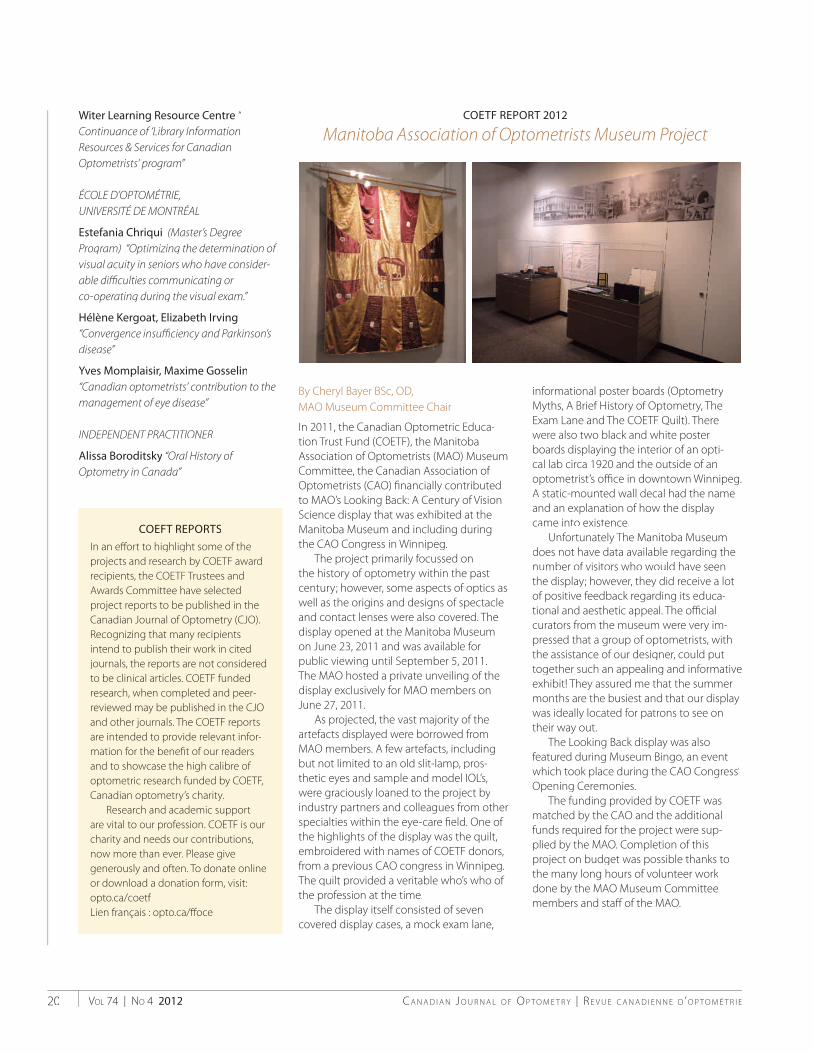
Manitoba Association of Optometrists Museum Project
By Cheryl Bayer BSc, OD, MAO Museum Committee Chair
In 2011, the Canadian Optometric Educa-tion Trust Fund (COETF), the ManitobaAssociation of Optometrists (MAO) Museum Committee, the Canadian Association of Optometrists (CAO) fi nancially contributedto MAO’s Looking Back: A Century of Vision Science display that was exhibited at theManitoba Museum and including duringthe CAO Congress in Winnipeg.
The project primarily focussed on the history of optometry within the past century; however, some aspects of optics as well as the origins and designs of spectacle and contact lenses were also covered. Thedisplay opened at the Manitoba Museumon June 23, 2011 and was available forpublic viewing until September 5, 2011.The MAO hosted a private unveiling of the display exclusively for MAO members on June 27, 2011.
As projected, the vast majority of theartefacts displayed were borrowed fromMAO members. A few artefacts, including but not limited to an old slit-lamp, pros-thetic eyes and sample and model IOL’s, were graciously loaned to the project by industry partners and colleagues from other specialties within the eye-care fi eld. One of the highlights of the display was the quilt,embroidered with names of COETF donors, from a previous CAO congress in Winnipeg. The quilt provided a veritable who’s who of the profession at the time.
The display itself consisted of sevencovered display cases, a mock exam lane,
informational poster boards (OptometryMyths, A Brief History of Optometry, TheExam Lane and The COETF Quilt). There were also two black and white poster boards displaying the interior of an opti-cal lab circa 1920 and the outside of anoptometrist’s offi ce in downtown Winnipeg. A static-mounted wall decal had the nameand an explanation of how the displaycame into existence.
Unfortunately The Manitoba Museum does not have data available regarding the number of visitors who would have seen the display; however, they did receive a lotof positive feedback regarding its educa-tional and aesthetic appeal. The offi cialcurators from the museum were very im-pressed that a group of optometrists, with the assistance of our designer, could puttogether such an appealing and informativeexhibit! They assured me that the summermonths are the busiest and that our display was ideally located for patrons to see ontheir way out.
The Looking Back display was alsofeatured during Museum Bingo, an eventwhich took place during the CAO Congress’ Opening Ceremonies.
The funding provided by COETF wasmatched by the CAO and the additionalfunds required for the project were sup-plied by the MAO. Completion of thisproject on budget was possible thanks to the many long hours of volunteer work done by the MAO Museum Committeemembers and staff of the MAO.
COEFT REPORTS
In an eff ort to highlight some of the projects and research by COETF awardrecipients, the COETF Trustees andAwards Committee have selectedproject reports to be published in theCanadian Journal of Optometry (CJO). Recognizing that many recipientsintend to publish their work in cited journals, the reports are not consideredto be clinical articles. COETF funded research, when completed and peer-reviewed may be published in the CJO and other journals. The COETF reportsare intended to provide relevant infor-mation for the benefi t of our readers and to showcase the high calibre of optometric research funded by COETF,Canadian optometry’s charity.
Research and academic support are vital to our profession. COETF is our charity and needs our contributions, now more than ever. Please givegenerously and often. To donate onlineor download a donation form, visit: opto.ca/coetfLien français : opto.ca/ff oce
Introducing HOYA iQ Amplitude – offering wider viewing areas across all points on the lens to provide comfortable reading and intermediate zones that satisfy various visual demands. This progressive lens features a flexible design, offering HOYA quality to progressive lens wearers that don’t have specific design demands.
What’s your iQ? Take advantage of an ideal solution to upgrade progressive lens patients to an affordable custom option. Offer your patients optimized sight through a more intelligent lens.
OPTIMIZING SIGHT THROUGH A MORE INTELLIGENT LENS
HOYA TorontoTel: 1 888-258-4692Fax: 1 888-258-6618
HOYA VancouverTel: 1 866-454-4692Fax: 1 888-454-9479
HOYA EdmontonTel: 1 888-291-0036Fax: 1 888-291-0736
HOYA LondonTel: 1 877-277-4745Fax: 1 877-277-4818
HOYA MontréalTel: 1 877-720-4692Fax: 1 877-820-0035
NEW ADVANCED PROGRESSIVE LENS
NEW!
To learn more about HOYALUX iQ SUMMIT or HOYA iQ AMPLITUDE lenses, contact your HOYA Territory Manager or visit www.iQTrueForm.ca
www.hoyavision.ca™ HOYA, TrueForm LENS TECHNOLOGY, FreeForm DESIGN TECHNOLOGY, HOYALUX iQ SUMMIT ECP & CD, HOYA iQ AMPLITUDE & MINI are registered trademarks of HOYA LENS CANADA INC. © 2011 HOYA LENS CANADA INC. 34030-08/11

C A N A D I A N J O U R N A L O F O P T O M E T R Y | R E V U E C A N A D I E N N E D ’ O P T O M É T R I EVOL 74 | NO 4 201222
SILVE
ROPEN YOUR EYES
PART
NER 2
012
EYE HEALTH COUNCIL OF C ANADA | LE CONSEIL C ANADIEN DE LA SANTÉ DE L'OEIL
Established in 1972, Essilor Canada isa subsidiary of Essilor International,world leader in ophthalmic opticalproducts. Essilor’s corporate missionis to enable everyone around theworld to access lenses that meet hisor her unique vision requirements inorder to improve everyone’s quality of life. To support this mission, theCompany allocates around €150 million to research and development every year, in a commitment to continuously bring new, more eff ective products to market.
Innovative ProductsWith three Innovation and TechnologyCenters and 550 researchers around the world developing the lenses of thefuture, Essilor puts innovation at theheart of its growth strategy. As a result,in 2011-12, Essilor launched three majorinnovations:
Optifog, the fi rst high-performance anti-fog lenses, winner of the Silmod’Or 2011 in the Vision category andvoted Product of the Year 2012 in the Personal Comfort category by Canadian consumers
Crizal UV, the fi rst corrective lens to protect the eyes from UV rays re-fl ected from the backside of the lens, and the E-SPF (Eye-Sun ProtectionFactor) rating system for lenses
Varilux S series, a revolution inprogressive lens technology, design and personalization, off ering presbyopes limitless vision; winner
of the Silmo d’Or 2012 in the Vision category and approved by wearers through tests conducted in accor-dance with a protocol endorsed bythe Research Center 968 INSERM,Université Pierre et Marie Curie.
Ongoing research – two major projects in CanadaIn its approach to innovation theGroup draws on an extensive network of international partnerships with universities, industrial companies and specialists in cutting-edge technolo-gies. In Canada, two major research projects are contributing: the NESRC-Essilor Industrial Research Chair in VisualPerception and Presbyopia at the Écoled'optométrie, Université de Montréal has introduced immersive virtual reality environments to study human behav-iour, transforming the existing techno-logy that had not been developed forscientifi c research, into an impressiveresearch tool. It has lead to the elabora-tion of the concept of a progressive lens adapted to the individual head-eye movements: Varilux® Ipseo®. The NSERC Multisectorial Industrial Research Chair in Coatings and Surface Engineering at Polytechnique Montréal, launched September 25, will focus on develop-ing a new generation of non-pollutingmanufacturing technologies fornanostructured coating materials whichwill allow for signifi cant progress and breakthroughs in the fi eld of antirefl ec-tive coatings and surface treatments.
Essilor in the CommunityIn addition, Essilor Canada is involvedat all levels in the Canadian optical community. By its consumer advertising and cultural event
sponsorship, it positions itself as a leader in developing the market. sponsorships,such as the Special Olympics and the « Participe pour Voir » program of LaFondation des Maladies de l’œil have been great opportunities to promote good visual health.
Essilor develops projects with allthe optical schools in Canada: it col-laborates very closely with the Écoled’Optométrie de l’Université de Mon-tréal where thanks to an edging and mounting lab headed by one of Essilor’s experts in the fi eld, optometry studentscan learn about new products, thelatest edging/mounting technologiesas well as remote edging and electronicordering. It has a long term partner-ship with the School of Optometry &Vision Science, University of Waterloo that will help enhance optometric education and further advance eye health and eye care in Canada. Essiloralso brings specialized business train-ing to eyecare professionals throughprograms such as the Management &Business Academy™ (MBA).
Essilor has been a sponsor of theEye Health Council since the year 2000, supporting its activities andthe diff usion of information on visual health.
Essilor Canada, fulfi lling its mission through innovation and partnerships
A UNIQUE TECHNOLOGY AGAINST FOG
LENS WITH OPTIFOGSTANDARD LENS
Limitless Vision
A revolution in progressive lenses:
For the best visual solution,
always ask your eyecare
professional for Varilux lenses.
www.varilux-s-series.ca
Refl ex vision: by respecting
the dominant eye’s leading role,
4D Technology improves the
reaction time
Stability of vision in motion:
swim effect reduced up to 90%
Wide angle vision:
fi elds of vision up to 50% wider
ORDINARY VISION CRIZAL VISION
www.crizal.ca
YOUR EYES NEED ULTRA PROTECTIONAGAINST DAMAGING ULTRAVIOLET LIGHT
CRIZAL LENSES: SO SAFE, SO CLEAR.
Introducing a new generation of Crizal® lenses designed to protect eyeglass wearers’ eyes from damaging UV light coming from the backside of the lens, ensuring their visual health over time.
Crizal is the only no-glare range on the market offering the most complete protection against the invisible and often irreversible dangers of
1, and the best enduring clarity of
glare, scratches, smudges, dust and water.
1. Measured by K. Citek, OD, PhD, FAAO, Professor of Optometry,
LENS WITH OPTIFOGSTANDARD LENS
Changing
Environment
Outdoor
Activities
Indoor
Activities
At Work
In daily activities at home, work and play, fogged lenses can be a real bother for your patients. Introducing OptifogTM a new breakthrough technology with superior anti-fog properties. Optifog is available with the latest Crizal innovations. This technologically advanced combination provides the incredible scratch, glare and dust protection you expect from Crizal lenses with the most effective and durable anti-fog solution on the market. Help your patients escape from annoying dangerous fog and introduce them to Crizal lenses with Optifog.
youtube.com/optifoglenses
Voted Product of the Year by Canadian consumers in the personal comfort category.

C A N A D I A N J O U R N A L O F O P T O M E T R Y | R E V U E C A N A D I E N N E D ’ O P T O M É T R I EVOL 74 | NO 4 201224

Vol 74 | No 4 2012C a N a d i a N J o u r N a l o f o p t o m e t r y | r e V u e C a N a d i e N N e d ’ o p t o m é t r i e 25
CliniCal study
Objet : La maladie de Fabry est considérée comme une maladie rare, de par sa prévalence. Cependant, ceci cache une réalité clinique toute autre en raison du nombre de cas non dépistés. Cet article vise à démontrer comment les optométristes, via un simple examen à la lampe à fente, peuvent aider à améliorer le dépistage des patients atteints, et ce, dans une perspective multi-disciplinaire.
Méthode :Un modèle de dépistage a été instauré, en se basant sur l’éducation continue des optométristes. Les patients suspects de Fabry sont référés à l’École d’optométrie de l’Université de Montréal pour des tests supplémentaires. Dans le cas où l’histoire de cas et/ou les signes cliniques sont caractéristiques de la maladie de Fabry, un test urinaire est demandé, visant l’identification de biomarqueurs
spécifiques. Si ce test s’avère positif, le sujet est alors référé à un spécialiste des maladies génétiques pour un test d’ADN et un suivi médical de sa condition.
Résultats : Des activités de formation continue ont été réalisées à travers tout le Québec, rejoignant près de 60% des optométristes. Après 16 mois d’implantation, ce modèle a permis l’identification de 10 suspects. De ce nombre, 2 patients Fabry ont été diagnostiqués, ce qui a conduit également à l’identification de la maladie chez 5 de leurs proches. Deux autres patients, atteints de Fabry mais sans suivi médical depuis des années, ont été à nouveau pris en charge par le médecin spécialiste. À ce jour, en raison de l’implication des optométristes, 7 nouveaux patients ont donc été dépistés et diagnostiqués alors que 2 reçoivent à nouveau les soins appropriés.
Conclusion : En se basant sur les résultats obtenus, le modèle de dépistage mis en place a été évalué comme positif. Il confirme le rôle crucial des optométristes dans le dépistage des maladies systémiques ayant des implications oculaires. L’éducation continue est essentielle à la remise à jour et en perspective de notions apprises il y a longtemps mais qui ne se rencontrent pas au quotidien dans les pratiques. De plus, ceci suggère que l’optométriste peut être impliqué dans des équipes multidisciplinaires visant le dépistage de patients à risque et de maladies, notamment celles qui entraînent des manifestations oculaires.
By Langis Michaud & christiane auray-BLais
introduction
Although Fabry disease has been known for more than a
century (1898), this lysosomal storage disorder remains poorly recognized. With a prevalence of 1/40,000 to 1/117,000 live male births,1 Fabry is considered one of 7,000 known rare diseases that exist in the US and reported. However, this number of patients seems to be underesti-mated.3
Many heterozygous subjects are affected without being diagnosed, no one seeing the globality of their symptoms. These are quite variable and unspecific, often leading to confusion with rheumatoid diseases or chronic inflammatory conditions.4 In some cases, the condition remains subclinical or is not characteristic of the full spectrum of the disease. A typical patient’s odyssey means multiple visits to more than ten different medical specialists before he or she achieves a confirmatory
Improved ways to screen for patients with Fabry disease, involving optometry in a multidisciplinary approach
Langis Michaud, OD, MSc, FAAO (Dipl) Associate Professor, École d’optométrie- Université de Montréal
Christiane Auray-Blais, LLM, PhDService of Genetics, Department of Pediatrics Faculty of Medicine and Health SciencesUniversité de Sherbrooke
Disclosure: Author has received research grants and speaker fees from Genzyme Canada.
RÉSUMÉ
CJO_Issue_4_Insides_2012.indd 25 12/12/12 3:08 PM
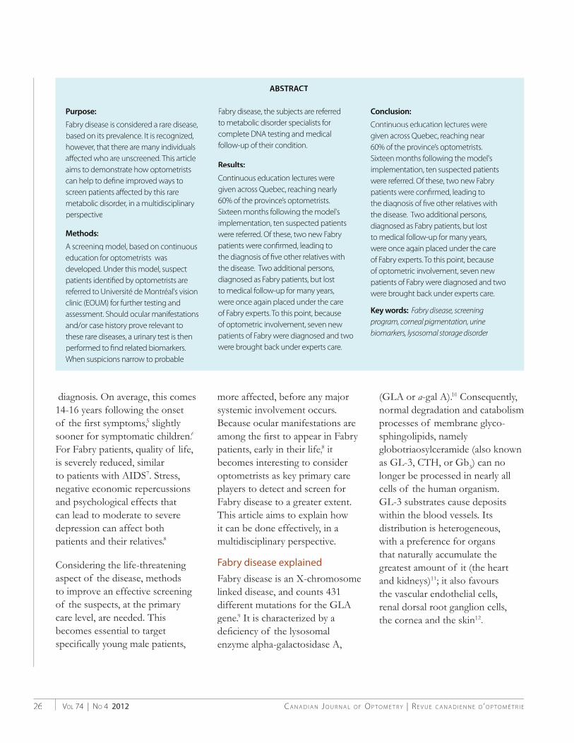
C A N A D I A N J O U R N A L O F O P T O M E T R Y | R E V U E C A N A D I E N N E D ’ O P T O M É T R I EVOL 74 | NO 4 201226
Purpose:
Fabry disease is considered a rare disease,based on its prevalence. It is recognized,however, that there are many individualsaff ected who are unscreened. This articleaims to demonstrate how optometristscan help to defi ne improved ways toscreen patients aff ected by this raremetabolic disorder, in a multidisciplinaryperspective
Methods:
A screening model, based on continuouseducation for optometrists wasdeveloped. Under this model, suspectpatients identifi ed by optometrists arereferred to Université de Montréal's visionclinic (EOUM) for further testing andassessment. Should ocular manifestationsand/or case history prove relevant tothese rare diseases, a urinary test is thenperformed to fi nd related biomarkers. When suspicions narrow to probable
Fabry disease, the subjects are referred to metabolic disorder specialists forcomplete DNA testing and medicalfollow-up of their condition.
Results:
Continuous education lectures weregiven across Quebec, reaching nearly60% of the province’s optometrists. Sixteen months following the model'simplementation, ten suspected patients were referred. Of these, two new Fabry patients were confi rmed, leading to the diagnosis of fi ve other relatives withthe disease. Two additional persons, diagnosed as Fabry patients, but lost to medical follow-up for many years, were once again placed under the careof Fabry experts. To this point, becauseof optometric involvement, seven new patients of Fabry were diagnosed and twowere brought back under experts care.
Conclusion:
Continuous education lectures weregiven across Quebec, reaching near60% of the province’s optometrists.Sixteen months following the model'simplementation, ten suspected patientswere referred. Of these, two new Fabrypatients were confi rmed, leading to the diagnosis of fi ve other relatives with the disease. Two additional persons, diagnosed as Fabry patients, but lostto medical follow-up for many years, were once again placed under the careof Fabry experts. To this point, because of optometric involvement, seven new patients of Fabry were diagnosed and twowere brought back under experts care.
Key words: Fabry disease, screening program, corneal pigmentation, urinebiomarkers, lysosomal storage disorder
ABSTRACT
diagnosis. On average, this comes 14-16 years following the onset of the fi rst symptoms,5 slightly sooner for symptomatic children.6
For Fabry patients, quality of life,is severely reduced, similarto patients with AIDS7. Stress,negative economic repercussionsand psychological effects that can lead to moderate to severedepression can affect both patients and their relatives.8
Considering the life-threatening aspect of the disease, methodsto improve an effective screening of the suspects, at the primary care level, are needed. Thisbecomes essential to target specifi cally young male patients,
more affected, before any majorsystemic involvement occurs.Because ocular manifestations areamong the fi rst to appear in Fabry patients, early in their life,8 it becomes interesting to consideroptometrists as key primary care players to detect and screen forFabry disease to a greater extent.This article aims to explain how it can be done effectively, in amultidisciplinary perspective.
Fabry disease explained
Fabry disease is an X-chromosomelinked disease, and counts 431different mutations for the GLAgene.9 It is characterized by adefi ciency of the lysosomalenzyme alpha-galactosidase A,
(GLA or a-gal A).10 Consequently, normal degradation and catabolism processes of membrane glyco-sphingolipids, namely globotriaosylceramide (also known as GL-3, CTH, or Gb3) can nolonger be processed in nearly allcells of the human organism. GL-3 substrates cause depositswithin the blood vessels. Itsdistribution is heterogeneous,with a preference for organsthat naturally accumulate thegreatest amount of it (the heart and kidneys)11; it also favoursthe vascular endothelial cells,renal dorsal root ganglion cells,the cornea and the skin12.
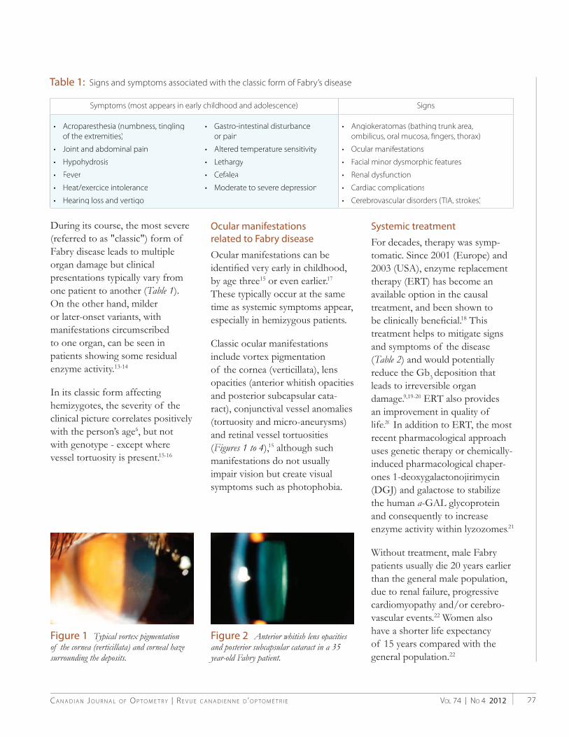
VOL 74 | NO 4 2012C A N A D I A N J O U R N A L O F O P T O M E T R Y | R E V U E C A N A D I E N N E D ’ O P T O M É T R I E 27
During its course, the most severe(referred to as "classic") form of Fabry disease leads to multiple organ damage but clinicalpresentations typically vary from one patient to another (Table 1).On the other hand, milderor later-onset variants, withmanifestations circumscribedto one organ, can be seen inpatients showing some residualenzyme activity.13-14
In its classic form affecting hemizygotes, the severity of theclinical picture correlates positively with the person’s age6, but not with genotype - except wherevessel tortuosity is present.15-16
Ocular manifestations related to Fabry disease
Ocular manifestations can beidentifi ed very early in childhood, by age three15 or even earlier.17
These typically occur at the sametime as systemic symptoms appear,especially in hemizygous patients.
Classic ocular manifestationsinclude vortex pigmentationof the cornea (verticillata), lens opacities (anterior whitish opacitiesand posterior subcapsular cata-ract), conjunctival vessel anomalies (tortuosity and micro-aneurysms)and retinal vessel tortuosities(Figures 1 to 4(( ),15 although such manifestations do not usually impair vision but create visualsymptoms such as photophobia.
Systemic treatment
For decades, therapy was symp-tomatic. Since 2001 (Europe) and2003 (USA), enzyme replacement therapy (ERT) has become anavailable option in the causal treatment, and been shown tobe clinically benefi cial.18 Thistreatment helps to mitigate signs and symptoms of the disease(Table 2) and would potentially 22reduce the Gb3 deposition that leads to irreversible organ damage.9,19-20 ERT also providesan improvement in quality of life.20 In addition to ERT, the most recent pharmacological approach uses genetic therapy or chemically-induced pharmacological chaper-ones 1-deoxygalactonojirimycin (DGJ) and galactose to stabilizethe human a-GAL glycoproteinand consequently to increaseenzyme activity within lyzozomes.21
Without treatment, male Fabry patients usually die 20 years earlierthan the general male population,due to renal failure, progressive cardiomyopathy and/or cerebro-vascular events.22 Women also have a shorter life expectancy of 15 years compared with the general population.22
Table 1: Signs and symptoms associated with the classic form of Fabry’s disease
Figure 1–Typical vortex pigmentation of the cornea (verticillata) and corneal haze surrounding the deposits.
Figure 2–Anterior whitish lens opacities –and posterior subcapsular cataract in a 35 year-old Fabry patient.
Symptoms (most appears in early childhood and adolescence) Signs
• Acroparesthesia (numbness, tinglingof the extremities)
• Joint and abdominal pain
• Hypohydrosis
• Fever
• Heat/exercice intolerance
• Hearing loss and vertigo
• Gastro-intestinal disturbanceor pain
• Altered temperature sensitivity
• Lethargy
• Cefalea
• Moderate to severe depression
• Angiokeratomas (bathing trunk area,ombilicus, oral mucosa, fi ngers, thorax)
• Ocular manifestations
• Facial minor dysmorphic features
• Renal dysfunction
• Cardiac complications
• Cerebrovascular disorders (TIA, strokes)

C A N A D I A N J O U R N A L O F O P T O M E T R Y | R E V U E C A N A D I E N N E D ’ O P T O M É T R I EVOL 74 | NO 4 201228
One way to reverse this naturalcourse is to implement effi cient screening strategies to identify suspects early in the diseaseprocess, and to refer them toFabry specialists in a timely manner. Such a screening strategy can start with the involvement of those who see these potentialpatients on a daily basis. Consider-ing that ocular manifestations areamong the fi rst to appear and theeasiest to assess,23 eye care profes-sionals should be targeted as key players.16
MethodsDefi ning the model
In 2009, a collaborative pattern forthe ocular follow-up of diagnosedFabry patients, under the require-ments of the Canadian Fabry Disease Initiative, was established between Université de Montréal, École d’optométrie (EOUM), andthe genetic center of one of its university hospital (Hôpital duSacré-Cœur de Montréal), treat-ing most of the Fabry patients in the province. The fi rst step inresponding to CFDI's request
was to review their requirementsfor oculo-visual examinationand follow-up (time, equipment,standards, etc.) Secondly, a faculty member (LM) from EOUM was designated to take charge of theproject, based on expertise in anterior segment. The third step was to secure a formal referralpathway that would functionreciprocally between EOUMand the CFDI research team(under Dr. Bichet, of Hôpital du Sacré-Cœur de Montréal).The fourth step was thendeveloped. It involved recruiting practising optometrists to screenfor Fabry patients on a large scale.Several continuing educationlectures were conducted across
the province. During these. events, emphasis was made on the clinicalcourse of the disease (age of onset and natural evolution), thesystemic symptoms and ocularmanifestations related to Fabry and the proposed screening referralpathway. Written documentation (manuscripts, posters with photos)and DVDs about Fabry and otherlysosomal storage disorders werealso provided as reminders forin-offi ce use.
Under the developed model,a patient presenting with spe-cifi c ocular manifestations and/orsymptoms of Fabry is considered a suspect. The situation is explainedto the patient who is offered to be
Table 2: Summary of the eff ects of enzyme replacement therapy (ERT) on Fabry’s patients
Symptoms ERT outcome
• Gastrointestinal disturbance /abdominal pain
• Renal function (overall)
• Proteinuria
• Cardiac complications
• Hearing
• Cerebrovascular events
• Alleviated
• Stabilize or slow the decline
• Not improved
• Benefi cial- reduction in left ventricular mass(if hypertrophy present before tx)
• Stabilize or improve (enzyme alpha)
• No improvement if tx started after event
• Unclear to prevent stroke on long term
Figure 3–Tortuosity and micro-aneurysms of blood vessels as they can be seen in the conjunctiva of a 42 year-old female patient.
Figure 4–Retinal vessel tortuosities, best RRseen in red-free photograph, in an 18 year-old Fabry patient.

VOL 74 | NO 4 2012C A N A D I A N J O U R N A L O F O P T O M E T R Y | R E V U E C A N A D I E N N E D ’ O P T O M É T R I E 29
seen at EOUM's vision clinic forconfi rmation and further assess-ment. If the clinical fi ndings proverelevant (presence of cornealpigmentation and at least manifes-tation of one systemic symptom),or in the case of a positive genetic background, a urine sample col-lected on fi lter paper is sent to the CHUS Expertise Centre inClinical Mass Spectrometry (Dr.Auray-Blais Waters' laboratory) for tandem mass spectrometry analysis for Gb3 levels (see below). In thecase of patients living outside of Montreal area, urinary test kits(fi lter papers and request forms)are sent to local optometrists to beadministrated. Based on clinicaland lab results, if enough suspicionpoints to Fabry (cornea verticillata,systemic symptoms and positiveurinary test result for Gb3 accu-mulation), a referral is made to the nearest genetic centre for completeDNA testing and follow-up.
Ocular data collected from confi rmed patients are part ofthe CFDI registry and constitute the basis of our current longitudinalstudy on ocular manifestationsrelated to Fabry.
Urinary biomarker testing
A method for screening high-risk individuals was developed todetect both male and femaleFabry disease patients.24 The methodology is reliable, effi cient and specifi c, if done after theage of 6 years. It is based on ananalysis of urinary Gb3 using tandem mass spectrometry,25-26
in a laboratory in Sherbrooke, QC. Urinary excretion of Gb3
is normalized to creatinine27 andpatients are always age-matchedto controls.28 Nevertheless, it must be taken into account that patientswith cardiac variant mutations whohave residual enzyme activity do
not excrete excessive amounts ofGb3in their urine.
Results Continuous education
During the fi rst 16 months of the project, 750 Quebec optometrists(out of 1,300 – 57.6%) participated in continuing education events,during evening and weekends.
Patient referrals
The fi rst examination of a patient referred to the EOUM clinicwas made in September 2009.Since that time, 10 patients havebeen seen, (Table 3), referred3by optometrists who attendedcontinuing education events.
The urinary test was performed on every suspect (6 out of 10)who showed signs (3 suspects) or symptoms (3 suspects) that suggested Fabry. This test
PatientSentby ODs
Sex Age Corneal pigmentationseen
Positive CaseHistory (Systemic symptoms)
Urinary test ordered
Patient sent for DNAtesting
Fabry’sconfi rmed
1 F 18 Yes–typical* Yes Yes Yes Yes
2 M 22 Yes (1) No No No No
3 F 34 No Yes Yes No No
4 F 51 No Yes Yes No No
5 M 39 No Yes Yes No No
6 M 23 Yes–typical Yes Yes Yes Yes
7 F 62 Yes (2) No No No No
8 F 51 Yes-atypical Yes(a) Yes Yes No
9 M 46 Yes (3) No No No No
10 F 35 Yes (4) No No No No
Table 3 : Demographics of the referred patients to U de M vision clinic for screening
• Typical means bilateral pigmentation, verticilattatype, asymetrical
• Hudson-Stahli type of pigmentation- monocular-Not related to Fabry
• Pigmentation secondary to amiodarone – symetrical OU
• Pigmentation secondary to chloroquine
• Pigmentation secondary to scarring
• Relatives aff ected (cousins)

C A N A D I A N J O U R N A L O F O P T O M E T R Y | R E V U E C A N A D I E N N E D ’ O P T O M É T R I EVOL 74 | NO 4 201230
allowed for the detection of 2 out of 3 suspects who showed cornealpigmentations and symptoms.The discovery of these patients led to cascade screening and thedetection of other Fabry patients (5)in the family, including two young homozygous patients fi ve andseven years old, respectively. Thelast patient with corneal signs hada negative result on the urinary test. However, because of herpositive family background forthe disease (cousins were knownFabry patients), she was referred for DNA testing which was also negative.
In addition to these patients, fouradditional suspects living in ruralareas were co-managed with theirlocal optometrists. Urinary testswere negative for the diseaseand consequently they were not referred for DNA testing.
Aside from these 19 individuals(10 suspects seen + 5 relatives+ 4 co-managed patients), twoother patients who had been lost to follow-up for many years werebrought back under the care ofeither Dr. Bichet’s team or a localgeneticist in Quebec City, following an optometric examination.
In total, 7 new patients (2 + 5relatives) were diagnosed asa direct consequence of thescreening model we have established, and 2 more were brought back under appropriatecare for their systemic condition.
DiscussionIn the past, several strategies have been proposed to increase screening for patients with rare diseases.29
These strategies have included developing and launching an in-teractive public website to informand communicate with the publicand with medical professionals; amail-based survey of all primary care physicians of a known area;systematic targeted screening ofthe relatives of patients already diagnosed, and a newborn/popu-lation-based urine testing programshould biomarkers be detected.For Fabry disease in particular, very few other initiatives havebeen implemented worldwide toincrease the screening rate forpatients. Some initiatives have attempted to look at specifi cgroups of patients consideredto be "at risk" based on theirsigns or symptoms. For example, studies have been conductedamong hemodialysis patients, with a positive-identifi cationsuccess rate of 0. 2 to 1.2%.30
A similar approach involving patients presenting with cryp-togenic stroke or unexplainedleft ventricular hypertrophy haveyielded a 3-6% success rate.31
In Argentina, blood sampled onfi lter paper (dry blood testing)from patients with signs andsymptoms has been systematically sent for analysis, with a yield of detection of 4.96%. Using thisapproach, 70 patients were foundpositive for the disease within 2.5years of implementation of the protocol.32 One other experiment
was conducted in Germany in2003: 615 ophthalmologists wererecruited and 125,908 patients were examined. Out of these, 44 subjects (3.5%) were suspectsand 21(1.75%) were confi rmed as Fabry patients. This result wasnot considered as successful asother strategies, due to the timeand effort needed to screen a small number of patients.33
As mentioned previously, in orderto increase the overall positiveresults from screening strategies, it is preferable to develop a targeted protocol and to constitute aninterdisciplinary group to identify confi rmed patients. In our case, several aspects of other screening programs were put into place with the uniqueness to includeoptometry in the multidisciplinary team and to be able to rely on biomarker testing at an affordablecost, compared to the higher cost of DNA testing. Referral patterns were set up considering availableresources.
The innovative feature of ourapproach was to involve opto-metrists, on a large scale, as teammembers for the screening effort. In North America, optometrists represent the most accessible re-sources in primary eye care. In theprovince of Quebec specifi cally,1,300 optometrists see 30-35%of the population every year.34
This high level of population penetration represents a unique opportunity for screening a largepopulation for Fabry disease.

Vol 74 | No 4 2012C a N a d i a N J o u r N a l o f o p t o m e t r y | r e V u e C a N a d i e N N e d ’ o p t o m é t r i e 31
Limitations of our analysis
We cannot estimate our rate of success as defined by the percentage of patients screened based on the total number of patients who consulted an optometrist who attended a continuing education event. We cannot estimate the male/female ratio of patients who consulted with a trained optometrist during this 16 months period of time. It is therefore difficult to compare our data with the German experiment or other screening projects.
On the other hand we can roughly approximate the value of the model. Since we know that those who attended continuing education. events represent 60% of Quebec optometrists, we can assume that they have seen as many as 2.4 million people, in theory, over the last 16 months (3 million/year/total ODs x 60% × 1.33 years). Considering that the prevalence of Fabry is 1/200,000 in the general population we can estimate that 12 Fabry patients (2.4 million × 1/200,000) should have been seen. The screening process helped to identify 7 individuals who were not diagnosed, priorly, and 2 that were lost to follow-up. In such a per-spective, 9 of 12 potential patients were identified. This gives an idea of the value of the screening effort.
On the other hand, most of the patients referred to EOUM for confirmation of the disease were not found positive for Fabry, based
on the lack of symptoms or due to other corneal pigmentation causes. This suggests that the educational process can be improved, or repeated. Also, sensitivity of the screening could be improved by modifying the referral criterion to only include patients with corneal deposits (unexplained by other commonly known drugs or other common causes of corneal pigmentation) and the presence of systemic symptoms or a family history.
Updated Data
The continuous education seminars were also conducted across Canada. As of December 2012, 18 other patients and relatives were found by optom-etrists and confirmed as Fabry patients. This brings the total of patient screened up to 25 in the last 2.5 years.
Conclusion Overall, a year and a half after its implementation, this screen-ing program met its goals, as it involved optometry and helped uncover new Fabry patients in Quebec.
This screening program was effective because it was based on a defined protocol, a multidis-ciplinary approach and a major effort to provide up-to-date information to optometrists as primary care providers.
Our ability to rely on an accessible resource, everywhere in the terri-tory, helped immeasurably. In
this sense, optometrists should be considered key players in the development of any large-scale screening program for rare diseases involving ocular manifestations.
References1. Meikle PJ, Hopwood JJ, Clague AE,
Carey WF. Prevalence of lysosomal storage disorders. JAMA 1999; 281: 249-54.
2. Spada M, Pagliardini S, Yasuda M, Tukel T, Thiagarajan G, Sakuraba H, Ponzone A, Desnick RJ. High incidence of later-onset Fabry disease revealed by newborn screening. Am J Hum Genet. 2006 Jul;79(1):31-40.
3. Hoffmann B, Mayatepek E. Fabry disease - often seen, rarely diagnosed. Dtsch Arztebl Intl. 2009; Jun, 106(26): 440-7.
4. Rozenfeld PA. Fabry disease: Treatment and diagnosis. IUMB Life 2009; 61(11): 1043-50.
5. Mehta A, Ricci R, Widmer U, Dehout F, Garcia de Lorenzo A, Kampmann C, Linhart A, Sunder-Plassmann G, Ries M, Beck M. Fabry disease defined: baseline clinical manifestations of 366 patients in the Fabry Outcome Survey. Eur J Clin Invest. 2004; 34: 838-44.
6. Ramaswani U, Whybra C, Parini R Pintos-Morell G, Mehta A, Sunder-Plassmann G, Widmer U, Beck M; FOS European Investigators. Clinical manifestations of Fabry disease in children: data from the Fabry Outcome Study. Acta Paediatr. 2006; 95: 86-92.
7. Gold KF, Pastores GM, Botteman MF, Yeh JM, Sweeney S, Aliski W, Pashos CL. Quality of life of patients with Fabry disease. Qual Life Res. 2002 Jun;11(4):317-27.
8. Cole AL, Lee PJ, Hughes DA, Deegan PB, Waldek S, Lachmann RH. Depression in adults with Fabry disease: a common and under-diagnosed problem.J Inherit Metab Dis. 2007 Nov;30(6):943-51.
9. Motabar O, Sidransky E, Goldin E, Zheng W. Fabry disease - current treatment and new drug development. Curr Chem Genomics 2010; 4:50-56.
CJO_Issue_4_Insides_2012.indd 31 12/12/12 3:00 PM

C A N A D I A N J O U R N A L O F O P T O M E T R Y | R E V U E C A N A D I E N N E D ’ O P T O M É T R I EVOL 74 | NO 4 201232
10. Desnick RJ, Iaconnu YA, Eng C. a-galactosidase A defi ciency: Fabry disease. In The Metabolic and Molecular Bases of Inherited Disease (Scriver CR, Beaudet AL, Sly WX and Valle D, eds).New York, McGraw Hill, 1995, pp 2741-84.
11. Askari H. Kaneski C. Semino-MoraC, Desai P, Ang A, Kleiner DE, Perlee LT, Quezado M, Spollen LE, WustmanBA, Schiffmann R. Cellular and tissuelocalization of globotriaosylceramide in Fabry disease. Virchows Arch.
12. Tavakoli M, Marshall A, Thompson L, Kenny, M, Waldek S, Efron N, Malik RA. Corneal confocal microscopy: anovel noninvasive means to diagnoseneuropathy in patients with Fabry disease. Muscle nerve 2009; Dec; 40(6): 976-84.
13. Nakao S, Takenaka T, Maeda M, Kodama C Tanaka A, Tahara M,Yoshida A, Kuriyama M, HayashibeH, Sakuraba H. An atypical variant of Fabry’s disease in men with left ventricular hypertrophy. N Engl J Med 1995; 333: 288-93.
14. Nagao Y, Nakashima H, Fukuhara Y, Shimmoto M, Oshima A, Ikari Y, Mori Y, Sakuraba H, Suzuki Y. Hypertrophic cardiomyopathy in late-onset variant of Fabry disease with high residual activity of alpha –galactosidase A. Clin Genet 1991; 39: 233-37.
15. Sodi A, Ioannidis AS, Mehta A, Davey C, Beck M, Pitz S.Ocular manifestationsof Fabry’s disease: data from the Fabry Outcome Survey. Br J. Ophthalmol.2007; 91: 210-14.
16. Allen LE, Cosgrave EM, Kersey JP, Ramaswani U. Fabry disease inchildren: correlation between ocular manifestations, genotype and systemic clinical severity. Br J Ophthalmol. 2010; 94: 1602-05.
17. Tsutsumi A, Uchida Y, Konai T. Corneal fi ndings in a fœtus with Fabry’s disease. Acta Ophthalmol (Copenh). 1984, 62: 923-31.
18. Eng CM, Guffon N, WIlcox WR, Germain DP, Lee P, Waldek S, Caplan L, Linthorst GE, Desnick RJ; International Collaborative Fabry Disease Study Group. Safety and effi cacy of recombinant human alpha-galactosidase
A- replacement therapy in Fabry’sdisease. N Engl J Med. 2001; 345: 9-16.
19. Lee K, Jin X, Zhang K, Copertino L, Andrews L, Baker-Malcolm J, Geagan L, Qiu H, Seiger K, BarngroverD, McPherson JM, Edmunds T. A biochemical and pharmacological comparison of enzyme replacement therapies for the glycolipid storage disorder Fabry disease. Glycobiology 2003; 13: 305-13.
20. Hoffmann B, Beck M, Sudner-Plassmann G, Borsini W Ricci R, MehtaA; FOS European Investigators. Natureand prevalence of apin in Fabry disease and its response to enzyme replacement therapy - a retrospective analysis fromthe Fabry otucome study. Clin J Pain 2007; 23: 535-42.
21.Rozenfeld P, Neumann PM. Treatment of Fabry disease: current and emergent strategies. Curr Pharm Biothechnol. 2011; 12(6): 916-22.
22. MacDermot KD, Holmes A, Miners AH. Anderson-Fabry disease: clinical manifestations and impact of disease in a cohort of 98 hemizygous males. J M’ed Genet 2001;38:750-60.
23. Sodi A, Ioannidis AS, Meltha A. Ophthalmological manifestations of Fabry disease. In: Methe A, Beck M,Sudner-Plassmann G. eds. Fabry disease. Perspectives from 5 years of FOS. Oxford UK: Oxford Pharmagenesis Ltd, 2006: 249-61.
24. Auray-Blais C, Clarke JTR, Young SP,Millington DS, Schiffmann R. Proposed high-risk screening protocol for Fabry disease in patients with renal and vascular disease, J. Inherit. Metab. Dis. 2009; 32(2) 303-308.
25. Auray-Blais C, Cyr D, Ntwari A, West ML, Cox-Brinkman J, Bichet DG, Germain DP, Laframboise R, Melançon SB, Stockley T, Clarke JTR, Drouin R.Urinary globotriaosylceramide excretioncorrelates with the genotype in childrenand adults with Fabry disease. Mol. Genet. Metab. 2008; 93(3): 331-340.
26.Auray-Blais C, Cyr D, Mills K, Giguère R, Drouin R. Development of a fi lter paper method potentially applicable to mass and high-risk urinary screeningsfor Fabry disease. J. Inherit. Metab. Dis.2007; 30(1): 106.
27. Auray-Blais C, Millington DS, Barr C, Young SP, Mills K, Clarke JTR. Gb3/creatinine biomarkers for Fabry disease:Issues to consider. Mol. Genet. Metab. 2009; 97, 237
28. Barr C, Clarke JTR, Ntwari A, DrouinR, Auray-Blais C. Fabry disease urinary globotriaosylceramide/creatinine biomarker evaluation by liquid chromatography-tandem massspectrometry in healthy infants from birth to 6 months. Mol. Genet. Metab. 2009; 97, 278–283.
29. Ranganath L, Taylor AM, Shenkin A, Fraser WD, Jarvis J, Gallagher JA, SireauN. Identifi cation of alkaptonuria in thegeneral population: a United Kingdomexperience describing the challenges,possible solutions and persistent barriers. J Inherit Metab Dis. Published online(http://www.springerlink.com/content/m1j1w2612h8471p1/) Feb 2011
30. Kotanko P, Kramar R, Devrnja D,Paschke E, Voigtländer T, Auinger M,Pagliardini S, Spada M, Demmelbauer K, Lorenz M, Hauser AC, Kofl er HJ,Lhotta K, Neyer U, Pronai W, WallnerM, Wieser C, Wiesholzer M, Zodl H, Födinger M, Sunder-Plassmann G. Results of a nationwide screening forAnderson-Fabry disease among dialysispatients. J Am Soc Nephr 2004;15: 1323-29.
31. Sachdev B, Takenaka T, Teraguchi H, Tej C, Lee P, McKenna WJ, Elliott PM. Prevalence of Anderson-Fabry disease in male patients with lateonset hypertrophic cardiomyopathy. Circulation 2002; 105: 1407-11.
32. Rozenfeld PA, Tarabuso A, Ebner R,Ramallo G, Fossati CA. A successful approach for the detection of Fabry patients in Argentina. Clin Genet 2006; 69: 344-48.
33. Houser AC, Lorenz M, Voigtlander T, Födinger M, Sunder-Plassmann G.Results of an ophthalmologic screening programme for identifi cation ofcases with Anderson-Fabry disease. Ophthalmologica 2004; 218: 207-9.
34. Association des Optométristesdu Québec, Montréal, Québec.Statistics on fi le.

VOL 74 | NO 4 2012C A N A D I A N J O U R N A L O F O P T O M E T R Y | R E V U E C A N A D I E N N E D ’ O P T O M É T R I E 33
CLINICAL REVIEW
BY DEREK MACDONALD, OD, FAAO
Introduction
For a disease recognized as acommon cause of irreversible
vision loss, a universally agreed-upon defi nition of glaucoma re-mains elusive. Glaucomatous opticneuropathy (GON) is characterizedby a progressive loss of retinalganglion cells (RGC), resulting in an excavated (cupped) optic nervehead and loss of visual fi eld sensi-tivity.1 Primary open-angle glaucoma(POAG), the most common formof the disease in North America with a prevalence of 2.1%, hasbeen described as “a multifactorialoptic neuropathy characterized by acquired loss of retinal ganglioncells and optic nerve atrophy”.2 This defi nition has evolved over time,with specifi c mention of intraocu-lar pressure (IOP) now conspicu-ously absent. This is at least in part in recognition of the paradox ofocular hypertension (OHT) without
accompanying GON, and of GONin the presence of ‘normal’ IOP; it could be stated that increased IOP is suffi cient, although not necessary, for the development of glaucoma.Normal-tension glaucoma (NTG)has been defi ned as POAG with untreated IOP within the statistically normal range of 15.5 +/-2.6mmHg;others specify that high-water IOP cannot exceed 21mmHg, at which point a diagnosis of POAG is established.3
Interestingly, while IOP no longerdefi nes POAG, it does defi ne NTG, and remains the single most impor-tant, and the only currently modifi -able, risk factor in the development of glaucoma. Further, patients with NTG may demonstrate a moreaggressive disease if left untreated,but often respond favourably toIOP-lowering treatment.4 This has4
led investigators to suggest that the glaucoma pendulum has swung too far away from IOP, and that the disease may be best defi ned as the
only pressure-dependent optic neuropathy.5
Indeed, many recommend that the concept of distinct clinical entitiesbe abandoned in favour of viewing glaucoma as a continuum from pri-marily IOP-dependent (POAG) toIOP-independent (NTG) disease.6
Given that as many as fi ve of every ten patients with glaucoma will pres-ent with statistically normal IOP, an understanding of the multifactorial nature of what this review will term NTG is of critical importance to the eye care practitioner.
Epidemiology and Risk FactorsEven more than POAG, NTGtends to be a disease of the elderly, with a prevalence of 1.6% in the population over the age of 75; up to 30% of patients with NTG, however, will be under the ageof 50.7 Upon diagnosis, the rateof progression and response to treatment appear unrelated to age.8
There is evidence that NTG is more common, more severe, and more resistant to treatment in females.9,10
There also appears to be an ethnicpredilection, as upwards of 90% of Japanese and Mongolian patientswith POAG present with IOPless than 21mmHg; Caucasians,however, tend to manifest moreserious disease.11-13 A family his-tory of glaucoma is reported by 30 to 40% of patients with NTG. Investigators have observed that patients with NTG tend to be of lower body weight and body-mass
Under pressure: a review of normal-tension glaucoma
En moyenne, un patient sur trois atteint de neuropathie optique glaucomateuse auraune pression intraoculaire se situant à l’intérieur des limites de la normale et recevra lediagnostic de glaucome à tension normale. Les professionnels des soins oculovisuels (etleurs patients) auront intérêt à bien connaître le diagnostic, le traitement et le pronosticde cette condition et à bien comprendre non seulement les similitudes et les diff érencesavec le glaucome primaire à angle ouvert mais les rôles importants joués par le systèmenerveux central et l’état vasculaire systémique.
Mots clés : Glaucome à tension normale (GTN), glaucome primaire à angle ouvert (GPAO),hystérèse cornéenne (HC), hémorragie discale (HD), atrophie péripapillaire de la zone bêta(APPβ), pression de perfusion oculaire (PPO), dysrégulation vasculaire, pression du liquidecéphalorachidien (PLCR), diff érence de pression à travers la lame criblée, neuroprotection
RÉSUMÉ

C A N A D I A N J O U R N A L O F O P T O M E T R Y | R E V U E C A N A D I E N N E D ’ O P T O M É T R I EVOL 74 | NO 4 201234
index (BMI).14 It has been hypo-4
thesized that patients with NTGtend to be more health-conscious(in fact, some would suggest health-anxious), and exhibit more proactivehealth behaviour. Myopic patientsmay demonstrate progressive GONin the presence of low IOP, andtend to have diffi cult to interpret,often tilted, optic nerve heads.15,16
While a discussion of genetics isbeyond the scope of this review,upwards of twenty genes associatedwith POAG have been identifi ed,and there is evidence that severalmay be specifi c for NTG. At least two gene loci are associated withNTG and exfoliative glaucoma;these loci infl uence transform-ing growth factor beta (TGF-β),perhaps suggesting a future neuro-protective target.17,18
Pathophysiology – Under PressureAlthough by defi nition NTGpresents with IOP within thestatistically normal range, admit-tedly an arbitrary construct with nopathophysiologic meaning, further reducing pressure tends to slow disease progression, albeit not uni-versally.19 Nocturnal IOP elevation, particularly in concert with noctur-nal systemic hypotension, is very signifi cant; sleep lab and telemetric studies demonstrate that as many as two out of every three patientsexhibit maximal IOP outside regular offi ce hours.20-22 In recognitionof the impact of corneal biome-chanical properties on applanationtonometry (AT), and potentially onocular integrity itself, these proper-ties have recently received greater
attention. Patients with NTG tend to have central corneal thicknesses (CCT) approximately 30 micronsbelow the population mean of550 microns, leading some tohypothesize that a subset of patientswith POAG are misdiagnosed with NTG.23,24 It has been proposed4
that the increased prevalence ofNTG among some ethnic groups(individuals of Japanese and African descent) may be partly attribut-able to thin CCT.25,26 Interestingly, reduced CCT was more common in patients with NTG and vasculardysregulation than in those without,suggesting more than simply anunderestimation of IOP.27 Whilethe Ocular Hypertension Treat-ment Study (OHTS) did lead to fewer patients with OHT and morepatients with ‘normal’ pressures be-ing treated, the association betweenCCT and glaucoma, specifi cally whether CCT may be considered a proxy for ONH biomechanicalintegrity, remains unclear.28 Recently,the role of corneal hysteresis (CH), refl ecting the cornea’s viscoelastic ability to dampen fl uctuations in IOP and reduce optic nerve head(ONH) strain, has received attention
as another potentially important biomechanical parameter.29,30 Whileinfl uenced by CCT, lower CH isconsistently and independently associated with an increased risk of GON.31 There is evidence that arelated parameter, corneal resistancefactor (CRF, a measure of ocular rigidity), is similarly reduced in casesof concurrently low but fl uctuating IOP – that is, NTG.32 Whereas attempts to ‘correct’ IOP for CCTalone have proven ineffective,‘corneal compensated IOP (IOPcc)’, cc
encompassing a more global corneal biomechanical analysis, may holdpromise: IOPcc was essentially equal to AT in POAG, but signifi cantly higher in NTG.33 Whether reduced CH and CRF are risk factors for, or a result of glaucoma, and whether they will prove to be better proxies for ONH biomechanical integrity than CCT alone is yet to be deter-mined; further study is necessary.34
Reduced ocular perfusion isfound in the majority of patientswith glaucoma, more so in the presence of NTG than POAG.35
Cardiovascular disease, including increased blood viscosity, diabetes,
On average, every third patient with glaucomatous optic neuropathy will present withintraocular pressure within the statistically normal range, manifesting normal-tensionglaucoma. Eye care practitioners (and their patients) will benefi t from a familiarity withthe diagnosis, treatment, and prognosis of this condition, including similarities to, anddiff erences from, primary open-angle glaucoma, and the important roles played by thecentral nervous system and systemic vascular status.
Key words: normal-tension glaucoma (NTG), primary open-angle glaucoma (POAG),corneal hysteresis (CH), disc hemorrhage (DH), beta-zone peripapillary atrophy (βPPA),ocular perfusion pressure (OPP), vascular dysregulation, cerebrospinal fl uid pressure (CSFP),trans-lamina cribrosa pressure diff erential, neuroprotection
ABSTRACT

VOL 74 | NO 4 2012C A N A D I A N J O U R N A L O F O P T O M E T R Y | R E V U E C A N A D I E N N E D ’ O P T O M É T R I E 35
and both systemic hypertension andhypotension, has been identifi ed asa risk factor for the development ofglaucoma, and may be predictive ofa poor response to treatment.36,37 In fact, patients tend to show increasedrisk of glaucoma at both extremesof blood pressure (BP), albeit moreso with hypotension, which resultsin generalized poor perfusion.Hypertension leads to atherosclero-sis, damaging endothelial cells andimpairing autoregulation, rendering the ONH more susceptible to decreased vascular perfusion,increased IOP, and metabolic demands.38 In the CollaborativeNormal-Tension Glaucoma Study (CNTGS), patients without cardio-tvascular disease tended to progressrapidly when untreated, but benefi t-ted from IOP reduction; vasospasticdisease was more predictive ofprogression than occlusive disease.Magnetic resonance imaging (MRI) of the brain has demonstrated vas-cular insuffi ciency in patients withNTG, while cardiac studies havereported an increased incidence ofsilent myocardial infarction.8,38 In patients with low IOP who show progressive visual fi eld (VF) andONH damage, systemic hypoten-sion causing low ocular perfusionpressure (OPP, a surrogate being the difference between brachial BP andIOP) may undermine the benefi tsof low IOP.39-41 The risk of GONincreases as much as six-fold in thepresence of low OPP; a diastolicOPP of less than 55mmHg hasbeen associated with a doubling of relative risk.42,43 A physiologicnocturnal BP dip secondary toreduced sympathetic nervous
system activity that coincides witha nocturnal IOP spike can cause a pronounced OPP trough.44,45
Patients with nocturnal BP dips ofgreater than 10 to 15% demonstrate more signifi cant retinal nerve fi ber layer (RNFL) and VF loss.46,47 Somepatients may experience iatrogenicsystemic hypotension secondary toaggressive treatment of systemichypertension.48,49 Indeed, aggressivelowering of BP has been shown to increase ONH cupping in patients without glaucoma. Signifi cant varia-tions in OPP, like IOP, may be anindependent risk factor for GONand VF deterioration within ten degrees of fi xation.50-52 OPP may be increased by lowering IOP andavoiding overtreatment of systemic hypertension (of course, deliberately elevating BP increases comorbidi-ties), and its variability reducedby smoothing IOP spikes and BP troughs.53
Patients with NTG often havehistories of tinnitus, migraine headache, and Raynaud’s phenom-enon, all manifestations of primary vasospastic vascular dysregulation,an imbalance between autoregula-tory vasoconstrictor and vasodilatorstimuli.4,14 Patients with migraine,4
especially women, seem particularly predisposed to rapid (2.6×) progres-sion of NTG, and lowering IOPin women with migraine may beless protective than in those with-out.8,12,54 Vasospastic disease is more 4
common in women, particularly post-menopause, and in patients ofJapanese descent, two populationsknown to be at higher risk of NTG.Hemorrhaging within the fi ngernail
capillary bed, an accepted sign of vascular dysregulation, is statistically more common in patients withglaucoma, particularly in those with a history of disc hemorrhage, and may be a helpful ancillary indica-tion of vascular insuffi ciency.55
Reduced arterial and peripapillary retinal capillary blood fl ow has been demonstrated in patientswith NTG; many of these patients exhibit vasospastic tendencies and asymmetric VF loss that correlates to interocular asymmetries in blood fl ow and velocity.56,57 Episodic vaso-spasm and rebound hyperperfusion can lead to local infl ammation andoxidative damage.35 Some patients with presumed GON and statisti-cally normal IOP will have a history of hemodynamic crisis (sudden andsevere systemic hypotension); such patients tend to show minimal ifany progression over time.36,37 Infact, in an early study, Drance noted that nearly 90% of patients with NTG had experienced transient orsustained systemic hypoperfusion.39
While eye care practitioners rou-tinely measure the trans-corneal pressure differential (the difference between IOP and atmosphericpressure), what truly infl uences the ONH through disruption ofRGC axoplasmic fl ow is the trans-lamina cribrosa pressure differential (the difference between IOP and orbital cerebrospinal fl uid pressure [CSFP]).58,59 The elevated trans-lamina cribrosa pressure differential of POAG caused by high IOP may be mimicked in NTG by a low CSFP within the optic nerve sub-arachnoid space (ON SAS).60 CSFP

C A N A D I A N J O U R N A L O F O P T O M E T R Y | R E V U E C A N A D I E N N E D ’ O P T O M É T R I EVOL 74 | NO 4 201236
is lower in patients with NTG than in patients with POAG; both groupsexhibit lower CSFP than controls (the average being between 5 and15mmHg), who, in turn, exhibit lower CSFP than patients with OHT.61,62
The inter-group CSFP differencesappear similar to the inter-group IOPdifferences observed in other studies.63
Low CSFP and high trans-laminacribrosa pressure differential areboth positively correlated with GON and glaucomatous VF loss.64 The4
thinning of the lamina known to occur in GON may exacerbate thetrans-laminar pressure differential.Given that pulsatile mechanical stressis more damaging than steady, therole of CSFP fl uctuation, akin to IOP fl uctuation, is also receiving attention.65 In patients with NTG,the density of CSF in the ON SASis signifi cantly lower than intracranialCSF; this impairs fl uid exchange and leads to relative CSF stagnation withinthe ON SAS, with potentially detri-mental impact upon RGC axons.66 All three pressures (IOP, OPP, and CSFP)are independent yet interrelated, andmay be simultaneously infl uencedby an as yet undetermined systemicmechanism.67 Indeed, one cannot discount the possibility that GON and VF loss attributed to low OPP isactually secondary to low CSFP, as thelatter is often found in the presenceof systemic hypotension. Neuroimag-ing has demonstrated a narrower ON SAS width in patients with NTG,suggesting lower CSFP in that space.68
Given that direct CSFP measurement through lumbar puncture (LP) isinvasive and not without risk, such asurrogate noninvasive means of as-sessment would certainly be of value.
Structural ChangeSome investigators feel that NTG exhibits an extreme amount ofONH cupping, typifi ed by a pale, gently sloping, moth-eaten appear-ance, with broad thinning of theinferior temporal aspect of theneuroretinal rim (NRR).69 Otherssuggest that the disc changes inNTG represent localized areas ofnonperfusion (a focal ischemicglaucoma), preceding or coinciding with adjacent wedge or slit RNFL loss that results in initial severe VFloss that is very close to fi xation.70
This type of damage appears more common in female patients with ahistory of systemic vasospasm andmigraine.71 Subsequent confocal scanning laser ophthalmoscopic(SLO) studies, however, found no signifi cant differences in optic disctopography in cases of POAG andNTG.72 The rate of progressive ONH damage may be greater inpatients with NTG than in those with POAG, particularly in patientswith already-advanced GON, wherelowering IOP may be of marginal benefi t.73
First described by Bjerrum over acentury ago, rising to prominencethrough the work of Drance some sixty years later, the etiology ofdisc hemorrhages (DH) remainsunclear. Rather than arguing cause versus effect (primary infarction versus secondary degeneration), a mixed-mechanism theory is gaining traction.74-77 These small pre-laminar radial fl ame- or splinter-shapedhemorrhages occur most commonly at the inferior temporal aspect ofthe ONH, adjacent to areas of focal
NRR thinning and RNFL loss, and within two clock hours of areas ofbeta-zone peripapillary atrophy.78-82
They are two- to fi ve-fold more common in patients with NTG thanin those with POAG or OHT, orwithout glaucoma. Indeed, 15 to42% of patients with NTG demon-strate DH at baseline or follow-up,versus 7 to 37% of patients withPOAG, 8% of those with OHT,and only 0.2 to 0.5% of the non-glaucomatous population.83-88
DH are found frequently in olderpatients with systemic hypertension, in patients with vasospastic disease, and in women with a history of migraine.89,90 They are more common in the presence of IOPinstability, and relatively rare inpatients with secondary OAG, whotypically present with signifi cant IOP elevation. In the Early Manifest Glaucoma Trial (EMGT), overhalf the participants demonstrated DH at least once over an averageof eight years; most will be foundwithin the fi rst three to fi ve years of diagnosis.91 However, given that theprevalence of glaucoma is 2 to 4%,up to 70% of isolated DH will befound in patients not (yet) diag-nosed with the disease.83 DH are best detected through photography:being transient and subtle, they are overlooked during clinical exam as often as 84% of the time. Concur-rent disease processes, including posterior vitreous detachment,diabetes, or venous occlusion, must be considered in the differentialdiagnosis.
DH have long been considered astrong and independent risk factor

VOL 74 | NO 4 2012C A N A D I A N J O U R N A L O F O P T O M E T R Y | R E V U E C A N A D I E N N E D ’ O P T O M É T R I E 37
for progressive GON, increasing the hazard rate by a factor of fourto six, more so in patients withNTG than POAG, particularly inelderly patients with pre-existing VF loss.92-98 In patients with OHT, DHwere strong indicators of futureconversion to POAG, and were upto fi ve times more common follow-ing conversion.94 In the CNTGS,4
DH was considered a reason to initiate or augment therapy, and wasa strong predictor of more rapidprogression (2.7×) of untreated NTG.99 Up to two-thirds of VFand three-quarters of ONH show progressive change following DH;VF loss may occur at two to eight times the rate, particularly whenDH are inferior temporal and/or multiple.74,77,81,90,94,98 Eyes with DHwere up to fourteen times morelikely to have a worsening of RNFL status within one year. RNFL, NRR,and VF loss can also precede DHby weeks or months; retrospectiveevaluation has indicated that all eyesdeveloping DH show evidence ofpreexisting NRR notching.100 Eyeswith enlarging RNFL defects arefour times more likely to demon-strate DH, with 80% occurring at the border between unhealthy andhealthy RNFL, suggesting that thisis the most active anatomical siteof glaucoma progression. Such RNFL defects enlarge toward thefovea nearly 90% of the time,causing more central VF change. Inlight of these relationships, someinvestigators now consider DH asign of, rather than a risk factor for,progression.101 DH become lesscommon in end-stage glaucoma,and then are found nasally, adjacent
to the only remaining viable NRR and peripapillary vasculature.90,95
Once DH is detected, careful docu-mentation and vigilant follow-up is critical; many investigators suggest every few months, given that theaverage duration of DH is eight to ten weeks. Recurrent bleeds, oftenwithin two years and two clock hours of the initial DH, are foundin up to 73% of patients with NTG; eyes that re-bleed tend to have asignifi cantly lower IOP than eyeswith isolated DH.76,77,87 A numberof studies suggest that patients withrecurrent DH have a higher prob-ability of progressive GON, RNFL loss, and more rapid rates of VFdeterioration.102,103 As a rule, patientswith DH do not respond as well to treatment as those without.12 In fact, moderate IOP lowering may not al-ter the rate of DH, indicating a lessIOP-dependent form of glaucoma requiring more aggressive pressure reduction even in the presence ofwhat would otherwise be consideredwell-controlled IOP.104
Beta-zone peripapillary atrophy (βPPA) is an absence of RPE and thinning of Bruch’s membrane andthe choriocapillaris immediately adjacent to the ONH; alpha-zone PPA is pigment irregularity just peripheral to the beta-zone whenthe latter is present. From a seman-tic perspective, some argue that the term parapapillary is more correct ythat peripapillary, as the atrophy may not completely encircle the ONH. While present in 15 to 20% of nor-mal eyes, βPPA has been noted tobe larger and more frequent in eyeswith glaucoma, and is considered
an independent, location-specifi c,and severity-dependent risk factor for the progression of GON.105-108
Many believe βPPA to be more common in NTG, particularly inyounger patients with moderate tosevere disease.109-111 Other investiga-tors feel that βPPA in NTG doesnot differ from that in POAG, but still helps differentiate NTG from non-glaucomatous optic neu-ropathy.112 Nasal βPPA is present in only 1 to 9% of normal eyes,but 15 to 71% of glaucomatouseyes; this may also aid in differential diagnosis.113-115 Assessing βPPAstability may be particularly valuable in the evaluation of small ONH inwhich intrapapillary glaucomatousdamage can be more diffi cult todetect.116 Conversely, βPPA may be less helpful in the evaluation ofoblique or highly myopic ONH and in patients of Asian ethnicity, where peripapillary alterations are moreprevalent to begin with; ironically, patients with NTG are commonly Asian and/or myopic.117 βPPA is often found adjacent to an area of focal NRR loss and/or DH, andlarge areas of βPPA are predictiveof future DH. Interestingly, βPPA and DH are associated even in theabsence of glaucoma, suggesting a shared etiology of local vascularinsuffi ciency and breakdown ofthe blood-retina barrier.115 Some hypothesize that a disturbance ofONH perfusion secondary to y βPPA may result in sectoral ischemia, or that leakage of vasoactive substances through compromised peripapillary vessels can damage the RNFL in the face of normal IOP.109
In these cases, βPPA is felt to be a

C A N A D I A N J O U R N A L O F O P T O M E T R Y | R E V U E C A N A D I E N N E D ’ O P T O M É T R I EVOL 74 | NO 4 201238
risk factor for, rather than a sequelae of, glaucoma. That being said, βPPA is not necessarily static; progressioncan be seen over time, three to fi vetimes more commonly in patientswith glaucoma, associated with increasing GON and VF loss.110
The presence and enlargement ofβPPA shows signifi cant correlationwith RNFL thickness and rate ofthinning (particularly in the inferiorquadrant), cup/disc ratio, mean VFloss, and NRR area.114 βPPA shows a strong correlation with VF defectswithin fi ve degrees of fi xationknown to be more common inNTG. Both the absolute scotomaof βPPA and the relative scotomaof alpha-zone PPA will cause an enlarged blind spot. βPPA can be detected and monitored qualita-tively through ophthalmoscopy andphotography, quantitatively throughimaging techniques including SLOand optical coherence tomography (OCT).
Functional ChangeAs already noted, as many as two-thirds of cases of NTG present with initial VF defects that threatenfi xation; these are strong predictorsof future VF deterioration andvisual acuity loss.118 VF defects that threaten fi xation are best monitoredwith both 24- or 30-degree and10-degree testing strategies. Signifi -cant VF deterioration appears to occur in one-sixth to one-third of patients with treated NTG.93 That being said, recall that the CNTGSshowed that over half the patientswith untreated NTG manifest nodiscernible deterioration over fi ve toseven years. While conventional
wisdom holds that most casesprogress slowly, there is signifi cant variability in rates of progression,even more so than in POAG: a ten-fold range from 0.2 to 2.0dB peryear.99 More VF loss is seen in NTG with higher IOP, but IOP variability over both short- and long-termappears to be an important predic-tor of, and perhaps independent risk factor for, glaucomatous VF progression, particularly in cases oflow IOP.119 The challenge, in bothNTG and POAG, is to identify those at risk of rapid progression, and initiate early and aggressive treatment. Particular attention must be paid to localized VF progression,which has been proven to be astrong predictor of future DH, andfocal GON.74 It has long been re-4
ported that thinning of the RNFL,documented through both qualita-tive and quantitative means, is an early sign of GON, often preceding VF loss.120-122 Spectral domain OCT (SD OCT) has indicated that RNFL thinning is most signifi cant at thesuperior and inferior temporal aspects of the ONH, and correlatesstrongly with VF deterioration.123
It has been proposed that loss of17 to 20% of age-matched averageRNFL thickness, to a level of 70to 75 microns, is the ‘tipping point’for structural change, whereas as many as half the RGC may needto be lost to manifest functional(VF) change.124,125 Given that RNFL thickness assessed through OCTdemonstrates a fl oor effect at approximately 50 microns, it may be best to monitor early GONthrough structural analysis, but advanced GON through functional
measures.126 Ideally, a combinedindex of structure and function would allow better detection, predic-tion, and follow-up at any stage ofthe disease continuum than eitherparameter in isolation.127
ManagementGiven that IOP remains important in the pathogenesis of NTG,the use of topical anti-glaucomadrugs remains the mainstay oftreatment.128 The CNTGS dem-onstrated that lowering IOP by 30% from baseline, to an average of 11mmHg, reduced the risk ofprogression nearly three-fold.19
That being said, 65% of untreatedeyes showed no progression overfi ve years of follow-up, while up to 20% of treated eyes did.8 The EMGT, a study in which over 50% of the cohort had NTG, indicated that reducing IOP halved the risk of glaucomatous damage, most signifi cantly in the face of already-low pressures.129,130 The conclusion that each 1mmHg IOP reduction reduced the risk of glaucomadamage by 10% emphasized the importance of vigilant monitoring,and that ‘last millimeter of mercury of effect’.107 This aggressiveness must be tempered, however, by the realization that glaucoma treatment is likely to continue for the durationof the patient’s life; the side effectsof medicine and surgery on quality of life must be carefully consid-ered.131-134 Lowering peak and mean 4
IOP and blunting IOP fl uctuationdecreases the risk and rate of glau-comatous VF loss.135 While dealing with an admittedly different popula-tion, the Advanced Glaucoma

VOL 74 | NO 4 2012C A N A D I A N J O U R N A L O F O P T O M E T R Y | R E V U E C A N A D I E N N E D ’ O P T O M É T R I E 39
Intervention Study (AGIS) indicatedthat patients with IOP consistently below 18mmHg demonstratedlittle if any VF progression over sixyears; even occasional elevations above 18mmHg resulted in more VF loss.135 Strict adherence to an individualized and appropriate target IOP appears to result in better VFpreservation.132 Particularly withNTG, clinicians must realize that in-offi ce IOP assessment is but amoment in time, and that structuraland functional damage may occurexponentially with undetected IOPspikes. This makes the goals oflowering mean and peak IOP, andsmoothing short- and long-termfl uctuations, equally critical. In thepresence of extreme GON, the disease may become essentially pressure-independent, emphasizing the importance of early and effec-tive intervention.
A review of clinical trials indicates that latanoprost, bimatoprost, timolol, and brimonidine areeffective in reducing IOP in patients with NTG: latanoprost seems most effective in reducing trough IOP and smoothing the diurnal curve,while brimonidine is most effectivein reducing peak IOP, but least effective at trough.136 The WorldGlaucoma Association recognizes topical carbonic anhydrase inhibi-tors (CAI) as having a benefi cialeffect on ONH perfusion throughincreasing blood fl ow velocity inthe short posterior ciliary arteries(SPCA); prostaglandin analogs (PA)appear to be hemodynamically neutral.137 PA and CAI lower bothdiurnal and nocturnal IOP, whereas
beta-blockers are ineffective dur-ing the nocturnal period. Among the beta-blockers, betaxolol may lower vascular resistance morethan timolol, leading to better VF preservation despite highertreated IOP. That being said, shouldtreatment of NTG be initiated, anaggressively low target IOP (ap-proaching episcleral venous pressureof approximately 10mmHg) may be preferable; this target may requiremultiple medications or the con-sideration of surgery.19 In addition to traditional topical management, it has been suggested that systemic calcium channel blockers may beprotective in cases of NTG throughreduction of vasospasm; othersargue against their use due to thepotential for nocturnal systemic hy-potension and reduced OPP.15,44,73,104
Systemic CAI may concurrently lower both IOP and CSFP, leaving the trans-lamina cribrosa pressuredifferential unchanged, providing little benefi t in the management ofchronic glaucoma.61
As an adjunct, moderate aerobic exercise may be benefi cial in bothstabilizing the cardiovascular systemand reducing IOP.138
Neuroprotection is defi ned as atherapeutic paradigm for slowing or preventing death of neurons (in the case of glaucoma, RGC and theiraxons) in order to maintain theirphysiologic function.139 Whethersecondary to excitotoxic neurotrans-mitters (glutamate), ischemic/oxidative injury and subsequent reperfusion infl ammation, blockageof growth factors/neurotrophins,
mitochondrial dysfunction, orsome other mechanism, apoptosis (programmed cell death) may continue independent of the levelof IOP.140 Glaucomatous damage isnot limited to the ONH; alterations in the visual pathway behind the globe (including lateral geniculatenucleus and visual cortex) havebeen noted in the absence of detectable RGC loss.141 Given that current treatments are limited to IOP-lowering, yet some patientswith glaucoma continue to progressdespite low pressures, a treatment that is independent of IOP iscertainly enticing. Some current glaucoma medications appear tohave neuroprotective activity: in the Low-Pressure Glaucoma Treat-ment Study (LoGTS), brimonidine demonstrated a benefi cial effect onVF preservation independent of IOP-lowering; as previously noted,dorzolamide has been proven to increase OPP.49,137 Memantine and bis(7)-tacrine (glutamate modifi ersused in the treatment of Alzheim-er’s disease, a disease that may sharesome basic mechanisms of celldeath with glaucoma) are among agrowing number of systemic agents being studied.142,143
In situations where there are atypical clinical fi ndings (age less than 50,visual acuity less than 20/40, ONHpallor, vertically aligned/neuro-logic VF defects, lack of correlationbetween structural and functionalchange, and/or progression at very low IOP) neuroimaging of patients with NTG to rule out compressive lesions of the optic nerve has beensuggested.16
C A N A D I A N J O U R N A L O F O P T O M E T R Y | R E V U E C A N A D I E N N E D ’ O P T O M É T R I EVOL 74 | NO 4 201240
ConclusionNormal-tension glaucoma is anincreasingly common, and certainly challenging, clinical presentation.The challenge begins with differen-tial diagnosis, and continues throughfollow-up. Practitioners must gather,integrate, and interpret a myriad ofdata: ophthalmic and systemic, past and present. Given that many un-treated patients show little progres-sion over time, careful observationprior to initiating therapy is certainly prudent. That being said, it may bewise to consider more aggressivetreatment of NTG in patients withmultiple risk factors – for example,a young female with a history ofmigraine presenting with a dischemorrhage. While the mainstay of contemporary management remains topical IOP-lowering, therecognition of ocular perfusion and cerebrospinal fl uid pressure asimportant contributing factors may lead to their modifi cation becoming part of the treatment paradigm.
References1. Wax MB, et al. Clinical and ocular
histopathological fi ndings in a patient with normal-pressure glaucoma. Arch Ophthalmol 1998;116:993-1001.l
2. Krupin T. Special considerations in low-tension glaucoma. Can J Ophthalmol2007;42:414-7.
3. Park SC, et al. Risk factors fornormal-tension glaucoma among subgroups of patients. Arch Ophthalmol2009;127:1275-83.
4. Nicolela MT. Clinical clues of vascular dysregulation and its associationwith glaucoma. Can J Ophthalmol2008;43:337-41.
5. Lichter PR. Expectations from clinical trials. Arch Ophthalmol 2002;120:1371-2.l
6. Mroczkowska S, et al. Primary open-angle glaucoma vs normal-tensionglaucoma. Arch Ophthalmol 2012 Sep l10:1-8;doi:10.1001/2013 (epub ahead of print).
7. Kamal D, Hitchings R. Normal tension glaucoma – a practical approach. Br J Ophthalmol 1998;82:835-40.
8. Drance S, et al. Risk factors for progression of visual fi eldabnormalities in normal-tension glaucoma. Am J Ophthalmol2001;131:699-708.
9. Krupin T, et al. The Low-Pressure Glaucoma Treatment Study (LoGTS). Ophthalmol 2005;112:376-85.l
10. Membrey WL, et al. Comparison ofvisual fi eld progression in patients withnormal pressure glaucoma betweeneyes with and without visual fi eld lossthat threatens fi xation. Br J Ophthalmol2000;84:1154-8.
11. Shields MB. Normal-tension glaucoma: is it different from primary open-angle glaucoma? Curr Opin Ophthalmol 2008;19:85-8.
12. Anderson DR, et al. Factors that predict the benefi t of lowering intraocular pressure in normal tension glaucoma. Am J Ophthalmol2003;136:820-9.
13. Araie M, et al. Risk factors for progression of normal-tension glaucoma under ß-blocker monotherapy. Acta Ophthalmol 2012;90:337-43.
14. Asrani S, et al. Clinical profi les ofprimary open angle glaucoma versus normal tension glaucoma patients: a pilot study. Curr Eye Research 2011;35:429-35.
15. Anderson DR, et al. Normal-tension glaucoma. J Glaucoma 2003;12:164-6.a
16. Mackenzie PJ, Mikelberg FS, Evaluating optic nerve damage: pearls and pitfalls.Open Ophthalmol J 2009;3:54-8.J
17. Gibson J, et al. Genome-wideassociation of primary open angle glaucoma risk and quantitative traits.Mol Vis 2012;18:1083-92.s
18. Wiggs JL, et al. Common variants at 9p21 and 8q22 are associated withincreased susceptibility to optic nerve
degeneration in glaucoma. PLoS Genetics 2008;8:e1002654.s
19. Palmberg P. Risk factors for glaucomaprogression. Arch Ophthalmol 2001;119:897-8.
20. Bagga H, et al. Intraocular pressuremeasurements throughout the 24 h.Curr Opin Ophthalmol 2009;20:79-83.l
21. Lee YR, et al. Circadian (24-hour)pattern of intraocular pressure andvisual fi eld damage in eyes withnormal-tension glaucoma. Invest Ophthalmol Vis Sci 2012;53:881-7.
22. Mansouri K, et al. Continuous 24-hour monitoring of intraocularpressure patterns with a contact lens sensor. Arch Ophthalmol2012 Aug 13;doi10.1001/archophthalmol.2012.2280:1-6.
23. Doyle A, et al. Central cornealthickness and vascular risk factors in normal tension glaucoma. Acta Ophthalmol Scand 2005;83:191-5.d
24. Gordon MO, et al. The Ocular Hypertension Treatment Study. Arch Ophthalmol 2002;120:714-20.l
25. Wadhwa SD, Higginbotham EJ. Ethnicdifferences in glaucoma: prevalence,management, and outcome. Curr Opin Ophthalmol 2005;16:101-6.l
26. Kosoko-Lasaki O, et al. Race, ethnicity and prevalence of primary open-angleglaucoma. J Nat Med Assoc 2006;98:1626-9.
27. Xu L, et al. Central corneal thicknessand optic disc hemorrhages: TheBeijing Eye Study. Arch Ophthalmol2008;126:435-6.
28. Robin AL, et al. The OcularHypertension Treatment Study. Arch Ophthalmol 2004;122:376-9.l
29. Pensyl D, et al. Combining corneal hysteresis with central corneal thicknessand intraocular pressure for glaucoma risk assessment. Eye 2012 Aug 10:1-e8;doi:10.1038/eye.2012.164 (epubahead of print).
30. Brown KE, Congdon HG. Cornealstructure and biomechanics: impact on the diagnosis and management of glaucoma. Curr Opin Ophthalmol 2006;17:338-43.
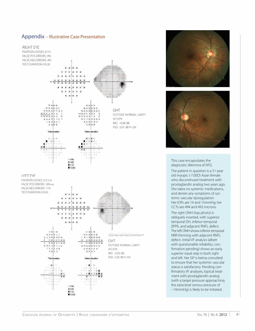
VOL 74 | NO 4 2012C A N A D I A N J O U R N A L O F O P T O M E T R Y | R E V U E C A N A D I E N N E D ’ O P T O M É T R I E 41
This case encapsulates the diagnostic dilemma of NTG.
The patient in question is a 51-year old myopic (-7.00D) Asian femalewho discontinued treatment withprostaglandin analog two years ago.She takes no systemic medications,and denies any symptoms of sys-temic vascular dysregulation.Her IOPs are 14 and 15mmHg; herCCTs are 494 and 493 microns.
The right ONH (top photo) is obliquely inserted, with superiortemporal DH, inferior temporal βPPA, and adjacent RNFL defect.The left ONH shows inferior temporalNRR thinning with adjacent RNFLdefect. Initial VF analysis (albeitwith questionable reliability; con-fi rmation pending) shows an earlysuperior nasal step in both rightand left. Her GP is being consultedto ensure that her systemic vascular status is satisfactory. Pending con-fi rmatory VF analyses, topical treat-ment with prostaglandin analog(with a target pressure approachingthe episcleral venous pressure of ~10mmHg) is likely to be initiated.
RIGHT EYEFIXATION LOSSES: 0/13FALSE POS ERRORS: 9%FALSE NEG ERRORS: 4%TTEST DURATION: 03:28
Appendix – Illustrative Case Presentation
LEFT EYEFIXATION LOSSES: 5/12 xxFALSE POS ERRORS: 18% xxFALSE NEG ERRORS: 11%TTEST DURATION: 03:45
EXCESSIVE HIGH FALSE POSITIVES***
GHTOUTSIDE NORMAL LIMITSVFI 97%MD –0.03 dBPSD 2.30 dB P<5%
GHTOUTSIDE NORMAL LIMITSVFI 97%MD –0.98 dBPSD 2.67 dB P<2%

C A N A D I A N J O U R N A L O F O P T O M E T R Y | R E V U E C A N A D I E N N E D ’ O P T O M É T R I EVOL 74 | NO 4 201242
31. Grise-Dulac A, et al. Assessment ofcorneal biomechanical propertiesin normal tension glaucoma andcomparision with open-angle glaucoma, ocular hypertension, and normal eyes. J Glaucoma 2012;21:486-9.a
32. Kaushik S, et al. Relationship between corneal biomechanicalproperties, central corneal thickness, and intraocular pressure across the spectrum of glaucoma.Am J Ophthalmol ..2012;153: 840-9.
33. Ehrlich JR, et al. Goldmann applanation tonometry compared with corneal-compensated intraocular pressure in the evaluation of primary open-angle glaucoma. BMC Ophthalmol2012 Sep 25;12(1):52 (epub ahead of print).
34. Ang GS, et al. Corneal biomechanical properties in primary open angleglaucoma and normal tension glaucoma. J Glaucoma 2008;17:259-62.a
35. Flammer J, et al. The impact of ocularblood fl ow in glaucoma. Prog Ret Eye Research 2002;21:359-93.h
36. Carter CJ, et al. Investigations intoa vascular etiology for low-tension glaucoma. Ophthalmol 1990;97:49-55.l
37. He Z, et al. The role of bloodpressure in glaucoma. Clin Exp Optom 2011;94:133-49.
38. Moore D, et al. Dysfunctionalregulation of ocular blood fl ow: a risk factor for glaucoma? Clin Ophthalmol 2008;4:849-61.
39. Drance SM, et al. Studies of factorsinvolved in the production of low tension glaucoma. Arch Ophthalmol1973;89:457-65.
40. Kaiser HJ, Flammer J. Systemichypotension: a risk factor for glaucomatous damage? Ophthalmologica1991;203:105-8.
41. Yanagi M, et al. Vascular risk factors in glaucoma: a review. Clin Experiment Ophthalmol 2011;39:252-8.l
42. Deokule S, Weinreb RN. Relationshipsamong systemic blood pressure,intraocular pressure, and open-angle glaucoma. Can J Ophthalmol2008;43:302-7.
43. Leske MC, et al. Risk factors for incident open-angle glaucoma.Ophthalmol 2008;115:85-93.
44. Caprioli J, Coleman AL. Blood pressure, perfusion pressure, and glaucoma. Am J Ophthalmol2010;149:704-12.
45. Liu JHK, et al. Twenty-four-hourintraocular pressure pattern associatedwith early glaucomatous changes. Invest Ophthalmol Vis Sci 2003;44:1586-90.i
46. Krasinska B, et al. A marked fall innocturnal blood pressure is associatedwith the stage of primary open-angle glaucoma in patients witharterial hypertension. Blood Pressure2011;20:171-81.
47. Tokunaga T, et al. Association between nocturnal blood pressure reduction and progression of visual fi eld defect in patients with primary open-angleglaucoma or normal-tension glaucoma. Jpn J Ophthalmol 2004;48:380-5.l
48. Hulsman, CAA et al. Blood pressure,arterial stiffness, and open-angleglaucoma. Arch Ophthalmol 2007;125:805-12.
49. De Moraes CG, et al. Risk factors forvisual fi eld progression in the Low-pressure Glaucoma Treatment Study. Am J Ophthalmol 2012;154:702-11.l
50. Sung KR, et al. Twenty-four hourocular perfusion pressure fl uctuation and risk of normal-tension glaucomaprogression. Invest Ophthalmol Vis Sci 2009;50:5266-74.
51. Sung KR, et al. Characteristics of visualfi eld progression in medically treated normal-tension glaucoma patients with unstable ocular perfusion pressure.Invest Ophthalmol Vis Sci 2011;52:737-43.i
52. Shoshani YZ, et al. Advances in theunderstanding of ocular blood fl ow in glaucoma. Expert Rev Ophthalmol 2010;5:189-200.
53. Plange N, et al. 24-h blood pressuremonitoring in normal tensionglaucoma: night-time blood pressurevariability. J Human Hypertension 2006;20:137-42.
54. Drance SM. What can we learn fromthe disc appearance about the risk factors in glaucoma? Can J Ophthalmol2008;43: 322-7.
55. Park HYL, et al. Nail bed hemorrhage. Arch Ophthalmol 2011;129:1299-1304.l
56. Chung HS, et al. Peripapillary retinal blood fl ow in normal tension glaucoma. Br J Ophthalmol 1999;83:l466-9.
57. Emre M, et al. Ocular blood fl ow alteration in glaucoma is related to systemic vascular dysregulation. Br J Ophthalmol 2004;88:662-6.l
58. Jonas JB. Role of cerebrospinal fl uid pressure in the pathogenesis of glaucoma. Acta Ophthalmol 2011;89:505-14.
59. Pasquale LR. Low intracranial pressure: a tipping point in our understanding of primary open-angle glaucoma?Ophthalmol 2008;115:761-2.l
60. Yang Y, et al. Role of cerebrospinalfl uid in glaucoma: pressure and beyond.Medical Hypotheses 2012;74:31-4.s
61. Ren R, et al. Cerebrospinal fl uidpressure in glaucoma. Ophthalmol2010;117:259-66.
62. Ren R, et al. Cerebrospinal fl uidpressure in ocular hypertension. Acta Ophthalmol 2011;89:142-8.l
63. Berdahl JP, et al. Intracranial pressurein primary open angle glaucoma, normal tension glaucoma, and ocular hypertension: a case-control study.Invest Ophthalmol Vis Sci 2008;49:5412-8.
64. Berdahl JP, et al. Cerebrospinal fl uid pressure is decreased in primary open-angle glaucoma. Ophthalmol2008;115:763-8.
65. Wostyn P, et al. Are intracranialpressure fl uctuations important in glaucoma? Medical Hypotheses2011;77:598-600.
66. Killer HE, et al. Cerebrospinal fl uid exchange in the optic nerve in normal-tension glaucoma. Br J Ophthalmol 2012;96:544-8.
67. Li Z, et al. Intraocular pressure vs intracranial pressure in diseaseconditions: a prospective cohort study (Beijing iCOP Study). BMC Neurology 2012;66:1-4.
68. Wang N, et al. Orbital cerebrospinalfl uid space in glaucoma: the Beijing Intracranial Pressure (iCOP) Study.Ophthalmol 2012;119:2065-73.l

VOL 74 | NO 4 2012C A N A D I A N J O U R N A L O F O P T O M E T R Y | R E V U E C A N A D I E N N E D ’ O P T O M É T R I E 43
69. Caprioli J, Spaeth GL. Comparisonof the optic nerve head in high- andlow-tension glaucoma. Arch Ophthalmol1985;103:1145-9.
70. Sugiyama K, et al. Disc haemorrhages in normal tension glaucoma. Asian J Ophthalmol 2000;2:6.l
71. Cheung W, et al. Neuroprotection inglaucoma: drug-based approaches. Optom Vis Sci 2008;85:406-16.i
72. Iester M, Mikelberg FS. Optic nervehead morphologic characteristicsin high-tension and normal-tension glaucoma. Arch Ophthalmol 1999;117:1010-3.
73. Tezel G, et al. Clinical factors associated with progression ofglaucomatous optic disc damage in treated patients, Arch Ophthalmol2001;119:813-8.
74. De Moraes CGV, et al. Spatially consistent, localized visual fi eld loss before and after disc hemorrhage. Invest Ophthalmol Vis Sci 2009;50:4727-33.i
75. Prata TS, et al. Factors affecting rates of visual fi eld progression in glaucomapatients with optic disc hemorrhage.Ophthalmol 2010;117:24-9.l
76. Drance SM. Disc hemorrhages in the glaucomas. Surv Ophthalmol 1989;33:l331-7.
77. Siegner SW, Netland PA. Optic dischemorrhages and progression of glaucoma. Ophthalmol 1996;103:1014-24.
78. de Beaufort HC, et al. Recurrent dischemorrhage does not increase the rate of visual fi eld progression. Graefes Arch Clin Exp Ophthalmol 2010;248:839-44.l
79. Susanna R, et al. Disc hemorrhagesin patients with elevated intraocularpressure. Arch Ophthalmol 1979;97:284-l5.
80. Jonas JB, et al. Morphologic predictivefactors for development of opticdisc hemorrhages in glaucoma. Invest Ophthalmol Vis Sci 2002;43:2956-61.i
81. Nitta K, et al. Does the enlargement ofretinal nerve fi ber layer defects relate to disc hemorrhage or progressive visualfi eld loss in normal-tension glaucoma? J Glaucoma 2011;20:189-95.a
82. Radcliffe NM, et al. Anatomicrelationship between disc hemorrhage
and parapapillary atrophy. Am J Ophthalmol 2008;146:735-40.l
83. Healey PR, et al. Optic disc hemorrhages in a population with and without signs of glaucoma. Ophthalmol 1998;105:216-23.
84. Budenz DL, et al. Detection and prognostic signifi cance of optic disc hemorrhages during the Ocular Hypertension Treatment Study.Ophthalmol 2006;113:2137-43.l
85. Diehl DLC, et al. Prevalence andsignifi cance of optic disc hemorrhagein a longitudinal study of glaucoma.Arch Ophthalmol 1990;108:545-50.l
86. Gloster J. Incidence of optic dischaemorrhages in chronic simpleglaucoma and ocular hypertension. Br J Ophthalmol 1981;65:452-6.l
87. Yeung DYL, et al. Disc hemorrhage: what do we know? HKI Ophthalmol 2011;14:5-7.
88. Suh MH, Park KH. Period prevalence and incidence of opticdisc haemorrhage in normal tensionglaucoma and primary open-angle glaucoma. Clin Experiment Ophthalmol2011;39:513-9.
89. Kim Y, et al. Risk factors associated with optic disc haemorrhage in patientswith normal tension glaucoma. Eye 2010;24:567-72.
90. Drance SM, et al. The importance ofdisc hemorrhage in the prognosis of chronic open angle glaucoma. Arch Ophthalmol 1977;95:226-8.l
91. Healy P. Optic disc haemorrhage: the more we look the more we fi nd. Clin Experiment Ophthalmol 2011;39:485-6.l
92. Collaborative Normal-Tension Glaucoma Study Group. Theeffectiveness of intraocular pressurereduction in the treatment of normal-tension glaucoma. Am J Ophthalmol1998;126:498-505.
93. Rasker MTE, et al. Rate of visual fi eldloss in progressive glaucoma. Arch Ophthalmol 2000;118:481-8.
94. Rasker MT, et al. Deterioration of visual fi elds in patients with glaucoma with and without optic disc hemorrhages. Arch Ophthalmol1997;115:1257-62.
95. Jonas JB, Xu L. Optic disk hemorrhages in glaucoma. Am J Ophthalmol 1994;118:1-8.
96. De Moraes CGV, et al. Risk factorsfor visual fi eld progression in treated glaucoma. Arch Ophthalmol 2011;129:l562-8.
97. Leske MC, et al. Factors for glaucomaprogression and the effect oftreatment. Arch Ophthalmol 2003;121:48-l56.
98. Ishida K, et al. Disk hemorrhage is asignifi cantly negative prognostic factor in normal-tension glaucoma. Am J Ophthalmol 2000;129:707-14.
99. Collaborative Normal-TensionGlaucoma Study Group. Naturalhistory of normal-tension glaucoma. Ophthalmol 2001;108:247-53.l
100. Guo Y, et al. Five-year follow-up ofparapapillary atrophy: The Beijing EyeStudy. PLoS ONE 2012;7:e32005.
101. Sung KR. Disc hemorrhage: is that a risk factor or sign of progression? J Glaucoma 2012;21:275-6.a
102. Kim SH, Park KH. The relationshipbetween recurrent optic disc hemorrhage and glaucoma progression. Ophthalmol 2006;113:598-602.
103. De Moraes CG, et al. Rate of visual fi eld progression in eyeswith optic disc hemorrhagesin the Ocular Hypertension Treatment Study. Arch Ophthalmol2012 Aug 13:1-6;doi:10.1001/archophthalmol.2012.2324 (epub ahead of print).
104. Shields MB. Hemorrhage of the opticdisk in normal-tension glaucoma. Am J Ophthalmol 2000;129:796-7.
105. Jonas JB, et al. Parapapillary chorioretinal atrophy in normal and glaucoma eyes. Invest Ophthalmol Vis Sci1989;30:919-26.
106. Park KH, et al. Correlation betweenperipapillary atrophy and optic nerve damage in normal-tension glaucoma.Ophthalmol 1996;103:1899-1906.l
107. Teng CS, et al. ß-zone parapapillary atrophy and the velocity of glaucoma progression. Ophthalmol 2010;117:l909-15.

C A N A D I A N J O U R N A L O F O P T O M E T R Y | R E V U E C A N A D I E N N E D ’ O P T O M É T R I EVOL 74 | NO 4 201244
108. Park HYL, et al. Optic disc torsiondirection predicts the location of glaucomatous damage in normal-tension glaucoma in patients withmyopia. Ophthalmol 2012;xx:xxx (article lin press).
109. Jonas JB, et al. Parapapillary atrophy in the chronic open-angle glaucomas. Graefes Arch Clin Exp Ophthalmol1999;237:793-9.
110. Uchida H, et al. Increasing peripapillary atrophy is associated with progressiveglaucoma. Ophthalmol 1998;105:1541-5.l
111. De Moraes CG, et al. Predictivefactors within the optic nerve complex for glaucoma progression: dischemorrhage and parapapillary atrophy.Asia-Pac J Ophthalmol 2012;1:105-12.l
112. Jonas JB, Xu L. Parapapillary chorioretinal atrophy in normal-pressure glaucoma. Am J Ophthalmol1993;115:501-5.
113. See JLS, et al. Rates of neuroretinal rimand peripapillary atrophy area change.Ophthalmol 2009;116:840-7.l
114. Ehrlich JR, Radcliffe NM. The role of clinical parapapillary atrophy evaluation in the diagnosis of open angle glaucoma. Clin Ophthalmol 2010;4:971-6.l
115. Lee EJ, et al. ß-zone parapapillary atrophy and the rate of retinal nerve fi ber layer thinning in glaucoma. Invest Ophthalmol Vis Sci 2011;52:4422-7.i
116. Jonas JB, et al. Parapapillary chorioretinal atrophy in normal and glaucoma eyes. Invest Ophthalmol Vis Sci1989;30:908-18.
117. Pan YZ, et al. The relationship betweenperipapillary atrophy and primary open angle glaucoma. Asian J Ophthalmol2008;10:114-7.
118. Collaborative Normal-TensionGlaucoma Study Group. Comparison of glaucomatous progression between untreated patients withnormal-tension glaucoma andpatients with therapeutically reduced intraocular pressures. Am J Ophthalmol 1998;126:487-97.
119. Caprioli J, Coleman AL. Intraocular pressure fl uctuation. Ophthalmol 2008;115:1123-9.
120. Sommer A, et al. Clinically detectable nerve fi ber atrophy precedes theonset of glaucomatous fi eld loss. Arch Ophthalmol 1991;109:77-83.
121. Sung KR, et al. Retinal nerve fi ber layer normative classifi cation by opticalcoherence tomography for prediction of future visual fi eld loss. Invest Ophthalmol Vis Sci 2011;52:2634-9.i
122. Makabe K, et al. Longitudinal relationship between retinal nerve fi berlayer thickness parameters assessed by scanning laser polarimetry (GDxVCC) and visual fi eld in glaucoma. Graefes Arch Clin Exp Ophthalmol 2012;250:575-81.
123. Leite MT, et al. Structure-functionrelationships using the Cirrus spectraldomain optical coherence tomographand standard automated perimetry. J Glaucoma 2012;21:49-54.a
124. Wollstein G, et al. Retinal nerve fi bre layer and visual function lossin glaucoma: the tipping point. Br J Ophthalmol 2012;96:47-52.
125. Ajtony C, et al. Relationship betweenvisual fi eld sensitivity and retinal nervefi ber layer thickness as measured by optical coherence tomography. Invest Ophthalmol Vis Sci 2007;48:258-63.i
126. Wheat JL, et al. Correlating RNFL thickness by OCT with perimetricsensitivity in glaucoma patients.J Glaucoma 2012;21:95-101.a
127. Medeiros F, et al. A combined indexof structure and function for staging glaucomatous damage. Arch Ophthalmol2012;130:1107-16.
128. Greenfi eld DS. Visual fi eld and intraocular pressure asymmetry in the Low-Pressure Glaucoma Treatment Study. Ophthalmol 2007;114:460-5.l
129. Heijl A, et al. Reduction of intraocularpressure and glaucoma progression.Arch Ophthalmol 2002;120:1268-79.l
130. Leske MC, et al. Predictors of long-term progression in the Early Manifest Glaucoma Trial. Ophthalmol2007;114:1965-72.
131. Janz NK, et al. The Collaborative InitialGlaucoma Treatment Study. Ophthalmol 2001;108:1954-65.
132. Pasquale LR. Optimizing therapy for newly diagnosed open-angle glaucoma. Arch Ophthalmol 2008;126:125-7.l
133. Lichter PR, et al. The investigators’ perspective on the Collaborative InitialGlaucoma Treatment Study (CIGTS). Arch Ophthalmol 2008;126:122-4.l
134. Danias J, Podos SM. Correspondence. Am J Ophthalmol 1999;127:623-6.
135. Goldberg I. Relationship betweenintraocular pressure and preservationof visual fi eld in glaucoma. Surv Ophthalmol 2003;48[suppl 1];S3-7.
136. Cheng JW, et al. Meta-analysis of medical intervention for normal tension glaucoma. Ophthalmol 2009;116:1243-9.
137. Zeitz O, et al. Effects of glaucoma drugs on ocular hemodynamics in normal tension glaucoma: a randomized trial comparing bimatoprost and latanoprost withdorzolamide. BMC Ophthalmol 2005;5:11.
138. Natsis K, et al. Aerobic exercise andintraocular pressure in normotensiveand glaucoma patients. BMC Ophthalmol 2009;9:6.
139. Weinreb RN, Levin LL. Is neuroprotection a viable therapy for glaucoma? Arch Ophthalmol 1999;117:1540-4.
140. Chang EE, et al. Glaucoma 2.0:neuroprotection, neuroregeneration, neuroenhancement. Ophthalmol 2012;119:979-86.
141. Gupta N, Yucel YH. Should we treat the brain in glaucoma? Can J Ophthalmol 2007;42:409-13.
142. Cordeiro MF, Levinn LA. Clinicalevidence for neuroprotectionin glaucoma. Am J Ophthalmol 2011;152:715-6.
143. Beidoe G, Mousa SA. Current primary open-angle glaucoma treatment and future directions. Clin Ophthalmol 2012;6:1699-1707.

ACU Trust_CND JRNL Optometry_EN_November.indd 1 12-10-12 11:22 AM
What keeps your practice growing? Referrals. From happy patients.
9 out of 10 ACUVUE® OASYS® Brand lens wearerssaid they were extremely or very satisfi ed with thequality of vision and comfort of their lenses.1
And it’s the #1 Recommended Brand for sphericalpatients by Canadian eye care professionals. 2
References: 1. Data on fi le. Johnson & Johnson Vision Care, Inc. 2009. 2. #1 Recommended Brand for Spherical Patients by Canadian Optometrists, Opticians and Ophthalmologists. Data on fi le. Johnson & Johnson Inc. 2011. 3. ACUVUE® is the most trusted brand of contact lenses by Canadian Optometrists, Opticians and Ophthalmologists. Data on fi le. Johnson & Johnson Inc. 2011.
ACUVUE® Brand Contact Lenses are indicated for vision correction. As with any contact lens, eye problems, including corneal ulcers, can develop. Some wearers may experience mild irritation, itching or discomfort. Lenses should not be prescribed if patients have any eye infection, or experience eye discomfort, excessive tearing, vision changes, redness or other eye problems. Consult the package insert for complete information. Complete information is also available from Johnson & Johnson Vision Care, Division of Johnson & Johnson Inc., by calling 1-800-267-5098 or by visiting www.jnjvisioncare.ca.
ACUVUE®, ACUVUE® OASYS® and INNOVATION FOR HEALTHY VISION™ are trademarks of Johnson & Johnson Inc. © Johnson & Johnson Inc. 2012. March 2012
//100/////////////////////////////////////////1///9
1#
MOSTTRUSTEDUUUUUUUUUUUUSSSSSSSSSSSSTTTTTTTTTTTTRRRRRRRRRRRRR TTTTTTTTTTTTEEEEEEEEEEEDDDDDDDDDDDDOOOOOOOOOOOOOSSSSSSSSSSSSMMMMMMMMMMMMM TTTTTTTTTTT
CANADIANBRAND3DDDDDDDDDDDDNNNNNNNNNNNNAAAAAAAAAAAAARRRRRRRRRRRBBBBBBBBBBBBB 3333
CACACACACACACACACACACACANANANANANANANANANANANANAN DIDIDIDIDIDIDIDIDIDID ANANANANANANNANANANANANAN
What keeps your practice growing?
ACU Oasys CND JRNL Optometry_EN_November.indd 1 12-10-12 11:22 AM

Now, bio-inspiration is... ...advancing lens care, too.
Bio-inspiration is a discipline that studies and learns
from nature’s best ideas to generate breakthrough
products and technologies. It’s how the butterfly’s
wing led to more energy-efficient LED displays.
Bausch + Lomb eye health scientists looked for
inspiration by studying how the eye works and
developed a new contact lens disinfectant...
WHAT IS BIO-INSPIRATION?
1. Based on a patient use study.
®/™ are trademarks of Bausch & Lomb Incorporated or its affiliates. All other brand/product names are trademarks of their respective owners. © Bausch & Lomb Incorporated. HL XXXX TP7551
Inspired by the biology of your eyes.
Biotrue goes beyond excellent disinfection by bringing
together three bio-inspired innovations:
Biotrue helps make contact lens wear easier on your
patients' eyes1.
For more information, visit www.bausch.ca
pH balanced to match tears
contains a lubricant, to help keep lenses moistand comfortable throughout the day
keeps certain beneficial tear proteins active
Introducing Biotrue™ multi-purpose solution.