circulation chapters 13 and 14 circulatory system transportation –nutrients, metabolic wastes...
TRANSCRIPT
Circulation
Chapters 13 and 14
Circulatory System• Transportation
– nutrients, metabolic wastes (excretion), water, ions and respiratory gases (O2, CO2),
• Regulatory– hormonal – transportation of hormones from endocrine glands to
target tissues – temperature – blood shunting to conserve or dissipate heat through
the skin
• Protection – blood clotting – protects against blood loss – immunity – leukocytes protect against disease
Subdivisions
• Cardiovascular system– heart and blood vessels– transport blood
• Lymphatic system– lymph vessels and lymphoid tissues (spleen
thymus, tonsils and lymph nodes)– transport lymph
Cardiovascular System
• Blood– transport medium (cells suspended in fluid)
• Heart – pump: forces blood through BV’s
• Blood vessels – tubing that carries blood (60,000 miles)
Blood• Transport medium for gases,
nutrients, wastes etc.• Volume
– Women = 5 liters– Men = 5.5 liters
• Blood is composed of: – Plasma (55% of blood volume)
• fluid portion
– Formed Elements (45%)• cells and cell fragments
Plasma• Water (90%)
– dissolves materials (gases, nutrients, etc.)– acts as fluid medium for transport of blood cells and
plasma proteins
• Proteins (7-9%)– Maintain osmotic pressure of blood (albumins)– Lipid transport (globulins)– Immunity (antibodies, complement proteins)– Clotting factors– Various enzymes
Formed Elements
• Erythrocytes – Red Blood Cells
• Leukocytes – White Blood Cells
• Thrombocytes– Platelets
Erythrocytes
• thin, disc-like cells• 25-30 trillion RBCs
circulating at a given time• Lack nuclei and
mitochondria– anaerobic respiration only
• transport respiratory gases (mainly O2)
Erythrocytes
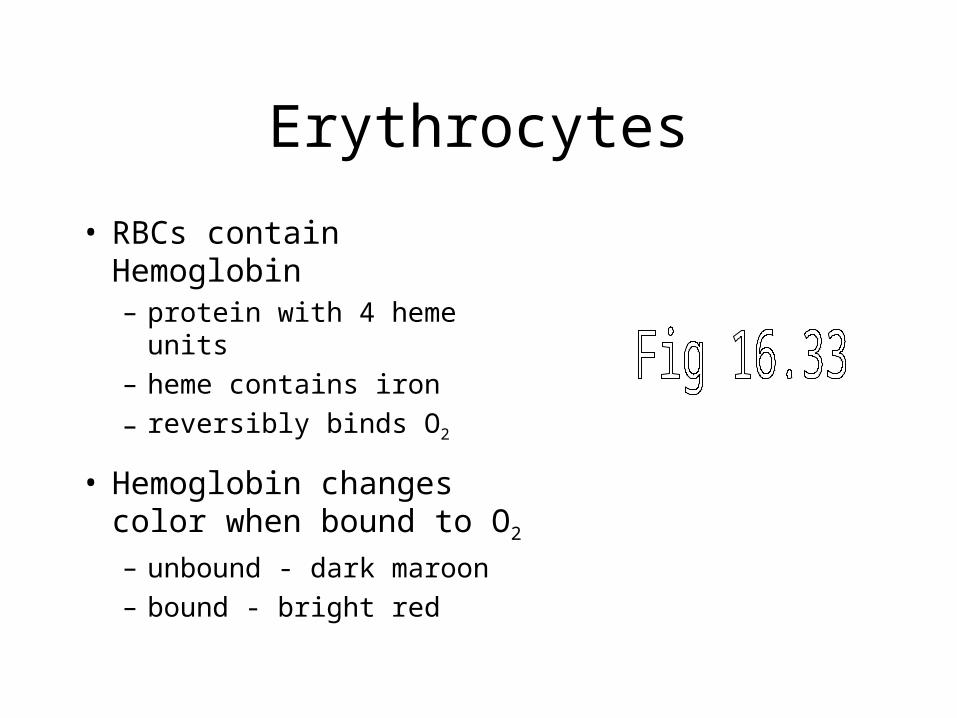
• RBCs contain Hemoglobin– protein with 4 heme units
– heme contains iron
– reversibly binds O2
• Hemoglobin changes color when bound to O2
– unbound - dark maroon
– bound - bright red
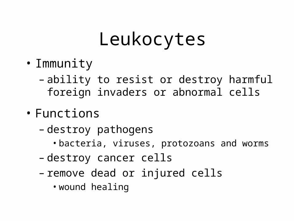
Leukocytes• Immunity
– ability to resist or destroy harmful foreign invaders or abnormal cells
• Functions– destroy pathogens
• bacteria, viruses, protozoans and worms
– destroy cancer cells – remove dead or injured cells
• wound healing
Leukocyte Types
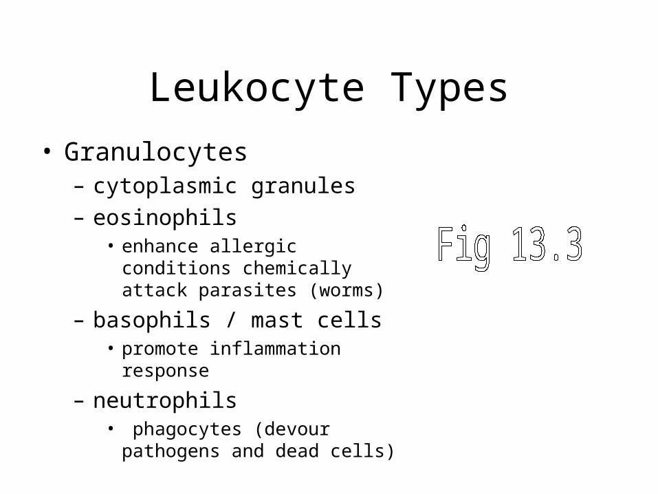
• Granulocytes – cytoplasmic granules
– eosinophils • enhance allergic conditions
chemically attack parasites (worms)
– basophils / mast cells• promote inflammation response
– neutrophils• phagocytes (devour pathogens and
dead cells)
Leukocyte Types
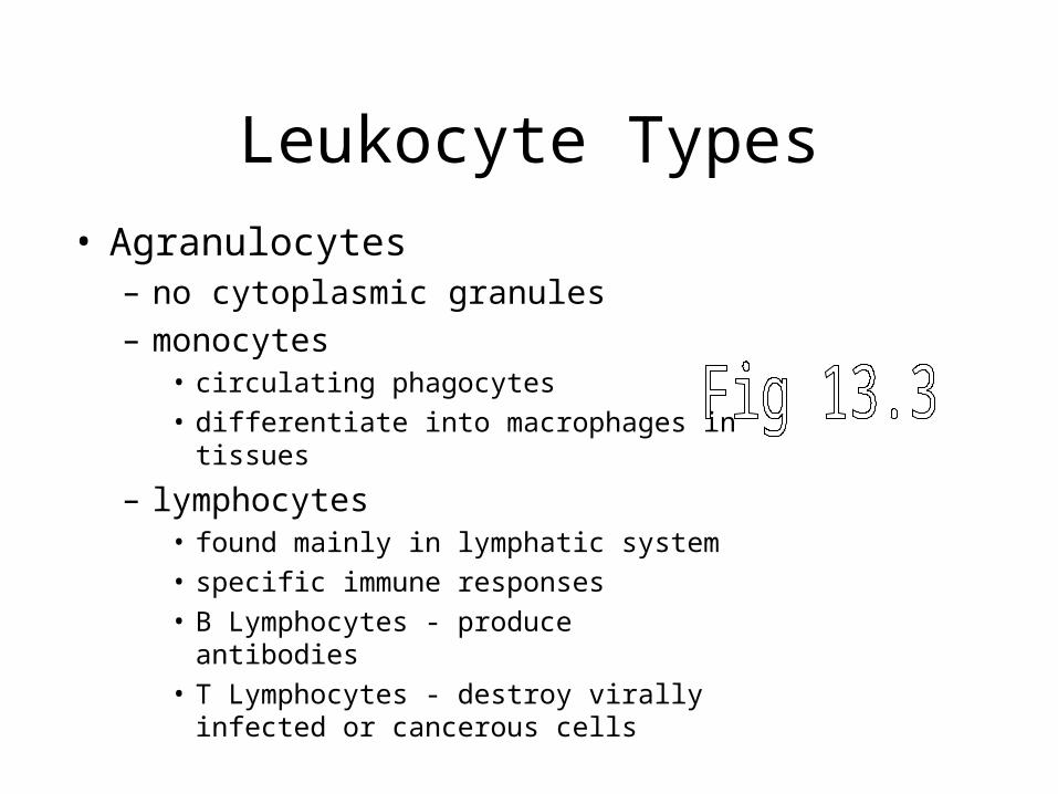
• Agranulocytes – no cytoplasmic granules
– monocytes• circulating phagocytes
• differentiate into macrophages in tissues
– lymphocytes • found mainly in lymphatic system
• specific immune responses
• B Lymphocytes - produce antibodies
• T Lymphocytes - destroy virally infected or cancerous cells

Platelets (Thrombocytes)
• cell fragments from megakaryocytes in myeloid tissue
• contain actin and myosin (contraction)
• Formation of a blood clot to stop bleeding
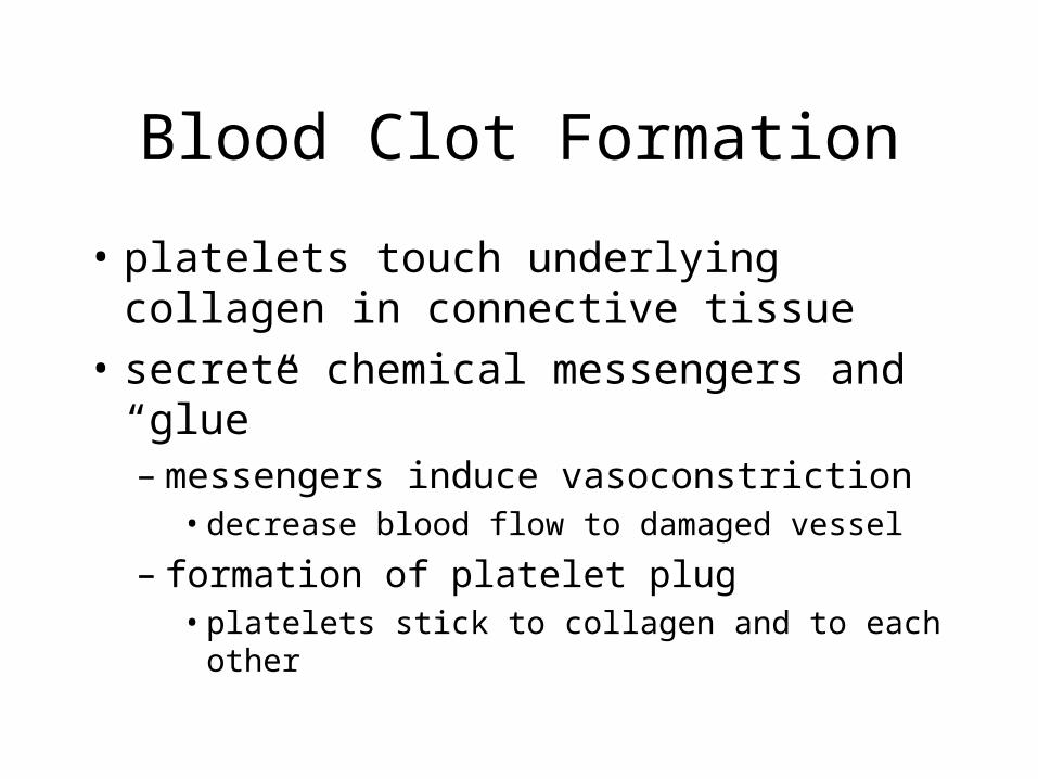
Blood Clot Formation
• platelets touch underlying collagen in connective tissue
• secrete chemical messengers and “glue” – messengers induce vasoconstriction
• decrease blood flow to damaged vessel
– formation of platelet plug • platelets stick to collagen and to each other
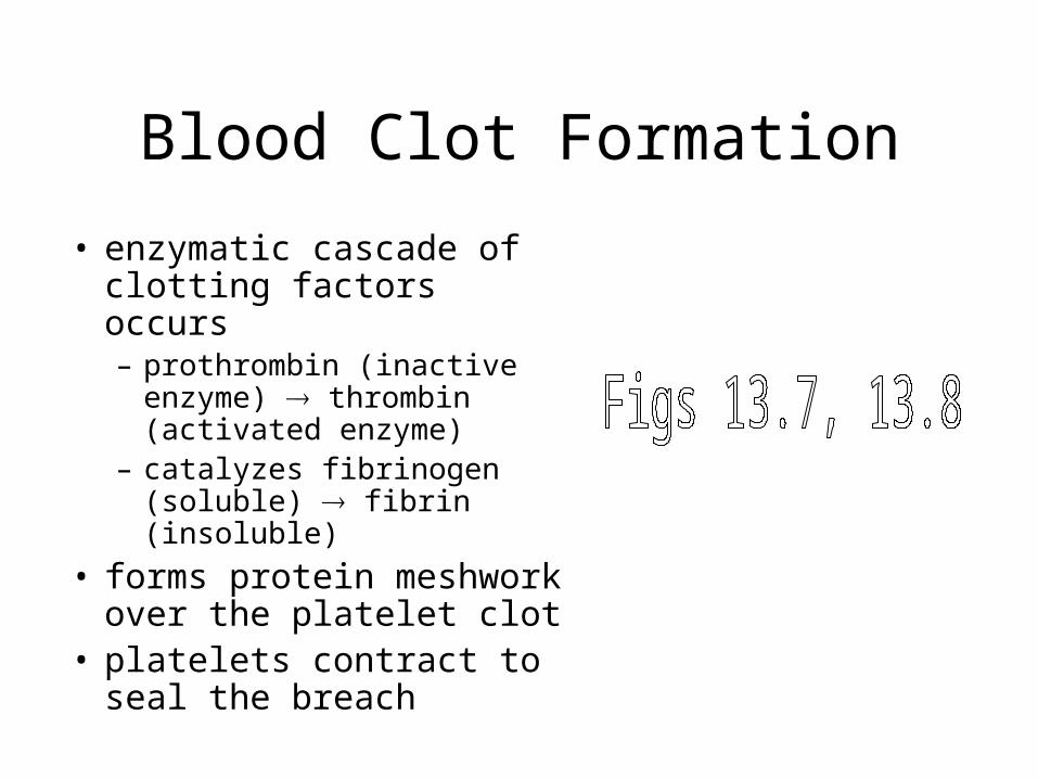
Blood Clot Formation
• enzymatic cascade of clotting factors occurs – prothrombin (inactive
enzyme) thrombin (activated enzyme)
– catalyzes fibrinogen (soluble) fibrin (insoluble)
• forms protein meshwork over the platelet clot
• platelets contract to seal the breach
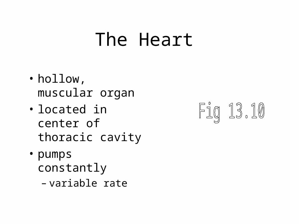
The Heart
• hollow, muscular organ
• located in center of thoracic cavity
• pumps constantly– variable rate
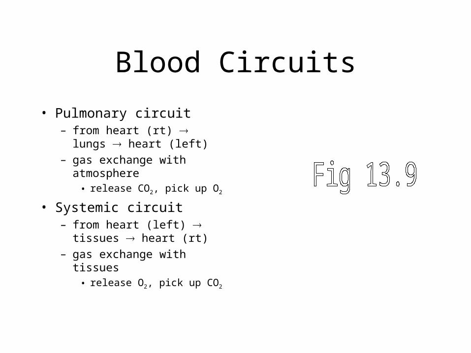
Blood Circuits
• Pulmonary circuit– from heart (rt) lungs
heart (left)
– gas exchange with atmosphere
• release CO2, pick up O2
• Systemic circuit– from heart (left) tissues
heart (rt)
– gas exchange with tissues • release O2, pick up CO2
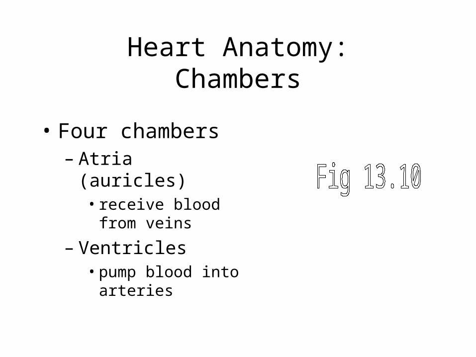
Heart Anatomy:Chambers
• Four chambers– Atria (auricles)
• receive blood from veins
– Ventricles• pump blood into
arteries
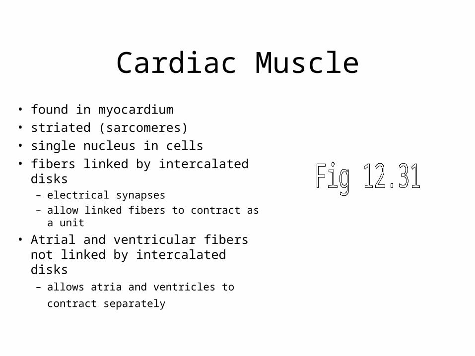
Cardiac Muscle
• found in myocardium
• striated (sarcomeres)
• single nucleus in cells
• fibers linked by intercalated disks– electrical synapses
– allow linked fibers to contract as a unit
• Atrial and ventricular fibers not linked by intercalated disks– allows atria and ventricles to contract
separately
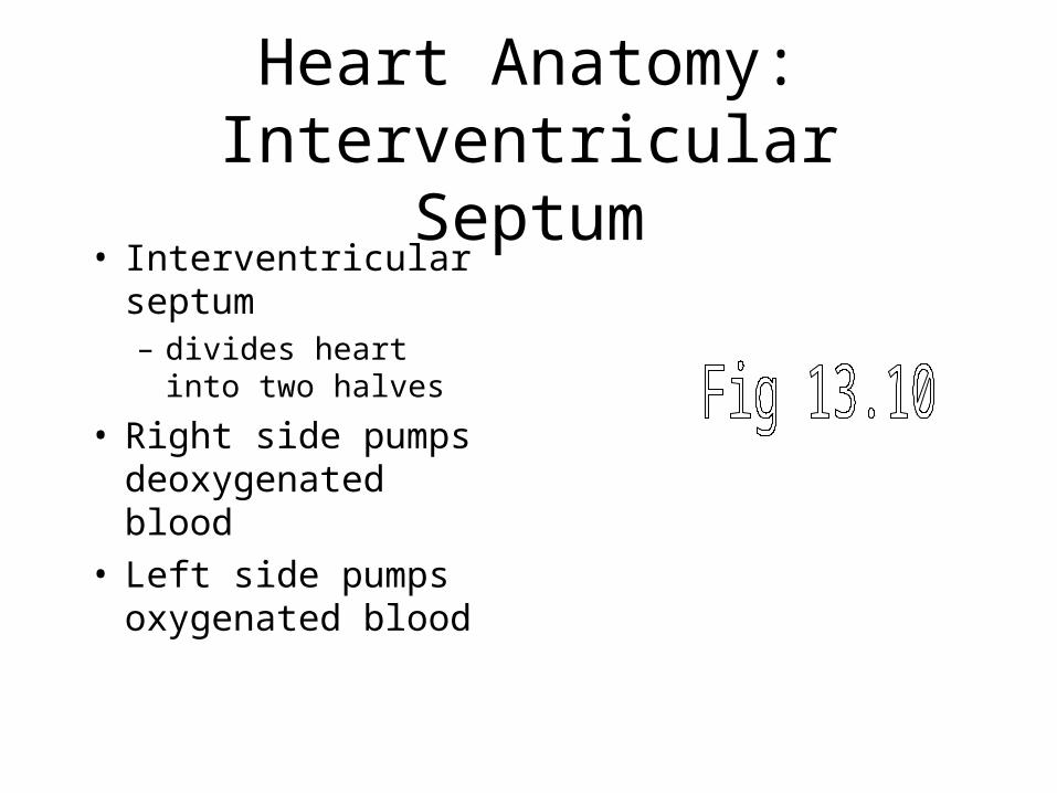
Heart Anatomy:Interventricular Septum
• Interventricular septum– divides heart into
two halves
• Right side pumps deoxygenated blood
• Left side pumps oxygenated blood
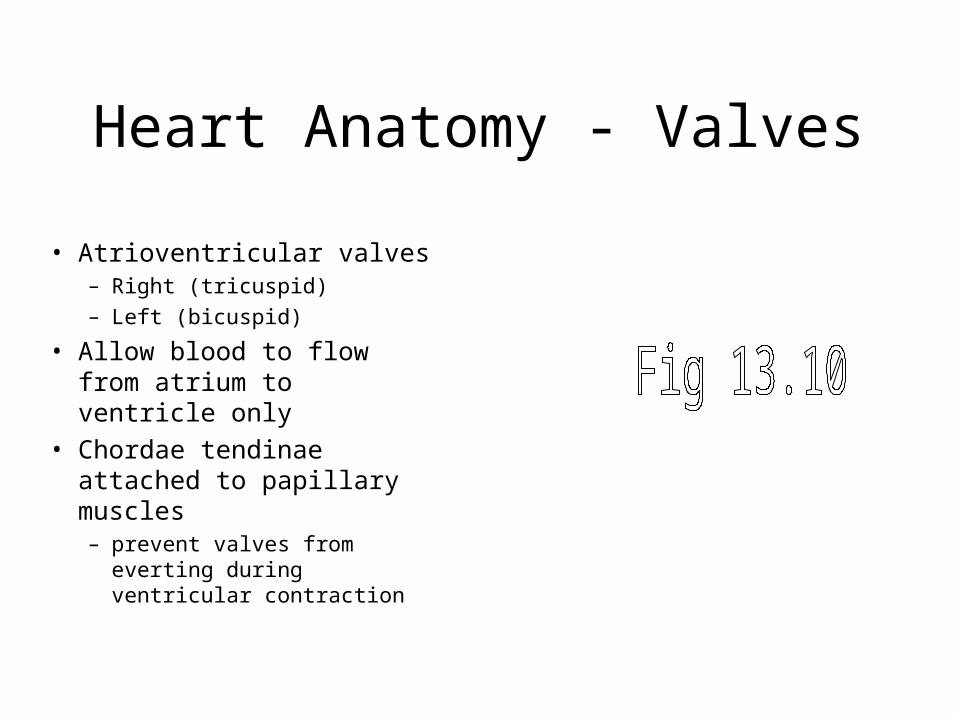
Heart Anatomy - Valves
• Atrioventricular valves– Right (tricuspid)
– Left (bicuspid)
• Allow blood to flow from atrium to ventricle only
• Chordae tendinae attached to papillary muscles – prevent valves from everting
during ventricular contraction
Heart Anatomy - Valves
• Semilunar valves– Pulmonary (Right)
– Aortic (Left)
• at openings of the arteries leaving the ventricles
• prevent backflow of blood during ventricular relaxation
Blood Flow Through the Heart
• Deoxygenated blood enters right side through vena cavae
• right atrium
• right AV valve
• right ventricle
• pulmonary semilunar valve
• pulmonary artery
• lungs (pulmonary circuit)

Blood Flow Through the Heart
• Oxygenated blood enters left side through pulmonary veins
• left atrium• left AV valve• left ventricle• aortic semilunar valve• aorta• tissues (systemic circuit)
Cardiac Cycle
• contraction (systole) + relaxation (diastole) of ventricles
• lasts 0.8 sec (based on 72 beats/min)
Cardiac Cycle - Blood Volumes
• End-diastolic volume– amt of blood in ventricles at end
of diastole
• End-systolic volume– amt of blood in ventricles at end
of systole– ~1/3 of end-diastolic vol.
• Stroke volume (SV)– Amt of blood ejected by ventricles– Equal to EDV – ESV
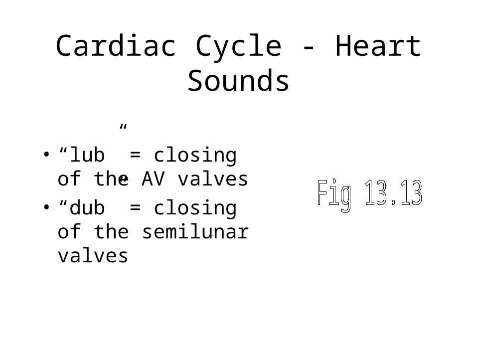
Cardiac Cycle - Heart Sounds
• “lub” = closing of the AV valves
• “dub” = closing of the semilunar valves
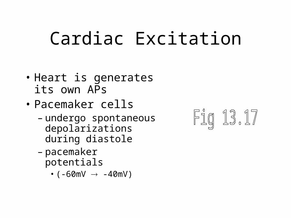
Cardiac Excitation
• Heart is generates its own APs
• Pacemaker cells– undergo spontaneous
depolarizations during diastole
– pacemaker potentials • (-60mV -40mV)
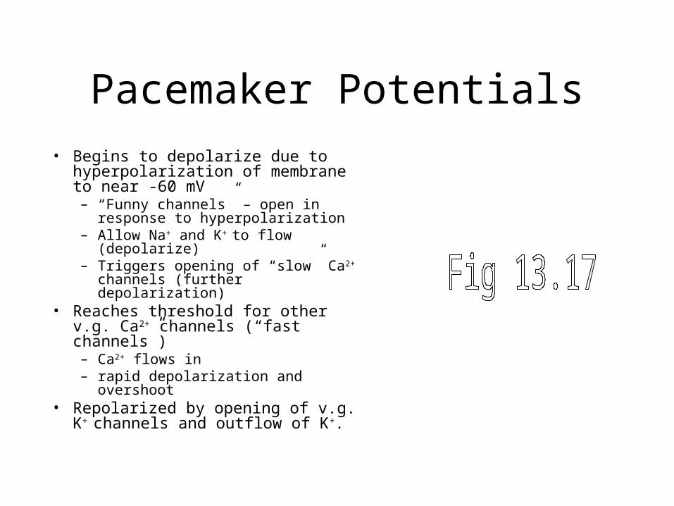
Pacemaker Potentials
• Begins to depolarize due to hyperpolarization of membrane to near -60 mV– “Funny channels” – open in response
to hyperpolarization– Allow Na+ and K+ to flow
(depolarize)– Triggers opening of “slow” Ca2+
channels (further depolarization)• Reaches threshold for other v.g. Ca2+
channels (“fast channels”) – Ca2+ flows in – rapid depolarization and overshoot
• Repolarized by opening of v.g. K+
channels and outflow of K+.
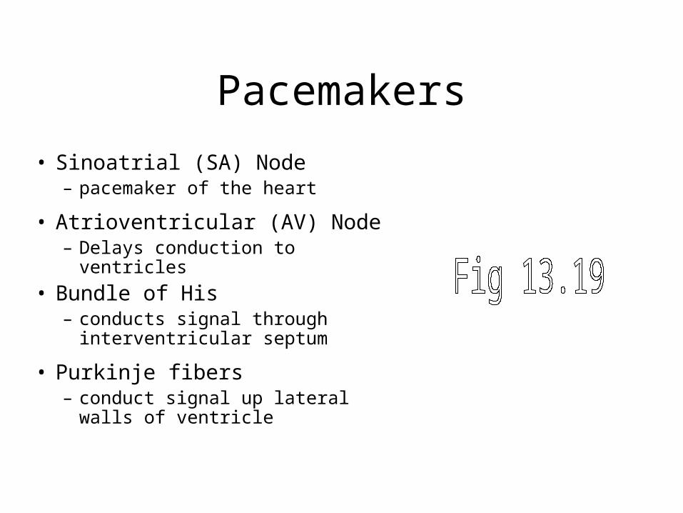
Pacemakers
• Sinoatrial (SA) Node – pacemaker of the heart
• Atrioventricular (AV) Node – Delays conduction to ventricles
• Bundle of His– conducts signal through
interventricular septum
• Purkinje fibers – conduct signal up lateral walls of
ventricle
Path of Cardiac Excitation
• SA node cells produce APs
• Atrial fibers activated– atrial contraction
• APs excite AV node– delay (complete atrial contract)
• APs of AV node travel down AV bundle to apex of heart
• signal conducted to Purkinje fibers throughout ventricles
• Myocardial fibers activated– ventricular contraction
Myocardial Action Potentials
• Prolonged action potential duration– Influx of Na+ - depolarization
– Plateau phase due to Ca2+ influx and slow opening of K+ channels
– Repolarization by delayed opening of v.g. K+ channels
• Prolonged refractory period ensures pumping action
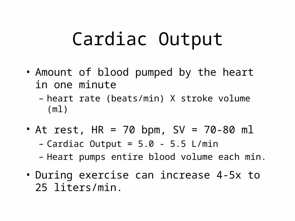
Cardiac Output
• Amount of blood pumped by the heart in one minute – heart rate (beats/min) X stroke volume (ml)
• At rest, HR = 70 bpm, SV = 70-80 ml – Cardiac Output = 5.0 - 5.5 L/min
– Heart pumps entire blood volume each min.
• During exercise can increase 4-5x to 25 liters/min.
Heart Rate Regulation
• Parasympathetic nervous system – Slows HR
• Sympathetic nervous system– Speeds up HR
• Hormones (Epinephrine)
Stroke Volume Regulation
• end-diastolic volume – ↑EDV, ↑SV– function of venous return
• total peripheral resistance– ↑Resistance, ↓SV– Function of blood vessel diameter
Stroke Volume Regulation
• contractility – function of sarcomere
length in muscle fibers
– sympathoadrenal stimulation (↑ Ca2+ concentrations)
Blood Vessels
• Tubes that conduct blood– Arteries– Aterioles– Capillaries– Venules– Veins

Blood Pressure
• Pressure blood exerts on blood vessel walls– produced by heart
contractions
– main driving force for the flow of blood through the blood vessels
– decreases as blood moves further from the heart
Arteries
• Large vessel receiving blood from the heart
• Functions– rapid transport of blood
• large radius, high pressure
– pressure reservoirs• walls expand upon systole• recoil during diastole
maintains blood flow
Arterial Blood Pressure
• Systolic blood pressure– pressure of blood in arteries
during ventricular systole
• Diastolic blood pressure– pressure of blood in arteries
during ventricular diastole
• Indicates – blood flow to the body– work load of the heart
Arterial Blood Pressure Regulation
• Blood pressure monitored by baroreceptors in carotid sinuses and aorta
• Signals sent to medulla– cardiac + vasomotor centers
• response sent out via autonomic MNs– sympathetic - HR,
vasoconstriction– parasympathetic - HR,
vasodilation
Arterioles
• Major resistance vessels fluctuations in blood pressure
btw systole & diastole
• Control of perfusion– smooth muscle in vessel walls
under autonomic control
– vasoconstriction diameter, blood flow
– vasodilation diameter, blood flow
Capillaries
• Single endothelial cell layer• Connect arterioles to venules• Exchange of materials between blood and
interstitial fluid
Properties of Capillaries
• Short diffusion distance – thin capillary walls
– narrow diameter
• High surface area– extensive branching (close
to all cells)
• Slow blood flow– increases time for
diffusion to occur
Ultrafiltration
• Blood passing through capillaries undergoes ultrafiltration– Blood pressure causes plasma fluid to
be pushed out (proteins remain)
– As fluid is pushed out, blood osmotic pressure increases
– Fluid drawn back into the capillaries when osmotic pressure exceeds blood pressure
– More fluid forced out than pulled in
Venules and Veins
• Return blood to the heart after exchanging materials in capillaries
• Hold most of the blood in the body– capacitance vessels
• regulate rate of blood return to the heart
Venous Return
Venous return – amt of blood veins deliver back to the heart
Factors influencing return• pressure gradient
– mean pressure is low– vein walls are highly elastic
• large radii of veins - little resistance – modified by vasoconstriction
Venous Return
• Venous Valves – prevent backflow of blood
• Skeletal Muscle Activity– contraction acts to “pump”
veins
– increases venous return with increased activity
Lymphatic System
• Blood passing through capillaries undergoes ultrafiltration– Net loss of fluid from the blood
– Must be returned to the blood
Lymphatic System
• Network of vessels – picks up lost fluid (lymph)
and returns it to circulation
• Pass through lymph nodes– Immune functions
• Vessels deposit lymph into subclavian veins