urinary system and excretion - napa valley college pages€¦ · urinary system and excretion...
TRANSCRIPT

Urinary System and Excretion
Bio105
Lecture 20
Chapter 16
1

Copyright © 2009 Pearson Education, Inc.
Outline – Urinary System
I. Function
II. Organs of the urinary system
A. Kidneys
1. Function
2. Structure
III. Disorders of the urinary system
IV.Metabolism
2

Copyright © 2009 Pearson Education, Inc.
Urinary System
The digestive system eliminated
waste from the digestive tract. But
we also need a way to eliminate
waste from the rest of the body.
Function of urinary system is:
Excretion of metabolic wastes and to
maintain homeostasis of blood.
3
Copyright © 2009 Pearson Education, Inc.
Which of the following system does not function to excrete waste?
1. Digestive
2. Urinary
3. Integumentary
4. Circulatory

Copyright © 2009 Pearson Education, Inc.
Urine
Urine contains:
Water
HCO3-
Inorganic salts
H+
Urea
Uric acid
Creatinine

Copyright © 2009 Pearson Education, Inc.
Excretion
Excretion - the majority of the
metabolic wastes removed from the
body is mainly due to the action of
the kidneys.
6
Copyright © 2009 Pearson Education, Inc.
Organs of the Urinary System
1. Kidneys – main organ in the urinary system,
produces urine.
2. Ureters - conduct urine from the kidneys to the
bladder by peristaltic contractions produced from
contractions of smooth muscles in ureter wall.
7

Copyright © 2009 Pearson Education, Inc.
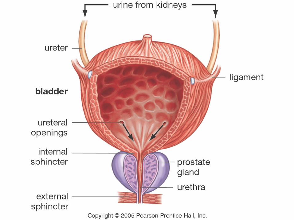
Organs of the Urinary System
3. Urinary bladder - stores urine until it is expelled
from the body.
4. Urethra - small tube that extends from the urinary
bladder to an external opening. In males the
urethra also functions as a reproductive tract
organ.
8

Copyright © 2009 Pearson Education, Inc.
Functions of the Kidneys
1. Filter waste from blood
2. Maintenance of water-salt balance of
the blood.
3. Regulates blood pressure
4. Maintenance of acid-base balance of
the blood.
5. Secretion of hormones = renin and
erythropoietin.
9

Copyright © 2009 Pearson Education, Inc. Figure 16.2 (1 of 2)
Urinary System
Kidney
• Produces urine• Conserves water• Regulates pH• Stimulates
production ofred blood cells
• Transforms vitamin D into active form
Ureter
• Transports urinefrom kidneysto bladder
Urinary
bladder
• Stores urine
Urethra
• Transports urine from urinary bladderto outside the body

Copyright © 2009 Pearson Education, Inc.
The kidneys are located in this cavity:
1. Cranial
2. Thoracic
3. Abdominopelvic
4. Pleural

Copyright © 2009 Pearson Education, Inc. Figure 16.2 (2 of 2)
Urinary System
Adrenal
gland
Renal
artery
Renal veinAorta
Heart
Diaphragm
Inferior
vena cava

Copyright © 2009 Pearson Education, Inc.
Vascularization
Aorta
Renal artery
Arterioles
Capillaries
Venules
Renal Vein
Vena Cava

Copyright © 2009 Pearson Education, Inc. Figure 16.3a
The Kidneys
Renal artery
Adrenal
gland
Renal vein
Ureter
Outermost
connective
tissue layer
Adipose
capsule Blood vessels and protective
layers around kidneys
Innermost
connective
tissue layer
(a)

Copyright © 2009 Pearson Education, Inc.
The Regions of the Kidneys
Each kidney has three regions:
1. Renal cortex
2. Renal medulla
3. Renal pelvis
Copyright © 2009 Pearson Education, Inc.
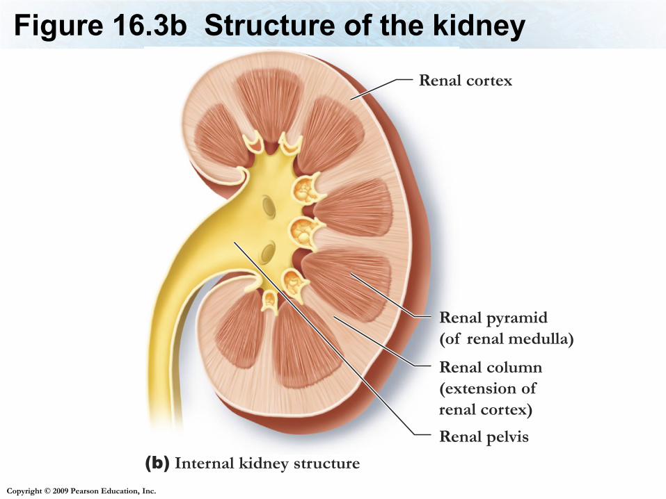
Figure 16.3b Structure of the kidney
Renal cortex
Renal pyramid
(of renal medulla)
Renal column
(extension of
renal cortex)
Renal pelvis
(b) Internal kidney structure

Copyright © 2009 Pearson Education, Inc.
Regions of the Kidney
1. Renal cortex - an outer granulated layer.
2. Renal medulla - consists of cone-shaped
tissue masses called renal pyramids.
3. Renal pelvis - a central cavity that is
continuous with the ureter.
17

Copyright © 2009 Pearson Education, Inc.
Nephrons
The functional units of the kidneys.
Over 1 million nephrons per kidney
Nephrons extend from the Renal cortex, into
the renal medulla
18

Copyright © 2009 Pearson Education, Inc. Figure 16.3b–c
The Nephron

Copyright © 2009 Pearson Education, Inc. Figure 16.4b
The Nephron
Afferent
(incoming)
arteriole
Glomerular
Capillaries
at start of
nephron
(b) A nephron and its blood supply
Distal
convoluted
tubule
Surrounding
capillaries
Loop of the
nephron
Collecting duct
Proximal
convoluted
tubule
Glomerular
capsule
Efferent
(outgoing)
arteriole

Copyright © 2009 Pearson Education, Inc.
Parts of the Nephron
1. The renal corpuscle
A. The glomerulus
B. The glomerular capsule
2. The renal tubule
A. Proximal convoluted tubule
B. Loop of the nephron
C. Distal convoluted tubule
3. The collecting Duct

Copyright © 2009 Pearson Education, Inc. Figure 16.4c
The NephronGlomerular capsule
(glomerulus within)
(c) Simplified view of a nephron, showing the
basic structural components but not the
associated capillaries
Proximal
convoluted tubule
Renal
tubule
Distal convoluted
tubule
Loop of the
nephron
Renal
corpuscle

Copyright © 2009 Pearson Education, Inc.
The renal corpuscle
The renal corpuscle is where fluid is
filtered from blood
Consists of
The glomerulus - The network of
capillaries
The glomerular capsule (Bowman’s
capsule) - Surrounds the glomerulus

Copyright © 2009 Pearson Education, Inc.
The Nephron
The nephron performs three functions
1. Glomerular filtration
2. Tubular reabsorption
3. Tubular secretion

Copyright © 2009 Pearson Education, Inc.
The Nephron - Glomerular filtration
Glomerular filtration occurs as blood
pressure forces water, ions, and other
small molecules in the blood through the
pores in the glomerulus and into the
glomerular capsule
The filtrate passes into the renal tubule

Copyright © 2009 Pearson Education, Inc.
26

Copyright © 2009 Pearson Education, Inc. Figure 16.5a
The Nephron
(a) The renal corpuscle consists of the
glomerular capsule and a ball of
capillaries called the glomerulus.
Afferent (incoming)
arteriole
Efferent (outgoing)
arteriole
Path of filtrate
Path of blood
Movement of water
and small solutes
Filtrate
Glomerulus
Glomerular
capsuleSpace within the
glomerular capsule
Capillary wall
Proximal
convoluted tubule

Copyright © 2009 Pearson Education, Inc.
The Nephron
Figure 16.5b

Copyright © 2009 Pearson Education, Inc.
The renal tubule
1. Proximal convoluted tubule (PCT) - where
reabsorption of filtrate components occurs,
tubular secretion can also occur here.
2. Loop of the Nephron (Loop of Henle) - consists of
a descending limb and an ascending limb that
regulates osmotic balance.
3. Distal convoluted tubule (DCT) – Further
absorption of water and salts; leads to the renal
pelvis
29

Copyright © 2009 Pearson Education, Inc.
Collecting Ducts
Collecting ducts - carry urine to the renal pelvis.
30

Copyright © 2009 Pearson Education, Inc.
The Nephron
Figure 16.7
Nitrogen-
containing
waste
Nitrogen-
containing
waste
Glomerulus
Glomerular
capsule
Vein
Artery
Step 1: Glomerular filtrationWater, ions, amino acids, glucose, nitrogen-containing wastes, and other small molecules move from the glomerulus to the inside of the glomerular capsule to form glomerular filtrate.
Urine
Renal pelvis
within kidney
Surrounding
capillaries
Path of filtrate
Path of blood
Movement of substances
from blood to filtrate
Movement of substances
from filtrate to blood
Collecting duct
Renal
medulla
Renal
cortex
Loop of the
nephron
Step 2: Tubular reabsorption Water, essential ions, and nutrients are reabsorbed from the proximal convoluted tubule into the surrounding capillaries. Some reabsorption of water and ions occurs along other sections of the renal tubule and collecting duct.
Efferent (outgoing)
arteriole
Afferent (incoming)
arteriole
Glucose
Amino acidsIons
H2O
Drugs
H+ NH4+
K+
H2O
Glucose
Ions
Step 3: Tubular secretion Wastes, excess ions, anddrugs are actively secreted into the distal (and proximal) convoluted tubules from the surrounding capillaries. Some secretion also occurs along the collecting duct.

Copyright © 2009 Pearson Education, Inc.
Urine Formation
Tubular reabsorption - many molecules are
reabsorbed – transported from the lumen into
the tissues then into capillaries. Occurs mainly in
the PCT(H2O, nutrients, salts)
Tubular secretion - substances are removed
from the blood and added to the tubular fluid,
mainly in the DCT. (H+, creatinine, and drugs like
penicillin)
32

Copyright © 2009 Pearson Education, Inc.

Copyright © 2009 Pearson Education, Inc.
34

Copyright © 2009 Pearson Education, Inc.
35

Copyright © 2009 Pearson Education, Inc.
This structure conducts urine from the kidneys to the bladder
1. Urethra
2. Ureters
36

Copyright © 2009 Pearson Education, Inc.
What is the functional unit of the kidney?
1. Renal medulla
2. Nephron
3. Renal cortex
37

A
D
F
B
C
E
A. Renal Cortex
B. Renal Medulla
C. Glomerular capsule,
contains glomerulus
D. Proximal Convoluted Tubule
E. Loop of Henle
F. Distal Convoluted Tubule
GG. Collecting Duct

Copyright © 2009 Pearson Education, Inc.
Regulation of Urine
Diuretics increase urinary output, making
more dilute urine
Examples:
Caffeine
Lasix
Alcohol
39
Copyright © 2009 Pearson Education, Inc.
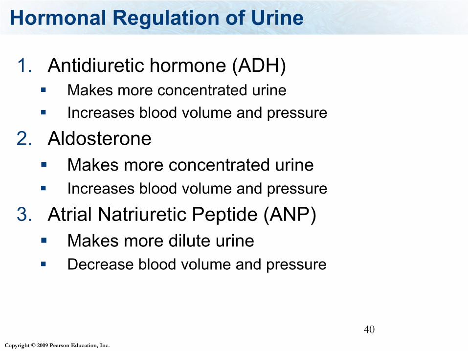
Hormonal Regulation of Urine
1. Antidiuretic hormone (ADH)
Makes more concentrated urine
Increases blood volume and pressure
2. Aldosterone
Makes more concentrated urine
Increases blood volume and pressure
3. Atrial Natriuretic Peptide (ANP)
Makes more dilute urine
Decrease blood volume and pressure
40

Copyright © 2009 Pearson Education, Inc.
Table 16.3 Review of Hormones

Copyright © 2009 Pearson Education, Inc.
Hormonal Regulation of Urine - ADH
Antidiuretic hormone (ADH)
Makes the collecting duct more permeable to
water therefore, increases the water
reabsorption in the collecting duct, making more
concentrated urine.
Produced by the hypothalamus, stored in the
posterior pituitary gland.
Site of action: collecting ducts.
42
Copyright © 2009 Pearson Education, Inc.
Diabetes insipidus
Diabetes insipidus is caused by producing
too little ADH
Symptoms: excrete large amounts of dilute
urine
43

Copyright © 2009 Pearson Education, Inc.
Kidney Function and ADH
Figure 16.9
Decrease in concentration
of water in blood is detected
by the hypothalamus.
Hypothalamus
Nerve cells
produce antidiuretic
hormone (ADH)
Anterior lobe of
pituitary gland
Posterior lobe of
pituitary gland
Antidiuretic hormone (ADH)
is produced by the
hypothalamus and released
by the posterior lobe of the
pituitary gland.
ADH prompts an increase in the
permeability to water of distal
convoluted tubules and collecting
ducts of nephrons.
An increase in the concentration
of water in the blood causes:
• Increase in blood volume
• Increase in blood pressure
• Decrease in urine volume
More water moves
from the filtrate back
into the blood.

Copyright © 2009 Pearson Education, Inc.
Hormonal Regulation of Urine - Aldosterone
Aldosterone
Hormone produced and released by the adrenal
cortex
Increases sodium reabsorption in the distal
convoluted tubule and the collecting duct, water
follows
Making more concentrated urine.
45
Copyright © 2009 Pearson Education, Inc.
Juxtaglomerular apparatus and Aldosterone
Aldosterone is released in response to blood
pressure monitored by the juxtaglomerular
apparatus
The cells in the juxtaglomerular apparatus
release the hormone renin.
Renin is converted to an active form that
stimulates the adrenal cortex to release
aldosterone

Copyright © 2009 Pearson Education, Inc.
Kidney Function and Hormones
Figure 16.10

Copyright © 2009 Pearson Education, Inc.
Hormonal Regulation of Urine
Atrial Natriuretic Peptide (ANP)
Hormone produced by the heart in response to
increased blood volume and pressure
Decreases sodium reabsorption in the distal
convoluted tubule and the collecting duct, water
stays in the filtrate
Also inhibits production of aldosterone and
renin
Making more dilute urine. Lowers blood
pressure and blood volume
48

Copyright © 2009 Pearson Education, Inc.
49

Copyright © 2009 Pearson Education, Inc.
Hormones secreted by the kidneys
1. Renin – Increases blood pressure by
triggering the release of aldosterone by
the adrenal cortex
2. Erythropoietin – speeds up the
maturation process of RBCs, target =
stem cells in bone marrow
50

Copyright © 2009 Pearson Education, Inc.
The Kidney’s role in Vit D
Vitamin D is produced in the skin in response
to sunlight, and provided by certain foods in
diet
The kidneys and liver transform Vitamin D
into the active form, calcitrol.
Calcitrol promotes the absorption of calcium
into the small intestine and re-absorption of
calcium in the kidneys.
51

Copyright © 2009 Pearson Education, Inc.
Kidney’s role in Acid-Base Balance
H+ is secreted into the tubules and
bicarbonate is reabsorbed out of the tubules
52

Copyright © 2009 Pearson Education, Inc.
When H+ is secreted into the tubules, this
lowers the pH of the blood.
1. True
2. False

Copyright © 2009 Pearson Education, Inc.
Kidney’s role in Salt-Water Balance
The kidneys reabsorb salt and water,
maintaining osmotic balance in the blood,
this also affects blood pressure
54

Copyright © 2009 Pearson Education, Inc.
Bladder
The urine goes from the kidneys into the
bladder where it is stored until it can be
released through the urethra.
55

9-5

Copyright © 2009 Pearson Education, Inc.
Urination
Urination is a reflex action controlled
by the brain
When the bladder fills to about 250ml
of urine then the motor nerve
impulses cause the bladder to
contract and the sphincters to relax
so that urination is possible.58

Copyright © 2009 Pearson Education, Inc.
Urinary Function Disorders
Urethritis—infection confined to the urethra.
Cystitis—infection of the urethra and
bladder.
Pyelonephritis—infection reaches the
kidneys.
59

Copyright © 2009 Pearson Education, Inc.
What hormone is secreted by the
kidneys to increase blood pressure:
1. ADH
2. Renin
3. Aldersterone
4. Erythropoietin
60

Copyright © 2009 Pearson Education, Inc.
Important Points
Read Chapter 17 for next lecture
Know the functions of the urinary system
Know the organs of the urinary system and their functions, including all the functions of the kidney
Know the structure of the kidney (see “kidney structure slide and preceding illustrations)
Know what a nephron is, what are the five parts of the nephron and the function of these parts.
61

Copyright © 2009 Pearson Education, Inc.
Important Points
Understand the re-absorption and secretion of
compounds in the nephron, what is re-absorbed
and secreted and where in the nephron are the
compounds re-absorbed or secreted.
How is urinary output regulated, what are
examples of diuretics. What hormones decrease or
increase urinary output. What effect on blood
pressure do these hormones have. Where are
these hormones produced, stored and released
from. What specific parts of the nephron do these
hormones effect. 62

Copyright © 2009 Pearson Education, Inc.
Important Points
What is diabetes insipidus
Know what renin is, what its function is, where it is produced.
Know what erythropoietin is, what its function is, where it is produced.
Know what the role the kidney has in calcium absorption
Definitions: diuretic, tubular reabsorption, tubular secretion, filtration, filtrate
63