chronic exposure to particulate chromate induces spindle assembly checkpoint bypass in human lung...
TRANSCRIPT

Chronic Exposure to Particulate Chromate Induces Spindle Assembly Checkpoint
Bypass in Human Lung Cells
Sandra WiseAmie L. Holmes
Hong XieW. Douglas ThompsonJohn Pierce Wise, Sr.
Chem. Res. Toxicology. 2006, 19, 1492-1498

Background

PG&E dumped Hexavalent Chromium into unlined ponds and polluted the ground water
The real Erin Brokovich

Hexavalent Chromium- Cr(VI)
Established lung carcinogen
An increased risk of lung cancer has been demonstrated in workers exposed to Cr(VI) compounds
Occupational exposures to Cr(VI) Occur during the production of stainless steel, chromate chemicals, and chromate pigments.
Also occur during other work activities such as stainless steel welding, thermal cutting, chrome plating, painting, and coating processes.
Occupational exposure to hexavalent chromium can occur from inhalation of dusts, mists, or fumes containing hexavalent chromium, or from eye or skin contact with hexavalent chromium
The Occupational Safety and Health Administration says 380,000 U.S. workers are exposed to the chemical on the job each year.


Hexavalent Chromium- Cr(VI) continued
Particulate form Most potent carcinogenic form of Cr(VI) is water-insoluble or particulate
form Epidemiological studies report a higher risk of cancer for particulate
Cr(VI) exposed workers Only particulate Cr(VI) compounds induce tumors in animal models &
neoplastic transformation of cultured mouse embryo cells
Lead chromate The most commonly studied particulate form of Cr(VI) In human lung cells, lead chromate induces chromosome aberrations
and DNA damage Including double and single-strand breaks and Cr adducts
Ions Genotoxicity results from particle dissolution outside of the cell releasing
both Cr and Pb ions

Lung Cancer Cr(VI) induces tumors at lung bifurcation sites
This is where Cr(VI) particles impact and persist Carcinogenic mechanisms are unknown
Hallmark of lung cancer is chromosome instability (CIN) Particularly tetraploid phenotype
Normally prevented by spindle assembly checkpoint Arsenic, another lung carcinogen induces spindle assembly
checkpoint bypass Chronic exposure of cells to arsenic induced premature anaphase
through an apparent disruption of the MAD2 protein
MAD2 is a key protein in the checkpoint A reduction in MAD2 levels is known to cause spindle assembly
checkpoint bypass

So.. Hypothesis is that chronic exposure to Cr (VI) also induces bypass of the spindle assembly checkpoint through a disruption of the MAD2 protein


Spindle Assembly Checkpoint Anaphase delayed until all of the chromosomes
are correctly bioriented. Prevents cells from developing an aneuploid
state
Microtubules exist in shrinking/growing state Probe 3D space around centromeres When encounter kinetochore, they become stabilized

What is the switch for the checkpoint?
1. Checkpoint regulated by microtubule occupancy at the kinetochores
&
2. Tension sensitive enzymes at kinetochores that send out negative regulators of anaphase
Before biorientation- unaligned chromosomes produce negative signals
After biorientation- tension from spindle fibers turns off negative signals

The spindle checkpoint detects the loss or impairment of functional connections between kinetochores and spindle microtubules during mitosis and disseminates signals that inhibit the APC/C
The Anaphase Promoting Complex/cyclosome (APC/C) is a ubiquitin ligase complex that starts a cascade of events that lead to the separation of chromatids
Cdc20 = Activator subunit

Genetic studies in yeast and mammals have implicated at least 7 genes in mitotic spindle checkpoint function:
BUB proteins: BUB1, BUBR1, BUB3
MAD proteins: MAD1, MAD2, MAD3
and CDC20
How these complexes work not fully understood
Agreed that functions of one or more of these genes must be compromised for spindle assembly abrogation

Spindle Assembly Checkpoint genes and Cancer Differential expression of the BUB1, BUB3, BUBR1, MAD1 and
MAD2 genes in various human tumors and cell lines (ex. ovarian cancer cells)
Reduced expression of MAD1, MAD2, BUB1 and BUBR1 has been found in different human cancers (ex. Gastric cancer)
Heterozygous MAD2, BUB3 or BUBR1 disruptions in mice result in partially downregulated checkpoint protein levels, an impaired spindle checkpoint and aneuploidy
Overexpression of CDC20 has been observed in several oral squamous cell carcinoma cell lines and primary head and neck tumors

MAD2 (mitotic arrest deficient)
MAD2 -- or a complex of checkpoint proteins -- inhibits the APC after it has sensed that the spindle attachments are defective.
MAD2 levels are high: In the presence of unrepaired DNA damage Chromosomes not ready for anaphaseSTOP MITOSIS
MAD2 levels are reduced for APC/C to receive a “go” signalALLOW MITOSIS
So MAD2 levels are a useful marker for the spindle assembly checkpoint
Can be detected with western blotting

Cohesin A molecule that holds the sister chromatids
together Located at the centromere during metaphase
http://www.sciencemag.org/feature/data/prizes/ge/2004/haering.dtl
Proteins: Smc1, Smc3, Scc1, Scc3
•subunits of a "cohesin" complex holding sisters together

Separase Cleaves Cohesin
Separase cleaves cohesin when the
spindle assembly checkpoint is turned off
2 separase cleavage sites in the SCC1 subunit of cohesin
cohesin


Materials and Methods
Hypothesis: Chronic exposure to particulate Cr (VI) induces
bypass of the spindle assembly checkpoint manifested as:
Premature progression into anaphase And a decrease in MAD2 protein levels

Cell line used:
Chose to study fibroblasts (WHTBF-6 cells) : Diploid cell line derived from normal human bronchial
fibroblasts Have similar clastogenic and cytotoxic responses to metals compared to
parent cells Ectopically express telomerase
Damaged fibroblasts contributes to unhealthy microenvironment
Chose not to study epithelial lung cell lines Derived from cancer cells Most are aneuploid so not suitable for study

Chromate Carcinogenicity of Cr(VI) related to its solubility
Insoluble particulate Cr(VI) compounds are the most potent carcinogens
Reasons for difference remain unknown
In this study:
Insoluble particulate Cr(VI) Lead chromate administered as suspension in acetone Main focus of the paper Most commonly studied particulate form of Cr(VI)
Soluble Cr(VI) Sodium chromate administered as a solution in water Used to determine lead was not causing deleterious effects

Arrested cells in Metaphase
Cells were arrested with colchicineActivates the spindle assembly checkpointPrevents progression of cells from anaphase to
metaphaseMechanism
Binds to tubulin Inhibits microtubule
polymerization

Cell Tissue Culture
Performed multiple cell tissue culture experiments
Exposed a monolayer of cells to varying concentrations of lead chromate (0.1, 0.5 and 1.0 ug/cm^2) for
Varying times (24, 48, 72, 96, and 120 hours)

Mitotic Stage Analysis
Used to compare the number of cells in the stages of mitosis with or without lead chromate Monolayer of cells treated with 0 and 0.5ug/cm^2 lead chromate for 96
and 120 h.
Mitotic figures were stained with Giemsa and analyzed under light microscopy
Scored by stage: Prophase Metaphase Anaphase Telophase

Examined Cells for CIN
Chromosome Instability1.Centromere spreading
Disassociation of chromatids at centromere but not at the rest of chromosome
2.Premature centromere divisionA cell in which at least one chromosome was still attached to its sister chromatid and at least one chromosome was completely separated from its sister chromatid
3.Premature anaphase Cells in which all of the sister chromatids were completely separated from each other

Centromere spreadingDisassociation of chromatids at centromere but not at the rest of chromosome

Premature centromere divisionA cell in which at least one chromosome was still attached to its sister chromatid and at least one chromosome was completely separated from its sister chromatid

Premature AnaphaseCells in which all of the sister chromatids were completely separated from each other

..and they examined cells for spindle assembly checkpoint bypass
MAD2 Protein Used as a marker to confirm involvement of spindle
assembly checkpoint bypass MAD2 protein levels determined by Western Blotting
Looked at cells treated with 0.5 ug/cm^2 lead chromate for 96 hours
Compared to Control cells just treated with acetone And cells treated with 10 Gy Ionizing Radiation (used to induce
spindle assembly checkpoint and raise levels of MAD2)

Western Blotting
Can be used to detect the protein of interest from a mixture of a great number of proteins
Can give information on protein expression when compared to a control such as an untreated sample.
Steps Obtain cell samples Lyse the cells to release protein contents Run these proteins on a gel that separates proteins by size
In this study, an SDS-PAGE was used to separate proteins Then transfer gel proteins onto a nitrocellulose membrane using electricity The membrane can be used to probe for proteins of interest using a primary antibody
In this study, membrane was probed with anti-MAD2 antibody The membrane is then probed with a secondary antibody: HRP-conjugated secondary
antibody The HRP converts a luminol substrate to a light releasing substance Light is detected as a spot on film Can determine how much protein is there relative to other spots

SDS-PAGE

Western Blotting

Reading a Western Blot
Lane 1- Marker Ladder which shows different known sizes of proteins
Lane 3- Cancer Sample Lane 5- Normal Sample
In this example, protein has a higher expression in the cancer sample than the normal sample
Compare protein spots in samples to the ladder in order to determine the protein size
If the size of the spots matches the known size of the protein in question then you know the blot worked.

Results Longer exposure to lead chromate induced spindle
assembly checkpoint bypass # of cells in each stage of CIN increased with both time
and concentration of lead chromate For example (premature anaphase):
Time No increase observed for 24 or 48 h exposures to 0.5 ug/cm^2 lead chromate But, increases were observed for 72, 96, or 120 h of exposure to 0.5 ug/cm^2
lead chromate at 6, 9, and 18% respectively
Concentration Increase at 72, 96, or 120 h as concentration increased from 0.1 to 1.0 ug/cm^2

CIN with Lead Chromate

• Centromere Spreading

Spindle Assembly Checkpoint Bypass is Not Due to a Cr-Colchicine Interaction
Analyzed the effect of lead chromate on mitosis in situ and without colchicine
Found Significant increase in the number of mitotic figures in anaphase
after lead chromate exposure 96 h 0.5 ug/cm^2 lead chromate
Increase from 19% (controls) to 31% with No increase in the other mitotic stages
120 h 0.5 ug/cm^2 lead chromate Increase from 18% (controls) to 41%
Reduction of number of cells in metaphase after chronic lead chromate exposure

Lead chromate leads to premature entry into anaphase – Mitotic Stage Analysis

Also, used alternative arresting agent, Nocodazole and no arresting agent
Increase in premature anaphase not a result of Cr interacting with colchicine
Higher levels of premature anaphase when no colchicine added or an alternative arresting agent is used.
Derivative of colchicine

Chronic exposure to Lead Chromate Causes Decreased MAD2 levels Examined MAD2 concentrations after chronic exposure
to lead chromate Highly clastogenic concentrations of lead chromate significantly decrease MAD2
levels
1 2 3 4Lane
Lane
234
Lane 1
Marker ladder which shows different known sizes of proteins
B-Actin
Used as a loading control
MAD2 expression
Lane 2-Control 100% (normal)
Lane 3- Lead Chromate 44% (low)
Lane 4- Ionizing Radiation 268% (high)

Chronic Exposure to Lead Chromate Causes Increased CIN
Found no increase in triploid or near triploid cells Found an increase in tetraploid cells
Increased with time of exposure to lead chromate By 72 h of exposure
increase in tetraploid cells from 1% in control to 5% at (1 ug/cm^2 lead chromate)
By 120 h of exposure Increase in tetraploid cells form
1% in control to 8% (0.1 ug/cm^2 lead chromate) 12% at (0.5 ug/cm^2 lead chromate) And 15 % at (1.0 ug/cm^2 lead chromate)

Chronic Exposure to Lead Chromate Causes Increased CIN

Cells with Lead Chromate-Induced CIN Cause a Permanent Tetraploid State
Fate of aneuploid cells determined by: Replating cells from the following groups
exposed to 0.5 ug/cm^2 lead chromate for 96 or 120-h treatments
Replated cells at colony forming densities on coverslips Allowed colonies to form
When colonies contained 25-50 cells harvested for chromosome analysis
Stained cells in situ Assessed for presence of tetraploid cells
Found that numerical tetraploid state was a permanent state

Permanent Tetraploid State

Spindle Assembly Checkpoint Bypass is Due to Chromium Treatment – Not Lead
Determined intracellular levels of Cr and Pb ions from exposure to lead chromate Then found similar levels of Cr ions using sodium chromate & Similar levels of Pb ions using lead glutamate
Exposure to 1 uM sodium chromate produces similar intracellular Cr ion concentrations as 0.5 ug/cm^2 lead chromate
Exposure to 50 uM lead glutamate produces similar intracellular Pb ion levels as 0.5 ug/cm^2 lead chromate
Results: Lead glutamate for 120 h
Did not increase disrupted metaphases No increase in number of polyploid cells
Sodium chromate for 120 h Did induce disrupted metaphases Increase of polyploid cells from 0.3% in control to 8% with sodium chromate

Sodium Chromate

Spindle Assembly Checkpoint Bypass is Not a Particle Effect To determine possible influence of the particle
Cells seeded on top and bottom layer of transwell plate Cells on bottom layer only treated with lead chromate particles
Saw no difference in percent of disrupted metaphases for either directly exposed cells (bottom layer)
or cells separated from lead chromate
by membrane (top layer)
Confirmed: This was a Cr ion effect Not a particle effect

Conclusions
Prolonged exposure to particulate Cr(VI) induces spindle assembly checkpoint bypass
Checkpoint is bypassed
Anaphase allowed to progress
MAD2 levels low
Increased levels of CIN
Checkpoint is active
Anaphase delayed
MAD2 levels high
No CIN
Exposure to Cr(VI)

Conclusions continued
Caused by Cr ions Not by
Colchicine Pb ions Particle
Mitotic stage analysis Consistent with bypass of the spindle assembly checkpoint Consistent with results reported for arsenic Indicates that Chromium causes more cells to move into
anaphase
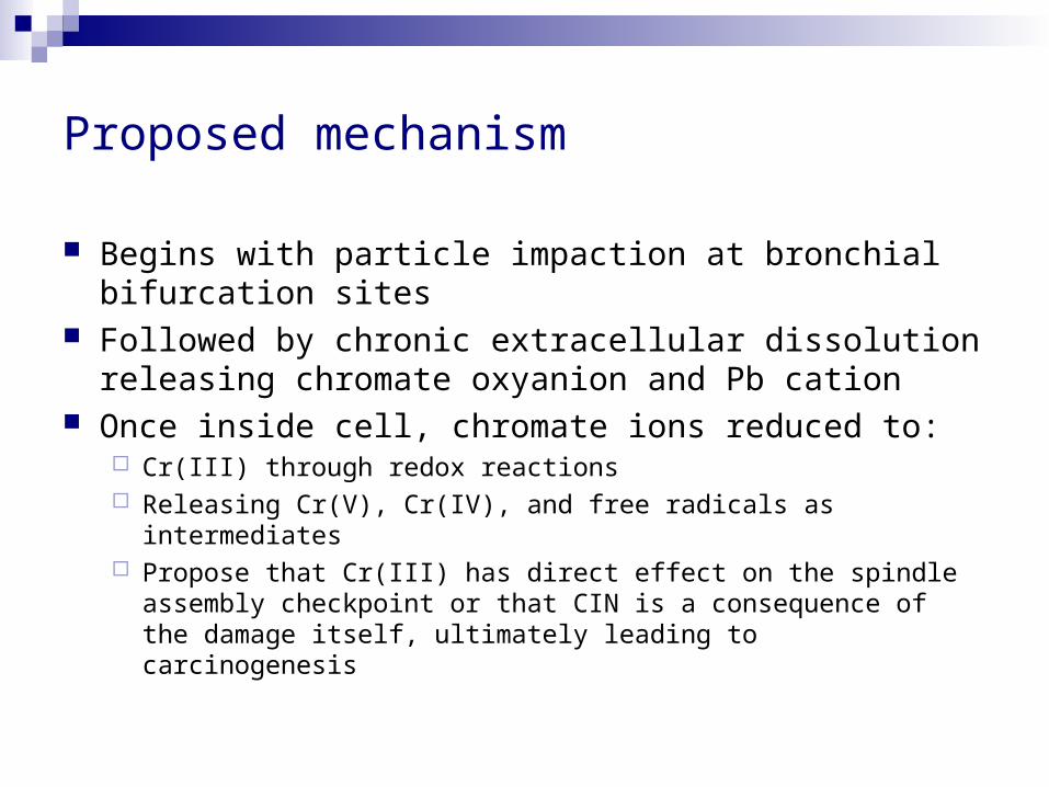
Proposed mechanism
Begins with particle impaction at bronchial bifurcation sites
Followed by chronic extracellular dissolution releasing chromate oxyanion and Pb cation
Once inside cell, chromate ions reduced to: Cr(III) through redox reactions Releasing Cr(V), Cr(IV), and free radicals as intermediates Propose that Cr(III) has direct effect on the spindle assembly
checkpoint or that CIN is a consequence of the damage itself, ultimately leading to carcinogenesis

Questions with the proposed mechanism Cr(VI) is reduced by the pulmonary epithelial lining fluid,
alveolar macrophages, and peripheral lung parenchyma cells to the essentially nontoxic and noncarcinogenic Cr(III) Why propose Cr(III) is
Respiratory cancer can be induced only at airborne concentrations of Cr(VI) that overwhelm the reductive capacity of these extracellular components

Future Directions
A look at some of the Papers that were published by the same group later in the summer of 2006

Particulate and soluble hexavalent chromium are cytotoxic and genotoxic to human lung epithelial cells
Studied the toxic effects of particulate and soluble Cr(VI) in immortalized human bronchial epithelial cells
Important because fibroblasts provide mechanistic insight and contribute to carcinogenesis but
Epithelial bronchial cells are the ones that transform and become tumors
Epithelial cell line was immortalized using HPV Instead of looking at spindle assembly checkpoint looked for cell death and
chromosomal aberrations: Chromatid lesions Isochromatid lesions Chromatid exchanges Dicentric chromosomes
Results Soluble Chromium
Produced same toxicity in epithelial cells as in fibroblasts Insoluble Chromium
Produced less toxicity in epithelial cells than in fibroblasts This may be a result of the different modes of immortalization Also, dissolution of Cr(VI) ions may be an intracellular event for epithelial cells while it
is an extracellular event for fibroblasts

The clastogenic effects of chronic exposure to particulate and soluble Cr(VI) in human lung cells
Elucidate the reason for the different potencies between soluble and insoluble Cr(VI) Hypothesis: The difference in potency between particulate and soluble
Cr(VI) is due to more chronic exposures with particulate chromate because it can deposit and persist in the lungs while soluble chromate is rapidly cleared
Used fibroblast cell line Results:
Chronic exposure to lead chromate induced a persistent amount of chromosome damage over time
Chronic exposure to sodium chromate induced a decrease in the amount of damage over the same time period
Produced similar trends in Cr ion uptake inducing both concentration- and time-dependent increases in intracellular Cr ion concentrations
Explanation: Pb cation in lead chromate may be causing the differences in
toxicity over longer time periods.

Questions?