chloroplasts of arabidopsis are the source and a primary ... · chloroplasts of arabidopsis are the...
TRANSCRIPT

Chloroplasts of Arabidopsis Are the Source and a PrimaryTarget of a Plant-Specific Programmed Cell DeathSignaling Pathway W
Chanhong Kim,a,b Rasa Meskauskiene,b Shengrui Zhang,a Keun Pyo Lee,b,1 Munusamy Lakshmanan Ashok,b
Karolina Blajecka,b Cornelia Herrfurth,c Ivo Feussner,c and Klaus Apela,b,2
a Boyce Thompson Institute for Plant Research, Ithaca, New York 14853-1801b Swiss Federal Institute of Technology Zürich, Institute of Plant Sciences, CH8092 Zurich, Switzerlandc Albrecht-von-Haller-Institute for Plant Sciences, Georg-August-University, D-37073 Gottingen, Germany
Enhanced levels of singlet oxygen (1O2) in chloroplasts trigger programmed cell death. The impact of 1O2 production inchloroplasts was monitored first in the conditional fluorescent (flu) mutant of Arabidopsis thaliana that accumulates 1O2 upona dark/light shift. The onset of 1O2 production is rapidly followed by a loss of chloroplast integrity that precedes the rupture ofthe central vacuole and the final collapse of the cell. Inactivation of the two plastid proteins EXECUTER (EX1) and EX2 in theflu mutant abrogates these responses, indicating that disintegration of chloroplasts is due to EX-dependent signaling ratherthan 1O2 directly. In flu seedlings, 1O2-mediated cell death signaling operates as a default pathway that results in seedlingscommitting suicide. By contrast, EX-dependent signaling in the wild type induces the formation of microlesions withoutdecreasing the viability of seedlings. 1O2-mediated and EX-dependent loss of plastid integrity and cell death in these plantsoccurs only in cells containing fully developed chloroplasts. Our findings support an as yet unreported signaling role of 1O2 inthe wild type exposed to mild light stress that invokes photoinhibition of photosystem II without causing photooxidativedamage of the plant.
INTRODUCTION
Programmed cell death (PCD) is a genetically regulated phy-siological process that is of central importance for the de-velopment and homeostasis of multicellular organisms (Greenand Reed, 1998). In several instances, the execution of PCDinvolves the participation of mitochondria that act as sensors ofcellular stress and initiate the onset of the cell death response(Green and Reed, 1998; Green and Kroemer, 2004). Permeabili-zation of mitochondrial membranes and the release of mito-chondrial proteins are hallmarks of these PCD processes (Adrainand Martin, 2001; Joza et al., 2001; Green and Kroemer, 2004).As shown in this work, another cell death response implicateschloroplasts as a source of a cell death signaling pathway. Inanalogy to PCD associated with impaired mitochondria, this celldeath program leads to a rapid loss of chloroplast integrity andthe subsequent collapse of the affected cell. In contrast withPCD associated with mitochondria, which has been linked to therelease of hydrogen peroxide (H2O2)/superoxide (Vacca et al.,2006), the plastid-derived PCD is initiated by the release ofsinglet oxygen (1O2).
Plants under oxidative stress suffer from damages that pre-viously have been interpreted as unavoidable consequences ofinjuries inflicted upon plants by toxic levels of reactive oxygenspecies (ROS) (Apel and Hirt, 2004.). For instance, plants undersevere light stress generate enhanced levels of ROS and maybleach. Based on the analysis of lipid peroxidation products, thisbleaching has been attributed to the cytotoxicity of 1O2 thatcauses extensive photooxidative damage (Triantaphylidès et al.,2008). However, as shown in this study, under less severe stressconditions that reflect more closely environmental fluctuationsoften experienced by plants in their natural habitat, 1O2 may alsoact as a signal, activating a PCD pathway that leads to theformation of microlesions, but does not seem to impair the vi-ability of the affected plant.The role of 1O2 as a trigger of cell death was initially revealed
in the fluorescent (flu) mutant of Arabidopsis thaliana (op denCamp et al., 2003). In the dark, plastids of the flu mutant ac-cumulate excess amounts of protochlorophyllide (Pchlide) dueto the absence of negative feedback control of tetrapyrrolebiosynthesis (Meskauskiene et al., 2001). In the light, Pchlideacts as a photosensitizer and generates 1O2, which leads toa rapid collapse of seedlings and growth inhibition of matureplants (op den Camp et al., 2003). The nucleus-encoded andchloroplast-localized EXECUTER1 (EX1) and EX2 proteins havebeen identified as essential components of 1O2 signaling. In-activation of EX proteins in an ex1 ex2 flu triple mutant is suffi-cient to suppress the upregulation of almost all 1O2-responsivegenes and to restore the wild-type phenotype (Wagner et al.,2004; Lee et al., 2007). In this work, this block of 1O2-mediatedresponses in an ex1 ex2 genetic background has been used to
1Current address: Department of Plant Science, University of Geneva,Geneva, Switzerland.2 Address correspondence to [email protected] author responsible for distribution of materials integral to the findingspresented in this article in accordance with the policy described in theInstructions for Authors (www.plantcell.org) is: Klaus Apel ([email protected]).WOnline version contains Web-only data.www.plantcell.org/cgi/doi/10.1105/tpc.112.100479
The Plant Cell, Vol. 24: 3026–3039, July 2012, www.plantcell.org ã 2012 American Society of Plant Biologists. All rights reserved.
identify a signaling role of 1O2 in wild-type plants and to definea genetically controlled PCD pathway unique to photosyntheticeukaryotes that operates under mild stress conditions thatimpede photosystem II (PSII) without causing photooxidativedamage of the plant.
RESULTS
1O2-Mediated Cell Death
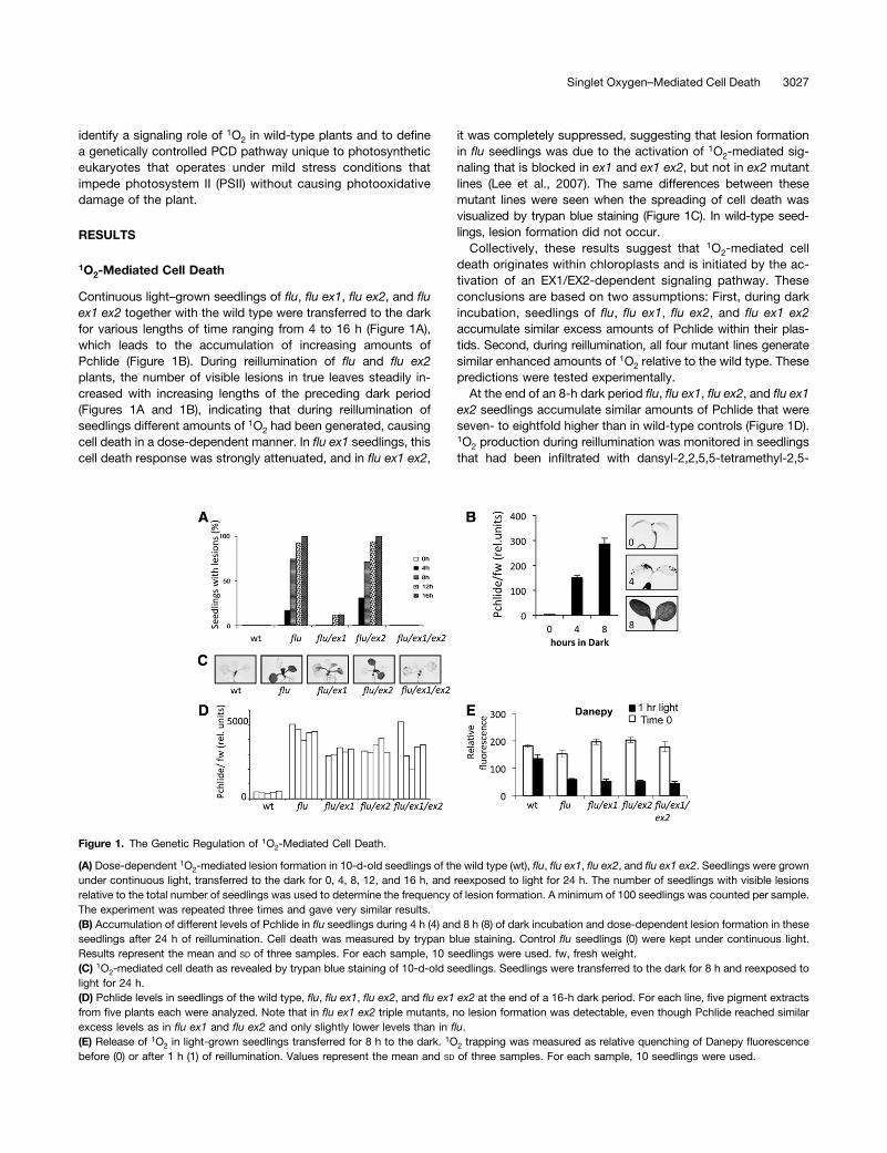
Continuous light–grown seedlings of flu, flu ex1, flu ex2, and fluex1 ex2 together with the wild type were transferred to the darkfor various lengths of time ranging from 4 to 16 h (Figure 1A),which leads to the accumulation of increasing amounts ofPchlide (Figure 1B). During reillumination of flu and flu ex2plants, the number of visible lesions in true leaves steadily in-creased with increasing lengths of the preceding dark period(Figures 1A and 1B), indicating that during reillumination ofseedlings different amounts of 1O2 had been generated, causingcell death in a dose-dependent manner. In flu ex1 seedlings, thiscell death response was strongly attenuated, and in flu ex1 ex2,
it was completely suppressed, suggesting that lesion formationin flu seedlings was due to the activation of 1O2-mediated sig-naling that is blocked in ex1 and ex1 ex2, but not in ex2 mutantlines (Lee et al., 2007). The same differences between thesemutant lines were seen when the spreading of cell death wasvisualized by trypan blue staining (Figure 1C). In wild-type seed-lings, lesion formation did not occur.Collectively, these results suggest that 1O2-mediated cell
death originates within chloroplasts and is initiated by the ac-tivation of an EX1/EX2-dependent signaling pathway. Theseconclusions are based on two assumptions: First, during darkincubation, seedlings of flu, flu ex1, flu ex2, and flu ex1 ex2accumulate similar excess amounts of Pchlide within their plas-tids. Second, during reillumination, all four mutant lines generatesimilar enhanced amounts of 1O2 relative to the wild type. Thesepredictions were tested experimentally.At the end of an 8-h dark period flu, flu ex1, flu ex2, and flu ex1
ex2 seedlings accumulate similar amounts of Pchlide that wereseven- to eightfold higher than in wild-type controls (Figure 1D).1O2 production during reillumination was monitored in seedlingsthat had been infiltrated with dansyl-2,2,5,5-tetramethyl-2,5-
Figure 1. The Genetic Regulation of 1O2-Mediated Cell Death.
(A) Dose-dependent 1O2-mediated lesion formation in 10-d-old seedlings of the wild type (wt), flu, flu ex1, flu ex2, and flu ex1 ex2. Seedlings were grownunder continuous light, transferred to the dark for 0, 4, 8, 12, and 16 h, and reexposed to light for 24 h. The number of seedlings with visible lesionsrelative to the total number of seedlings was used to determine the frequency of lesion formation. A minimum of 100 seedlings was counted per sample.The experiment was repeated three times and gave very similar results.(B) Accumulation of different levels of Pchlide in flu seedlings during 4 h (4) and 8 h (8) of dark incubation and dose-dependent lesion formation in theseseedlings after 24 h of reillumination. Cell death was measured by trypan blue staining. Control flu seedlings (0) were kept under continuous light.Results represent the mean and SD of three samples. For each sample, 10 seedlings were used. fw, fresh weight.(C) 1O2-mediated cell death as revealed by trypan blue staining of 10-d-old seedlings. Seedlings were transferred to the dark for 8 h and reexposed tolight for 24 h.(D) Pchlide levels in seedlings of the wild type, flu, flu ex1, flu ex2, and flu ex1 ex2 at the end of a 16-h dark period. For each line, five pigment extractsfrom five plants each were analyzed. Note that in flu ex1 ex2 triple mutants, no lesion formation was detectable, even though Pchlide reached similarexcess levels as in flu ex1 and flu ex2 and only slightly lower levels than in flu.(E) Release of 1O2 in light-grown seedlings transferred for 8 h to the dark. 1O2 trapping was measured as relative quenching of Danepy fluorescencebefore (0) or after 1 h (1) of reillumination. Values represent the mean and SD of three samples. For each sample, 10 seedlings were used.
Singlet Oxygen–Mediated Cell Death 3027

dehydro-1H-pyrrole (Danepy) (Hideg et al., 1998; Kálai et al.,1998). Danepy is used as a double (spin and fluorescent) 1O2
sensor, consisting of a fluorophore and a nitroxide precursor. Inthe absence of 1O2, Danepy is highly fluorescent but shows noelectron paramagnetic resonance signal. Upon reaction with1O2, conversion of the spin trap moiety into an electron para-magnetic resonance–active nitroxide results in partial fluores-cence quenching (Kálai et al., 1998). Prior to reexposure to light,infiltrated seedlings of the wild type and the four mutant linesdisplayed similar Danepy fluorescence levels (Figure 1E). After1 h of light exposure, this fluorescence was strongly quenchedin flu, flu ex1, flu ex2, and flu ex1 ex2, whereas in wild-typeseedlings, the fluorescence was only slightly reduced (Figure1E). These results indicate that in all mutant lines, Pchlide actsas a photosensitizer and produces similar amounts of 1O2. Sincecell death responses were observed only in flu and flu ex2 butnot in flu ex1 or flu ex1 ex2, photooxidative damage of chloro-plasts by 1O2 seems unlikely to be the cause of cell death.
Monitoring peroxidation of polyunsaturated fatty acids duringreillumination of predarkened flu seedlings also supported thisconclusion. Linolenic acid is the most prominent polyunsaturatedfatty acid in chloroplast membranes (Murakami et al., 2000) andhas been shown to be a preferred target of ROS. After 1 h ofreillumination of predarkened flu seedlings, peroxidation of li-nolenic acid occurred almost exclusively enzymatically by lip-oxygenases and not by direct attack of 1O2, as illustrated by therapid accumulation of the S-enantiomer of 13-hydroxy octade-catrieonic acid (13-HOT), a specific marker of enzymatic per-oxidation of linolenic acid in flu (Berger et al., 2001; op denCamp et al., 2003). At this early time of reillumination, the non-enzymatic peroxidation products of linolenic acid 10-HOT and15-HOT are hardly detectable (Figure 2A).
1O2-Mediated Chloroplast Leakage in the flu Mutant
The first consequence of 1O2 production in the flu mutant visibleto the eye was a rapid loss of chloroplast integrity. Chloroplastintegrity was assessed under the confocal microscope by mon-itoring the fluorescence distribution of the green fluorescentprotein (GFP) in transgenic plants that express a chimeric reporterprotein consisting of the nucleus-encoded and chloroplast-localized small subunit of the ribulose-1,5-bisphosphate car-boxylase (SSU) and GFP. In nontransgenic flu seedlings kept inthe dark or transferred from the dark to light, no GFP fluores-cence signals were detectable (Figures 3A1 and 3A2). In trans-genic wild-type plants, the fusion protein was confined to theplastid compartment (Figure 3A3). An identical distribution of thefusion protein was also seen in transgenic flu plants grown un-der continuous light (Figure 3A4). Under these growth con-ditions, flu seedlings do not overaccumulate Pchlide and do notexhibit enhanced 1O2 production (op den Camp et al., 2003).However, after keeping transgenic flu plants in the dark for 4 h,chloroplast integrity was impaired during reillumination and thefusion protein was released from the chloroplast to the sur-rounding cytoplasm (Figure 3A5). The molecular weight of theSSU-GFP fusion protein as revealed by its electrophoretic mo-bility during SDS-PAGE and its relative amount did not changeduring the first hour of reillumination (Figure 3B). Thus, during
initiation of 1O2-mediated cell death, the presence of the re-porter protein outside of chloroplasts does not seem to be dueto an enhanced accumulation of nonprocessed higher molec-ular weight precursors of SSU-GFP prior to its translocationinto chloroplasts. A similar change in the intracellular distributionof GFP fluorescence as in flu occurred also in flu ex2 seedlings(Figure 3A7), whereas in transgenic flu ex1 and flu ex1 ex2seedlings subjected to the same dark/light shift, the fusionprotein was retained within chloroplasts (Figures 3A6 and 3A8).Since all four mutant lines generate similar amounts of 1O2
during reillumination (Figure 1E), loss of chloroplast integrity influ and flu ex2 seedlings cannot be accounted for by a rupture ofchloroplast envelopes by direct attack of 1O2 but rather seemsto be the result of 1O2 signaling that is blocked in ex1 and ex1ex2 lines.Loss of chloroplast integrity could be one of the initial steps
triggering 1O2-mediated PCD, or it could merely be a secondaryconsequence of cellular disintegration during the spreading of1O2-mediated PCD. In the former case, loss of chloroplast in-tegrity should precede the collapse of cells, whereas in the lattercase, it should occur together with the disintegration of otherintracellular compartments. To address this question, we ex-ploited the unique properties of the flu mutant. Because of theprecision with which generation of 1O2 can be triggered in flu,
Figure 2. Enzymatic and Nonenzymatic Peroxidation of Linolenic Acid in10-d-Old Seedlings of flu and the Wild Type.
(A) Continuous light–grown flu seedlings were shifted to the dark for 8 hand reexposed for 1 or 24 h to light. f.w., fresh weight.(B) Wild-type (wt) seedlings shifted from low light (15 µmol m22 s21)/room temperature (22°C) to high light (270 µmol m22 s21)/low temper-ature (12°C) for 5 or 72 h. The 1O2-specific nonenzymatic peroxidationproducts 10-HOT, 15-HOT, and 13-HOT generated both enzymaticallyand nonenzymatically were determined. Results in (A) and (B) representthe mean and SD of three biological samples.(C) and (D) Lipid peroxidation signatures of flu (8 h dark/24 h light) (C)and the wild type (72 h of low-temperature/high-light stress) (D) indicatethat nonenzymatic peroxidation occurs primarily through 1O2.
3028 The Plant Cell

Figure 3. Genetic Control of 1O2-Mediated Chloroplast Leakage.
(A) Ten-day-old wild-type (wt), flu, flu ex1, and flu ex1 ex2 seedlings expressing the SSU-GFP fusion protein were grown under continuous light, shiftedto the dark for 8 h, and reexposed to light for 1 h. The green fluorescence of GFP and red fluorescence of chlorophyll (Chl) were monitored separately byCLSM, and the two fluorescence images were merged. Bar = 10 µm.(B) Size and relative concentration of the SSU-GFP fusion protein in total extracts of flu seedlings. Ten and 30 µg of total protein extracted fromseedlings before (0) or 1 h (1) after reillumination were separated electrophoretically by SDS-PAGE and blotted, and the fusion protein was detectedimmunologically. Only the mature-sized SSU-GFP fusion protein was detected using the GFP antiserum. As a loading control, the stained protein gelprior to blotting is shown. Arrow marks the position of the fusion protein.(C) Kinetics of 1O2-mediated chloroplast leakage, vacuole rupture, and initiation of cell death in flu. Ten-day-old flu seedlings expressing the SSU-GFPfusion protein were initially grown under continuous light, shifted to the dark for 8 h, and reexposed to light for various lengths of time (0 to 12 h).Chloroplast leakage (asterisk) and intactness of vacuoles (triangle) were assessed by the intracellular distribution of the GFP fusion protein, and theonset of cell death (arrowhead) was visualized by staining with PI. The green fluorescence of GFP, the red fluorescence of chlorophyll, and the bluefluorescence of PI were monitored separately by CLSM, and the three fluorescence images were merged. Bars = 50 µm.(D) 1O2-mediated changes of the maximum quantum efficiency (Fv/Fm) of PSII in flu and flu ex1 seedlings during reillumination. Results represent themean of three samples. For each sample, at least 50 seedlings were analyzed.
Singlet Oxygen–Mediated Cell Death 3029

the sequence of intracellular changes that occur during initiationof 1O2-mediated cell death can be analyzed and early targets of1O2-mediated signaling can be identified and distinguished fromsecondary changes at a later time during disintegration of cells.
The kinetics of chloroplast leakage, rupture of the centralvacuole, and onset of cell death were determined in flu seed-lings. Continuous light-grown flu seedlings were transferred tothe dark for 4 h and reexposed to light for various lengths oftime. Generation of 1O2 in flu seedlings has been shown pre-viously to occur within <1 min following the dark/light shift (opden Camp et al., 2003). During the first 15 min of reillumination,the SSU-GFP fusion protein remained within the plastid com-partment and chloroplast integrity was not visibly perturbed, butduring the following 15 min, widespread chloroplast leakagestarted to occur (Figure 3C, asterisk). At this time the majority ofaffected cells still maintained their central vacuole intact (Fig-ure 3C, triangle). Loss of chloroplast integrity clearly precededvacuole rupture and cellular collapse. Over the next 15 min, thenumber of cells that not only lost chloroplast integrity but alsocontained ruptured vacuoles gradually increased. However,these cells still appeared intact. The onset of cell death in thesecells was determined by propidium iodine (PI) staining. This re-agent is excluded from intact cells but penetrates dying ordead cells and intercalates into double-stranded nucleic acids(Oparka and Reed, 1994; Kirik et al., 2001). PI staining was firstdetected in flu seedlings 2 h after the beginning of reillumination(Figure 3C, arrowhead). During the next few hours, the numberof cells with positive PI staining rapidly increased throughout theseedling, and this was accompanied by visible lesion formation.At the same time, the intensities of GFP and chlorophyll fluo-rescence declined.
The kinetics of these 1O2-mediated changes that lead to thecollapse of the affected cells were in line with rapid changes ofthe functional state of PSII that was determined by measuringthe maximum quantum efficiency of PSII expressed as the ratioof variable to maximum fluorescence of chlorophyll (Fv/Fm)(Figure 3D). During the first 15 to 20 min following the dark/lightshift, the Fv/Fm ratios dropped slightly in flu and flu ex1 seed-lings, suggesting that in both lines PSII activity was similarlyaffected by the release of 1O2. Afterwards, the Fv/Fm ratio con-tinued to decline rapidly in flu seedlings, whereas in flu ex1seedlings, it remained constant over the next 24 h (Figure 3D).The time at which Fv/Fm ratio changes in flu and flu ex1 seedlingsstarted to diverge coincides with the beginning of chloroplastleakage in flu. Hence, the initial steps of 1O2-mediated signalingin the flu mutant that induce the loss of chloroplast integrity anddepend on the activity of EX1 and EX2 must be completed priorto this divergence of PSII activity changes.
With the progression of cell death, nonenzymatic peroxidationproducts of linolenic acid started to accumulate. Two non-enzymatic reaction types may lead to peroxidation of poly-unsaturated fatty acids (Stratton and Liebler, 1997; Mueller et al.,2006). Type I reactions are initiated by free radicals, such ashydroxyl radicals, and type II reactions are affected by 1O2. H2O2
and superoxide radical do not directly oxidize polyunsaturatedfatty acids, but in the presence of transition, metal ions such asFe2+ are converted to hydroxyl radical (Halliwell and Gutteridge,2007). Nonenzymatic peroxidation of linolenic acid may result in
the formation of six possible hydroxy fatty acid isomers. Twoof them, 10-HOT and 15-HOT, are exclusively formed by 1O2
during a type II reaction, whereas both reaction types generatethe other four, 9-HOT, 12-HOT, 13-HOT, and 16-HOT. Thesignature of these various HOT isomers has been used to de-termine the relative impact of 1O2 and H2O2 and superoxideradical on lipid peroxidation (Triantaphylidès et al., 2008). Therelative abundance of the 1O2-specific peroxidation products oftype II reactions, 10-HOT and 15-HOT, to that of 12- and 16-HOTdemonstrates that in flu seedlings during this late stage of celldeath progression nonenzymatic peroxidation can be attributedpredominantly to the cytotoxicity of 1O2 (Figures 2A and 2C).
1O2-Mediated Release of Chloroplast Proteins
So far, 1O2-mediated release of chloroplast proteins to the sur-rounding cytoplasm had been deduced from intracellular changesof GFP fluorescence distribution in transgenic flu plants. Asecond independent approach was used to verify the rapid1O2-mediated loss of chloroplast integrity by comparing theproportion of SSU-GFP recovered in the cytosol fractions of fluand wild-type seedlings subjected to a dark/light shift. Forsuch an experiment, isolated protoplasts rather than intactseedlings were used to minimize chloroplast rupture by me-chanical stress during the homogenization and fractionationsteps. As shown previously (Danon et al., 2005), protoplasts offlu subjected to a dark/light shift also generate 1O2 that inducescell death. At the end of the dark period, protoplasts containedintact chloroplasts as indicated by the localization of GFPfluorescence within the chloroplast compartment (Figures 4Aand 4B). During reillumination, the number of flu protoplastswith intact chloroplasts rapidly declined, and after 3 h of re-illumination, it had dropped to <20% (Figure 4B). By contrast,throughout reillumination of wild-type protoplasts, the majorityof chloroplasts remained intact (Figures 4A and 4B). As in thecase of seedlings, in flu protoplasts, chloroplast leakage andthe release of SSU-GFP also seemed to be genetically de-termined and a result of photooxidative damage. In protoplastsof flu ex1 seedlings subjected to the same dark/light shift,chloroplasts remained intact and did not release SSU-GFP tothe surrounding cytoplasm (see Supplemental Figure 1 online).Death of protoplasts as revealed by staining with PI occurredonly after chloroplast leakage and rupture of vacuoles hadtaken place (Figure 4A).After various lengths of time of reillumination, aliquots of the
protoplast suspension were collected and protoplasts werelysed. The intactness of chloroplasts within a given protoplastsample was determined by centrifuging lysed protoplasts andcomparing the amounts of the SSU-GFP fusion protein in thesupernatant by immunoblot analysis using an antiserum againstGFP. Control experiments with protoplasts isolated from wild-type and flu plants grown under continuous light demonstratedthat during protoplast lysis most chloroplasts remained intact.Only small fractions of the reporter protein were recovered inthe supernatant of both protoplast samples (see SupplementalFigure 2 online).During reillumination of predarkened wild-type protoplasts,
minor amounts of SSU-GFP in the supernatant increased over
3030 The Plant Cell

time (Figure 4C). Much higher levels of SSU-GFP were found inthe supernatant fraction of lysed flu protoplasts, regardless ofwhether proteins were probed with antisera against GFP or SSU(Figure 4C). Differences in the integrity of chloroplasts betweenthe wild type and flu were also evident when chloroplasts wereisolated from protoplasts before and after the beginning of re-illumination and examined under the confocal microscope.Prior to reillumination, chloroplasts isolated from both protoplastsamples retained the bright fluorescence of GFP. The majorityof chloroplasts isolated from wild-type protoplasts 3 h afterthe beginning of reillumination still retained GFP fluorescence,whereas chloroplasts isolated from flu protoplasts had lost theGFP fluorescence and upon excitation showed only the brightred fluorescence of chlorophyll. At the same time, their size hadincreased due to swelling (see Supplemental Figure 3 online).Previously, the release of cytochrome f (cyt f) from chloroplast
membranes of stressed plants had been proposed to triggera cell death response, similar to cytochrome c (cyt c) that acts asa trigger of PCD associated with impaired mitochondria (Petersand Chin, 2005; Zuppini et al., 2009). During the first hour ofreillumination of predarkened flu protoplasts, when chloroplastleakage was first detected, no cyt f was found in the supernatantfraction of lysed protoplasts. The release of trace amounts of cytf from thylakoid membranes was seen only after the onset ofcell death as revealed by PI staining of protoplasts (Figures 4Aand 4D, asterisk). The concentrations of cyt f in pellet fractionsof wild-type and flu protoplasts were the same and did notchange significantly throughout reillumination (Figure 4D). As therelease of minute amounts of cyt f from thylakoid membranesof the flu mutant occurs only after cells start to disintegrate, cytf is not a likely candidate for a molecular trigger that initiates1O2-mediated cell death but rather seems to be a marker for thefinal stage of cellular collapse.
1O2-Mediated Cell Death in Wild-Type Plants
Previously, cell death and bleaching of plants exposed to highlight stress had been attributed to photooxidative damagecaused by the cytotoxicity of 1O2 (Triantaphylidès et al., 2008).These results raise the question of whether in the wild type anenhanced generation of 1O2 in chloroplasts may also activatea genetically controlled PCD signaling pathway as seen in the flumutant. Seedlings that express the SSU-GFP reporter proteinwere initially grown for 5 d at room temperature (22°C) under lowlight (15 µmol m22 s21) and were then transferred to a combi-nation of higher light (270 µmol m22 s21) and lower temperature(12°C) (Figure 5A). As shown previously, at reduced temper-atures, moderate light intensities are sufficient to cause severelight stress for photosynthetic membranes and to induce bleach-ing of seedlings that resembles closely the bleaching of flu
Figure 4. The Release of Chloroplast Proteins during 1O2-MediatedChloroplast Leakage.
(A) 1O2-mediated chloroplast leakage and cell death in protoplasts iso-lated from flu seedlings expressing the SSU-GFP fusion protein. Chlo-roplast leakage and cell death were monitored by CLSM as described inFigure 3. Arrows indicate PI staining of nuclei of dead cells. Wild-type (wt)protoplasts after 3 h of reillumination are shown as controls. Bar = 10 µm.(B) Changes of the percentage of protoplasts with intact chloroplasts offlu and the wild type during reillumination. Average and SD of three rep-licates are shown.(C) The release of SSU-GFP and SSU from chloroplasts to the cytosolduring reillumination of wild-type and flu protoplasts. After variouslengths of time, protoplasts were lysed and the concentrations of SSU-GFP and SSU in the supernatant (sup) were assessed by immunoblotanalysis using antisera against GFP and SSU.(D) The release of thylakoid membrane-bound cyt f from chloroplasts tothe cytosol during reillumination of wild-type and flu protoplasts. After
various lengths of time, lysed protoplasts were centrifuged and theconcentration of cyt f in the supernatant and pellet fractions was de-termined by immunoblot analysis, using an antiserum against cyt f. Traceamounts of cyt f were seen only in the supernatant fractions of flu pro-toplasts 2 h after the beginning of reillumination (asterisk).
Singlet Oxygen–Mediated Cell Death 3031

seedlings exposed to nonpermissive light/dark cycles (op denCamp et al., 2003; Meskauskiene et al., 2009). Shortly afterthe beginning of stress treatment, seedlings released enhancedlevels of 1O2 and H2O2 as indicated by the rapid upregulation ofthe 1O2-responsive AAA-ATPase followed by the enhancedexpression of the H2O2-responsive FERRITIN1 (FER1) markergene (Figure 5B). The upregulation of AAA-ATPase was stronglysuppressed in ex1 ex2 seedlings, whereas the enhanced ex-pression of FER1 was not affected (Figure 5B), in line with ourearlier finding that enhanced levels of 1O2 and H2O2 in chloro-plasts activate two separate and distinct signaling pathways (opden Camp et al., 2003).During this initial phase of stress treatment, photoinhibition of
PSII occurs (Figure 5C), but photooxidative damage was hardlydetectable as indicated by the very low levels of the non-enzymatic peroxidation products of linolenic acid 10- and 15-HOT after 5 h of stress treatment (Figure 2B). The presence of13-HOT at this time was primarily due to the accumulation of itsS-enantiomer that has previously been identified as a product ofenzymatic peroxidation of linolenic acid by lipoxygenases inchloroplasts in response to enhanced levels of 1O2 (op denCamp et al., 2003) (Figure 2B). Similar to the flu mutant, also inthe wild type loss of chloroplast integrity occurred prior to thestress-induced collapse of seedlings. However, contrary to flu,loss of chloroplast integrity in the wild type did not immediatelyfollow stress-induced 1O2 production but was delayed and oc-curred only after an extensive light stress treatment (Figure 5D).Initiation of cell death started 4 d after the beginning of low-temperature/high-light treatment with chloroplast leakage pre-ceding the rupture of the vacuole and the collapse of the cell(Figures 5D and 5E).ex1 ex2 seedlings were less susceptible to the combined low-
temperature/high-light stress than the wild type as shown bydifferences in lesion formation and the decline of the maximumquantum efficiency of PSII (Fv/Fm) (Figures 5C and 5E). Prior tostress, seedlings of both lines showed Fv/Fm values of 0.85 to0.87. After 24 h of stress treatment, this ratio had dropped to 0.6(Figure 5C). In wild-type seedlings, the quantum efficiency ofPSII continued to decline further. After 4 d of stress, the firstwild-type seedlings started to collapse and the number ofbleached seedlings increased rapidly during the next 3 d (Figure5E). In contrast with the wild type, ex1 ex2 seedlings retainedtheir chlorophyll for up to 5 d of stress treatment and were stillviable with Fv/Fm values > 0.4 (Figure 5C).
Figure 5. Stress-Induced Cell Death of Wild-Type and ex1 ex2 Seed-lings.
(A) Seedlings were initially grown for 5 d at 22°C and 15 µmol m22 s21
and were then exposed to a combined low-temperature (12°C)/high-light(270 µmol m22 s21) stress program. Prior to the beginning of the low-temperature/high-light treatment, seedlings were placed for 30 min in thedark at 12°C to avoid high light–induced stress preacclimation during thelowering of the temperature.(B) Stress-induced changes in the expression of the 1O2-responsiveAAA-ATPase and the H2O2-responsive FER1 during low-temperature/high-light treatment of wild-type (wt) and ex1 ex2 seedlings as revealedby quantitative PCR analysis. Results represent means of three in-dependent biological replicates. Actin2 was used as a control for nor-malization.
(C) Stress-induced decline of the maximum quantum efficiency (Fv/Fm) ofPSII in wild-type and ex1 ex2 seedlings kept for various lengths of timeunder the combined low-temperature/high-light stress. Results representthe mean and SD of three samples. For each sample, 25 seedlings wereanalyzed.(D) GFP and chlorophyll (Chl) autofluorescence images of leaf cells ofwild-type seedlings expressing SSU-GFP were examined by CLSMbefore (0) and 3, 4, and 7 d after the beginning of low-temperature/high-light stress. Bars = 30 µm.(E) Stress-induced bleaching of wild-type (white bars) and ex1 ex2seedlings (black bars). Values represent the mean and SD of four sam-ples. For each sample, 50 seedlings were analyzed.
3032 The Plant Cell

However, during the following days, ex1 ex2 seedlings keptunder low-temperature/high-light stress also started to bleach(Figure 5E). Hence, the block of 1O2 signaling in ex1 ex2 seedlingsexposed to low temperature/high light did not abrogate but onlydelayed the final collapse of seedlings by 1 to 2 d. 1O2-dependentPCD seems to be involved in triggering a cell death response inlow-temperature/high-light-treated wild-type seedlings, but pro-gression of cell death in these plants is not exclusively driven by1O2-mediated and EX1/EX2-dependent signaling.
Prior to the bleaching of seedlings, the concentration of non-enzymatic peroxidation products increased drastically. The rel-ative abundance of 1O2-specific markers of type II reactions,10- and 15-HOT, and that of 12- and 16-HOT that are also formedby type I reactions indicates that this nonenzymatic lipid perox-idation is primarily due to 1O2 (Figures 2B and 2D). Hence, undersevere light stress conditions, the genetically determined PCDresponse of the wild type seems to be masked and superimposedby photooxidative damage caused by the cytotoxicity of 1O2.
If correct, this notion would suggest that under less severestress conditions photooxidative damage should be attenuatedand activation of the genetically determined 1O2-mediated celldeath response should prevail. This prediction was tested by ini-tially growing wild-type and ex1 ex2 seedlings at room tempera-ture for 5 d under moderate (90 µmol m22 s21) rather than low lightintensities, before transferring them to the same low-temperature/high-light stress program used in the previous experiment (Figure6A). Similar to seedlings grown under low light, moderate light–grown seedlings also started to activate the 1O2-dependent sig-naling pathway right after the beginning of the stress treatment, asindicated by the activation of the 1O2-responsive marker geneAAA-ATPase and its suppression in ex1 ex2 seedlings (Figure 6B).However, moderate light–grown wild-type and ex1 ex2 seedlingsdid not bleach but retained their chlorophyll and remained viablethroughout the stress treatment. There was a minor initial re-duction of the Fv/Fm values from 0.86 to 0.74 (the wild type) and0.79 (ex1 ex2) after 24 h of low-temperature/high-light treatment,but during the following stress treatment, the maximum quantumefficiency of PSII in both lines remained stable and did not furtherdecline (Figure 6C). Chloroplast leakage in wild-type seedlingswas first seen between 48 and 72 h of stress treatment (Figure 6D)and preceded formation of microlesions that were detected bystaining with trypan blue (Figures 6D and 6E). In ex1 ex2 seed-lings, these cell death responses were completely blocked. Evenwhen kept for an extended period of time under stress, ex1 ex2seedlings did not develop microlesions and their chloroplasts re-mained intact (Figures 6D and 6E).
1O2-Mediated PCD Is Activated in Green Leaf Sectors of thevariegated2 Mutant with Fully Developed Chloroplasts butNot in Its White Sectors with Undifferentiated Plastids
To confirm the proposed role of chloroplasts as a source ofa plant-specific PCD pathway, seedlings of the leaf-variegatedmutant variegated2 (var2) were first grown for 10 d at roomtemperature and 90 µmol m22 s21. Cells in the green leaf sectorsof this mutant contained morphologically normal chloroplasts,whereas in the white sectors, they contained undifferentiatedplastids that were reduced in size and lacked pigments and
developed lamellar structures (Sakamoto et al., 2009). In whitesectors, undifferentiated plastids were still able to accumulatethe SSU-GFP fusion protein. Chloroplasts in the green leafsectors of var2 like those in the wild type accumulated highlevels of the fusion protein (Figure 7A).Seedlings of var2 and var2 ex1 ex2 grown at room tempera-
ture under moderate light (90 µmol m22 s21) for 10 d weretransferred to the low-temperature/high-light stress programdescribed in Figure 6A. Soon after the beginning of the stresstreatment, 1O2-dependent signaling was activated in green coty-ledons of var2 seedlings, as indicated by the rapid upregulation ofthe 1O2-responsive marker gene AAA-ATPase and its suppres-sion in var2 ex1 ex2 (Figure 7B). AAA-ATPase transcripts accu-mulated faster in var2 and reached higher levels during the first12 h of stress treatment than in the wild type (Figure 7B), in linewith the reported enhanced susceptibility of var2 to light stress(Rosso et al., 2009; Liu et al., 2010). In green sectors of variegatedtrue leaves, chloroplast leakage occurred like in the wild type(Figure 7C) and preceded the final collapse of the cell, whereas inwhite leaf sectors, plastids remained intact (Figure 7C).Previously, enhanced levels of superoxide radical and H2O2
had been shown to accumulate in green leaf sectors of varie-gated mutants (Kato et al., 2009). These ROS do not seem to beinvolved in triggering the cell death response of var2 seedlingsexposed to the low-temperature/high-light stress; cell death ingreen leaf sectors of var2 ex1 ex2 seedlings was completelysuppressed (Figure 7D). Hence, PCD in var2 seedlings takes itsorigin in chloroplasts and seems to be under strict control of1O2-mediated and EX-dependent signaling.
DISCUSSION
The main finding of our study implicates chloroplasts as beingthe source and a primary target of a genetically determined,1O2-mediated cell death response unique to plants. In the flumutant, activation of 1O2-mediated signaling ends with seed-lings committing suicide. The analysis of lipid peroxidation inthese seedlings revealed two distinct, sequentially occurringbiological activities of 1O2. During initiation of the cell deathresponse, 1O2 acts as a signal without causing photooxidativedamage. Lipid peroxidation at this stage occurs almost ex-clusively enzymatically. However, during the final collapse andbleaching of flu seedlings, the cytotoxicity of 1O2 prevails,causing massive nonenzymatic lipid peroxidation and photo-oxidative damage. The initiation of 1O2-mediated cell deathsignaling in flu following a dark-to-light shift was due to thephotosensitizing activity of Pchlide that had accumulated in thedark. As overaccumulation of this photosensitizer stops in illu-minated flu seedlings (Meskauskiene et al., 2001; Goslings et al.,2004), generation of 1O2 during the final stage of the cell deathresponse is likely to occur via another photosensitizer, chloro-phyll, that is released during the 1O2-induced and EX-dependentdisintegration of thylakoid membranes (Matile et al., 1999). The1O2-mediated bleaching of flu seedlings is completely blocked influ ex1 ex2 seedlings that in the dark reach similar excess levelsof free Pchlide and during reillumination generate similar amountsof 1O2 as the parental flu line (Lee et al., 2007).
Singlet Oxygen–Mediated Cell Death 3033

Wild-type plants exposed to very severe light stress alsobleach, very similar to flu seedlings (Meskauskiene et al., 2009).Under such stress conditions, similar sets of 1O2-responsivegenes are activated in the wild type as in the flumutant followinga dark/light shift (Bechtold et al., 2008; Triantaphylidès et al.,2008; González-Pérez et al., 2011). Furthermore, inactivationof EX1 and EX2 suppresses the expression of 1O2-responsivegenes, and lesion formation in the wild type and flu coincide withdrastic nonenzymatic lipid peroxidation that could be ascribedto 1O2. Collectively, these results seem to suggest that also inthe wild type during severe light stress the 1O2-dependent PCDpathway is active and responsible for the collapse of stressedseedlings. However, contrary to what has been found in fluseedlings, bleaching of the wild type does not mark the finalconsequence of activating a PCD pathway that depends strictlyon the activities of EX1 and EX2. Nonenzymatic lipid peroxidationand lesion formation in the wild type occur under exceedinglyhigh light stress that surpasses the plant’s photochemical andnonphotochemical scavenging capacities to protect photo-synthetic membranes from photooxidative damage. Undersuch severe light stress, activation of the 1O2-dependent PCDpathway is masked and superimposed by an EX1- and EX2-independent cell death response that is mainly caused by thetoxicity of 1O2 and leads to the rapid bleaching and collapse ofseedlings. Under less severe light stress conditions, photo-oxidative damage caused by the cytotoxicity of 1O2 was sup-pressed, but genetically regulated 1O2-mediated cell deathsignaling was still operating in wild-type seedlings and was fullydependent on EX1 and EX2. This latter finding is consistent withthe stress-induced cell death response of the var2 mutant ex-posed to the same mild stress program. PCD was induced onlyin cells containing fully developed chloroplasts. In neighboringcells of white leaf sectors that contain undifferentiated plastidsand lack pigments, cell death did not occur. Hence, activation ofthis PCD pathway in green parts of the leaf does not affectadjacent areas that lack chloroplasts and seems to operate cellautonomously. Even though higher levels of H2O2 and super-oxide radical accumulate constitutively in green leaf sectors ofvariegated mutants (Kato et al., 2009), these ROS do not seemto interfere with the stress-induced onset of PCD. Blocking 1O2-mediated signaling in var2 ex1 ex2 seedlings was sufficient tosuppress the stress-induced cell death response.Unlike in flu, in the wild type not Pchlide but chlorophyll acts
as a photosensitizer that generates 1O2 and initiates cell death.Generation of 1O2 may happen either in light-harvesting an-tenna complexes (Rinalducci et al., 2004) or in the reactioncenters of PSII (Krieger-Liszkay et al., 2008). Chlorophylls in light-harvesting complexes are in direct contact with carotenoids thatefficiently quench excess light energy and suppress 1O2 formation(Niyogi, 1999). Only when the absorbed light strongly exceeds thescavenging capacity of light-harvesting structures or in caroten-oid-deficient plants does generation of 1O2 cause photooxidativedamage and nonenzymatic lipid peroxidation (Rebeiz et al., 1988;
Figure 6. Activation of the 1O2-Dependent PCD Pathway in Wild-TypeSeedlings Exposed to Moderate Light Stress.
(A) Seedlings were initially grown for 5 d at 22°C and 90 µmol m22 s21
and were then exposed to a combined low-temperature (12°C)/high-light(270 µmol m22 s21) stress program as described in Figure 5.(B) A qualitative assessment of stress-induced changes in the expres-sion of the 1O2-responsive AAA-ATPase (At3g28580) during moderatelow-temperature/high-light stress treatment of wild-type (wt) and ex1 ex2seedlings by RT-PCR analysis. Actin2 expression was taken as a control.This experiment was repeated three times with independent biologicalsamples and gave similar results.(C) Stress-induced decline of the maximum quantum efficiency (Fv/Fm) ofPSII in wild-type and ex1 ex2 seedlings kept for various lengths of timeunder the combined low-temperature/high-light stress. Results representthe mean and SD of three samples. For each sample, 25 seedlings wereanalyzed.(D) GFP and chlorophyll (Chl) autofluorescence images of leaf cells ofwild-type seedlings expressing SSU-GFP were examined by CLSM 3,48, 72, and 96 h after the beginning of the moderate low-temperature/high-light stress treatment. As a control, images of ex1 ex2 seedlingsafter 96 h of stress treatment are included. Bars = 30 µm.(E) Stress-induced activation of the 1O2-dependent PCD pathway inwild-type seedlings during the moderate low-temperature/high-lightstress treatment. Cell death was visualized by trypan blue staining. Itaffected only single cells or cell clusters without impairing the viability of
seedlings. This cell death response was abrogated in ex1 ex2 seedlingsthat are unable to activate 1O2-mediated signaling.
3034 The Plant Cell

Chamovitz et al., 1991; Triantaphylidès et al., 2008). In animals,some of the nonenzymatic peroxidation products are biolog-ically active and may act as signaling molecules (Lindshieldet al., 2007; Kalariya et al., 2008). In plants, signaling under suchsevere stress conditions could be associated with responses toinsults detrimental to the organism.
In the reaction center of PSII, carotenoids are far less involvedin light scavenging. b-Carotenes bound to the PSII reactioncenter chlorophyll would compete for light energy, thereby re-ducing the efficiency of light-driven electron transport. Notsurprisingly, they are localized away from the reaction centerP680 chlorophyll (Ferreira et al., 2004; Loll et al., 2005). 1O2
production by this special chlorophyll seems to be part ofa trade-off of this optimization step and has become an inherentproperty of PSII (Vass and Cser, 2009). 1O2 is produced via in-teraction of 3O2 with the excited triplet reaction center chloro-phyll that is formed whenever the electron acceptor site of PSIIis reduced and unable to oxidize the excited P680 chlorophyll.This may frequently occur under fluctuating higher light conditions
but also, at a reduced rate, under dim light (Tyystjärvi and Aro,1996; Edelman and Mattoo, 2008).One of the first visible consequences of 1O2-mediated sig-
naling in flu seedlings was the release of plastid proteins to theextraplastidic cytoplasm. This 1O2-mediated loss of chloro-plast integrity bears a striking resemblance to the release ofmitochondrial proteins during the onset of PCD associatedwith an impairment of mitochondrial activity (Kroemer andReed, 2000). Whereas in numerous organisms the release ofmitochondrial proteins, such as cyt c, has been shown to ac-tivate a proteolytic cascade responsible for the breakdown ofcells (Adrain and Martin, 2001; Tait and Green, 2010), in plants,the possible role of mitochondrial proteins as a trigger of celldeath is still under debate (Balk and Leaver, 2001; Balk et al.,2003; Yao et al., 2004; Vacca et al., 2006; Reape et al., 2008).Similarly, it is not clear yet whether the 1O2-mediated chloro-plast leakage and the release of chloroplast proteins to thesurrounding cytoplasm have any bearing on the 1O2-mediatedcollapse of cells.
Figure 7. 1O2-Mediated Cell Death and Loss of Plastid Integrity in Leaves of the var2 Mutant Are Confined to Cells Containing Fully DevelopedChloroplasts.
(A) The distribution of intact chloroplasts in green and undifferentiated plastids in white leaf sectors of 10-d-old var2/SSU-GFP seedlings grown at 22°Cand 90 µmol m22 s21. The green fluorescence of GFP (1) and red fluorescence of chlorophyll (2) were monitored separately by CLSM. (3) Lightmicroscopy image of the leaf sector. (4) Merged images of chlorophyll and GFP fluorescence images. Bar = 60 µm.(B) 1O2-mediated gene expression changes in cotyledons of wild-type (wt), ex1 ex2, var2, and ex1 ex2 var2 seedlings. Unlike true leaves, cotyledons ofvar2 are uniformly green. A qualitative assessment of transcript levels of the 1O2-responsive marker gene AAA-ATPase and the reference gene Actin2was done by RT-PCR. This experiment was repeated three times with independent biological samples and gave similar results.(C) Stress-induced plastid leakage. Ten-day-old var2/SSU-GFP seedlings grown as in (A) were exposed for up to 4 d to the combined low-temperature/high-light regime described in Figure 6A. Loss of plastid integrity occurred in cells with fully developed chloroplasts in green leaf sectors but not in cellsof the white leaf sector with undifferentiated plastids. The distribution of GFP fluorescence (1) and chlorophyll fluorescence (2) was determined byCLSM. (3) Light microscopy image of the leaf sector and (4) merged images of GFP and chlorophyll fluorescence. Bar = 60 µm.(D) Stress-induced cell death was monitored by trypan blue staining in leaves of ex1 ex2 var2 and var2 seedlings kept under the same growth/stressconditions as described in (C). Cell death occurred in green leaf sectors of var2 but not of ex1 ex2 var2 seedlings.
Singlet Oxygen–Mediated Cell Death 3035

Current forms of PCD associated with an impaired mito-chondrial activity have been traced back to an ancient hypo-thetical prokaryotic form acquired by primitive eukaryotic hostcells following the integration of the prokaryotic ancestor ofmitochondria as an endosymbiont (Blackstone and Green,1999). It is tempting to speculate that the chloroplast-associatedcell death program as defined in this work may originate froma second, independent endosymbiotic event, in which chloro-plasts arose from a cyanobacterial ancestor acquired by aeukaryotic host (Koonin and Aravind, 2002). Under stressconditions that interfere with the photosynthetic electron trans-port, hyperreduction of the photosynthetic electron transportchain promotes the release of 1O2 by the reaction center of PSII(Krieger-Liszkay et al., 2008). During the subjugation of the cya-nobacterial endosymbiont, the host cell might have refined byselection the function of 1O2 as a stress signal and subsequentlyused it to activate a cell death program that targets the chloro-plast and impairs its integrity. Strikingly, the two plastid proteinsEX1 and EX2, essential components of the 1O2-dependent PCDpathway, are highly conserved among all plants for which ge-nome sequence information is available, but they are not presentin prokaryotic cyanobacteria (Lee et al., 2007).
As a genetically regulated cell death response must haveevolved under selective pressure, it should be beneficial to theplant. However, at present, possible benefits of the 1O2-dependentPCD pathway have not yet been identified. Previously, formationof microlesions was shown to be closely associated with anenhanced resistance against pathogens (Alvarez et al., 1998;�Simková et al., 2012), and many of the 1O2-responsive genes areexpressed in response to abiotic and biotic stress (op den Campet al., 2003). Hence, it seems likely that activation of the 1O2-dependent PCD pathway in wild-type plants may be part of anacclimation response that enhances stress resistance.
METHODS
Plant Material and Growth Conditions
Transgenic Arabidopsis thaliana Columbia-0 plants expressing the SSU-GFP transgene under the control of the cauliflower mosaic virus 35Spromoter (Kim and Apel, 2004) were crossed with flu (Meskauskiene et al.,2001), flu ex1 (Wagner et al., 2004), flu ex2, flu ex1 ex2 (Lee et al., 2007),and var2 (Sakamoto et al., 2009), and the resulting double, triple, andquadruple mutants were selected from segregating F2 generations.Arabidopsis (Columbia-0), var2, var2 ex1 ex2, and ex1 ex2 seedlings wereused for the low-temperature/high-light stress experiments as describedpreviously (Meskauskiene et al., 2009).
Determination of the Subcellular GFP Distribution
The subcellular GFP distributions in protoplasts and seedlings wereanalyzed using a Leica TCS SP5 confocal laser scanning microscope(CLSM). GFP was excited with the 488-nm line of an argon laser, and theemission was monitored using a 510- to 540-nm band-pass filter. Thechlorophyll autofluorescence and bright-field images were obtained aspreviously described (�Simková et al., 2012). To detect concurrent celldeath, protoplasts and seedlings were incubated with 50 mM PI (Mo-lecular Probes) diluted with water. After removing the excess dye bywashing with water, samples were observed under the CLSM. The DNAand PI complexes in cells undergoing cell death were visualized with the
561 diode-pumped solid-state laser, and the emission was capturedusing a 613- to 630-nm band-pass filter.
Determination of 1O2 Levels
Danepy solution was prepared as described earlier (Hideg et al., 2007).Danepy was excited with a 404 UV laser line and traced using a 510- to625-nm band-pass filter. Relative fluorescence of each plant sample wasanalyzed and processed with CLSM.
Staining of Dead Cells
Trypan blue staining of seedlings was performed as described (op denCamp et al., 2003).
RNA Extraction and RT-PCR
Total RNA was extracted from seedlings using an RNeasy plant mini kit(Qiagen). cDNA was synthesized from 0.7 mg RNA, treated with DNase(Promega) using oligo(dT)15 primers (Promega) and Improm II reversetranscriptase (Promega) according to the manufacturer’s instructions. RT-PCR was performed with equal amounts of cDNA using the GeneAmp PCRsystem 9700 (Applied Biosystems). AAA-ATPase was selected as an early1O2-responsive gene (Gene Expression Omnibus accession number formicroarray data, GSE10509; http://www.ncbi.nlm.nih.gov/geo/query/acc.cgi?acc=GSE10509).FER1was selected as aH2O2-responsivegene. Primersequences for these genes are shown in Supplemental Table 1 online.
Isolation of Protoplasts and Chloroplasts
Transgenic wild-type and flu seedlings expressing SSU-GFP under thecontrol of the cauliflower mosaic virus 35S promoter were used to isolateintact protoplasts to monitor chloroplast integrity under the CLSM.Progression of cell death was determined using Evans Blue (Danon et al.,2005). Seedlings initially grown under continuous light for 4 d wereharvested under green safe light and incubated in protoplast extractionbuffer for 15 h in the dark. Afterwards, protoplasts were isolated undergreen safe light as previously described (Danon et al., 2005). Isolatedintact protoplasts in culture medium were illuminated for 1, 2, or 3 h andused for protein analysis or were lysed and used for chloroplast isolation.
Identification of Intact versus Broken Protoplasts or Chloroplasts
At time 0, aliquots were taken from the protoplast suspension prior toillumination. During illumination (100 µmol m22 s21), aliquots were takenat various time intervals (0, 0.5, 1, 2, and 3 h) and intact and nonintactprotoplasts were counted under the CLSM. For chloroplast isolation, theprocedure of Aronsson and Jarvis (2011) was slightly modified. For eachtime point, protoplasts were resuspended (400 mM sorbitol, 20 mMMES-KOH, pH 6, and 0.5mM CaCl2) and centrifuged at 60g, and subsequentlythe protoplast pellet was resuspended in lysis buffer (300 mM sorbitol,20 mM Tricine-KOH, pH 8.4, 10 mM EDTA, 10 mM NaHCO3, and 0.1%BSA). The lysed protoplast suspension was centrifuged at 355g, andthe pellet was resuspended either in HS buffer (50 mM HEPES-KOH,pH 8.0, and 0.33 M D-sorbitol) to monitor the intactness of chloroplastsunder CLSM or in 90% acetone for pigment extraction.
Protein Analysis
During reillumination of predarkened protoplasts of the wild type and fluexpressing SSU-GFP, aliquots were taken at various time points andlysed to harvest pellets and the corresponding supernatant fractions.Pellets were washed two times with lysis buffer, and supernatants were
3036 The Plant Cell

directly mixed with 23 protein SDS loading buffer. Electrophoretic proteinseparation and immunoblot analysis were done as described previously(�Simková et al., 2012). SSU and cyt fwere detected immunologically usingrabbit polyclonal antibodies (Stephan Greiner, Max Planck Institute, Golm,Germany). GFP fusion proteins were detected using a GFP monoclonalantibody (Roche).
Extraction and Measurement of Pchlide
Pchlide measurement was done as described previously (Kim and Apel,2004). Pchlide was extracted with 90% acetone and 10% 0.1MNH4OH inwater and separated by HPLC on a C18 reverse-phase silica gel column(Nucleosil ODS 5 µm, 250 3 4.6 mm; Machery Nagel). The relativefluorescence unit of Pchlide was determined using 430-nm excitation and630-nm emission (ex/em) wavelengths.
Measurement of the Maximum Quantum Efficiency ofPSII Photochemistry
Fv/Fmwas determined before and during stress treatment with a FluorCam800MF system (Photon Systems Instruments). Fv/Fm was determinedusing the standard quenching analysis protocol provided by PhotonSystems Instruments.
Determination of Peroxidation Products of Linolenic Acid
Enzymatic and nonenzymatic peroxidation products of linolenic acid weremeasured as described previously (Przybyla et al., 2008). Free oxylipinswere analyzed by HPLC or by gas chromatography–mass spectrometry.For the analysis of esterified oxylipins, the samples were transmethylated(op den Camp et al., 2003). Esterified compounds were quantified usingtriricinoleate as an internal standard to determine the recovery of es-terified hydroxyl fatty acids, whereas free compounds were quantifiedusing 13g-HOT as an internal standard.
Accession Numbers
Sequence data from this article can be found in the Arabidopsis GenomeInitiative or GenBank/EMBL databases under the following accessionnumbers: At1g27510 (EX2), At2g30950 (VAR2) At3g14110 (FLU),At3g18780 (Actin2), At3g28580 (AAA-ATPase), At4g33630 (EX1), andAt5g01600 (FER1).
Supplemental Data
The following materials are available in the online version of this article.
Supplemental Figure 1. Genetic Control of 1O2-Mediated ChloroplastLeakage in flu Protoplasts.
Supplemental Figure 2. Probing the Intactness of Chloroplasts duringIncubation of Protoplasts.
Supplemental Figure 3. The Intactness of Chloroplasts Isolated fromWild-Type and flu Seedlings Expressing the SSU-GFP Fusion Protein.
Supplemental Table 1. List of Selected Genes and Primer Sequences.
ACKNOWLEDGMENTS
We thank Karin Krupinska (University of Kiel, Germany) and members ofour group at the Boyce Thompson Institute for helpful discussions, EvaHideg (Szeged, Hungary) for the gift of Danepy, Stephan Greiner (MaxPlanck Institute, Golm, Germany) for the cyt f antiserum, and Wataru
Sakamoto (Okayama University, Japan) for seeds of the var2 mutant. Wealso thank the Boyce Thompson Institute Plant Cell Imaging Centersupported by the National Science Foundation (DBI-0618969) and theTRIAD Foundation. This study was supported by the Swiss ScienceFoundation, the Boyce Thompson Institute for Plant Research, theNational Institutes of Health (Grant R01-GM085036 to K.A.), and theGerman Research Council (I.F.).
AUTHOR CONTRIBUTIONS
C.K. and K.A. designed the research. C.K., R.M., S.Z., K.P.L., M.L.A.,K.B., C.H., and I.F. performed research. All the authors analyzed data.C.K. and K.A. wrote the article.
Received May 14, 2012; revised June 18, 2012; accepted June 25, 2012;published July 12, 2012.
REFERENCES
Adrain, C., and Martin, S.J. (2001). The mitochondrial apoptosome: Akiller unleashed by the cytochrome seas. Trends Biochem. Sci. 26:390–397.
Alvarez, M.E., Pennell, R.I., Meijer, P.J., Ishikawa, A., Dixon, R.A.,and Lamb, C. (1998). Reactive oxygen intermediates mediate asystemic signal network in the establishment of plant immunity. Cell92: 773–784.
Apel, K., and Hirt, H. (2004). Reactive oxygen species: Metabolism,oxidative stress, and signal transduction. Annu. Rev. Plant Biol. 55:373–399.
Aronsson, H., and Jarvis, R.P. (2011). Rapid isolation of Arabidopsischloroplasts and their use for in vitro protein import assays. Meth-ods Mol. Biol. 774: 281–305.
Balk, J., Chew, S.K., Leaver, C.J., and McCabe, P.F. (2003). The in-termembrane space of plant mitochondria contains a DNase activitythat may be involved in programmed cell death. Plant J. 34: 573–583.
Balk, J., and Leaver, C.J. (2001). The PET1-CMS mitochondrialmutation in sunflower is associated with premature programmedcell death and cytochrome c release. Plant Cell 13: 1803–1818.
Bechtold, U., Richard, O., Zamboni, A., Gapper, C., Geisler, M.,Pogson, B., Karpinski, S., and Mullineaux, P.M. (2008). Impactof chloroplastic- and extracellular-sourced ROS on high light-responsivegene expression in Arabidopsis. J. Exp. Bot. 59: 121–133.
Berger, S., Weichert, H., Porzel, A., Wasternack, C., Kühn, H., andFeussner, I. (2001). Enzymatic and non-enzymatic lipid perox-idation in leaf development. Biochim. Biophys. Acta 1533: 266–276.
Blackstone, N.W., and Green, D.R. (1999). The evolution of a mech-anism of cell suicide. Bioessays 21: 84–88.
Chamovitz, D., Pecker, I., and Hirschberg, J. (1991). The molecularbasis of resistance to the herbicide norflurazon. Plant Mol. Biol. 16:967–974.
Danon, A., Miersch, O., Felix, G., Camp, R.G., and Apel, K. (2005).Concurrent activation of cell death-regulating signaling pathwaysby singlet oxygen in Arabidopsis thaliana. Plant J. 41: 68–80.
Edelman, M., and Mattoo, A.K. (2008). D1-protein dynamics in pho-tosystem II: The lingering enigma. Photosynth. Res. 98: 609–620.
Ferreira, K.N., Iverson, T.M., Maghlaoui, K., Barber, J., and Iwata,S. (2004). Architecture of the photosynthetic oxygen-evolvingcenter. Science 303: 1831–1838.
González-Pérez, S., Gutiérrez, J., García-García, F., Osuna, D.,Dopazo, J., Lorenzo, O., Revuelta, J.L., and Arellano, J.B. (2011).Early transcriptional defense responses in Arabidopsis cell suspensionculture under high-light conditions. Plant Physiol. 156: 1439–1456.
Singlet Oxygen–Mediated Cell Death 3037

Goslings, D., Meskauskiene, R., Kim, C., Lee, K.P., Nater, M., andApel, K. (2004). Concurrent interactions of heme and FLU with GlutRNA reductase (HEMA1), the target of metabolic feedback in-hibition of tetrapyrrole biosynthesis, in dark- and light-grown Arab-idopsis plants. Plant J. 40: 957–967.
Green, D.R., and Kroemer, G. (2004). The pathophysiology of mito-chondrial cell death. Science 305: 626–629.
Green, D.R., and Reed, J.C. (1998). Mitochondria and apoptosis.Science 281: 1309–1312.
Halliwell, B., and Gutteridge, J. (2007). Free Radicals in Biology andMedicine, 4th ed. (Oxford, UK: Oxford University Press).
Hideg, E., Kálai, T., Hideg, K., and Vass, I. (1998). Photoinhibition ofphotosynthesis in vivo results in singlet oxygen production de-tection via nitroxide-induced fluorescence quenching in broad beanleaves. Biochemistry 37: 11405–11411.
Hideg, E., Kós, P.B., and Vass, I. (2007). Photosystem II damageinduced by chemically generated singlet oxygen in tobacco leaves.Physiol. Plant. 131: 33–40.
Joza, N., et al. (2001). Essential role of the mitochondrial apoptosis-inducing factor in programmed cell death. Nature 410: 549–554.
Kálai, T., Hideg, É., Vass, I., and Hideg, K. (1998). Double (fluores-cent and spin) sensors for detection of reactive oxygen species inthe thylakoid membrane. Free Radic. Biol. Med. 24: 649–652.
Kalariya, N.M., Ramana, K.V., Srivastava, S.K., and van Kuijk, F.J.G.M. (2008). Carotenoid derived aldehydes-induced oxidative stresscauses apoptotic cell death in human retinal pigment epithelial cells.Exp. Eye Res. 86: 70–80.
Kato, Y., Miura, E., Ido, K., Ifuku, K., and Sakamoto, W. (2009). Thevariegated mutants lacking chloroplastic FtsHs are defective in D1degradation and accumulate reactive oxygen species. Plant Physiol.151: 1790–1801.
Kim, C., and Apel, K. (2004). Substrate-dependent and organ-specificchloroplast protein import in planta. Plant Cell 16: 88–98.
Kirik, V., Bouyer, D., Schöbinger, U., Bechtold, N., Herzog, M.,Bonneville, J.M., and Hülskamp, M. (2001). CPR5 is involved in cellproliferation and cell death control and encodes a novel trans-membrane protein. Curr. Biol. 11: 1891–1895.
Koonin, E.V., and Aravind, L. (2002). Origin and evolution of eu-karyotic apoptosis: The bacterial connection. Cell Death Differ. 9:394–404.
Krieger-Liszkay, A., Fufezan, C., and Trebst, A. (2008). Singlet ox-ygen production in photosystem II and related protection mecha-nism. Photosynth. Res. 98: 551–564.
Kroemer, G., and Reed, J.C. (2000). Mitochondrial control of celldeath. Nat. Med. 6: 513–519.
Lee, K.P., Kim, C., Landgraf, F., and Apel, K. (2007). EXECUTER1-and EXECUTER2-dependent transfer of stress-related signals fromthe plastid to the nucleus of Arabidopsis thaliana. Proc. Natl. Acad.Sci. USA 104: 10270–10275.
Lindshield, B.L., Canene-Adams, K., and Erdman, J.W. Jr. (2007).Lycopenoids: Are lycopene metabolites bioactive? Arch. Biochem.Biophys. 458: 136–140.
Liu, X., Yu, F., and Rodermel, S. (2010). An Arabidopsis penta-tricopeptide repeat protein, SUPPRESSOR OF VARIEGATION7, isrequired for FtsH-mediated chloroplast biogenesis. Plant Physiol.154: 1588–1601.
Loll, B., Kern, J., Saenger, W., Zouni, A., and Biesiadka, J. (2005).Towards complete cofactor arrangement in the 3.0 A resolutionstructure of photosystem II. Nature 438: 1040–1044.
Matile, P., Hörtensteiner, S., and Thomas, H. (1999). Chlorophylldegradation. Annu. Rev. Plant Physiol. Plant Mol. Biol. 50: 67–95.
Meskauskiene, R., Nater, M., Goslings, D., Kessler, F., op denCamp, R., and Apel, K. (2001). FLU: A negative regulator of
chlorophyll biosynthesis in Arabidopsis thaliana. Proc. Natl. Acad.Sci. USA 98: 12826–12831.
Meskauskiene, R., Würsch, M., Laloi, C., Vidi, P.A., Coll, N.S.,Kessler, F., Baruah, A., Kim, C., and Apel, K. (2009). A mutation inthe Arabidopsis mTERF-related plastid protein SOLDAT10 activatesretrograde signaling and suppresses (1)O(2)-induced cell death.Plant J. 60: 399–410.
Mueller, M.J., Mène-Saffrané, L., Grun, C., Karg, K., and Farmer,E.E. (2006). Oxylipin analysis methods. Plant J. 45: 472–489.
Murakami, Y., Tsuyama, M., Kobayashi, Y., Kodama, H., and Iba,K. (2000). Trienoic fatty acids and plant tolerance of high temper-ature. Science 287: 476–479.
Niyogi, K.K. (1999). Photoprotection revisited: Genetic and molecularapproaches. Annu. Rev. Plant Physiol. Plant Mol. Biol. 50: 333–359.
Oparka, K.J., and Reed, N.D. (1994). The use of fluorescent probesfor studies of living plant cells. In Plant Cell Biology: A PracticalApproach, N. Harris and K.J. Oparka, eds (New York: Oxford Uni-versity Press), pp. 27–50.
op den Camp, R., Przybyla, D., Ochsenbein, C., Laloi, C., Kim, C.,Danon, A., Wagner, D., Hideg, E., Göbel, C., Feussner, I., Nater,M., and Apel, K. (2003). Rapid induction of distinct stress re-sponses after the release of singlet oxygen in Arabidopsis. PlantCell 15: 2320–2332.
Peters, J.S., and Chin, C. (2005). Evidence for cytochrome f in-volvement in eggplant cell death induced by palmitoleic acid. CellDeath Differ. 12: 405–407.
Przybyla, D., Göbel, C., Imboden, A., Hamberg, M., Feussner, I., andApel, K. (2008). Enzymatic, but not non-enzymatic, 1O2-mediatedperoxidation of polyunsaturated fatty acids forms part of the EX-ECUTER1-dependent stress response program in the flu mutant ofArabidopsis thaliana. Plant J. 54: 236–248.
Reape, T.J., Molony, E.M., and McCabe, P.F. (2008). Programmedcell death in plants: Distinguishing between different modes. J. Exp.Bot. 59: 435–444.
Rebeiz, C.A., Montazer-Zouhoor, A., Mayasich, J.M., Tripathy, B.C., Wu, S.M., and Rebeiz, C. (1988). Photodynamic herbicides.Recent developments and molecular basis of selectivity. CRC Crit.Rev. Plant Sci. 6: 385–436.
Rinalducci, S., Pedersen, J.Z., and Zolla, L. (2004). Formation ofradicals from singlet oxygen produced during photoinhibition ofisolated light-harvesting proteins of photosystem II. Biochim. Bio-phys. Acta 1608: 63–73.
Rosso, D., Bodo, R., Li, W., Krol, M., Saccon, D., Wang, S.,Schillac, L.A., Rodermel, S.R., Maxwell, D.P., and Hüner, N.P.A.(2009). Photosynthetic redox imbalance governs leaf sectoringin the Arabidopsis thaliana variegation mutants immutans, spotty,var1, and var2. Plant Cell 21: 3473–3492.
Sakamoto, W., Uno, Y., Zhang, Q., Miura, E., Kato, Y., and Sodmergen.(2009). Arrested differentiation of proplastids into chloroplasts invariegated leaves characterized by plastid ultrastructure and nucle-oid morphology. Plant Cell Physiol. 50: 2069–2083.
�Simková, K., Kim, C., Gacek, K., Baruah, A., Laloi, C., and Apel, K.(2012). The chloroplast division mutant caa33 of Arabidopsis thalianareveals the crucial impact of chloroplast homeostasis on stress ac-climation and retrograde plastid-to-nucleus signaling. Plant J. 69:701–712.
Stratton, S.P., and Liebler, D.C. (1997). Determination of singletoxygen-specific versus radical-mediated lipid peroxidation in pho-tosensitized oxidation of lipid bilayers: Effect of beta-carotene andalpha-tocopherol. Biochemistry 36: 12911–12920.
Tait, S.W., and Green, D.R. (2010). Mitochondria and cell death:Outer membrane permeabilization and beyond. Nat. Rev. Mol. CellBiol. 11: 621–632.
3038 The Plant Cell
Triantaphylidès, C., Krischke, M., Hoeberichts, F.A., Ksas, B.,Gresser, G., Havaux, M., Van Breusegem, F., and Mueller, M.J.(2008). Singlet oxygen is the major reactive oxygen species involvedin photooxidative damage to plants. Plant Physiol. 148: 960–968.
Tyystjärvi, E., and Aro, E.M. (1996). The rate constant of photoinhibition,measured in lincomycin-treated leaves, is directly proportional to lightintensity. Proc. Natl. Acad. Sci. USA 93: 2213–2218.
Vacca, R.A., Valenti, D., Bobba, A., Merafina, R.S., Passarella, S.,and Marra, E. (2006). Cytochrome c is released in a reactive oxygenspecies-dependent manner and is degraded via caspase-like pro-teases in tobacco Bright-Yellow 2 cells en route to heat shock-in-duced cell death. Plant Physiol. 141: 208–219.
Vass, I., and Cser, K. (2009). Janus-faced charge recombinations inphotosystem II photoinhibition. Trends Plant Sci. 14: 200–205.
Wagner, D., Przybyla, D., Op den Camp, R., Kim, C., Landgraf, F.,Lee, K.P., Würsch, M., Laloi, C., Nater, M., Hideg, E., and Apel, K.(2004). The genetic basis of singlet oxygen-induced stress re-sponses of Arabidopsis thaliana. Science 306: 1183–1185.
Yao, N., Eisfelder, B.J., Marvin, J., and Greenberg, J.T. (2004). Themitochondrion—An organelle commonly involved in programmedcell death in Arabidopsis thaliana. Plant J. 40: 596–610.
Zuppini, A., Gerotto, C., Moscatiello, R., Bergantino, E., andBaldan, B. (2009). Chlorella saccharophila cytochrome f and its in-volvement in the heat shock response. J. Exp. Bot. 60: 4189–4200.
Singlet Oxygen–Mediated Cell Death 3039