structures, functions, and interactions of clpt1 and … · structures, functions, and interactions...
TRANSCRIPT

Structures, Functions, and Interactions of ClpT1 andClpT2 in the Clp Protease System of Arabidopsis Chloroplasts
Jitae Kim,a Matthew S. Kimber,b Kenji Nishimura,a Giulia Friso,a Lance Schultz,b Lalit Ponnala,c
and Klaas J. van Wijka,1
a Department of Plant Biology, Cornell University, Ithaca, New York 14853bDepartment of Molecular and Cellular Biology, University of Guelph, Guelph, Ontario N1G 2W1, CanadacComputational Biology Service Unit, Cornell University, Ithaca, New York 14853
ORCID ID: 0000-0001-9536-0487 (K.J.v.W.)
Plastid ClpT1 and ClpT2 are plant-specific proteins that associate with the ClpPR protease. However, their physiologicalsignificance and structures are not understood. Arabidopsis thaliana loss-of-function single clpt1 and clpt2 mutants showedno visible phenotypes, whereas clpt1 clpt2 double mutants showed delayed development, reduced plant growth, andvirescent, serrated leaves but were viable and produced seed. The clpt1 and clpt1 clpt2 mutants showed partialdestabilization of the ClpPR complex, whereas clpt2 null mutants showed only marginal destabilization. Comparativeproteomics of clpt1 clpt2 plants showed a proteostasis phenotype similar to viable mutants in ClpPR core subunits, indicatingreduced Clp protease capacity. In vivo and in vitro assays showed that ClpT1 and ClpT2 can independently interact with thesingle ClpP ring and ClpPR core, but not with the single ClpR ring. We determined ClpT1 and ClpT2 structures (2.4- and 2.0-Åresolution) and detailed the similarities to the N-domains of bacterial ClpA/C chaperones. The ClpT structures suggesteda conserved MYFF motif for interaction with the ClpPR core near the interface between the P- and R-rings. In vivocomplementation showed that ClpT function and ClpPR core stabilization require the MYFF motif. Several models arepresented that may explain how ClpT1,2 contribute to ClpPR protease activity.
INTRODUCTION
The Clp protease system is the most abundant and complexserine-type protease in the soluble stromal phase of chlo-roplasts in Arabidopsis thaliana and likely most other higherplants (reviewed in Yu and Houry, 2007; Kato and Sakamoto,2010; Olinares et al., 2011a; Nishimura and van Wijk, 2014; vanWijk, 2015). It consists of a 350-kD barrel-shaped ClpPR corecomplex formed by a heptameric ring with ClpP3, ClpP4, ClpP5,and ClpP6 (the P-ring) and a heptameric ring with ClpP1, ClpR1,ClpR2, ClpR3, and ClpR4 (the R-ring). Complementation of nullmutants of CLPR4 and CLPP3 using StrepII-tagged versions ofClpR4 and ClpP3, respectively, allowed purification of individualClpR and ClpP rings (Olinares et al., 2011b). By spiking theseisolated P- and R-rings with stable isotope-labeled proteotypicpeptides, the absolute stoichiometry of ClpPR subunits in eachring could be determined. This showed that the heptameric ClpRring consists of ClpP1, R1, 2, 3, and 4 in a 3:1:1:1:1 ratio andthat the P-ring consists of ClpP3, 4, 5, and 6 in a 1:2:3:1 ratio(Olinares et al., 2011b). The ClpPR core complex was initiallyobserved by native gel electrophoresis in Arabidopsis chlo-roplasts and in nonphotosynthetic plastids of roots of Brassicarapa and flower petals of Brassica oleracea (Peltier et al., 2001,2004). Moreover, native isoelectric focusing showed that the
ClpPR core complex formed a single complex, rather than amixture of ClpPR core complexes with different compositions(Peltier et al., 2004).Mass spectrometry (MS) analysis and immunoblotting showed
that the ClpPR core complex also contains copies of ClpT1 andClpT2, two ;20-kD proteins with high sequence identity to theN-terminal domain of ClpA/C chaperones (reviewed in Nishimuraand van Wijk, 2014). Image analysis of silver-stained and Coo-massie blue-stained gels and titration experiments suggestedthat ClpPR core complexes contain on average one copy of eachClpT1 and ClpT2 (Peltier et al., 2004; Sjögren and Clarke, 2011).Homology modeling suggested that ClpT1 and ClpT2 are un-likely to be part of the ring structure, but rather associate with theaxial side of the ClpP ring involving the so-called P1 pocket. ClpTproteins are not found in prokaryotes or nonphotosynthetic eu-karyotes. Thus ClpT is specific for plastids in photosyntheticspecies and likely represents an adaptation to the plastid pro-teome and/or the Clp system (Peltier et al., 2004). Various hy-potheses have been proposed for ClpT function: (1) selection ofsubstrates, in particular those that do not require unfolding, suchas cleaved chloroplast transit peptide (Peltier et al., 2004); (2)ClpPR complex assembly factors (Sjögren and Clarke, 2011); (3)tethering the association of other protein interactors, such asClpS or Clp chaperones (Peltier et al., 2004); and, recently, (4)a regulator/stimulator of ClpC2 and ClpD chaperones (Colomboet al., 2014).In an attempt to determine ClpT1,2 function, Arabidopsis
single CLPT mutants (clpt1-1 and clpt2-1) without visible growthphenotypes were analyzed (Sjögren and Clarke, 2011). Nativegel electrophoresis indicated partial destabilization of the ClpPRcore complex into the individual heptameric rings, in particular in
1 Address correspondence to [email protected] author responsible for distribution of materials integral to the findingspresented in this article in accordance with the policy described in theInstructions for Authors (www.plantcell.org) is: Klaas J. van Wijk ([email protected]).www.plantcell.org/cgi/doi/10.1105/tpc.15.00106
The Plant Cell, Vol. 27: 1477–1496, May 2015, www.plantcell.org ã 2015 American Society of Plant Biologists. All rights reserved.

the clpt1-1 mutants. No double mutants could be identified, andit was concluded that ClpT1 and ClpT2 are essential for ClpPRcore assembly; hence, the double mutants would be embryo orseedling lethal (Sjögren and Clarke, 2011). However, as we willshow and explain in this study, double mutants for ClpT1 (in-cluding with a stronger clpt1-2 allele) and ClpT2 can be ob-tained, and they can complete their life cycle in soil, produceviable seed, and be maintained as a homozygous line. This vi-ability allowed us to determine the ClpPR assembly state insingle and double mutants and show that ClpT1,2 do contributeto ClpPR core stability, but that they are not strictly essential forClpPR core stabilization/assembly. Perhaps even more in-teresting is that the strong growth phenotype of the doublemutant cannot easily be explained by the partial ClpPR de-stabilization observed on native gels. In vivo protein interactionanalysis that showed that ClpT1 and ClpT2 can independentlyinteract with the intact core, but no support was found for in-teraction between ClpT1,T2 and ClpC chaperones, in contrastto a recent report in case of ClpT1 (Colombo et al., 2014).Comparative proteomics of clpt1-2 clpt2-1 double mutantsshowed a molecular phenotype that shared several key featuresof mutants for the ClpPR subunits, clpr2-1 (Rudella et al., 2006;Zybailov et al., 2009), clpp3-1 (Kim et al., 2013), and clpr4-1 (Kimet al., 2009). Following crystallization, structures for ClpT1 andClpT2 were determined and compared with structural featuresto N-domains of bacterial Clp chaperones; the in vivo signifi-cance of two domains was tested. A functional model forClpT1,2 is discussed.
RESULTS
Phylogeny of ClpT Proteins
The chloroplast ClpT proteins ClpT1 (At4g25370) and ClpT2(At4g12060) show high sequence similarity (SupplementalFigure 1A) and are likely derived from ClpC chaperones giventheir significant sequence similarity (31% sequence identifyacross 93 to 98 residues) to the a-helical N-domain of chloro-plast ClpC1,2 chaperones (see below). The N-domains in pro-karyotic and plastid Clp chaperones contain two repeats and areinvolved in substrate binding and interaction with adaptor pro-teins (e.g., MecA, YpbH, and McsB for ClpC and ClpS for ClpA)in bacteria (Erbse et al., 2008; Kirstein et al., 2009; Kress et al.,2009; Striebel et al., 2009). To better understand ClpT proteins,we performed an in-depth phylogenetic analysis with two se-quences from the moss Physcomitrella patens and 48 sequencesfrom 31 vascular plants (41 sequences from 25 eudicots andseven sequences from six monocots). ClpT proteins in vascularplants clearly separated into two large clades, which we as-signed ClpT1 and ClpT2 clades (Figure 1A; Supplemental DataSets 1 and 2 provide full sequences and the sequence align-ment used for generation of the cladogram). P. patens pos-sesses two ClpT-like proteins, forming a separate clade (Figure1A). All higher plants that we investigated contain ClpT1 and/orClpT2 proteins. ClpT1 proteins were found in all higher plantspecies except Medicago truncatula and Solanum tuberosum,whereas some species contained two or three paralogs of
ClpT1. ClpT1 sequences in monocots form a single subcladedistinct from those in dicots. ClpT2 proteins are missing in allsix monocots analyzed and are also missing in some dicots(Cucumis sativus, Citrus sinensis, Ricinus communis, and Vitisvinifera). The Brassicaceae form smaller subclades for eachClpT1 and ClpT2 (shown by five species in Figure 1A). Otherproteins with homology to the N-domain Clp chaperone se-quences have been found in the algae Chlamydomonas andVolvox, but not other algae; however, because they are sodifferent from ClpT1 and ClpT2, these proteins were assignedas ClpT3 and ClpT4 (Derrien et al., 2012). It remains to bedetermined if these distant ClpT homologs have similarfunctions as ClpT1 and ClpT2. Some cyanobacteria alsopossess some N-domain-containing proteins that are notAAA+ chaperones, but these are very distant from ClpT pro-teins in algae and plants (Derrien et al., 2012). Thus, ClpT1and ClpT2 proteins are consistently present in higher plants,but not in cyanobacteria or algae, even if both cyanobacteriaand green algae form heteromeric ClpPR complexes (seeNishimura and van Wijk, 2014). In the remainder of the article,we focus on the structure and function of ClpT1 and ClpT2 inArabidopsis.
Accumulation of ClpPR Bound and Free ClpT1 and ClpT2
For understanding ClpT structure and function, it is important toknow the N and C termini for ClpT, as well as the molar ratiobetween the ClpPR core complex and free and ClpPR-boundforms of ClpT1 and ClpT2. We note that both ClpT1 and ClpT2each have only one predicted gene model (or splice form). MSanalysis of stromal extracts and purified ClpPR core complexesidentified the likely N termini of ClpT1 and ClpT2 (SupplementalFigure 1B). Mature ClpT1 protein starts with S65, whereas ma-ture ClpT2 protein starts with S76. In the case of ClpT2, we alsoobserve likely N-terminal peptides starting two residues down-stream (Pro-78); such “ragged” N-terminal ends are quitecommon in the chloroplast proteome (E. Rowland and K.J. vanWijk, unpublished data). Projecting these N termini onto thesequence logos of 30 ClpT1 and 18 ClpT2 protein sequencesfrom higher plants shows that these N termini coincide witha transition of lower sequence similarity to higher similarity(Supplemental Figure 2). This is consistent with the notion thatcTPs are less conserved than the mature regions of proteinhomologs. Also, the C terminus is present in the mature ClpT1and ClpT2 proteins without C-terminal trimming (SupplementalFigure 1B). Based on silver and Coomassie blue staining ofstromal ClpPRT complexes, we previously estimated that ClpT1and ClpT2 were present on average with one copy each perClpPR core complex (Peltier et al., 2004). Extensive label-freespectral counting analysis of denatured chloroplast stromalproteomes by high-resolution tandem mass spectrometry (MS/MS) analysis also suggested an ;1:1 ratio between total ClpT1and ClpT2 (Zybailov et al., 2008) and on average ;2.5 copieseach of ClpT1 and ClpT2 per ClpPR core complex, when as-suming that all ClpPR proteins are assembled in the tetrade-cameric core. We note that these latter experiments provide noinformation as to whether ClpT proteins are indeed associatedto the ClpPR core. Based on spectral counting analysis of total
1478 The Plant Cell

wild-type leaf rosette denatured proteomes (after SDS-PAGE)with nine independent replicates, we estimate that the ClpPRsubunits and the ClpT1 and ClpT2 subunits represent 0.36 and0.08%, respectively, of the total leaf protein mass (SupplementalTable 1). Assuming that all ClpPR proteins are part of the ClpPRtetradecamer and correcting for the number of available trypticpeptides for quantification, this suggests an average approxi-mately four copies of total ClpT1 and ClpT2 per ClpPR core(Supplemental Table 1). Thus, both stromal and total leaf pro-teome analysis suggest that there are four to five ClpT proteins
for each ClpPR core, with comparable amounts of ClpT1 andClpT2.
Growth and Developmental Phenotypes of CLPT1 andCLPT2 Single and Double Mutants
We screened the various T-DNA insertion collections in theColumbia-0 background for potential null and knockdownmutants in CLPT1 and CLPT2. After extensive genotyping byPCR, DNA sequencing of the T-DNA inserts, and RT-PCR, we
Figure 1. Phylogenetic Analysis of ClpT and Characterization of clpt1 and clpt2 Single and Double Mutants.
(A) Nonrooted phylogenetic tree of 50 ClpT proteins from six different monocots, 25 different dicots, and the moss P. patens. Bootstrap values areindicated for key branch points. Three main types or ClpT are assigned, namely, ClpT1 (both monocots and dicots), ClpT2 (only dicots), and ClpT-likeprotein in moss.(B) Gene model structures and position of T-DNA inserts in the CLPT mutants used in this study. Exons (black boxes for coding sequence; open boxesfor 59 and 39 untranslated regions [UTRs]) and T-DNA insertions (triangles) are indicated.(C) Transcript accumulation levels in the leaves of the CLPT single and double mutants used in this study. Transcript levels were determined by RT-PCR(25 cycles) using gene-specific primer pairs; ACTIN2 was used as internal control. At least three biological replicates were performed for each RT-PCRanalysis (primers are listed in Supplemental Table 2).(D) Growth and development of wild-type and homozygous clpt mutants grown on soil for 23 d under an 18/6-h light/dark cycle at 120 mmol photonsm22 s21. Bar = 3 cm.(E) ClpT1 and ClpT2 protein levels in the single and double clpt mutants. Asterisks mark a protein recognized by the anti-ClpT1 serum. Ten microgramsof stromal protein was loaded in each lane for the upper panels. Total soluble protein was loaded in each lane for the lower panel. 1x, 3x, and 10xindicate that 3, 9, and 30 mg protein, respectively, was loaded.
ClpT Structure and Function in Chloroplasts 1479

identified two CLPT1 mutants and two CLPT2 mutants. Themutants are clpt1-1 (SALK_052772), clpt1-2 (GK_285A05),clpt2-1 (SAIL_340A10), and clpt2-2 (SALK_132943). The re-spective gene models and the position of the confirmed T-DNAinsertions are shown in Figure 1B. RT-PCR of the homozygousclpt2-1 and clpt2-2 did not detect any CLPT2 transcript, andthey are thus null mutants. Transcript levels for CLPT1 were 60and 20% of wild-type levels in clpt1-1 and clpt1-2, respectively(Figure 1C). Interestingly, transcript levels for CLPT1 were about;1.6-fold of wild-type levels in both clpt2 alleles, and for CLPT2;1.6-fold of wild-type levels in both clpt1 alleles, indicative ofa small transcriptional compensatory response (Figure 1C). Thefour homozygous alleles grown on soil were indistinguishablefrom wild-type plants (Figure 1D). Immunoblot analysis of leafextract of both clpt1 mutant alleles and clpt2-1 showed thatClpT1 level was reduced to 25% in clpt1-1 but was undetect-able in clpt1-2, whereas ClpT2 was not detected in the clpt2mutant allele (Figure 1E). Interestingly, ClpT1 protein levels in-creased ;3-fold in clpt2-1 and ClpT2 protein levels increased;4-fold in clpt1-2, indicative of a strong compensatory re-sponse and suggesting (partial) functional redundancy betweenClpT1 and ClpT2 (Figure 1E). When the anti-ClpT1 immunoblotswere overexposed or when using higher protein loading, weobserved a higher molecular mass band (;2 kD higher) specif-ically in the clpt1-2 backgrounds (Figure 1E). Because theT-DNA insertion in clpt1-2 was at the stop codon, we verifiedwhether read-through translation could explain this higher massband. Sequencing the genomic DNA for this allele showed lossof the original stop codon and a new stop codon 189 nucleo-tides downstream. RT-PCR of wild-type and clpt1-2 cDNAindeed detected this longer transcript in the clpt1-2 allele(Supplemental Figure 3). However, this should result in an ad-ditional 63 amino acids (;7 kD), which is much longer than theestimated protein size deduced from the anti-ClpT1 immunoblot(;2 kD increase). Therefore, it appears unlikely that the highermolecular mass band represents a read-through product. Itshould be noted that also in the clpp3-1 null mutant a highermolecular mass form of ClpT1 (;1 kD higher) accumulates, butnever ClpT2 (Kim et al., 2013). Despite significant efforts wehave not been able to determine what the higher molecular massbands represent.
To test the genetic interactions and possible functional re-dundancy between ClpT1 and ClpT2, we generated four differ-ent homozygous clpt1 clpt2 double mutants using these clpt1and clpt2 alleles. All four clpt1 clpt2 mutant combinationsshowed delayed development, reduced growth and biomass,and a yellow to pale-green phenotype (Figure 1D). Consistentwith the stronger reduction of ClpT1 expression, clpt1-2 clpt2-1and clpt1-2 clpt2-2 plants showed more severe phenotypesthan clpt1-1 clpt2-1 and clpt1-1 clpt2-2 plants (Figure 1D), il-lustrating a strong dosage effect of ClpT1. We did not detectphenotypic differences between the contributions of the twoclpt2 alleles, consistent with them both being null alleles. Theclpt1-2 clpt2-1 mutant could be fully complemented (no visiblegrowth phenotype) with constructs that express either ClpT1 orClpT2 (both StrepII-tagged) (Supplemental Figures 4A and 4B),indicating that ClpT1 and ClpT2 are at least partially redundant.Immunoblot analysis of leaf extracts of clpt1-2 clpt2-1 plants did
not detect ClpT1 or ClpT2 protein, except for two higher massmolecular bands in case of ClpT1 at ;2 to 2.5 kD (Figure 1E).One band aligned to the higher mass band observed in theclpt1-2 single mutant, whereas the other was slightly higher inmass (Figure 1E). MS/MS analysis of total leaf extracts of clpt1-2clpt2-1 mutants neither detected ClpT1 nor ClpT2, whereasClpT1 and ClpT2 were detected with high confidence in thewild-type plants (Supplemental Data Set 3). A developmentalseries of clpt1-2 clpt2-1 plants is shown in Supplemental Figure4C. The clpt1-2 clpt2-1 double mutant was smaller in stature (20to 30% in rosette cross section) as the leaky clpr2-1 mutant with;20% ClpR2 levels conditions (Rudella et al., 2006), but leafcolor and shape were very similar (Supplemental Figures 4D and4E). Size differences were greater under short-day than long-dayconditions. However, the phenotype of the clpt1-2 clpt2-1double mutant was much weaker than null mutants for CLPP3and CLPR4 (both arrested in development in the cotyledonstage) and the embryo-lethal CLPP4 and CLPP5 null mutants;this is summarized in Figure 2A. In the remainder of the article,we use the stronger CLPT1 allele (clpt1-2), rather than theweaker allele (clpt1-1) for further analysis.
Molecular Phenotyping of clpt1-2 clpt2-1 byComparative Proteomics
To gain insight in the consequences of the loss of ClpT1 andClpT2, we compared the total denatured leaf proteomes of theclpt1-2 clpt2-1 double mutant and wild-type plants in de-velopmental stage 1.11 (Figure 2B). Total leaf proteomes wereextracted with SDS, and each proteome was separated by SDS-PAGE, followed by in-gel trypsin digestion, protein identification,and quantification by nano-liquid chromatography-MS/MS, re-sulting in the identification of 2360 proteins, quantified as 1775individual proteins and 126 protein groups (SupplementalFigures 5A and 5B and Supplemental Data Set 3). The averagepairwise correlation coefficients among the three biologicalreplicates within the wild-type and clpt1 clpt2 data sets were0.937 and 0.968, respectively, indicating high reproducibilitybetween the independent replicates for each genotype(Supplemental Figure 5C). Principle component analysis alsoshowed that the variation between genotypes was larger thanbetween replicates within each genotype (Figure 2C). Together,this shows that the quantitative proteome data are of highquality with little noise and that the clpt1-2 clpt2-1 mutant hasa measurable proteome phenotype. Based on our recent refer-ence Arabidopsis chloroplast proteome (Huang et al., 2013) andadditional updates, 795 proteins were chloroplast localized (in-cluding 38 dual localized proteins), representing 56% of theprotein mass in the double mutant and 59% in the wild type.Compared with the wild type, the clpt1-2 clpt2-1 thylakoidproteome mass decreased by 23%, but the proteome mass ofthylakoid-associated plastoglobuli (Lundquist et al., 2012) (20identified proteins) more than doubled (2.35-fold higher), re-flecting loss of photosynthetic electron transport capacity andincreased thylakoid stress. Statistical analysis using the con-sensus results of two different statistical packages (GLEE withP < 0.01 and QSPEC at false discovery rate < 0.05; see Meth-ods) showed that among the 51 significantly changed proteins in
1480 The Plant Cell

clpt1-2 clpt2-1 leaves, chloroplast proteins were overrepresented(37/51), consistent with the location of the Clp system. Signifi-cantly changed extraplastidic proteins were located in differentsubcellular compartments and did not show any specific func-tional trends.
Evaluation of the clpt1-2 clpt2-1 functional chloroplast pro-teome phenotype suggests particular bottlenecks or defects inprotein import, chloroplast translation, protein folding stress,and loss of photosynthetic capacity (Table 1, Figures 2D and2E). Below, we highlight the most interesting aspects of theclpt1-2 clpt2-1 chloroplast proteome phenotype.
Loss of Photosynthetic Capacity
Relative protein mass investments in the photosynthetic elec-tron transport chain and Calvin cycle decreased very similarly by24 and 22%, respectively. Within the thylakoid-bound photo-synthetic apparatus, the photosystem II core, photosystem Icore, the cytochrome b6f complex, the NDH complex, and theATP-synthase decreased by 26, 31, 48, 36, and 35%, whereaslight-harvesting complex II (LHCII) and LHCI did not significantly
change (Figure 2D). Also in the other Clp core mutants, thedownregulation of LHCs is much less than for the photosystemI and photosystem II cores or other thylakoid complexes(Supplemental Figure 5D).
Protein Synthesis and Folding Stress
Protein translation initiation and elongation factors togetherincreased by 59% and tRNA synthases increased by 75%,whereas ribosome protein mass increased by just 9% (Figure2E). Stromal protein chaperones (e.g., CPN60, HSP70, andHSP90), isomerases (e.g., ROC4), and the unfoldase ClpB3 in-creased collectively by 73% (Figure 2E). Within these functions,several individual proteins significantly increased, in particular,the chaperones and three elongation factors (Table 1).
Protein Import Bottleneck
The abundance level of the chloroplast envelope TOC and TICprotein import apparatus increased by 65 and 69%, respectively,indicative of a bottleneck in protein import (Figure 2E).
Figure 2. Comparison of clp Mutants and Comparative Proteomics of clpt1-2 clpt2-1 and the Wild Type.
(A) Summary of phenotypes of CLPPRT mutants. ^, clpr2-1 leaky allele with ;20% mRNA and protein. *, clpt1-2 clpt2-1. a, Nomarski microscopyshows an early block in embryogenesis (no abortion); b, sugars can be sucrose or glucose (1 to 3%); c, after initial growth on sugar-containing medium;d, requires sugar in the medium; e, strongly delayed flowering.(B) The clpt1-2 clpt2-1 and wild-type (wt) plants used for proteome analysis. Total leaves were harvested at growth stage 1.11, 41 d for clpt1-2 clpt2-1and 27 d for the wild type. Plants were grown on soil under a short-day cycle (10-h/14-h light/dark) at 100 mmol photons m22 s21. The Coomassie blue-stained SDS-PAGE gel with extracted proteomes is shown in Supplemental Figure 5A.(C) Principal component analysis based on NadjSPC of individual replicates for the wild type and double mutant.(D) and (E) Figures illustrating the molecular proteome phenotype of the thylakoid photosynthetic machinery (D) and plastid gene expression andproteotasis (E). Black bars indicate the wild type, and open bars represent clpt1-2 clpt2-1 plants.
ClpT Structure and Function in Chloroplasts 1481

Table 1. Chloroplast Proteins Significantly Up- or Downregulated in clpt1-2 clpt2-1 Compared with the Wild Type and Comparison to ClpPR Core andClpC1 Mutants
Best Model Protein Annotation FunctionTotaladjSPCa
clpt1xclpt2/Wild Typea
clpr2-1/Wild Typeb
clpp3/Wild Typec
clpc1-1/Wild Typed
AT1G06950.1 Tic110 Protein import 280 1.6 3.6 2.1 [ND inmutant]
ATCG01130.1 Tic214 (YCF1.2) Protein import 32 12.1 (#) [ND] [ND inwild type]
[ND]
AT1G55490.1AT3G13470.1
Cpn60-b-1,2 Protein folding 708 1.9 2.4** 2.2 [1.7]
AT5G56500.1 Cpn60-b-3 Protein folding 64 5.4 2.4** 8.3 ND inwild type
AT2G28000.1 Cpn60-a-1 Protein folding 656 1.8 2.2 2.1 [1.4]AT4G24280.1 cpHSP70-1 Protein folding 213 1.8 2.0 2.5 [1.0]AT5G49910.1 cpHSP70-2 Protein folding 187 2.0 3.2 2.5 [1.1]AT2G04030.1 cpHSP90 Protein folding 232 1.9 2.3 2.4 [1.6]AT5G15450.1 ClpB3 Protein unfolding 52 3.3 6.6 5.5 3.4AT3G62030.1 Peptidylprolyl isomerase (ROC4; CYP20-3) Protein folding 312 1.4 2.3 1.5 [0.8]AT4G12060.1 ClpT2 Clp system 31 ND in
mutant[2.2] [1.9] [1.4]
AT4G25370.1 ClpT1 Clp system 20 ND inmutant
[2.8] [1.7] 2.1
AT3G19170.1 Prep1-Zn metalloprotease Peptidases 310 1.6 4.2 2.4 [1.4]AT5G42390.1 Stromal processing peptidase (SPP) Peptidases 74 2.4 [ND] 5.5 [1.6]AT5G26742.2 DEAD box RNA helicase (RH3) RNA splicing 124 2.7 8.0 2.4 [ND]AT4G16390.1 PPR protein P67 (SVR7) RNA metabolism 29 ND in
wild type[ND] 3.8 [1.0]
AT1G70070.1 DEAD/DEAH box helicase RNA metabolism 29 ND inwild type
[ND] 4.0 [2.0]
AT4G20360.1 Elongation factor Tu (EF-Tu-1), plastid Protein synthesis 703 1.7 1.5 1.7 [1.6]AT5G13650.1 Elongation factor typeA/bipA-like (SVR3) Protein synthesis 111 3.1 4.6 3.5 2.7AT1G62750.1 Elongation factor Tu-G (EF-G; SCO1) Protein synthesis 387 1.4 [2.0] 2.3 [1.5]AT3G26650.1 Glyceraldehyde 3-phosphate
dehydrogenase A-1 (GAPA-1)Calvin cycle 432 0.5 [0.9]* [0.9] 0.4
AT5G38420.1AT5G38410.1
Rubisco small subunit 2b/3b Calvin cycle 397 0.5 [1.0]* 0.4 0.6
AT5G60600.1 4-Hydroxy-3-methylbutyl diphosphatesynthase (HDS; also GcpE, CLB4, IspG)
MEP pathway 147 3.2 10.6 3.2 2.1
AT1G69740.1 d-Aminolevulinic acid dehydratase-1(ALAD-1)
Tetrapyrrolesynthesis
66 3.0 [1.7] [1.94] 1.6
AT5G53460.1 NADH-GOGAT or NADH-glutamatesynthase (GLT1)
N-metabolism 215 5.6 1.6 21.9 3.2
AT4G31990.1 Aspartate aminotransferase (AAT1; Asp5) Amino acidmetabolism
130 2.0 [1.2] 2.8 [0.7]
AT3G11630.1 2-Cys Peroxiredoxin A (Prx A) Redox and stress 155 2.0 [1.4] [1.4] [0.9]AT1G76080.1 Thioredoxin (CDSP32) Redox and stress 42 3.7 [3.0] [2.0] [1.2]AT3G26060.1 Lumenal peroxiredoxin Q (Prx Q) Redox and stress 72 0.3 [0.9] 0.3 [1.6]AT4G04020.1 Fibrillin 1a (FBN1a) Plastoglobules 123 2.6 4.0 4.3 ND in
wild typeAT4G22240.1 Fibrillin 1b (FBN1b) Plastoglobules 62 5.3 5.8 15.5 ND in
wild type
Single asterisk indicates that ratios are significantly decreased at stage 1.07. Double asterisks indicate group with Cpn60-b1,2,3. #, This ratio is artificially highdue because YCF1 was only observed once in the wild type (two missing values) and one high outlier value in the mutant. ND, not detected.aProtein abundance ratio for clpt1-2 clpt2-1/wild type (growth stage 1.11; this study). Statistically significant changes. AdjSPC, number matchedadjusted MS/MS spectra across all replicates and both genotypes.bProtein abundance ratio for clpr2-1/wild type (average growth stage 1.07 and 1.14; from Zybailov et al., 2009). Statistically significant changes. Proteinratios in brackets are not significantly changed.cProtein abundance ratio for clpp3-1/wild type (growth stage 1.14; from Kim et al., 2009). Statistically significant changes. Protein ratios in brackets arenot significantly changed.dProtein abundance ratio for clpc1-1/wild type (average growth stage 1.08 to 1.09; from Nishimura et al., 2013). Statistically significant changes. Proteinratios in brackets are not significantly changed.
1482 The Plant Cell

Feedback on Chloroplast Proteases
The abundances of the ClpP ring, the ClpR ring, and ClpC1 andClpC2 were not significantly changed, whereas stromal pepti-dases SPP (Teixeira and Glaser, 2013) and PREP1 (Kmiec et al.,2014) both significantly increased by 136 and 59%, respectively(Table 1). Other relatively abundant stromal proteases, such aspeptidases CGEP (Forsberg et al., 2005), OOP (Kmiec et al.,2013), and DEG2 (Schuhmann and Adamska, 2012), did notsignificantly change in clpt1-2 clpt2-1 mutants. The thylakoidFTSH heterooligomeric complex (FTSH1,2,5,8) decreased by16%, but after normalization for thylakoid protein mass, thyla-koid FTSH was increased by 9%. The lack of significant increasein thylakoid FTSH capacity was also observed in other Clp coremutants (Kim et al., 2013).
No Differential Effect on Chloroplast-Encoded Comparedwith Nuclear-Encoded Proteins, Despite Significant Increasein Chloroplast Elongation Factors
The Arabidopsis chloroplast genome contains 79 genes; most ofthese code for ribosomal subunits (26 proteins) or thylakoidproteins of the photosynthetic apparatus (43 proteins). Becauseribosomes and the thylakoid photosynthetic apparatus alsocontain many nuclear-encoded proteins, we determined if nuclear-encoded and chloroplast-encoded proteins were differentiallyaffected within each of these two main functions. Chloroplast-encoded and nuclear-encoded ribosomal proteins increased by19 and 6%, respectively, but this difference was not significant.Chloroplast-encoded and nuclear-encoded thylakoid photosyn-thetic proteins decreased by 30 and 17%, respectively, but thisdifferential response was due to the lack of decrease in LHCproteins (all nuclear-encoded) and is not related to nuclear versuschloroplast gene expression. This can be illustrated by the com-parison between chloroplast-encoded and nuclear-encoded pro-teins within photosystem II core; these were 23 and 33%downregulated, respectively, in the mutant. In summary, the clpt1clpt2mutant shows no systematic differential effect on the levels ofchloroplast-encoded versus nuclear-encoded proteins within thesame function or complex. Moreover, chloroplast-encoded pro-teins were clearly not affected in the same manner (e.g., ribosomalproteins levels are barely changed while thylakoid proteins arestrongly downregulated), suggesting that despite the strong in-crease in protein translation factors and tRNA synthases, plastidgene expression was not systematically affected.
Direct comparison of the significantly changed plastid pro-teins with significantly changed chloroplast proteins in CLPPRcore and CLPC1 mutants (Rudella et al., 2006; Kim et al., 2009,2013; Zybailov et al., 2009; Nishimura et al., 2013) showed greatconsistency as is illustrated in Table 1. This suggests that theclpt1-2 clpt2-1 molecular phenotype results mainly from reducedClp protease capacity (see Discussion).
Assembly State of the ClpPR Core Complex in the CLPTSingle and Double Mutants
The ClpPR core complex consists of a ClpP and a ClpR ring,as demonstrated extensively in previous studies (reviewed in
Nishimura and van Wijk, 2014). Most of the ClpPR complex isstable when analyzed by one-dimensional native gel electro-phoresis (1D-BN-PAGE) and migrates as a single 350-kD com-plex, but destabilization (between the P- and R-ring but notwithin the rings) can occur during the protein isolation and/orelectrophoresis process. Indeed, complete dissociation of theClpPR core into individual, intact P- and R-rings can be obtainedby incubation with NaCl in the absence of stabilizers such asglycerol (Olinares et al., 2011b; Derrien et al., 2012). To de-termine the consequences of the loss of ClpT1 and/or ClpT2 onthe stability/assembly state of the ClpPR core complex, chlo-roplast stromal proteomes of the wild type, clpt1-2, clpt2-1, andclpt1-2 clpt2-1 were extracted under nondenaturing conditionsand proteins were separated by 1D-BN-PAGE, followed by im-munoblotting with specific antisera against ClpP6 and ClpP4 (asrepresentatives of the P-ring) and ClpR2 (as representative ofthe R-ring). This showed that in the wild type, indeed, ;70 to90% of the ClpPR proteins accumulated in the 350-kD complexwith the remainder accumulating in ;200-kD heptameric ClpPor ClpR rings (Figure 3A). In contrast, in both clpt1-2 and clpt1-2clpt2-1, the ClpPR complex was partially destabilized and only;20 to 30% of the ClpPR subunits were found in the 350-kDcomplex, with the remainder in the individual P- and R-rings.However, most of the ClpPR core (60 to 70%) remained stable inclpt2-1 (Figure 3A). This shows that (1) ClpT1 is more importantin the stabilization of the ClpPR core than ClpT2 and, surpris-ingly, that (2) loss of both ClpT1 and ClpT2 leads to similar levelsof destabilization as loss of only ClpT1, despite the muchstronger phenotype of the double mutants compared with thesingle mutants. This immediately suggests that the observeddestabilization on the native gels cannot be the only causativeeffect of the strong developmental and growth phenotype of theclpt1 clpt2 double mutants. Consequently, ClpT1 and ClpT2must have a function in addition to stabilizing the interactionsbetween the ClpP and ClpR rings (as visualized by native gels),such as activation through structural changes within the core(see Discussion).To quantify the association of ClpT1 and ClpT2 to the ClpPR
core, the 1D-BN-PAGE lanes for the wild type and the doublemutant were directly transferred to membranes and immuno-blotted with anti-T1 and anti-T2 (Figure 3A), or the native gelslanes were first denatured and proteins separated in the seconddimension by SDS-PAGE, followed by immunoblotting with anti-ClpT1 and anti-ClpT2 (Figure 3B). In the case of the wild-typeplants, this showed that both ClpT1 and ClpT2 were found in the350-kD complex, as well as in a low molecular mass free form.Relatively more of ClpT1 than of ClpT2 was associated with thecore complex. In clpt1-2 and the double mutant, ClpT1 wasdetected at ;20% of wild-type levels, indicating that there isindeed a low level of residual ClpT1 in clpt1-2 backgrounds andthat this ClpT1 does associate with the ClpPR core and Clp ring.The ClpT1 signal was a bit stronger in the double mutant thanthe clpt1-2 mutant, consistent with the immunoblot shown inFigure 1E.To test if addition of ClpT1 and/or ClpT2 to the clpt1-2 clpt2-1
mutant stimulated ClpPR core assembly/stability, we overex-pressed and purified ClpT1 and ClpT2 from Escherichia coli(designated rClpT1 and rClpT2). Stroma from clpt1-2 clpt2-1
ClpT Structure and Function in Chloroplasts 1483
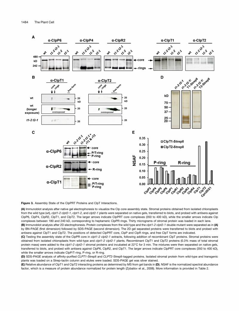
Figure 3. Assembly State of the ClpPRT Proteins and ClpT Interactions.
(A) Immunoblot analysis after native gel electrophoresis to visualize the Clp core assembly state. Stromal proteins obtained from isolated chloroplastsfrom the wild type (wt), clpt1-2 clpt2-1, clpt1-2, and clpt2-1 plants were separated on native gels, transferred to blots, and probed with antisera againstClpP6, ClpP4, ClpR2, ClpT1, and ClpT2. The larger arrows indicate ClpPRT core complexes (350 to 400 kD), while the smaller arrows indicate Clpcomplexes between 180 and 240 kD, corresponding to heptameric ClpPR rings. Thirty micrograms of stromal protein was loaded in each lane.(B) Immunoblot analysis after 2D electrophoresis. Protein complexes from the wild type and the clpt1-2 clpt2-1 double mutant were separated as in (A)by BN-PAGE (first dimension) followed by SDS-PAGE (second dimension). The 2D gel separated proteins were transferred to blots and probed withantisera against ClpT1 and ClpT2. The positions of detected ClpPRT core, ClpP and ClpR rings, and free ClpT forms are indicated.(C) Testing the assembly state of the ClpPR core in clpt1-2 clpt2-1 extracts, following addition of recombinant ClpT proteins. Stromal proteins wereobtained from isolated chloroplasts from wild-type and clpt1-2 clpt2-1 plants. Recombinant ClpT1 and ClpT2 proteins (0.3% mass of total stromalprotein mass) were added to the clpt1-2 clpt2-1 stromal proteins and incubated at 22°C for 3 min. The mixtures were then separated on native gels,transferred to blots, and probed with antisera against ClpP6, ClpR2, and ClpT1. The larger arrows indicate ClpPRT core complexes (350 to 400 kD),while the smaller arrows indicate ClpP/T-ring, P-ring, or R-ring.(D) SDS-PAGE analysis of affinity-purified CLPT1-StrepII and CLPT2-StrepII-tagged proteins. Isolated stromal protein from wild-type and transgenicplants was loaded on a Strep-tactin column and elutes were loaded. SDS-PAGE gel was silver stained.(E) Relative abundance of ClpT1 and ClpT2 interacting proteins as determined by MS from gel bands in (D). NSAF is the normalized spectral abundancefactor, which is a measure of protein abundance normalized for protein length (Zybailov et al., 2008). More information is provided in Table 2.
1484 The Plant Cell

was incubated with rClpT1 and rClpT2, and extracts were sub-sequently run on 1D-BN-PAGE, transferred to blots, and probedwith antisera against ClpP6 (representing the P-ring), ClpR2(representing the R-ring), and ClpT1 (Figure 3C). This resulted inan increase of the ClpPR core and accumulation of a ClpP ringwith associated rClpT, but never a ClpR ring with associatedrClpT. Thus, exogenous ClpT can help to stabilize the ClpPRcore complex in 1D-BN-PAGE.
ClpT1- and ClpT2-Interacting Proteins
To determine if proteins other than the ClpPR core interact withClpT1 or ClpT2 and to determine if ClpT1 and ClpT2 can interactwith the ClpPR core independently of each other, we generatedtransgenic Arabidopsis lines expressing StrepII-tagged ClpT1 orClpT2 subunits in the clpt1-2 clpt2-1 double homozygous mu-tant background. The eight-residue StrepII tag (Schmidt andSkerra, 2007) was attached to the C terminus rather than the Nterminus to prevent interference with the N-terminal chloroplasttargeting peptide. Previously we used StrepII-tagged ClpR4 andClpP3 lines successfully to purify ClpPR cores (Olinares et al.,2011b). The StrepII-tagged ClpT1 and ClpT2 lines grew well onsoil and, in contrast to the clpt1-2 clpt2-1 line, exhibited wild-type-like phenotypes (Supplemental Figure 4A). PCR of genomicDNA (data not shown) and immunoblotting using an anti-StrepIIantibody confirmed the complementation (Supplemental Figure4B). For affinity purification, chloroplast stromal proteins wereisolated from the CLPT1-StrepII and CLPT2-StrepII-taggedcomplemented lines, as well as from the clpt1-2 clpt2-1 doublehomozygous line serving as negative control. After affinity pu-rification using Strep-Tactin superflow columns, protein eluateswere run on an SDS-PAGE gel and then stained with silver ni-trate (Figure 4D). MS analyses of the gel bands identified all nineClpP and ClpR subunits, as well as the StrepII-tagged baits inboth CLPT1-StrepII and CLPT2-StrepII complemented lines;none of these Clp proteins were present in the negative control(Figure 3E). Importantly, this shows that ClpT1 and ClpT2 caneach independently interact with the ClpPR core. Whereas theClpT2-StrepII purification overall yielded less MS/MS spectra,the ratio of matched MS/MS spectra for the ClpPR subunitsper ClpT was the same for ClpT1-StrepII and ClpT2-StrepII(Supplemental Table 3). We also note that the P-ring and R-ringsubunits were present in a roughly equal ratio, indicating that wepurified mostly intact ClpPR cores (Figure 3E). We identified oneadditional candidate interacting protein, namely, the cochaper-onin CPN20 (AT5G20720) (Koumoto et al., 1999). This proteinwas present with a significant number of MS/MS spectra in bothClpT1 and ClpT2 StrepII purifications but not in the negativecontrols (Table 2). In previous experiments, extensive StrepIIpurification of the ClpPR core using complemented null mutantsfor CLPP3 and CLPR4 with StrepII-tagged ClpP3 and ClpR4proteins (Olinares et al., 2011b), we found either very low levelsor no CPN20 in these StrepII purifications, indicating thatCPN20 enrichment is not a common, unspecific interactor inStrepII affinity purifications and/or a direct interactor with theClpPR rings. Finally, it was recently reported that ClpT interactswith ClpC2 and ClpD chaperones and stimulates ClpD chaper-one activity (Colombo et al., 2014). However, our StrepII-tagged
ClpT1/2 purification did not identify ClpC/D chaperones as in-teracting proteins.
ClpT Structure Determination
ClpT1 and T2 are unique to higher plants and their interactionwith the ClpPR core (and perhaps other proteins) likely repre-sents a specific adaptation to the plastid proteome and/orhigher plant plastid Clp protease system. In order to gainfunctional understanding of ClpT1 and ClpT2 and how theymight interact with the ClpP ring, we determined their structuresby x-ray crystallography. We crystallized a ClpT1 (S65-Q238)and a ClpT2 (S76-E241) construct representing the matureproteins and determined their structures at a resolution of 2.4and 2.0 Å, respectively (Figure 4A, Table 2). Both structures hadtwo monomers per asymmetric unit; the two independent chains(molecules) in the ClpT1 structure can be superimposed witha root mean square deviation (r.m.s.d.) of 0.21 Å, while those ofClpT2 can be superposed with an r.m.s.d. of 0.25 Å, implyingminimal difference between chains. (Note: ClpT1 and ClpT2have differing numbering due to differing lengths of the cTP;equivalent numbering for mature ClpT2 can be derived fromClpT1 by adding 8 starting with Lys-90 in ClpT2 [SupplementalFigure 1A].)The structure of each ClpT paralog is organized around a set
of eight a-helices, which show a clear internal repeat organi-zation where helices aA to aD (repeat 1 in the N-domain) arestructurally equivalent to helices aE to aH (repeat 2 in theN-domain) (Figure 4A). The ClpT1 and ClpT2 structures closelyresemble one another and can be superimposed with an r.m.s.d.of 0.55 Å (Figure 4B). Because they are very similar, in the fol-lowing discussion, we focus on the more complete and betterresolved ClpT2 structure (Figure 4A), pointing out differences inClpT1 where pertinent. Searching the Protein Data Bank (http://www.rcsb.org/pdb/) with ClpT2 in PDBeFold shows that theseproteins are most closely related to the N-terminal domain ofBacillus subtilis ClpC (2y1r), with r.m.s.d. of 1.57 Å over 137residues (Wang et al., 2011); this structural similarity reinforcesthe idea that ClpT may be derived from the bacterial ClpCN-domain. The bacterial protein cg2963 from Corynebacteriumglutamicum is also structurally closely related (3fh2; 1.82 Å over137 residues), as well as the N-terminal domains of E. coli ClpB(1khy; 2.2 Å over 133 residues) and the N-terminal domain ofE. coli ClpA (1r6q; 2.37 Å over 128 residues) (Xia et al., 2004).Figure 4C (and Supplemental Figure 5A) shows the overlaybetween the structures of ClpT1, ClpT2, and N-domains ofB. subtilis ClpC (2y1r) and E. coli ClpA (1r6q).
Candidate Binding Sites of ClpT
Studies of complexes formed by homologous Clp N-domainstructures, including E. coli ClpA, B. subtilis ClpC, and Vibriocholera ClpV, show that at least four independent surfaces in theN-domain structures are utilized to recognize (different) bindingpartners. The most commonly utilized site is centered on theconvergence of the N-terminal ends of helix aB, aE, and aG(Supplemental Figure 6B). Here, the amide nitrogen atoms at theN-terminal end of aB coordinate with two conserved threonine
ClpT Structure and Function in Chloroplasts 1485

Figure 4. Structural Information on ClpT1 and ClpT2 and Docking to the ClpP Ring.
(A) ClpT2 monomeric structure, colored in a gradient of blue to red (blue-green-yellow-orange-red) from the N to C terminus. This view is oriented toshow the pseudo 2-fold symmetry that here lies horizontal in the plane; 68 residues of the two related halves of ClpT2 can be superimposed with a 2.6-År.m.s.d. and are 15% identical at the sequence level. For ClpT1, 64 residues can be superposed with a 2.1-Å r.m.s.d. and are 22% identical. In ClpT2,the region (22 amino acids) N-terminal of Lys-93 in ClpT2 (corresponding to Lys-85 in ClpT1) is disordered in chain B, while in chain A, residuesN-terminal to Lys-90 are disordered, except residues Ser-76 to Asn-81, which are weakly stabilized by crystal packing interactions; therefore, theseN-terminal ClpT2 regions are not displayed. The position of the MYFF motif in the aD-aE loop is marked with small spheres (marked with an arrow).(B) Superposition of ClpT paralogs. ClpT1 is shown in yellow and ClpT2 in cyan. These structures superpose closely, with the main differences in theaD-aE loop, and the disorder of the C terminus in ClpT1. In the ClpT1 structure, residues N-terminal to Pro-84 (14 residues) and C-terminal to Glu-227(11 residues) are disordered in both chains and are therefore not displayed.(C) Superposition of ClpT homologs. ClpT1 is shown in yellow, ClpT2 in cyan, E. coli ClpA N-terminal domain in blue, and B. subtilis ClpC N-terminaldomain in orange. Note that despite the general organizational similarity of these proteins, differences in the positions of individual helices and longconnecting loops can result in very different interaction surfaces.(D) to (F) Three conserved interaction surfaces in ClpT2. The upper left insets show sequence conservation (left panel; with plum being the mostconserved and cyan the most variable) and upper right insets show electrostatic ([D] and [E]; blue is electropositive, red electronegative) or
1486 The Plant Cell

residues to form a binding pocket that specifically recognizessurface-exposed glutamic acid residues, while surroundingmotifs extend the interaction surface to recognize other featuresof the cognate ligand, most often an a-helix (Erbse et al., 2008;Kirstein et al., 2009; Kress et al., 2009; Striebel et al., 2009). Inthe B. subtilus ClpC N-domain, Thr-31 and Thr-81 in this glu-tamate binding pocket binds Glu-184 of adaptor MecA (Wanget al., 2011) (Supplemental Figure 6D). Similarly, in E. coli ClpA,Glu-28, Thr-81, and the backbone amines at the N-terminal endof aB recognize Glu-79 of ClpS (Guo et al., 2002; Zeth et al.,2002) (Supplemental Figure 6E). The equivalent residues inClpT2 (Thr-119 and Glu-120) (Figure 4D, lower panel) at the
N-terminal end of aB along with Thr-172 are conserved in ClpT2(and also ClpT1), suggesting a conserved role in recognizingglutamate. In ClpT1, this pocket binds Glu-159 of the adjacentClpT1 molecule in each asymmetric unit within the crystal(Supplemental Figure 6C). This region of the protein containssome additional conserved residues (e.g., Lys-154, Lys-158,and Asp-160) and is flanked in the structure by the conservedhydrophobic MYFF motif (Figures 4A and 4B; next section) in theaD- aE loop (see sequence logo in Supplemental Figure 2). Totest the in vivo significance of the predicted glutamate bindingpocket in ClpT1 and ClpT2, we transformed the clpt1-2 clpt2-1double mutant with StrepII-tagged variants of ClpT1 (T111V and
Figure 4. (continued).
hydrophobicity ([F]; orange is hydrophobic and green hydrophilic, Eisenberg scale) with the location of the detailed view (lower panels) marked witha dashed box.(D) The glutamate binding surface of ClpT2. This figure is oriented very similar as (A) to (C). The glutamate binding pocket in ClpT2 is comprised of Thr-119 and Glu-120 at the N-terminal end of aB, along with Thr-172; glutamate has been observed to mediate biologically relevant interactions with thehomologous pocket in the N-domains of ClpA (with ClpS) and ClpC (with MecA) (Supplemental Figures 6B, 6D, and 6E). In ClpT1, the equivalentresidues bind a glutamate from a neighboring molecule, showing that the essential recognition elements remain intact (Supplemental Figure 6C).(E) Candidate basic binding pocket on the face of ClpT2. This face has a large number of conserved hydrophobic (e.g., Met-104, Leu-107, and Ile-128)and basic (e.g., Lys-100, Arg-110, Lys-111, Lys-113, and Arg-149) residues and is flanked by the hydrophobic MYFF motif. This pocket in E. coli ClpAN-domain mediates interactions between the N-domain and the rest of the chaperone structure (Supplemental Figure 5G).(F) The hydrophobic binding pocket formed between aA and aE; view is down the quasi 2-fold axis. The equivalent surface is similarly hydrophobic andis used to bind substrates in B. subtilis ClpC and V. cholera ClpV (Supplemental Figures 6H and 6I).
Table 2. Data Collection, Model Refinement, and Final Structure Statistics for ClpT1 and ClpT2
ClpT1 ClpT2
Crystallographic data collection statisticsSpace group P212121 C2Cell dimensions:
a = (Å) 30.4 104.15b = (Å) 109.2 57.77c = (Å) 120.5 61.65b = (°) 90 98.17
Wavelength (Å) 0.979098 1.54158Resolution (Å) 2.4 2Unique observations 16,579 23,802Completeness (last shell)a 0.998 (1.00) 0.954 (0.678)Redundancy (last shell)a 7.9 (8.1) 4.9 (2.6)<I/S(I)> (last shell)a 12.7 (2.1) 21.6 (3.5)Rsym (last shell)a 0.089 (0.99) 0.066 (0.457)X-ray structure refinement statisticsRcryst 0.206 0.173Rfree
b 0.253 0.207Asymmetric unit contents
Water molecules 24 288Other molecules 2 Cl2 2 SO4, 1 Cl2
Average ADPs (Å2)Protein 75.3 28.4Water 51.9 31.1r.m.s.d. bond lengths (Å) 0.011 0.0085r.m.s.d. bond angles (°) 1.461 1.134Ramachandran favored (%) 95.2 96.1Ramachandran outliers (%) 0.4 0.3aThe last shell includes all reflections between 2.46 and 2.40 Å for ClpT1 and between 2.1 and 2.0 Å for ClpT2.bRfree calculated using 5% of the data that were chosen randomly.
ClpT Structure and Function in Chloroplasts 1487

T164V) and ClpT2 (T119V and T172V). Surprisingly, all fourClpT1 or ClpT2 mutant constructs fully complemented thedouble mutant (Supplemental Figure 7). This indicates that de-spite the conservation of these residues, this pocket is not re-quired for essential ClpT functions and perhaps that ClpT doesnot bind adaptors, such as ClpS1, or as yet unidentified ones.However, it cannot be totally excluded that the relatively con-servative Thr→Val (similar sized uncharged polar → apolar)mutations retain sufficient interaction strength to suppress thegrowth phenotype seen in the mutants.
Due to the internal rotational symmetry of Clp N-domains, thearrangement of helices in the glutamate binding pocket centeredon aB is repeated on the opposite side of the molecule, cen-tered on the N-terminal end of aF. In B. subtilis ClpC, thispocket also has the appropriate T/E/T motif and is used to bindGlu-189 of MecA (Wang et al., 2011). In ClpT, the T/E/T motif isabsent and the packing of nearby loops narrows the pocket,implying that this pocket is unlikely to mediate any analogousinteractions; this is consistent with our previous (Nishimuraet al., 2013) and current observations that ClpT proteins do notinteract with adaptor proteins.
A third interaction surface in chaperone N-domains and ClpT1and ClpT2 is found on the reverse face of the glutamate bindingpocket, and this surface is characterized by a band of basicresidues flanked on either side by conserved hydrophobic res-idues (Figure 4E, upper panel). The top hydrophobic band isformed by the MYFF motif, while the lower hydrophobic band isformed by residues including Met-104, Leu-107, and Ile-128.Among the conserved basic residues are Arg-110, Lys-111, Lys-113, Arg-149, and Lys-158, while Glu-108 and Glu-167 areacidic residues interspersed among the basic residues (Figure4E, lower panel). In E. coli ClpA, this surface is used by theN-domain to bind to the AAA domain. The strong conservationof this pocket in ClpT implies that it may also have an importantfunction in ClpT.
The fourth potential binding pocket in ClpT2 is a large ex-posed hydrophobic patch located between the long helices aAand aE (Figure 4F). Trp-94, Ile-99, Phe-102, Leu-171, Leu-179,and Leu-183 are all conserved as hydrophobic residues, whilesome adjacent hydrophilic residues including Gln-176 and Asp-180 are also absolutely conserved in ClpT2 (and very conservedin ClpT1). The equivalent surface, similarly hydrophobic, is usedby the N-domain of the AAA protease V. cholera ClpV to rec-ognize its substrate VipB in type VI protein secretion (Pietrosiuket al., 2011). In ClpA, this region interacts with ClpS and is alsoimplicated in peptide binding (Xia et al., 2004), while in Myco-bacterium ClpC, this region is the target of the antimicrobialcyclomarin A, implying a possible role in substrate interaction(Vasudevan et al., 2013) (Supplemental Figures 6H and 6I).Unless required for function, proteins generally avoid exposingextensive hydrophobic patches as they are prone to aggrega-tion; the presence of this patch here implies a likely role inprotein-protein interaction, albeit with an unknown partner.
The MYFF Motif Is Functionally Important
The long aD-aE loop contains a hydrophobic, aromatic-richsequence motif, MYFF, in Arabidopsis ClpT1 and ClpT2 and
generally conserved as (M/L)(Y/F)(F/Y/W)F in both ClpT1 andClpT2 (Supplemental Figure 2). In our structures, this motif is notpart of the hydrophobic core of the protein, but instead is or-dered by packing interactions in chain A of ClpT2, partially or-dered in both ClpT1 monomers and disordered in ClpT2 chain B.This very hydrophobic motif is therefore exposed on the proteinsurface in the middle of a flexible loop, with a strong tendencytoward disorder. Its lack of interaction with the core ClpTstructure, along with the position of this motif at the periphery oftwo candidate binding surfaces (the glutamate binding and ba-sic sites) suggests that this motif may be involved in an externalinteraction.To test the in vivo significance of the MYFF motif of ClpT1 or
ClpT2, we transformed the clpt1-2 clpt2-1 or clpt1-2 clpt2-2double mutant with variants of ClpT1 and ClpT2 (MYFF→AAAA).Unlike clpt1 clpt2 CLPT1 or clpt1 clpt2 CLPT2 lines, whichshowed full complementation, clpt1 clpt2 CLPT1(MYFF→AAAA)or clpt1 clpt2 CLPT2(MYFF→AAAA) did not exhibit a wild-type-like phenotype (Figure 5A; see Supplemental Figure 8 for olderplants). They showed the same small, yellow, pale-green phe-notype like clpt1-2 clpt2-2, implying that they could not com-plement the double homozygous mutant. The overexpressionof the variant ClpTs form was confirmed by immunoblottingagainst ClpT antisera (Figure 5B). The lack of complementationof the mutant phenotype shows that the MYFF motif of ClpT iscrucial for Clp protease and chloroplast function and plant de-velopment. We then tested the ClpPR core assembly state and ifthe variant ClpT1 and ClpT2 proteins could interact with theClpPR core complex or ClpP ring. To that end, chloroplaststromal proteins were extracted under nondenaturing conditionsand proteins were separated by 1D-BN-PAGE, followed by im-munoblotting with ClpR2 and ClpT1 serum (Figure 5C). Theclpt1-2 clpt2-2 CLPT1(MYFF→AAAA) mutants failed to stabilizethe ClpPR core and showed about the same level of Clp coreassembly as the clpt1-2 clpt2-2 double mutant. In contrast,expression of the StrepII-tagged wild-type-like ClpT1 did restorethe ClpPR assembly state determined by blue native PAGE (BN-PAGE; Figure 5C). Using BN-PAGE and two-dimensional PAGE,we showed that variant ClpT could only interact with the in-dividual ClpP ring and not with the 350-kD core (Figures 5C and5D). These data demonstrate that the MYFF motif is required forstabilization of ClpP and ClpR ring interactions, but not strictlyfor interaction to the ClpP ring. These data also show that ClpT1and ClpT2 binding to the ClpP ring is not enough for core sta-bilization, and we suggest that binding of this extended hydro-phobic MYFF motif to the P-ring results in an allosteric changein the P-ring, leading to stabilization of the overall complex.
DISCUSSION
ClpT Proteins Are Unique to Higher Plants
Sequence analysis strongly suggests that ClpT proteins arederived from ClpC chaperones but that these ClpT proteins areabsent in cyanobacteria; this is significant because the functionof ClpT concerns ClpPR protease activity and these chloroplastprogenitors also have ClpR proteins and a mixed ClpP3R
1488 The Plant Cell

complex. However, the organization of this cyanobacterialcomplex is different in that the two heptameric rings in theClpP3R complex are symmetrical (Stanne et al., 2007), while theClpP and ClpR rings in chloroplasts are asymmetrical with eachhaving their own set of proteins (reviewed in Nishimura and vanWijk, 2014). Moreover, the ClpC chaperones in higher plantchloroplasts likely only interact with the R-ring (based on se-quence comparison of key residues in cyanobacteria ClpP3and ClpR), whereas ClpT proteins directly bind to the P-ring(Nishimura and van Wijk, 2014). This suggests that higher plantClpT proteins have specifically coevolved with the ClpPR pro-teins to ensure a fully functional ClpPR complex, by facilitating
the formation/stabilization/activation of the ClpPR complex. Analternative hypothesis for the presence of ClpT proteins is thatthey facilitate the proteolysis of a specific set of proteins, e.g.,by binding to specific intrachloroplast membranes, binding tochloroplast-specific adaptors, or binding to specific proteasesubstrates. The biochemical, structural, and proteomics analysisin this study strongly suggests that ClpT evolved to ensurea functional ClpPR complex, rather than facilitating degradationof specific proteins. Finally, monocots only have ClpT1 proteinsand also a few dicots have either only ClpT1 or ClpT2, sug-gesting that ClpT1 and ClpT2 proteins have largely redundantfunctions.
Figure 5. Complementation of clpt1 clpt2 with 35S-Driven cDNAs for CLPT1, CLPT1(MYFF→AAAA), CLPT2, and CLPT2(MYFF→AAAA) and AssemblyState of the ClpPRT Proteins.
(A) Comparison of the wild type, clpt1-2 clpt2-1, clpt1-2 clp2-2, and clpt1-2 clpt2-2 transformed with 35S-driven cDNA for CLPT1 or CLPT1(MYFF->AAAA), and clpt1-2 clp2-1 transformed with 35S-driven CLPT2 or CLPT2(MYFF→AAAA). Plants were grown for 11 d on half-strength Murashige andSkoog agar plates + 2% sucrose under continuous light at 100 mmol photons m22 s21. Older, more developed plants grown further after transfer to soilare shown in Supplemental Figure 8 and further illustrate the lack of phenotypic complementation for the MYFF constructs. Bar = 5 mm.(B) Accumulation levels of ClpT1 protein in clpt1-2 clpt2-1 CLPT1 and clpt1-2 clpt2-2 CLPT1(MYFF→AAAA) compared with the wild type. Total solubleleaf proteins were extracted from each line. After SDS-PAGE, proteins were transferred to blots and probed with ClpT1 antiserum. Ten micrograms wasloaded in each lane.(C) Assembly state of the ClpPRT proteins determined by immunoblot analysis after native gel electrophoresis. Stromal proteins obtained from isolatedchloroplasts from the wild type (wt), clpt1-2 clpt2-2, clpt1-2 clpt2-1/T1, and clpt1-2 clpt2-2 CLPT1(MYFF→AAAA) were separated on native gels,transferred to blots, and probed with antisera against ClpR2 and ClpT1. The larger arrows indicate ClpPRT core complexes (350 to 400 kD), while thesmaller arrows indicate ClpP/T1-ring, P-ring, or R-ring. Thirty micrograms of stromal protein was loaded in each lane.(D) Immunoblot analysis after 2D electrophoresis. Protein complexes from the wild type, clpt1-2 clpt2-1 double mutant, clpt1-2 clpt2-1 CLPT1-StrepII,and clpt1-2 clpt2-2 CLPT1(MYFF→AAAA)-StrepII were separated as in (C) by BN-PAGE (first dimension) followed by SDS-PAGE (second dimension).The 2D gel was electroblotted and probed with antisera against ClpT1. The positions of detected ClpPRT core, ring, and free form are indicated.
ClpT Structure and Function in Chloroplasts 1489

Double Homozygous CLPT Mutants Are Viable but Showa Clear Growth and Developmental Phenotype
Based on the screening of 100 progeny, it was previouslyreported that double homozygous mutants (clpt1-1 clpt2-1)are not viable (Sjögren and Clarke, 2011). However, when wecrossed and then selfed each CLPT1 and CLPT2 allele, weproduced double homozygous mutants that survived well onsoil, flowered, and generated viable seeds. Since the twoCLPT genes are located on the same chromosome (CLPT1,At4g25370; CLPT2, At4g12060; physical distance betweenCLPT1 and CLPT2 is ;5.7Mb), we screened hundreds ofseedlings to identify the double homozygous mutants. For ex-ample, in the case of clpt1-1 clpt2-1, we identified 15 doublehomozygous mutants out of 1151 seedlings. Considering a ge-netic distance of 1% recombination corresponds, on average, toa physical distance of ;250 kb in Arabidopsis (Lukowitz et al.,2000), the chance of recombination between CLPT1 and CLPT2is ;23%. Thus, the probability of double homozygous (aabb)mutant based on 23% recombination is 0.013. This matcheswell with our finding of 15 double homozygous mutants out of1151 seedlings. This explains why the previous report failedto identify double homozygous mutants. Importantly, the vi-able double mutants allowed us to analyze the role of ClpT1and ClpT2 in ClpPR stability and proteostasis and to de-termine (1) if ClpT1 and ClpT2 can interact independently withthe ClpPR core (as opposed to an interdependent interaction),(2) if ClpT1 and ClpT2 have redundant functions, and (3) themolecular consequences for the lack of ClpT1 and ClpT2 onleaf development and chloroplast biogenesis, function, andproteostasis.
Side-by-side comparison of the clpt1-2 clpt2-1 double mutantto the CLPR2 knockdown line clpr2-1 (with ;20% of ClpR2levels) showed that the clpt double mutant was somewhatsmaller in stature, with quite similar patterns of leaf virescenceand serration, independent of leaf development and light regime.However, whereas null mutants for the genes encoding ClpR2,ClpP3, and ClpR4 are seedling lethal and null mutants for thegenes encoding ClpP4 and ClpP5 are embryo lethal, the doublemutant in CLPT1 and CLPT2 does not require sucrose and isviable on soil. This demonstrates that ClpT1 and ClpT2 are notstrictly required for Clp protease function and indicates that intheir absence ClpPR capacity is reduced to ;20%. However,unlike both clpt2 alleles, the strongest clpt1 mutant (clpt1-2) didaccumulate a ClpT1 cross-reacting band, albeit at higher mo-lecular mass. Whereas we could not detect ClpT1 in this clpt1-2allele by MS/MS analysis, clpt1-2 may not be a strict null mutant; itis thus possible that complete loss of ClpT1, together with com-plete loss of ClpT2, does lead to seedling (or embryo) lethality.
The Proteome Phenotype Suggests Limited ClpProtease Capacity
Comparison of the proteome phenotype of the clpt1-2 clpt2-1double mutant versus loss-of-function mutants of CLPR2,CLPR4, and CLPP3 (reviewed in Nishimura and van Wijk, 2014)showed very similar patterns of under- and overaccumulation ofproteins. For instance, the unfoldase ClpB3, maturase HSP90,
the CPN60 chaperones, as well as the three chloroplast elon-gation factors were all significantly upregulated. The strongupregulation of plastoglobular proteins indicated significantstress levels similar as observed in the other Clp mutants. Wedid not observe proteins that were specifically over- or under-accumulating in the ClpT mutant but not in the ClpPR coremutants. Hence, the ClpT mutant proteome phenotype is easi-est explained by a strongly reduced ClpPR protease capacity.However, clearly, the ClpT double mutant must have significantClp protease activity because complete loss of ClpPR capacityleads to embryo or seedling lethality. We thus conclude thatClpT proteins do not appear to facilitate the proteolysis ofa specific class of proteins, unlike, for example, the adaptorNblA for degradation of phycobilisomes in cyanobacteria (Karradtet al., 2008; Baier et al., 2014) but rather facilitate ClpPR activity,as discussed further below.
The Double Mutant Phenotype Cannot Be Explained by Lossof ClpPR Core Assembly
In a recent study (Sjögren and Clarke, 2011), it was proposedthat individual ClpP and ClpR rings exists in the stroma and thatClpPR activity is regulated through (reversible) association ofClpP and ClpR rings by sequential activity of ClpT1 and ClpT2.This model was essentially based on results from BN-PAGEanalysis of wild-type proteins showing that <50% of ClpPR ringsare assembled in a ClpPR core complex, but rather exist asindividual rings. The percentage of assembly ClpPR core waseven lower in the weak clpt1-1 (;30%) and the clpt2-1 nullmutants (;10%). Upon addition of recombinant ClpT1 andClpT2, the percentage of observed ClpPR cores increased. Theirmodel was further inspired by the erroneous observation that nodouble mutants for ClpT1 and ClpT2 could be obtained, evenwhen using the weak clpt1-1 allele, as we discussed above.This study, combined with our previous studies (reviewed inNishimura and van Wijk, 2014), contradicts this model based onthe following observations: (1) in-house colorless native PAGE(CN-PAGE), gel staining, and MS analysis (Peltier et al., 2004,2006), as well as immunoblotting of CN-PAGE gels with anti-polyHis serum of the clpr2-1mutant complemented with polyHIS6-tagged ClpR2 (Rudella et al., 2006), indicated that in wild-typechloroplasts, a single ClpPR complex accumulates without de-tectable individual free ClpP and ClpR rings; and (2) the ClpTdouble mutant clpt1-2 clpt2-1 shows a strong growth pheno-type, yet the observed level of ClpPR core accumulation is atleast as high as the clpt1-2 single mutant, which lacks a growthphenotype.
ClpT1 and ClpT2 Structure, Modeling, and ExperimentalTesting the Interactions between ClpT and the ClpProtease Core
The high-resolution structures of ClpT1 and ClpT2 clearlydemonstrated the similarity to N-domains of Clp chaperones.These structures allowed us to suggest, and subsequently ex-perimentally test, possible interaction domains and motifs be-tween ClpT1, ClpT2, and the ClpPR core. Out of the three mostobvious interaction surfaces, we showed that the MYFF motif is
1490 The Plant Cell

essential for ClpT function. The hydrophobic nature of this motifis reminiscent of the hydrophobic IG(F/L) motif used by E. coliClpA/X (conserved in Arabidopsis ClpC1, ClpC2, and ClpD;Peltier et al., 2004) to bind to hydrophobic grooves in the adaxialside of the ClpP ring (Kim et al., 2001). This motif in ClpT may bemore hydrophobic as only one such motif will be utilized forinteraction, versus the six interactions used to recruit ClpA/Chexamers (one interaction contact for each ClpA/C) (Kim et al.,2001); the more extended hydrophobic region in ClpT may inturn require a longer hydrophobic binding groove on the ClpPring. Thus, the structure suggests that MYFF motif of ClpT mightbe a key motif for interacting with the ClpPR core. The MYFFmotif is likely to act in synergy with either the glutamate bindingpocket (though the nonessentiality of the Thr-119 and Thr-172possibly argues against this) or the basic pocket located on theopposite face. This involvement of a larger binding pocket couldexplain why ClpT is able to interact with the ClpP ring. The MYFFmotif might then stabilize the 350-kD core through two possiblemodes of action, as discussed in the next section.
ClpT1 and ClpT2 Affect ClpPR Protease Capacity
The observed loss of ClpPR core assembly on native gels likelyreflects the weakened interactions in absence of (or at low lev-els) of ClpT proteins. Depending on the forces during native gelelectrophoresis (different protocols), different levels of intactClpPR core are observed. Based on all available information, wesuggest two possible mechanisms for the stabilization and
activation of ClpPR protease by ClpT (Figure 6). In the mostattractive model (Figure 6A), ClpT binds to the ClpP ring andinserts the MYFF loop into a hydrophobic pocket. This in-teraction then allosterically stabilizes the ClpP-ClpR ring in-teraction. Inter-ring allostery is a well-established phenomenonin ClpP; for example, in the bacterial ClpAP system, ClpAbinding to one ClpP ring also activates the protease activity ofthe distal ClpP ring through in large conformational changes(Maglica et al., 2009; Liu et al., 2014). Furthermore, various smallmolecules (e.g., ADEPs) have recently been identified that acti-vate the ClpP complex by conformational changes to the ringstructure visualized by cryoEM and single particle averaging(Alexopoulos et al., 2012; Liu et al., 2014). In the second, lessfavored model (Figure 6B), ClpT binds primarily to the ClpP ring,while the MYFF loop binds a hydrophobic pocket on the ClpRring, directly stabilizing the ClpP-ClpR interaction. We thuspropose that ClpT1 and ClpT2 stabilize the ClpP and ClpR ringinteractions and likely activate the ClpPR complex through al-losteric effects (structural conformations).
Interactions between ClpT1, ClpT2, and Adaptors andAccumulation of Free ClpT Proteins
Both the primary sequences and structures of ClpT1 and ClpT2demonstrate high similarity with the N-domains of bacterial (andplant) ClpA/C chaperones. The main function of this N-domain isbinding of adaptor proteins (e.g., ClpS and MecA) and sub-strates (see Introduction and Sauer and Baker [2011]). Based on
Figure 6. Models for the Role of ClpT1 and ClpT2 in the Clp Protease System in Plastids.
It is conceivable that the outer adaxial surface of the R-ring could be affected indirectly by ClpT binding through the structural dynamics of the corecomplex, thus influencing ClpC affinity and/or access to the ClpPR pore.(A) ClpT binds to the ClpP ring and inserts the MYFF loop into a hydrophobic pocket. This interaction then allosterically stabilizes the ClpP-ClpR ringinteraction.(B) ClpT binds primarily to the ClpP ring, while the MYFF loop binds a hydrophobic pocket on the ClpR ring, directly stabilizing the ClpP-ClpRinteraction.
ClpT Structure and Function in Chloroplasts 1491
this homology, we previously speculated that ClpT1 and ClpT2could interact with chloroplast ClpS1 (or other unknown adap-tors) and perhaps substrates (Peltier et al., 2004). However,StrepII affinity purifications, coimmunoprecipitation with thespecific anti-ClpT1 and ClpT2 anti-sera, or previous interactionanalysis with recombinant ClpS1 and ClpT (Nishimura et al.,2013) did not detect such interactions. Nevertheless, one canstill not entirely exclude that such functional interactions mightexist, since perhaps ClpS1-ClpT interactions may only occurwhen ClpS1 interacts with specific substrates. Chloroplast ClpTproteins were initially identified as members of the ClpPR basedon native page (CN-PAGE, BN-PAGE, and native isoelectricfocusing) followed by staining and mass spectrometry (Peltieret al., 2001, 2004) with on average one ClpT1 and one ClpT2 percomplex as estimated by silver and Coomassie blue stainingand theoretical correction for amino acid composition (Peltieret al., 2004). The in vivo StrepII tagging and affinity experimentsshowed that ClpT1 and ClpT2 independently bind to the ClpPRcore complex. Current and previous label-free spectral countinganalysis of stroma and total leaf extracts suggests approxi-mately four to five ClpT proteins per ClpPR core complex. Withtwo ClpT proteins bound to each ClpPR complex, this wouldsuggest that 50% of ClpT proteins are unbound. Recent esti-mates by Sjögren and Clarke (2011) suggest that only 5% ofClpT is bound to the ClpPR core; however, given the relativelyweak and salt sensitive interactions between ClpT and theClpPR core and the relatively harsh nature of BN-PAGE (de-pending on the procedure used), this strongly underestimatesthe fraction of bound ClpT. Nevertheless, the significance of freestromal ClpT proteins remains to be determined. The suggestionthat recombinant ClpT interacts with ClpC2 and ClpD chaper-ones and stimulates ATP hydrolysis activity by ClpD (Colomboet al., 2014) also warrants further investigation.
Interactions between ClpT and CPN20
The in vivo ClpT StrepII tagging and affinity experiments alsoidentified a CPN20 as a candidate interacting protein with ClpT1and ClpT2, most likely when these ClpT proteins do not interactwith the ClpPR core. CPN20 is part of the family of Type Ichaperonins that function in protein folding together with CPN60proteins and CPN10 proteins (Koumoto et al., 1999; Sharkiaet al., 2003; Vitlin Gruber et al., 2013). It remains to be de-termined if and how CPN20 has a functional relationship toClpT1 and ClpT2.
Future Directions
The structures of ClpT1 and ClpT2 allowed us to assess theirpossible interaction domains and surfaces with the ClpPR core.However, better understanding of the docking and functionalinteraction of ClpT to the ClpP ring, and perhaps the ClpR ring,is necessary to determine how ClpT influences ClpPR stabili-zation and activity. This will require a better understanding of theorganization of the ClpP/R rings, in particular the order of theClpP and ClpR subunits within each ring, as well as experi-mental determination of direct ClpPR binding partners for ClpT1and ClpT2. The availability of Arabidopsis StrepII-tagged ClpP3,
ClpR4, ClpT1, and ClpT2 lines in their respective null back-grounds should allow such interaction mapping by affinity pu-rification, cross-linking, and mass spectrometry. Based on suchinformation, better homology models for the P-ring and R-ringcan then be created, thus allowing more efficient testing ofdocking models for ClpT1 and ClpT2. This could explain mo-lecular mechanisms involving the conserved ClpT proteins inhigher plants.
METHODS
Plant Growth, Mutant Isolation, and RT-PCR Analysis
T-DNA insertion lines in Columbia-0 for CLPT1 (AT4G25370) and CLPT2(AT4g12060) are SALK_052772 (clpt1-1), GK-285A05 (clpt1-2), SAIL_340A10 (clpt2-1), and SALK_132943 (clpt2-2). The location of the T-DNAinsertions was confirmed by DNA sequencing. Genotyping and RNAextraction were performed as described previously (Rudella et al., 2006).Various growth conditions are detailed in the figure legends. For RT-PCR,total RNA was isolated with an RNeasy plant mini kit (Qiagen). The firststrand was synthesized from equal amounts of total RNAwith SuperscriptIII reverse transcriptase (Invitrogen). We tested 15, 20, 25, and 30 cyclesfor the primer pairs. Fifteen cycles were insufficient to visualize all tran-scripts, while 20 and 25 cycles best allowed us to visualize the transcripts,and we observed good linearity for 20 and 25 cycles. Primers for genomicPCR and RT-PCR analysis and various complementations are listed inSupplemental Table 2. Transcripts were quantified using ImageJ (http://imagej.nih.gov/ij/).
Complementation
Full-length and various mutagenized forms of CLPT1 and CLPT2 cDNAfragments were PCR amplified, and the C-terminal StrepII sequence wasattached using Taq polymerase. Primers for complementation are alsolisted in Supplemental Table 2. The PCR products were subcloned intopCR8/GW/TOPO vector (Invitrogen). Using LR clonase, the DNA wasintroduced into pEARLEYGATE100 Gateway destination plant binaryvector (Curtis and Grossniklaus, 2003). Competent cells of Agrobacteriumtumefaciens strain GV3101 were transformed with the binary vector. Theclpt1 clpt2 double homozygous plants were used for Agrobacterium-mediated plant transformation by the floral dipping method. Trans-formants were screened using 10 mg/mL DL-phosphinothricin (CrescentChemical). Complemented plants were selected and verified by PCRgenotyping.
Phylogenetic Analysis
Protein sequences were collected from Phytozome v9 (http://www.phytozome.net/). Fifty ClpT proteins frommoss and 31 angiosperms werealigned using Muscle (http://toolkit.tuebingen.mpg.de/muscle) with 30times of iterations and ClustalW output format. The aligned sequenceswere edited to remove their predicted chloroplast transit peptide portions,gaps, insertions, and extensions with Jalview (http://www.jalview.org/)and then converted to the PHYLIP format. A phylogenetic tree wasgenerated using the RAxMLHPCBlackBox interface with the general timereversal model of the protein substitution matrix on the CIPRES ScienceGateway (http://www.phylo.org/index.php/portal/). The resulting phylo-genetic tree was visualized by FigTree (http://tree.bio.ed.ac.uk/software/figtree/), and significant RAxML bootstrap values (>50) were shown at thenodes of the tree. Original protein sequences and multiple sequencealignment are available as Supplemental Data Sets 1 and 2.
1492 The Plant Cell
Chloroplast Stroma and Total Leaf Proteome Isolation for Analysisof Clp Assembly States
For chloroplast stroma isolations, leaves of the wild-type and variousmutant alleles were briefly homogenized in grinding medium (50 mMHEPES-KOH, pH 8.0, 330 mm sorbitol, 2 mM EDTA, 5 mM ascorbic acid,5 mM cysteine, and 0.03% BSA) and filtered through a nylon mesh. Thecrude plastids were then collected by a 2-min spin at 1100g and furtherpurified on 35 to 85% Percoll cushions (Percoll in 0.6% Ficoll and 1.8%polyethylene glycol) by a 10-min spin at 3750g and one additional wash inthe grinding medium without ascorbic acid, cysteine, and BSA. Chloro-plasts were subsequently lysed in 10 mM HEPES-KOH, pH 8.0, 5 mMMgCl2, and 15% glycerol with a mixture of protease inhibitors under mildmechanical disruption. The lysate was then subjected to ultracentrifu-gation (100,000g) to pellet the membrane components. The supernatant(stroma) was then collected. Protein amounts were determined using theBCA protein assay kit (Thermo Scientific). For total leaf proteome isolationunder nondenaturing conditions, total leaf material was ground in liquidnitrogen and solubilized in 50 mM HEPES-KOH, pH 8.0, 15% glycerol,and 10 mM MgCl2 with protease inhibitor cocktail. The suspension wasthen filtered through Miracloth and spun at 100,000g.
Affinity Purification of StrepII-Tagged ClpT1 and ClpT2
Isolated stromal protein (2 mg) from the clpt1-2 clpt2-1 double mutant(negative control) and transgenic StrepII-tagged plants was loaded ona Strep-Tactin superflow high capacity column (IBA). Glycerol (15%) wasincluded in all the buffers to preserve the Clp core complex. After washingwith 1 mL of washing buffer (50 mM HEPES-KOH, pH 8.0, 10 mM MgCl2,75 mM NaCl, and 15% glycerol) five times, elution was performed with0.5 mL Buffer E (washing buffer with 2.5 mM Desthiobiotin) six times.
Native PAGE and BN-SDS-PAGE (2D) Analysis
Light-blue native PAGE was performed for separation of stromal and totalleaf extracts under nondenaturing conditions using the NativePAGENovex gel system (Invitrogen) with precast 4 to 16% acrylamide Bis-Trisgels (Invitrogen). For 2D electrophoresis, the lanes on the blue nativePAGE gel were cut out and incubated in SDS-PAGE equilibration buffer(6 M urea, 2% SDS, 50 mM Tris-HCl, pH 6.8, 30% glycerol, and 5 mMDTT) at room temperature for 1 h. The gel was loaded on top of the gel andwas separated by SDS-PAGE.
Immunoblot Analysis
For immunoblots, proteins were blotted onto nitrocellulose or poly-vinylidene fluoride membranes and probed with antibodies usingchemiluminescence for detection, following standard procedures. Anti-sera against ClpP6, ClpT1, and ClpT2 were generated in rabbits againstprotein domains [ClpP6 (Δ1-51), ClpT1 (Δ1-64), and ClpT2 (Δ1-75)]overexpressed in Escherichia coli. Crude antisera were affinity purifiedusing these overexpressed antigens as bait. StrepII polyclonal antibodywas purchased from GenScript.
Cloning, Overexpression, and Purification of Mature ClpT1 andClpT2 in E. coli
The cDNAs encoding ClpT1 and ClpT2 were cloned out of Arabidopsisthaliana cDNA. The fragments encoding mature ClpT1 (ClpT1 D1-64) andmature ClpT2 (ClpT2 D1-75) were cloned into the expression vector pET-28b (Novagen). The plasmids pET28-ClpT1 D1-64 and pET28-ClpT2D1-75 were transformed into BL21 (DE3) E. coli cells. Overnight small-scalecultures were inoculated into Luria-Bertani media containing kanamycin(50 µg/mL), grown at 3°C until OD600 reached 0.6, and then induced with
isopropyl b-D-1-thiogalactopyranoside (1 mM final concentration) at 37°Cfor 3.5 h. Cells were harvested by centrifugation and resuspended in lysisbuffer (10 mM imidazole, 50 mM NaH2PO4, pH 8.0, and 300 mM NaCl).After sonicating on ice for six 10-s bursts, the lysate was centrifuged at10,000g for 20 min at 4°C. The cleared lysate was then mixed with the Ni-NTA His Bind slurry by shaking at 4°C for 2 h. The lysate-Ni-NTA His Bindmixture was loaded into a column and then the column was washed withwash buffer (20 mM imidazole, 50 mM NaH2PO4, pH 8.0, and 300 mMNaCl) three times and then finally eluted with a step gradient (40 to250 mM imidazole with 30 mM increments, 50 mM NaH2PO4, pH 8.0, and300 mM NaCl). The protein eluted maximum at 130 and 160 mM imidazoleconcentration.
Structure Determination
ClpT1 was crystallized from 10% polyethylene glycol 6000 and 0.2 Mtripotassium citrate; ClpT2was crystallized from 1.6M ammonium sulfate,10% dioxane, and 0.1 M MES, pH 6.5. Crystals were frozen in thecryostream after the surface water was removed by paratone N-oil. ClpT2data were collected on a Rigaku rotating anode (45 kV, 30 mA) witha Rigaku 4k CCD detector; data were processed using the Proteumpackage and scaled using XPREP. ClpT1 data were collected at theCanadian Synchrotron Light Source (beam line CMCB) and processed inXDS (Kabsch, 2010). The structure of ClpT2 was determined by molecularreplacement with Phaser (McCoy et al., 2007), using the structure 3FH2(Corynebacterium glutamicum) as a search model. ClpT1 was then de-termined using the ClpT2 structure as a search model. Both structureswere rebuilt in Coot (Emsley and Cowtan, 2004) and refined in Phenix(Adams et al., 2002), using TLS parameters. The ClpT1 refinement in-cluded torsion angle NCS restraints. Residues 76 to 81 of ClpT2 showsome weak electron density in chain A, but the nonconserved nature ofthis region and their disorder in chain B argue that the partial order of thisregion may be an artifact of the crystallization process. The structures andx-ray diffraction data sets of ClpT1 and ClpT2 have been deposited to theProtein Data Bank (www.rcsb.org) with reference codes 4Y0B and 4Y0C,respectively.
Proteomics and Mass Spectrometry
Three biological replicates per genotype were analyzed resulting in 66MS/MS runs. Homozygous clpt1-2 clpt2-1 plants were grown undera short-day cycle (10 h/14 h of light/dark at 100mmol photonsm22 s21) for41 d. Total leaf proteins were extracted under denaturing conditions aftergrinding 250 to 350 mg fresh leaves in liquid N2 into a fine powder,followed by removal of cell debris on Fritt spin columns (pore size 30 mm)as described (Friso et al., 2011). This was performed in three biologicalreplicates. Protein concentrations were determined using the BCAProteinAssay Kit (ThermoFisher). Fifty micrograms of total leaf protein of clpt1-2clpt2-1 and wild-type samples were each run out on a Bio-Rad CriterionTris-HCl precast gels (10.5 to 14% acrylamide gradient) using the threebiological replicates. Each of the six gel lanes were cut into 11 bandsfollowed by reduction, alkylation, and in-gel digestion with trypsin asdescribed (Shevchenko et al., 2006; Friso et al., 2011).
The resuspended peptide extracts were analyzed by data-dependentMS/MS using an on-line LC-LTQ-Orbitrap (Thermo Electron). Peptidesamples were automatically loaded on a guard column (LC PackingsMGU-30-C18PM) via an autosampler followed by separation on a Pep-Map C18 reverse-phase nanocolumn (LC Packings nan75-15-03-C18PM)using 90-min gradients with 95%water, 5% acetonitrile, and 0.1% formicacid (solvent A), and 95% acetonitrile, 5% water, and 0.1% formic acid(solvent B) at a flow rate of 200 nL/min. Two blanks were run after everysample (see Zybailov et al. [2009] for the gradient and sample injectionscheme). The acquisition cycle consisted of a survey MS scan in theOrbitrap with a set mass range from 350 to 1800 m/z at the highest
ClpT Structure and Function in Chloroplasts 1493
resolving power (100,000) followed by five data-dependent MS/MS scansacquired in the LTQ. Dynamic exclusion was used with the followingparameters: exclusion size, 500; repeat count, 2; repeat duration, 30 s;exclusion time, 180 s; exclusion window, 66 ppm or 6100 ppm. Targetvalues were set at 5 3 105 and 104 for the survey and MS/MS scans,respectively. MS survey scan in the Orbitrap was acquired in one mi-croscan. Fragment ion spectra were acquired in the LTQ as an average ofthree microscans. Mass spectrometry data processing, data searchingagainst TAIR10 using Mascot, and subsequent filtering and quantificationbased on normalized and adjusted spectral counts was performed asdescribed (Friso et al., 2011) and as outlined in Supplemental Figure 5.Mass spectrometry-derived information and annotation of protein name,location, and function for the identified proteins can be found in the PlantProteome Database (http://ppdb.tc.cornell.edu/). The 66 RAW MS files(Thermo), peak lists (MGF files), and search results (as Pride XML files) areavailable through ProteomeXchange (http://www.proteomexchange.org/).
Significance Analysis of Large-Scale SpectralCounting-Based Quantification
The GLEE software was developed using Python (with packages such asNumpy, SciPy, and Matplotlib), and a standalone executable version ofthe software was created using the PyInstaller program (A. Poliakov, L.Ponnala, P.D. Olinares, and K.J. van Wijk, unpublished data). GLEE wasrun in a Windows platform with a cubic polynomial equation fitting and10,000 iterations for estimation of variation. QSpec analysis was per-formed in LINUX platform using the QPROT software provided by Choiet al. (2008). Markov chain Monte Carlo simulations were performed witha burn-in period of 5000 and 20,000 iterations to ensure convergence of thealgorithm. No normalization by protein length or peptide length was included.
Assignment of Functional Categories
Protein functions were assigned using the MapMan bin system (Thimmet al., 2004) that we further curated and incorporated into the PlantProteome Database at http://ppdb.tc.cornell.edu.
Accession Numbers
Sequence data from this article can be found in the Arabidopsis GenomeInitiative under the following accession numbers: AT5G50920, ClpC1;AT3G48870, ClpC2; ATCG00670 ClpP1; AT1G66670, ClpP3; AT5G45390,ClpP4; AT1G02560, ClpP5; AT1G11750, ClpP6; AT1G49970, ClpR1;AT1G12410, ClpR2; AT1G09130, ClpR3; AT4G17040, ClpR4; AT4G25370,ClpT1; AT4G12060, ClpT2; and AT5G20720, CPN20.
Supplemental Data
Supplemental Figure 1. Primary Arabidopsis ClpT1 and ClpT2sequences, their alignment, and mass spectrometry-based identification.
Supplemental Figure 2. Sequence conservation of ClpT1 and ClpT2homologs in higher plants illustrated by sequence logos.
Supplemental Figure 3. Analysis of CLPT1 transcript accumulation inthe wild type and clpt1-2.
Supplemental Figure 4. ClpT single and double mutant phenotypes,complementations with StrepII-tagged transgenes, and comparison ofthe growth phenotype to clpr2-1.
Supplemental Figure 5. Comparative proteome analysis of wild-typeand clpt1-2 clpt2-1 seedlings grown on soil.
Supplemental Figure 6. Structural conservation of the glutamatebinding pocket in ClpT1, CLPT2, and N-domains of bacterial ClpA/Cchaperones.
Supplemental Figure 7. Probing the in vivo significance of theglutamate binding pocket domain through complementation.
Supplemental Figure 8. Probing the in vivo significance of the MYFFdomain through complementation.
Supplemental Table 1. Determination of the approximate molar ratiobetween ClpT proteins and the ClpPR core in total Arabidopsis leafextracts based on label-free mass spectrometry and spectral counting.
Supplemental Table 2. Primers used in this article.
Supplemental Table 3. Identification and quantification of proteinsinteracting in vivo with StrepII-tagged version of ClpT1 or ClpT2expressed in the clpt1-2 clpt2-1 mutant.
Supplemental Data Set 1. Fifty ClpT plant and two moss proteinsequences with their accession numbers from Phytozome used for thephylogenetic analysis (cladogram) in Figure 1A.
Supplemental Data Set 2. Sequence alignments (after removal ofgaps) used for generation of the cladogram in Figure 1A.
Supplemental Data Set 3. Large-scale comparative proteomics of thewild type and the clpt1-2 clpt2-1 double homozygous mutant.
ACKNOWLEDGMENTS
M.S.K. thanks P. Grochulski and S. Labiuk at Canadian Light Source forhelp in synchrotron data collection. This research was supported bya grant from the National Science Foundation (MCB1021963) to K.J.V.W.and by a Discovery Grant from the National Science and EngineeringResearch Council of Canada (327280) to M.S.K.
AUTHOR CONTRIBUTIONS
J.K., M.S.K., K.N., G.F., and L.S. designed and performed the experi-mental analysis. L.P. carried out the statistical and correlation analyses.G.F. carried out all mass spectrometry analyses. M.S.K. and L.S. carriedout the structural analysis. K.J.V.W., J.K., and M.S.K. wrote the article.K.J.V.W. obtained the funding for this project, recruited the coauthors,and provided general oversight.
Received February 4, 2015; revised March 19, 2015; accepted April 8,2015; published April 28, 2015.
REFERENCES
Adams, P.D., Grosse-Kunstleve, R.W., Hung, L.W., Ioerger, T.R.,McCoy, A.J., Moriarty, N.W., Read, R.J., Sacchettini, J.C.,Sauter, N.K., and Terwilliger, T.C. (2002). PHENIX: building newsoftware for automated crystallographic structure determination.Acta Crystallogr. D Biol. Crystallogr. 58: 1948–1954.
Alexopoulos, J.A., Guarné, A., and Ortega, J. (2012). ClpP: a struc-turally dynamic protease regulated by AAA+ proteins. J. Struct. Biol.179: 202–210.
Baier, A., Winkler, W., Korte, T., Lockau, W., and Karradt, A. (2014).Degradation of phycobilisomes in Synechocystis sp. PCC6803:evidence for essential formation of an NblA1/NblA2 heterodimerand its codegradation by A Clp protease complex. J. Biol. Chem.289: 11755–11766.
Choi, H., Fermin, D., and Nesvizhskii, A.I. (2008). Significanceanalysis of spectral count data in label-free shotgun proteomics.Mol. Cell. Proteomics 7: 2373–2385.
1494 The Plant Cell

Colombo, C.V., Ceccarelli, E.A., and Rosano, G.L. (2014). Charac-terization of the accessory protein ClpT1 from Arabidopsis thaliana:oligomerization status and interaction with Hsp100 chaperones.BMC Plant Biol. 14: 228.
Curtis, M.D., and Grossniklaus, U. (2003). A Gateway cloning vectorset for high-throughput functional analysis of genes in planta. PlantPhysiol. 133: 462–469.
Derrien, B., Majeran, W., Effantin, G., Ebenezer, J., Friso, G., vanWijk, K.J., Steven, A.C., Maurizi, M.R., and Vallon, O. (2012). Thepurification of the Chlamydomonas reinhardtii chloroplast ClpPcomplex: additional subunits and structural features. Plant Mol.Biol. 80: 189–202.
Emsley, P., and Cowtan, K. (2004). Coot: model-building tools for mo-lecular graphics. Acta Crystallogr. D Biol. Crystallogr. 60: 2126–2132.
Erbse, A.H., Wagner, J.N., Truscott, K.N., Spall, S.K., Kirstein, J.,Zeth, K., Turgay, K., Mogk, A., Bukau, B., and Dougan, D.A.(2008). Conserved residues in the N-domain of the AAA+ chaperoneClpA regulate substrate recognition and unfolding. FEBS J. 275:1400–1410.
Forsberg, J., Ström, J., Kieselbach, T., Larsson, H., Alexciev, K.,Engström, Å., and Åkerlund, H.-E. (2005). Protease activities in thechloroplast capable of cleaving an LHCII N-terminal peptide. Physiol.Plant. 123: 21–29.
Friso, G., Olinares, P.D.B., and van Wijk, K.J. (2011). The workflowfor quantitative proteome analysis of chloroplast development anddifferentiation, chloroplast mutants, and potein interactions byspectral counting. In Chloroplast Research in Arabidopsis, R.P.Jarvis, ed (New York: Humana Press), pp. 265–282.
Guo, F., Esser, L., Singh, S.K., Maurizi, M.R., and Xia, D. (2002).Crystal structure of the heterodimeric complex of the adaptor, ClpS,with the N-domain of the AAA+ chaperone, ClpA. J. Biol. Chem.277: 46753–46762.
Huang, M., Friso, G., Nishimura, K., Qu, X., Olinares, P.D., Majeran,W., Sun, Q., and van Wijk, K.J. (2013). Construction of plastidreference proteomes for maize and Arabidopsis and evaluation oftheir orthologous relationships; the concept of orthoproteomics. J.Proteome Res. 12: 491–504.
Kabsch, W. (2010). Xds. Acta Crystallogr. D Biol. Crystallogr. 66: 125–132.Karradt, A., Sobanski, J., Mattow, J., Lockau, W., and Baier, K.
(2008). NblA, a key protein of phycobilisome degradation, interactswith ClpC, a HSP100 chaperone partner of a cyanobacterial Clpprotease. J. Biol. Chem. 283: 32394–32403.
Kato, Y., and Sakamoto, W. (2010). New insights into the types andfunction of proteases in plastids. Int. Rev. Cell Mol. Biol. 280: 185–218.
Kim, J., Olinares, P.D., Oh, S.H., Ghisaura, S., Poliakov, A., Ponnala, L.,and van Wijk, K.J. (2013). Modified Clp protease complex in the ClpP3null mutant and consequences for chloroplast development and func-tion in Arabidopsis. Plant Physiol. 162: 157–179.
Kim, J., Rudella, A., Ramirez Rodriguez, V., Zybailov, B., Olinares,P.D., and van Wijk, K.J. (2009). Subunits of the plastid ClpPRprotease complex have differential contributions to embryogenesis,plastid biogenesis, and plant development in Arabidopsis. Plant Cell21: 1669–1692.
Kim, Y.I., Levchenko, I., Fraczkowska, K., Woodruff, R.V., Sauer,R.T., and Baker, T.A. (2001). Molecular determinants of complexformation between Clp/Hsp100 ATPases and the ClpP peptidase.Nat. Struct. Biol. 8: 230–233.
Kirstein, J., Molière, N., Dougan, D.A., and Turgay, K. (2009).Adapting the machine: adaptor proteins for Hsp100/Clp and AAA+proteases. Nat. Rev. Microbiol. 7: 589–599.
Kmiec, B., Teixeira, P.F., and Glaser, E. (2014). Phenotypical con-sequences of expressing the dually targeted presequence protease,AtPreP, exclusively in mitochondria. Biochimie 100: 167–170.
Kmiec, B., et al. (2013). Organellar oligopeptidase (OOP) providesa complementary pathway for targeting peptide degradation inmitochondria and chloroplasts. Proc. Natl. Acad. Sci. USA 110:E3761–E3769.
Koumoto, Y., Shimada, T., Kondo, M., Takao, T., Shimonishi, Y.,Hara-Nishimura, I., and Nishimura, M. (1999). Chloroplast Cpn20forms a tetrameric structure in Arabidopsis thaliana. Plant J. 17:467–477.
Kress, W., Maglica, Z., and Weber-Ban, E. (2009). Clp chaperone-proteases: structure and function. Res. Microbiol. 160: 618–628.
Liu, K., Ologbenla, A., and Houry, W.A. (2014). Dynamics of the ClpPserine protease: a model for self-compartmentalized proteases.Crit. Rev. Biochem. Mol. Biol. 49: 400–412.
Lukowitz, W., Gillmor, C.S., and Scheible, W.R. (2000). Positionalcloning in Arabidopsis. Why it feels good to have a genome initiativeworking for you. Plant Physiol. 123: 795–805.
Lundquist, P.K., Poliakov, A., Bhuiyan, N.H., Zybailov, B., Sun, Q.,and van Wijk, K.J. (2012). The functional network of the Arabi-dopsis plastoglobule proteome based on quantitative proteomicsand genome-wide coexpression analysis. Plant Physiol. 158: 1172–1192.
Maglica, Z., Kolygo, K., and Weber-Ban, E. (2009). Optimal effi-ciency of ClpAP and ClpXP chaperone-proteases is achieved byarchitectural symmetry. Structure 17: 508–516.
McCoy, A.J., Grosse-Kunstleve, R.W., Adams, P.D., Winn, M.D.,Storoni, L.C., and Read, R.J. (2007). Phaser crystallographicsoftware. J. Appl. Cryst. 40: 658–674.
Nishimura, K., and van Wijk, K.J. (2014). Organization, function andsubstrates of the essential Clp protease system in plastids. Bio-chim. Biophys. Acta pii: S0005–S2728.
Nishimura, K., Asakura, Y., Friso, G., Kim, J., Oh, S.H., Rutschow,H., Ponnala, L., and van Wijk, K.J. (2013). ClpS1 is a conservedsubstrate selector for the chloroplast Clp protease system in Ara-bidopsis. Plant Cell 25: 2276–2301.
Olinares, P.D., Kim, J., and van Wijk, K.J. (2011a). The Clp proteasesystem; a central component of the chloroplast protease network.Biochim. Biophys. Acta 1807: 999–1011.
Olinares, P.D., Kim, J., Davis, J.I., and van Wijk, K.J. (2011b).Subunit stoichiometry, evolution, and functional implications of anasymmetric plant plastid ClpP/R protease complex in Arabidopsis.Plant Cell 23: 2348–2361.
Peltier, J.B., Ytterberg, J., Liberles, D.A., Roepstorff, P., and vanWijk, K.J. (2001). Identification of a 350-kDa ClpP protease com-plex with 10 different Clp isoforms in chloroplasts of Arabidopsisthaliana. J. Biol. Chem. 276: 16318–16327.
Peltier, J.B., Ripoll, D.R., Friso, G., Rudella, A., Cai, Y., Ytterberg,J., Giacomelli, L., Pillardy, J., and van Wijk, K.J. (2004). Clpprotease complexes from photosynthetic and non-photosyntheticplastids and mitochondria of plants, their predicted three-dimensionalstructures, and functional implications. J. Biol. Chem. 279: 4768–4781.
Peltier, J.B., Cai, Y., Sun, Q., Zabrouskov, V., Giacomelli, L.,Rudella, A., Ytterberg, A.J., Rutschow, H., and van Wijk, K.J.(2006). The oligomeric stromal proteome of Arabidopsis thalianachloroplasts. Mol. Cell. Proteomics 5: 114–133.
Pietrosiuk, A., Lenherr, E.D., Falk, S., Bönemann, G., Kopp, J.,Zentgraf, H., Sinning, I., and Mogk, A. (2011). Molecular basis forthe unique role of the AAA+ chaperone ClpV in type VI protein se-cretion. J. Biol. Chem. 286: 30010–30021.
Rudella, A., Friso, G., Alonso, J.M., Ecker, J.R., and van Wijk, K.J.(2006). Downregulation of ClpR2 leads to reduced accumulation ofthe ClpPRS protease complex and defects in chloroplast bio-genesis in Arabidopsis. Plant Cell 18: 1704–1721.
ClpT Structure and Function in Chloroplasts 1495

Sauer, R.T., and Baker, T.A. (2011). AAA+ proteases: ATP-fueledmachines of protein destruction. Annu. Rev. Biochem. 80: 587–612.
Schmidt, T.G., and Skerra, A. (2007). The Strep-tag system for one-step purification and high-affinity detection or capturing of proteins.Nat. Protoc. 2: 1528–1535.
Schuhmann, H., and Adamska, I. (2012). Deg proteases and theirrole in protein quality control and processing in different subcellularcompartments of the plant cell. Physiol. Plant. 145: 224–234.
Sharkia, R., Bonshtien, A.L., Mizrahi, I., Weiss, C., Niv, A., Lustig,A., Viitanen, P.V., and Azem, A. (2003). On the oligomeric state ofchloroplast chaperonin 10 and chaperonin 20. Biochim. Biophys.Acta 1651: 76–84.
Shevchenko, A., Tomas, H., Havlis, J., Olsen, J.V., and Mann, M.(2006). In-gel digestion for mass spectrometric characterization ofproteins and proteomes. Nat. Protoc. 1: 2856–2860.
Sjögren, L.L., and Clarke, A.K. (2011). Assembly of the chloroplastATP-dependent Clp protease in Arabidopsis is regulated by theClpT accessory proteins. Plant Cell 23: 322–332.
Stanne, T.M., Pojidaeva, E., Andersson, F.I., and Clarke, A.K.(2007). Distinctive types of ATP-dependent Clp proteases in cya-nobacteria. J. Biol. Chem. 282: 14394–14402.
Striebel, F., Kress, W., and Weber-Ban, E. (2009). Controlled de-struction: AAA+ ATPases in protein degradation from bacteria toeukaryotes. Curr. Opin. Struct. Biol. 19: 209–217.
Teixeira, P.F., and Glaser, E. (2013). Processing peptidases in mito-chondria and chloroplasts. Biochim. Biophys. Acta 1833: 360–370.
Thimm, O., Blasing, O., Gibon, Y., Nagel, A., Meyer, S., Kruger, P., Selbig,J., Muller, L.A., Rhee, S.Y., and Stitt, M. (2004). MapMan: a user-driventool to display genomics data sets onto diagrams of metabolic pathwaysand other biological processes. Plant J. 37: 914–939.
van Wijk, K.J. (January 12, 2015). Protein maturation and proteolysisin plant plastids, mitochondria and peroxisomes. Ann. Rev. PlantBiol., in press.
Vasudevan, D., Rao, S.P., and Noble, C.G. (2013). Structural basis ofmycobacterial inhibition by cyclomarin A. J. Biol. Chem. 288:30883–30891.
Vitlin Gruber, A., Nisemblat, S., Azem, A., and Weiss, C. (2013). Thecomplexity of chloroplast chaperonins. Trends Plant Sci. 18: 688–694.
Wang, F., Mei, Z., Qi, Y., Yan, C., Hu, Q., Wang, J., and Shi, Y.(2011). Structure and mechanism of the hexameric MecA-ClpCmolecular machine. Nature 471: 331–335.
Xia, D., Esser, L., Singh, S.K., Guo, F., and Maurizi, M.R. (2004).Crystallographic investigation of peptide binding sites in the N-domainof the ClpA chaperone. J. Struct. Biol. 146: 166–179.
Yu, A.Y., and Houry, W.A. (2007). ClpP: a distinctive family of cylin-drical energy-dependent serine proteases. FEBS Lett. 581: 3749–3757.
Zeth, K., Ravelli, R.B., Paal, K., Cusack, S., Bukau, B., and Dougan,D.A. (2002). Structural analysis of the adaptor protein ClpS incomplex with the N-terminal domain of ClpA. Nat. Struct. Biol. 9:906–911.
Zybailov, B., Rutschow, H., Friso, G., Rudella, A., Emanuelsson, O.,Sun, Q., and van Wijk, K.J. (2008). Sorting signals, N-terminalmodifications and abundance of the chloroplast proteome. PLoSOne 3: e1994.
Zybailov, B., Friso, G., Kim, J., Rudella, A., Rodríguez, V.R.,Asakura, Y., Sun, Q., and van Wijk, K.J. (2009). Large scalecomparative proteomics of a chloroplast Clp protease mutant re-veals folding stress, altered protein homeostasis, and feedbackregulation of metabolism. Mol. Cell. Proteomics 8: 1789–1810.
1496 The Plant Cell

DOI 10.1105/tpc.15.00106; originally published online April 28, 2015; 2015;27;1477-1496Plant Cell
van WijkJitae Kim, Matthew S. Kimber, Kenji Nishimura, Giulia Friso, Lance Schultz, Lalit Ponnala and Klaas J.
Arabidopsis ChloroplastsStructures, Functions, and Interactions of ClpT1 and ClpT2 in the Clp Protease System of
This information is current as of June 29, 2018
Supplemental Data /content/suppl/2015/04/29/tpc.15.00106.DC1.html
References /content/27/5/1477.full.html#ref-list-1
This article cites 51 articles, 21 of which can be accessed free at:
Permissions https://www.copyright.com/ccc/openurl.do?sid=pd_hw1532298X&issn=1532298X&WT.mc_id=pd_hw1532298X
eTOCs http://www.plantcell.org/cgi/alerts/ctmain
Sign up for eTOCs at:
CiteTrack Alerts http://www.plantcell.org/cgi/alerts/ctmain
Sign up for CiteTrack Alerts at:
Subscription Information http://www.aspb.org/publications/subscriptions.cfm
is available at:Plant Physiology and The Plant CellSubscription Information for
ADVANCING THE SCIENCE OF PLANT BIOLOGY © American Society of Plant Biologists