chelan county paramedic protocols - lifeline...
TRANSCRIPT

Chelan County Protocols 9/2005
1
CHELAN COUNTY
PARAMEDIC PROTOCOLS

Chelan County Protocols 9/2005
2
I
RECEIPT OF PROTOCOL ACKNOWLEDGEMENT This is to certify that the undersigned has received the Chelan County Paramedic Protocols, and accepts the responsibility for knowing and practicing in accordance with these protocols. A copy of this page shall be kept on file at the agency in which the paramedic is employed, for each paramedic working in Chelan County. ________________________________________________________________ Name (please print) Date __________________________________________________ Signature

Chelan County Protocols 9/2005
3
II
ACKNOWLEDGEMENTS These protocols have been developed for use in Chelan County by certified paramedics. I am indebted to the Denver Metro Physicians Advisor Group & EMS Protocols Committee for their hard work on the Denver Metro Protocols which were a template for these protocols. The Continuous Quality Improvement (CQI) committee will continue to edit and revise these protocols on an ongoing basis to reflect the dynamic role of Emergency Medical Services within our community. At this time, only the medical and trauma treatment portions are completed. The previous protocols should be followed for pediatric and obstetrical patients. As soon as those portions of the protocols have been revised a new copy will be made available. Lance W. Jobe, MD Medical Program Director, Emergency Medical Service

Chelan County Protocols 9/2005
4
TREATMENT PROTOCOLS: MEDICAL TREATMENT
TABLE OF CONTENTS Page Number I Treatment Protocols: Medical Treatment Abdominal Pain 5 Anaphylaxis 6,7 Arrhythmias: General 8,9 Algorithms: Premature Ventricular Contractions (PVCs) 10 Ventricular Fibrillation/Pulseless Ventricular Tachycardia 11,12 Asystole 13 Pulseless Electrical Activity (PEA) 14 Bradycardia with Pulse 15 Wide Complex Tachycardia with Pulse 16 Narrow Complex Tachycardia, with Pulse 17 Cardiac Arrest 18 Chest Pain 19,20,21 Coma/Altered Mental Status/Neurologic Deficit 22 Hypertension 23 Poisons and Overdoses 24 Psychiatric/Behavioral 25 Respiratory Distress 26,27 Seizures 28 Shock: Medical 29,30 Syncope 31 Vomiting 32

Chelan County Protocols 9/2005
5
Medical Treatment ABDOMINAL PAIN Specific information needed A Pain: nature (crampy or constant), duration, location; radiation to back, groin, chest, shoulder B. Associated symptoms: nausea, vomiting (bloody or coffee-ground), diarrhea, constipation,
black or tarry stools, urinary difficulties, menstrual history, fever C Past history: previous trauma, abnormal ingestions, medications, known diseases, surgery Specific objective findings A. Vital signs B. General appearance: restless, quiet, sweaty, pale C. Abdomen: tenderness, guarding, distention, rigidity, pulsatile mass D. Emesis, stool, or urine, describe, amount Treatment A. Position of comfort B. NPO C. If BP < 90 systolic and signs of hypovolemic shock:
1. Administer oxygen. 2. Establish venous access: NS fluid bolus (20cc/kg up to 1000cc)
D. Establish venous access if vital signs normal. E. Monitor vitals during transport. Specific precautions A. Causes of abdominal pain can rarely be determined in the field. Pain medication is seldom indicated and may change details of the physical exam necessary to diagnose the patient in the Emergency Department. B. The most important diagnoses to consider are those associated with catastrophic internal bleeding:
ruptured aneurysm, liver, spleen, ectopic pregnancy, etc. Since the bleeding is not apparent, you must think of the volume depletion and monitor patient closely to recognize shock. Field report to ED should clearly indicate if signs of shock are present.
C. Elderly patients may have significant hypovolemic shock with systolic blood pressures above 90 mm Hg. With signs of hypovolemia (see Shock: Medical) contact base and treat with fluids.
D. Upper abdomen and lower chest pain may reflect thoracic pathology such as myocardial
infarction, etc. Massive fluid resuscitation may be contraindicated.

Chelan County Protocols 9/2005
6
Revised 11/2001 Medical Treatment ANAPHYLAXIS Specific information needed
A. History: current sequence of events, exposure to allergens (bee stings, drugs, nuts, seafood most
common), prior allergic reactions B. Current symptoms: itching, wheezing, respiratory distress, nausea, weakness, rash, anxiety C. Medications, past medical history Specific objective findings
A. Vital signs, level of consciousness B. Respirations: wheezing, upper airway noise, effort C. Mouth: tongue or upper airway swelling D. Skin: hives, swelling, flushing
Treatment
A. Ensure airway. Suction as needed. Prepare to assist ventilations. - ,) J*v
B. Position of comfort (upright if respiratory distress predominates; supine if shock prominent) C. Administer 02.
D. Establish venous access. E. Monitor cardiac rhythm. F. If signs of shock and/or altered LOC:
1. Fluid bolus: IV, NS (10-20cc/kg) 2. Diphenhydramine (1-2 mg/kg up to 50 mg), IV (slow) or IM administration. 3. Epinephrine 1:1000, 0.01cc/kg up to 0.5cc SC (standard adult dose 0.3cc SC). 4. Contact base to consider: Epinephrine 0.1mg IV of 1:10,000 followed by epinephrine
1 mg in 250cc NS titrate to blood pressure >90 systolic starting at 2 mcg/min (0.5cc/min).
G. For respiratory distress and BP > 90 systolic consider:
1. Epinephrine, 1:1,000, (0.3cc SQ) 2. Diphenhydramine (1-2 mg/kg up to 50 mg) IV (slow) or IM administration 3. Consider Albuterol nebulized for bronchospasm.
H. For all other acute, allergic reactions, consider diphenhydramine (25-50 mg), slow IV push or
IM administration. I. If bee stinger is still present, scrape with straight edge to remove. J. Transport rapidly if patient unstable.

Chelan County Protocols 9/2005
7
3 Specific precautions A. Allergic reactions can take multiple forms. If in doubt, early consult with base physician is
encouraged. B. Anxiety, tremor, palpitations, tachycardia, and headache are not uncommon with administration
of epinephrine. These may be particularly severe when given IV. In children, epinephrine may induce vomiting. In elderly patients, angina, MI, or dysrhythmias may be precipitated.
C. Two forms of epinephrine are carried as part of Paramedic equipment. The standard ampules of
aqueous epinephrine contain a 1:1,000 dilution appropriate for SQ or IM injection. IV epinephrine should be given in a 1:10,000 dilution. Use the 1:10,000 premix for IV dosing to avoid mistakes. Be sure you are giving the proper dilution to your patient, and give slowly.
D. Before treating anaphylaxis, be sure your patient has objective signs as well as subjective
symptoms and history. Hyperventilators will occasionally think they are having an allergic reaction. Epinephrine will.just aggravate their anxiety.
E. Lethal edema may be localized to the tongue, uvula, or other parts of the upper airway. Examine
closely, and be prepared for early intubation before swelling precludes this intervention.

Chelan County Protocols 9/2005
8
Medical Treatment ARRHYTHMIAS: GENERAL Specific information needed
A. Present symptoms: sudden or gradual onset, palpitations B. Associated symptoms: chest pain, dizziness or fainting, trouble breathing, abdominal pain,
fever C. Prior history: arrhythmias, cardiac disease, exercise level, pacemaker D. Current medications, particularly cardiac Specific objective finding
A. Vital signs B. Signs of poor cardiac output:
1. Altered level of consciousness 2. "Shocky" appearance: cool/clammy skin, pallor 3. Blood pressure < 90 systolic
C. Signs of cardiac failure (increased back-up pressure): 1. Neck vein distention 2. Lung congestion, rales 3. Peripheral edema: sign of chronic failure, not acute
D. Signs of hypoxia: marked respiratory distress, cyanosis, tachycardia Advanced treatment, general
A. Administer 02, position of comfort. B. Establish venous access. C. Evaluate the patient. Is the patient perfusing adequately or are there signs of inadequate
perfusion? D. Apply cardiac monitor and evaluate arrhythmia.
1. Is there a pulse corresponding to monitor rhythm? 2. Rate: tachycardia, bradycardia, normal? 3. Are the ventricular complexes wide or narrow? 4. What is the relation between atrial activity (P waves) and ventricular activity? 5. Is the arrhythmia potentially dangerous to the patient? (see Specific precautions D).
E. Document the arrhythmia by paper tape recording. F. Treat if needed according to pulse rate (see protocols) or as directed by base physician. G. Document results of treatment (or lack thereof) by checking pulse and recording change on
paper tape. H. Transport nonemergent if patient has stabilized. Monitor condition en route. Specific precautions
A. Treat the patient, not the arrhythmia! If the patient is perfusing adequately, he may not need emergency treatment. This is true of bradyarrhythmias as well as tachyarrhythmias. What is normal for one person may be fatal to another.

Chelan County Protocols 9/2005
9
B. Documentation of arrhythmias is extremely important. Field treatment of an arrhythmia
maybe life-saving but long-term treatment requires knowing what the problem was. Documentation also allows for learning anddiscussion after the case. These cases are not common, and should be reviewed and used as learning tools by a s many persons as possible.
C. Correct arrhythmia diagnosis based only on monitor strip recordings is difficult and often not possible. Treatment must be based on observable parameters: rate, patient condition and distance from the hospital.
D. Dangerous rhythms are those which do not necessarily cause poor perfusion, but are likely to
deteriorate. They require recognition and treatment to prevent degeneration to mechanically significant arrhythmias. Among dangerous rhythms are: multifocal PVCs, ventricular tachycardia, and Mobitz II 2nd degree block.
E. Cardiac arrest and life-threatening arrhythmias can be successfully treated in the field, and
show the benefits of "stabilization before transfer" in prehospital care. The patient is better off when the duration of arrest or poor perfusion is minimized.

Chelan County Protocols 9/2005
10
Medical Treatment PREMATURE VENTRICULAR CONTRACTIONS (PVCs)
INITIATE SUPPORTIVE MEASURES: - ABCs - Airway management as indicated - Initiate oxygen therapy - Establish venous access - Cardiac monitor
Ø
Are there 3 or more PVCs seen together or in a "run?"
Ø ____________________________________________________ YES NO Ø Ø See Wide Complex Is the patient complaining of Tachycardia with chest pain (presumed cardiac Pulse protocol _____________________________ etiology)?________ YES NO Ø Ø
Do you see: INITIATE - multifocal pvcs TRANSPORT - couplets - bigeminy or trigeminy
Ø ________________________________________________
YES NO Ø Ø LIDOCAINE See Chest Pain 1.5 mg/kg IV push protocol Ø INTIATE TRANSPORT AND CONTACT BASE
Special notes A. Determine if atrial fibrillation/flutter or any conduction block exists (I st, 2nd, or 3rd degree).
If so, avoid treatment and discuss options with base. B. PVCs are common in elderly patients who are seen for any reason. Treatment should only be
initiated in the presence of acute cardiac symptoms, and drug dosages may need to be modified with elderly patients.

Chelan County Protocols 9/2005
11
Medical Treatment VENTRICULAR FIBRILLATION/ PULSELESS VENTRICULAR TACHYCARDIA
INITIATE SUPPORTIVE MEASURES: ABCs CPR until cardiac monitor attached
Ø VF/VT PRESENT - Defibrillate up to 3 times if needed (200 J, 300 J, 360 J: if biphasic unit, follow manufacturer recommendation.)
RHYTHM AFTER FIRST 3 COUNTERSHOCKS? Ø ____________________________________________________________________
Ø Pulseless VT/VF Ø
CPR Endotracheal intubation Hyperventilate 100% 02 Establish venous access Ø EPINEPHRINE (1:10,000) 1 mg IV push repeat every 3-5 min, or vasopressin 40u IV X1 Ø DEFIBRILLATE 360 J (unless biphasic)
Ø LIDOCAINE 1.5 mg/kg IV push Ø
DEFIBRILLATE 360 J (unless biphasic) Ø MAGNESIUM SULFATE 2 gm IV push Ø REPEAT CYCLE AS INDICATED. INITIATE TRANSPORT AND CONTACT BASE Special Notes: A. Hemodialysis patients give CaCl 10ml of 10% solution IV pushafter epinephrine if no ROSC or if
deterioration following ROSC. B. High dose epinephrine, in general, is not indicated.
8
Ø NSR
Ø Other Dysrhythmias
Ø Reassess Support ABCs
Ø Support ABCs Treat per protocol
Ø INITIATE TRANSPORT AND CONTACT BASE Ø
LIODOCAINE 1.5 mg/kg IV push
Ø Consider lidocaine gtt @ 1-2mg/min if >10 min from receiving facility
Ø INITIATE TRANSPORT AND CONTACT BASE

Chelan County Protocols 9/2005
12
C. Consider Sodium Bicarbonate in prolonged resuscitation (two amps IV followed by repeat drugs). D. If vasopressin is given it is a one time dose. If ROSC is not obtained, continue down the protocol. If still unsuccessful, then use standard epinephrine.

Chelan County Protocols 9/2005
13
Medical Treatment ASYSTOLE
INITIATE SUPPORTIVE MIEASURES: - ABCs - CPR - Endotracheal intubation - Establish venous access - Confirm asystole in at least two leads
Ø
EPINEPHRINE (1:10,000) 1mg IV push, repeat every 3-5 min
Ø
ATROPINE 1mg IV push, repeat every 3-5 min up to 3 mg or .04 mg/kg whichever is greater
Ø
Consider sodium bicarbonate 1mEq/kg IV Repeat above drugs. Ø
CONTACT BASE to discuss termination of efforts.

Chelan County Protocols 9/2005
14
Medical Treatment PULSELESS ELECTRICAL ACTPVITY (PEA)
INHIATE SUPPORTIVE MIEASURES: -ABCs -CPR -Endotracheal intubation -Establish venous access
Ø
CONSIDER POSSIBLE CAUSES: -Hypovolemia ------------- IV fluid bolus (20 ml/kg NS) -Tension pneumothorax --- Chest decompression (per protocol) -Hypoxia ----------------- Check tube placement -Acidosis ----------------- Ventilation -Hyperkalemia (Renal Failure)---Calcium Chloride 10ml of 10% sol’n -Cardiac tamponade -Hypothermia -Pulmonary embolism -Myocardial infarction -Drug overdose Ø EPINEPHRINE (1:10,000) 1 mg IV push, repeat every 3-5 min Ø If HR < 60, Atropine 1 mg IV q 3-5min, up to 0.04 mg/kg Ø INITIATE TRANSPORT AND CONTACT BASE

Chelan County Protocols 9/2005
15
Medical Treatment BRADYCARDIA WITH PULSE
INITIATE SUPPORTIVE MEASURES: - ABCs - Airway management as indicated - Initiate oxygen therapy - Establish venous access
Ø
Is the patient conscious, alert without signs of poor perfusion? Ø
____________________________________________________________ YES NO Ø Ø INITIATE TRANSPORT ATROPINE AND CONTACT BASE 0.5 – 1 mg IV push
Ø EVALUATE RESPONSE Ø
______________________________________________________________________________ Ø Ø Ø BP > 90 mm Hg Heart rate normal Bradycardia
persists BP < 90 min Hg Ø INITIATE TRANSPORT Fluid bolus to INITIATE AND CONTACT BASE 250 cc's maximum TRANSCUTANEOUS Ø PACING (TCP) Consider Dopamine gtt Ø At 5mcg/kg/min. Titrate INITIATE To BP, up to 20mcg/kg/min. TRANSPORT Ø AND
CONTACT INITATE TRANSPORT BASE AND CONTACT BASE Special notes
A. Do not delay TCP while awaiting IV access or for atropine to take effect if the patient is showing signs of poor perfusion.
B. When pacing, verify mechanical capture and patient tolerance. Versed 2-4mg IV for comfort with TCP. Contact base if further medication required.
C. Differentiate premature ventricular beats from escape beats, which are wide complexes occurring late after preceding beat as a lower pacemaker cell takes over. Escape beats are beneficial to the patient and should be treated by increasing the underlying rate and conduction, not by suppressing the escape beats.
Chelan County Protocols 9/2005
16
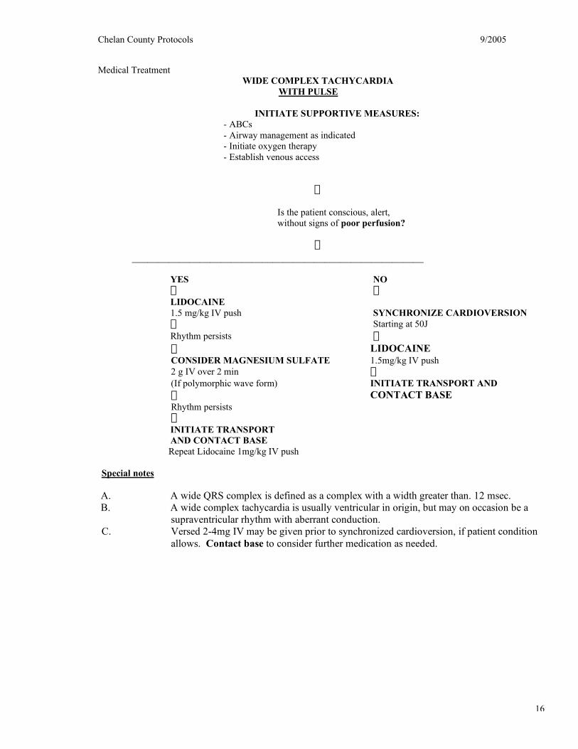
Medical Treatment WIDE COMPLEX TACHYCARDIA WITH PULSE INITIATE SUPPORTIVE MEASURES:
- ABCs - Airway management as indicated - Initiate oxygen therapy - Establish venous access
Ø
Is the patient conscious, alert, without signs of poor perfusion?
Ø
________________________________________________________
YES NO Ø Ø LIDOCAINE 1.5 mg/kg IV push SYNCHRONIZE CARDIOVERSION Ø Starting at 50J Rhythm persists Ø Ø LIDOCAINE CONSIDER MAGNESIUM SULFATE 1.5mg/kg IV push 2 g IV over 2 min Ø (If polymorphic wave form) INITIATE TRANSPORT AND Ø CONTACT BASE Rhythm persists Ø INITIATE TRANSPORT AND CONTACT BASE
Repeat Lidocaine 1mg/kg IV push Special notes
A. A wide QRS complex is defined as a complex with a width greater than. 12 msec. B. A wide complex tachycardia is usually ventricular in origin, but may on occasion be a
supraventricular rhythm with aberrant conduction. C. Versed 2-4mg IV may be given prior to synchronized cardioversion, if patient condition
allows. Contact base to consider further medication as needed.

Chelan County Protocols 9/2005
17
Medical Treatment
INITIATE SUPPORTIVE MEASURES: - ABCs - Airway management as indicated - Initiate oxygen therapy - Establish venous access
Ø
Is the patient conscious, alert, without signs of poor perfusion?
Ø ______________________________________________________
Ø YES Ø OBTAIN 12 LEAD EKG IF AVAILABLE ___________________ Ø may attempt Valsalva maneuver Ø ADENOSINE 6mg IV push and May repeat at 12 mg IV push Ø INITIATE TRANSPORT AND CONTACT BASE:
Special notes
A. A narrow QRS complex is less than .12 sec. in duration. B. Tachycardia is most likely a secondary problem when the pulse is less than 160. Treat hypoxia,
hypovolemia,pain, and other problems first.
NARROW COMIPLEX TACHYCARDIA WITH PULSE
Ø NO Ø
Ø Patient AWARE
Ø Patient UNRESPONSIVE
Ø Ø IMMEDIATE SYNCHRONIZED CARDIOVERSION Starting at 100J Ø INITIATE TRANSPORT AND CONTACT BASE
ADENOSINE 12 mg IV rapid push Ø INITIATE TRANSPORT AND CONTACT BASE

Chelan County Protocols 9/2005
18
Medical Treatment CARDIAC ARREST Specific information needed A. History of arrest: onset, preceding symptoms, bystander CPR, other treatment, duration of arrest B. Past history: diseases, medications C. Surroundings: evidence of drug ingestion, trauma, other unusual presentations Specific objective findings A. Absence of consciousness B. Terminal or no respirations C. Absence of pulse D. Signs of trauma, blood loss E. Air temperature; skin temperature Treatment A. Check surroundings for safety to rescuers. B. Initiate CPR. C. Call for back-up if needed. D. Check rhythm with monitor or quick look paddles; treat rhythm by protocol. Specific precautions
A. Cardiac arrest in a trauma situation is not treated according to this protocol (see Trauma Arrest
protocol). In trauma situation, transport should be rapid, with IV, CPR en route (see Multiple Trauma Overview protocol).
B . Survival from cardiac arrest is related to the time to BOTH BLS and ALS treatment. Don’t forget CPR in the rush for advanced equipment. A call for back-up should be initiated promptly by any BLS unit. Likewise, standing order administration of the first steps in treatment is recommended to minimize time delays to ALS.
D. Large peripheral veins (antecubital or external jugular) are preferred IV sites in an arrest. E. Quick-look paddles preferred for initial rhythm check. Change to patches for more secure
reading. Be sure machine is set to record from whichever mode is in use. F. Be sure to recheck for pulselessness and unresponsiveness upon arrival, even if CPR is in
progress. This will avoid needless and dangerous treatment of "collapsed" patients who are inaccurately diagnosed initially, or who have spontaneous return of cardiac function after an arrhythmia or vasovagal episode.

Chelan County Protocols 9/2005
19
Revised 4/2004 Medical Treatment CHEST PAIN Specific information needed Symptoms: Patient of either gender, more than 20 years old with any of the following chief complaints.
A. Chest Pain Complex:
1. Pressure 2. Tightness, heaviness 3. Radiating in neck, jaw, shoulders, back, one or both arms, left-sided, or both 4. Chest discomfort 5. Chest ache, pressure, stabbing, or squeezing 6. "Elephant on chest" 7. Burning 8. Indigestion or heartburn/nausea and/or vomiting, unexplained indigestion/belching 9. Persistent shortness of breath 10.Weakness/dizziness/lightheadness/loss of consciousness
11. Patient of any age or gender with classic symptoms (acute onset, nausea, diaphoresis, radiation of pain to arm, neck, jaw, to the back)
12.No pain or discomfort; but, patient may experience painless syncope, change in mental status, or dyspnea. Associated symptoms may include nausea, vomiting, diaphoresis, indigestion, belching, etc
13. Cocaine use B. Respiratory
1. Acute onset of shortness of breath 2. Wheezing 3. Significant hemoptysis with shortness of breath
C. Cardiac Surgery * H/X Cardiac Disease 1. CABG 2. Vascular Surgery
Specific objective findings A. Vital signs B. General appearance: color, apprehension, sweating C. Signs of heart failure: neck vein distention, peripheral edema, respiratory distress D. Lung exam by auscultation: rales, wheezes or decreased sounds E. Chest wall tenderness, abdominal tenderness Treatment
A. Reassure and place patient at rest, position of comfort (usually semifowlers). B. Administer 02. C. If patient's history suggests a cardiac origin to the chest pain:
1. Monitor cardiac rhythm 2. Establish venous access 3.Normalize pulse by treating tachycardia > 160 or bradycardia < 50 according to protocols. 4. ASA 240mg chew and swallow. Administration of ASA should not preclude other ACLS and life saving measures. 5. If capable obtain 12 Lead EKG.

Chelan County Protocols 9/2005
20
6. Administer nitroglycerin SL if BP > 100 systolic. Repeat until pain relieved every 5 min up to 3
doses, or systolic BP < 100 (includes patient administered nitroglycerin within last 15 minutes) 7. Administer Lidocaine if PVCs are significant (multifocal PVCs couplets, bigeminyor
trigeminy) a. Lidocaine bolus 1.5 mg/kg; max 3 mg/kg b. If more than 10 minutes out, consider Lidocaine drip 1-2 mg/min.
8. If SBP <90 contact base to consider fluid challenge of NS, and/or vasopressor. 9. If pain persists after third nitroglycerin consider morphine sulfate 2-4 mg IV push, up to 10mg, as
long as SBP >100. Contact base for additional morphine sulfate after first 10 mg have been administered.
D. Enroute to hospital, if EKG indicates acute myocardial infarction, initiate AMI thrombolysis screen
(next page) without delaying patient transport. E. Monitor cardiac rhythm and vitals enroute. F. If the EKG reads AMI, report to the receiving hospital with the following information as soon as
possible. 1. Speak with the physician on duty. 2. Patient age and gender 3. Brief clinical presentation 4. Clearly communicate EKG shows AMI 5. Report any exclusionary criteria for thrombolytic therapy.
Specific Precautions A. Suspicion of an acute MI is based on history. Do not be reassured by a "normal" monitor strip.
Conversely, "abnormal" strips (particularly ST and T changes) can be due to technical factors or nonacute cardiac diseases ST elevation that changes after nitroglycerin administration can be significant and should be documented.
B . Constant monitoring is essential. As many as 50% of patients with acute MI who develop ventricular fibrillation may have no warning arrhythmias.
C. Lidocaine should not be given if: 1. Blood pressure < 90 systolic, or 2. Heart rate < 50, or 3. Periods of sinus arrest or any A-V block are present
D. If patient develops depressed respirations following morphine sulfate administration, be prepared to actively support airway and ventilation or administer naloxone.
E. Remember there are many causes for chest pain. Consider pulmonary embolus, pneumonia, aneurysm, pneurnothorax.
F. Beware of IV fluid overload in the potential cardiac patient. G. Patients who have taken Viagra within the last 48 hours should not be given nitroglycerin.

Chelan County Protocols 9/2005
21
AMI Thrombolysis Screen
Patient Name ________________________
EMS Agency ________________________
1. Identify time of onset of signs and symptoms of acute MI: ____________
Has the chest pain been present at least 30 minutes, but less than 6 hours and unrelieved by nitrates? (circle one)
Yes No
If answer to #1 is Yes, attempt to obtain the following information:
2. CONTRAINDICATIONS:
____Suspected aortic dissection
____ Active GI bleeding ____ Recent stroke (within six months) ____ Major head and/or spinal surgery or trauma (within two months) ____ Recent major surgery, trauma, GI bleeding, or GU bleeding (within 10 days) ____ Brain tumor ____ Known aneurysm ____ Known bleeding tendency ____ Severe uncontrolled high blood pressure (Systolic BP > 200 and/or diastolic >- 120) ____ Pregnancy
3. RELATWE CONTRAFNDICATIONS
____ Active peptic ulcer disease ____ History of CVA, tumor, injury, or brain surgery ____ Significant liver dysfunction ____ Acute pericarditis ____ Major trauma or surgery (within two months) ____ Patients currently receiving oral anticoagulants or with a known bleeding disorder ____ Hypertension (Systolic BP > 180 and/or diastolic BP >100) ____ Subclavian or internal jugular venous cannulation ____ Prolonged or known traumatic CPR ____ Altered level of consciousness ____ Any other condition in which bleeding constitutes a significant hazard or would be particularly difficult to manage because of its location

Chelan County Protocols 9/2005
22
Medical Treatment COMA/ALTERED MENTAL STATUS/NEUROLOGIC DEFICIT Specific Information needed A. Present history: duration of illness, onset and progression of present state; antecedent symptoms such as
headaches, seizures, confusion, trauma, etc. B. Past history: previous medical or psychiatric problems C. Medications: use or abuse D. Surroundings: check for pill bottles, syringes, etc., and bring with patient. Note odor in house. Specific objective findings A. Safety to rescuer: check for gases or other toxins. B. Vital signs C. Level of consciousness and neurological status D. Signs of trauma: head, body E. Breath odor F. Needle tracks G. Medical alert tag Treatment A. Airway: protect as needed (positioning, nasopharyngeal or oropharyngeal airway, suctioning,
intubation). B. Administer02. C. Monitor cardiac rhythm. D. Establish venous access and fluid bolus as indicated. E. Draw appropriate tube and test for blood glucose, if available. F. Administer dextrose 50% (25 gm slow IV), if blood glucose reading < 60, and if clinically indicated. G. Administer naloxone (2 mg IV), if clinically indicated. H. Transport in lateral recumbent position. (If trauma suspected, transport supine with cervical collar
and back-board; logroll as necessary.) I. Monitor vitals during transport. Specific precautions A. Be particularly attentive to airway. Difficulty with secretions, vomiting, and inadequate tidal volume are
common. B. Hypoglycemia may present as focal neurologic deficit or coma (stroke-like picture). C. Coma in the diabetic may be due to hypoglycemia or to hyperglycemia (diabetic ketoacidosis). IV dextrose
should be given to all unconscious diabetics, as well as patients with coma of unknown origin unless a blood glucose reading in the high range is obtained. The treatment may be life-saving in hypoglycemic patient, and will do no harm in the normal or hyperglycemic patient. Do not give oral sugar to an unconscious patient (see Dextrose).
D. Stroke patients may be alert but unable to respond (aphasic); therefore, communicate with the patient and explain what you are doing. Avoid inappropriate comments.
E. Naloxone is useful in any potential overdose situation, but be sure the airway and the patient are controlled before giving naloxone to a known drug addict. The acute withdrawal precipitated in an addict may result in violent combativeness.
F. For patients being treated with narcotics for chronic pain syndromes, use small doses of naloxone (0.1 mg) in order to titrate to improved respirations and mental status. Avoid large doses which may precipitate severe pain in these patients.

Chelan County Protocols 9/2005
23
Revised 4/2004 Medical Treatment HYPERTENSION Specific information needed A. History of hypertension and current medications B. New symptoms: dizziness, nausea, confusion, visual impairment, paresthesia, weakness C. Drug use: phenylpropanolamine(found in a wide variety of over-the-counter anorexiants),
amphetamines, cocaine D. Other symptoms: chest pain, breathing difficulty, abdominal/back pain, severe headache Specific objective finding
A. Evidence of encephalopathy: confusion, seizures, coma, vomiting B. Presence of associated findings: pulmonary edema, neurologic signs, neck stiffness, unequal
peripheral pulses Treatment
A. Administer 02. B. Place patient at rest in position of comfort. C. Recheck BP, with special attention to diastolic pressure, correct cuff size and placement. D. Treat chest pain, pulmonary edema, seizure activity as per usual protocols. E. Establish venous access. F. If diastolic remains above 130 on repeated readings and patient has symptoms of
encephalopathy, chest pain, pulmonary edema, consider. 1. Nitroglycerin sublingual up to 3 doses q 5 minutes. 2. Morphine sulfate 2-5mg IV 3. Contact base if systoms continue and/or BP not improved.
G. Monitor cardiac rhythm. H. Monitor vital signs and mental status during transport. Specific precaution
A. Secondary hypertension (high BP in response to stress or pain) is commonly seen in the field. It
does not require field treatment, and may not even mean the patient has chronic hypertension requiring ongoing treatment.
B. Hypertensive encephalopathy is rare, but can be treated with nitroglycerin or morphine.
Hypertension is more common in association with other problems (pulmonary edema, seizures, chest pain, coma, or altered mental states). It should be managed by treating the other problem, which is usually primary.
C. Diastolic pressures and mean arterial pressures are much more important in determining danger
of severe hypertension than is systolic pressure. These are poorly measured in the field. The diagnosis of "malignant"hypertension is not based on numerical levels, but rather on microscopic changes in blood vessels and damage to organs, which place this disease beyond the scope of pre-hospital care.
D. Don't forget that false elevation of BP can result from a cuff which is too small for the patient.
The cuff should cover 1/3 to 1/2 of the upper arm, and the bladder should completely encircle the arm.
E. Hypertension is seen in severe head injury and intracranial bleeding, and is thought to be a
protective response which increases perfusion. to the brain. Treatment should be directed at the intracranial process, not the blood pressure.

Chelan County Protocols 9/2005
24
Revised 4/2004 Medical Treatment POISONS AND OVERDOSES Specific information needed A. Type of ingestion. What, when, and how much was ingested? Bring the poison, the container,
description of emesis, all medications and everything questionable in the area with the patient to the emergency department.
B. Reason for exposure: think of child neglect, depression, etc. C. Symptoms: respiratory distress, sleepiness, nausea, agitation or decreased level of consciousness D. Past history- medications, diseases E. Action taken by bystanders: induced emesis? "antidote" given? Specific objective findings A. Vital signs B. Airway: patency and adequacy of ventilation C. Level of consciousness and neurologic status: check frequently. D. Breath odor, increased salivation, oral burns E. Skin: sweating, cyanosis F. Systemic signs: vomitus, arrhythmias, lung findings Treatment A. Evaluate and actively support the airway as needed. B. Administer 02. C. Establish venous access. D. Support patient on side and protect airway. E. Test for blood glucose, if available. Administer dextrose 50% (25 gm), IV in secure vein, if
clinically indicated. F. Administer naloxone (2.0 gm) in patients with decreased respiratory effort and observe patient for
improved ventilations. G. Monitor cardiac rhythm. H. May need to administer sodium bicarbonate (I mEq/kg) if widened QRS, or ventricular arrhythmias
on monitor after excessive tricyclic antidepressant(s) ingested. Contact Base in these circumstances.
I. Frequent monitoring of vital signs during transport. Specific precautions A. There are few specific "antidotes." Product labels and home kits can be misleading and dangerous.
Watch the ABCs: these are important. B. Do not neutralize acids with alkalis. Do not neutralize alkalis with acids. These "treatments" cause
heat-releasing chemical reactions which can further injure the GI tract. C. Inhalation poisoning is particularly dangerous to rescuers. Recognize an environment with
continuing contamination and extricate rapidly. D. Organophosphate exposure may require massive doses of atropine (5-10mg). Contact Base for
direction.
Poison Control Phone #: 1-800-732-6985

Chelan County Protocols 9/2005
25
Revised 4/2004 Medical Treatment PSYCHIATRIC/BEHAVIORAL Specific information needed
A., Obtain history of current event, inquire about recent crisis, toxic exposure, drugs, alcohol,
emotional trauma, suicidal ideations. B. Obtain past history; inquire about previous psychiatric and medical problems, medications.
Specific objective findings
A. Evaluate vital signs. B. Note medic alert tags, odor to breath. C. Determine orientation to person, place or time D. Determine ability to relate to reality E. Note hallucinations and behavior.
Treatment
A. Attempt to establish rapport. B. Assure airway. C. If patient refusing transport and is an apparent danger to self or others, contact community mental
health for permission to restrain, if necessary (see Restraints protocol). D. Out of control patients, who are a danger to themselves or others, may be chemically restrained
with haloperidol 2.5 to 5mg IV or IM. E. Monitor vital signs. F. If altered mental status or unstable vital signs:
1. Administer02. 2. Establish venous access. 3. Draw appropriate blood tubes. 4. Consider D50 and Narcan.
Specific precautions
A. Psychiatric patients often have an organic basis for mental disturbances. Beware of hypoglycemia,
hypoxia, head injury, intoxication, or toxic ingestion. B. If emergency treatment is unnecessary, do as little as possible except to reassure while transporting.
Try not to violate the patient's personal space. C. If the situation appears threatening, consider a show of force involving police before attempting to
restrain. D. Beware of weapons. These patients can become very violent. E. If not already available, request police backup and mental health involvement (Chelan-Douglas
Behavioral Health Phone #: 509-662-7105) for patients who are clearly a danger to self or others, if there is any resistance or concern of potential problems. EMS personnel safety, without question, precludes that of the involved parties.

Chelan County Protocols 9/2005
26
Medical Treatment RESPIRATORY DISTRESS
Specific information needed A. History: acute change or injury, slow deterioration, possible foreign body aspiration B. Past history: chronic lung or heart problems or known diagnosis, medications,
home 02, past allergic reactions, recent surgery. C. Associated symptoms- chest pain, cough, fever, hand or mouth paresthesias Specific objective findings
A. Vital signs B. Oxygenation: level of consciousness, cyanosis C. Ventilatory effort: accessory muscle use, forward position, pursed lips D. Neurologic signs: slurred speech, impaired consciousness, evidence of drug/alcohol
ingestion E. Signs of upper airway obstruction: hoarseness, drooling, exaggerated chest wall
movements, inspiratory stridor, inability to talk F. Signs of congestive failure: neck vein distention in upright position, rales, peripheral
edema G. Breath sounds: clear, decreased, rales, wheezing, or rhonchi H. Hives, upper airway edema I. Evidence of trauma: crepitus of neck or chest, bruising, steering wheel damage,
penetrating wounds Treatment
A. Put patient in position of comfort (usually upright). B. Identify and treat upper airway obstruction if present
1. if patient can talk, give reassurance and have him try to cough 2. if awake, but unable to talk, attempt Heimlich maneuver 3. if unsuccessful, suction, nasopharyngeal airway, direct visualization and removal
with forceps, endotracheal intubation C. Administer 02 (high flow for moderate to severe respiratory distress.)
D. Assist ventilation with pocket mask or bag valve mask if necessary. E. Assess and consider treatment for the following problems if respiratory distress is severe
and patient does not respond to proper positioning and administration of 02. 1. Asthma: a. Establish venous access. b. Monitor cardiac rhythm C. Albuterol (2.5 mg GPN) via nebulizer d. Atrovent may be considered especially in older patients (possible COPD)
or critical asthmatics 2. Pulmonary edema (except in patients with evidence of COPD):
a. Sit patient up, legs dangling if possible. b. Establish venous access. C. Monitor cardiac rhythm. d. If SBP > 110, give Nitroglycerin 0.4 mg SL. e. Contact base to consider:
1. Lasix (discuss dose with medical control) 2. Morphine sulfate 2-5 mg/IV.
f. Assist ventilations and consider intubation if patient has altered mentation.

Chelan County Protocols 9/2005
27
3. COPD with deterioration: a. Administer02. b. Monitor cardiac rhythm C. Albuterol via nebulizer (2.5 mg GPN). d. Atrovent via nebulizer e. Establish venous access.
4. Pneumothorax: watch for signs of tension. If patient deteriorating rapidly, consider chest decompression.
F. If diagnosis unclear, place patient in position of comfort, and administer oxygen, transport.
G. Prepare to assist ventilations if patient fatigues or develops altered mentation, or if respiratory arrest occurs.
Specific precautions
A. Consider cardiac ischemia in diabetic and elderly patients B. Wheezing in older persons may be due to pulmonary edema ("cardiac asthma"). C. Don’t overdiagnose “hyperventilation” in the field which could cause you to miss
pulmonary emboli or other serious problems. Treatment with oxygen will not harm the hyperventilator.
D. Use epinephrine very cautiously in patients > 35 years and/or known coronary artery disease.

Chelan County Protocols 9/2005
28
Medical Treatment SEIZURES Specific information needed A. Seizure history: onset, time interval, previous seizures, type of seizure B. Medical history: especially head trauma, diabetes, headaches, drugs, alcohol, medications, pregnancy. Specific objective finding A. Vital signs B. Description of seizure activity C. Level of consciousness D. Head and mouth trauma E. Incontinence F. Air temperature; patient temperature G. Skin color and moisture Treatment A. Airway: ensure patency - nasopharyngeal airways are useful. NOTE: Do not force anything between the
teeth. B. Administer 02. C. Suction as needed. D. Protect patient from injury. E. Check pulse immediately after seizure stops. F. Keep patient on side. G. Establish venous access. H. Administer diazepam 5-10mg IV for ongoing seizure. Repeat in 3-4 minutes for continued seizure activity.
This drug may be also given PR, IO, or ET using double the IV dose. I. Draw appropriate tube; test for blood glucose if available. J. Administer dextrose 5 0% (25 gm), if clinically indicated. K. Administer naloxone (2.0 mg), if clinically indicated. L. Monitor cardiac rhythm. M. Keep in lateral recumbent position for transport. N. Monitor vitals. Specific precautions A. Move hazardous materials away from patient. Restrain the patient only if needed to prevent injury. Protect
patient's head. B. Trauma to tongue is unlikely to cause serious problems, however, trauma to teeth may. Attempts to force
an airway into the patient's mouth can completely obstruct airway. Do not use bite sticks. C. Seizure can be due to lack of glucose or oxygen to the brain, as well as to the irritable focus we associate
with epilepsy. Hypoxia from transient arrhythmia or cardiac arrest (particularly in younger patients) may cause seizure and should be treated promptly. Don't forget to check for pulse once a seizure terminates.
D. Hypoxic seizures can also be caused by simple faint, either when the tongue obstructs the airway in the supine position, or when overly helpful bystanders prop the patient or elevate the head prematurely.
E. Alcohol related seizures are common, but cannot be differentiated from other causes of seizure in the field. Assessment in the intoxicated patient should still include consideration of hypoglWemia and all
other potential causes. Field management is as for any seizure. F. Seizures may be due to arrhythmias or stroke. Of these, arrhythmia is the most important to recognize in
the field. G. Medical personnel are often called to assist epileptics who seize in public. If patient clears completely, is
taking his/her medications, has his/her own physician, and is experiencing his/her usual frequency of seizures, transport may be unnecessary. Consult your base physician.
H. Lorazepam has a tendency to decrease respiratory effort. I. Seizures in pregnant patients (or even those who are recently delivered) may be the presenting sign of
eclampsia or toxemia of pregnancy. Seizures in those patients will respond better to administration of magnesium sulfate.

Chelan County Protocols 9/2005
29
Medical Treatment SHOCK: MEDICAL Specific information needed
A. Onset: gradual or sudden; precipitating cause or event B. Associated symptoms: itching, peripheral or facial edema, thirst, weakness, respiratory distress,
abdominal or chest pain, dizziness on standing C. History: allergies, medications, bloody vomitus or stools, significant medical diseases, history of
recent trauma, last menstrual period, vaginal bleeding, fever Specific objective finding
A. Vital signs: pulse > 120 (occasionally< 50); BP < 90 systolic C. Mental status: apathy, confusion, restlessness, combativeness D. Skin: flushed, pale, sweaty, cool or warm, hives, or other rash E. Signs of trauma F. Signs of cardiogenic shock: jugular venous distention in upright position, rales, peripheral edema
Treatment
A. Administer 02. B. Cover patient to avoid excess heat loss. Do not over bundle. C. Assess for cardiogenic cause:
1. If P > 150, treat tachyarrhythmia according to protocol. 2. If P < 60, treat bradyarrhythmia according to protocol. 3. If distended neck veins, chest pain, or other evidence of cardiac cause:
a. Position of comfort b. Be prepared to assist ventilations or initiate CPR. c. Establish venous access. d. Monitor cardiac rhythm. e. Evaluate for possible tension pneumothorax. Treat as appropriate - see
protocol. D. Consider dopamine. Begin 5 mcg/kg/min and titrate to SBP of 100 E. Transport rapidly for definitive diagnosis and treatment. F. If no evidence of cardiogenic cause, institute general treatment measures:
1. Place patient supine, elevate legs 10- 12 inches. (If respiratory distress results, leave patient in position of comfort.)
2. Fluid bolus: IV, NS. G. Assess and treat for specific cause, such as anaphylaxis, if this can be determined.
H. Monitor VS, cardiac rhythm, and level of consciousness during transport Specific precautions A. Shock in a cardiac patient may be caused by hypovolemia; however, contact should be made
with base prior to administering fluid boluses. B. Mixed forms of shock are treated as hypovolemia, but the other factors contributing to the low
perfusion should be considered. Neurogenic shock is caused by relative hypovolemia as blood vessels lose tone, either from spinal cord trauma, drug overdose, or sepsis. Cardiac depressant factors can also be involved. Anaphylaxis is a mixed form of shock with hypovolemic, neurogenic, and cardiac depressant components. Epinephrine is used in addition to fluid load.
C. Cardiogenic shock from various causes is difficult to treat even in a hospital setting. Rapid transport is recommended.

Chelan County Protocols 9/2005
30
SHOCK: MEDICAL Mechanism/Causes Differential/Symptoms HYPOVOLEMIA Dehydration suggestive illness Vomiting, diarrhea Diabetes with hyperglycemia Diabetes; acute illness, increased urine or blood loss, thirst, fever Ectopic pregnancy female, 12-50 years, abdominal pain GI bleed vomitus: black or red stool Ruptured abdominal aneurysm severe back/abdomen pain, age, history of high blood pressure Vaginal bleeding suggestive history, miscarriage, abortion or delivery Intra-abdominal bleeding minor trauma, abdomen, back or shoulder pain CARDIOGENIC .....
X Arrhythmia palpitations Pericardial tamponade chest area cancer, blunt or penetrating trauma Tension pneumothorax respiratory distress, COPD, trauma Myocardial failure chest pain, history of congestive failure Pulmonary embolus sudden respiratory distress, chest pain MIXED
Sepsis symptoms fever, elderly, urinary symptoms Drug overdose suggestive history Anaphylaxis SOB, itching, mouth swelling, dizziness, exposure to allergen

Chelan County Protocols 9/2005
31
Medical Treatment SYNCOPE Reviewed 3/2004 Specific information needed
A. History of the event onset, duration, seizure activity, precipitating factors. Was the patient
sitting, standing or lying? Pregnant? B. Past history: medications, diseases, prior syncope C. Associated symptoms: dizziness, nausea, chest or abdominal/back pain, headache, palpitations
Specific objective findings
A. Vital signs B. Neurological status: level of consciousness, residual neurological deficit C. Signs of head trauma, mouth trauma, incontinence D. Neck stiffness
Treatment
A. Position of comfort, do not sit patient up prematurely; supine or lateral positioning if not
completely alert. B. Cardiac monitor. C. Establish venous access. D. Monitor vital signs and level of consciousness closely for changes or recurrence; if abnormal
administer O2. E. If signs of hypoglycemia are present: 1. Determine blood glucose or obtain appropriate blood tubes. 2. Administer IV bolus of dextrose 50% (25 gm) if indicated. F. Treat cardiac arrhythmias and shock per appropriate protocols.
Specific precautions
A. Syncope is by definition a transient state of unconsciousness from which the patient has
recovered. If the patient is still unconscious, treat as Coma. If the patient is confused, treat according to Coma/Altered Mental Status/Neurologic Deficit protocol.
B. Most syncope is vasovagal, with dizziness progressing to syncope over several minutes. Recumbent position should be sufficient to restore vital signs and level of consciousness to normal.
C. Syncope which occurs without warning or while in a recumbent position is potentially serious, and often caused by arrhythmia.
D. Patients over the age of 40 with syncope, even though apparently normal, should be transported In middle-aged or elderly patients, syncope can be due to a number of potentially serious problems. The most important of these to monitor and recognize are: arrhythmias, occult GI bleeding, seizure, or ruptured abdominal aortic aneurysm.
E. Any elderly patient with syncope and back pain should be considered to have a ruptured abdominal aortic aneurysm until proven otherwise.

Chelan County Protocols 9/2005
32
Revised 4/2004
Medical Treatment VOMITING Specific information needed A. Frequency, duration of vomiting B. Presence of blood in vomitus C. Associated symptoms: abdominal pain, weakness, confusion D. Medication ingestion E. Past medical history: diabetes, cardiac disease,, abdominal problems, alcoholism
Specific objective finding
A. Vital signs B. Color of vomitus: presence of blood C. Abdomen: tenderness, guarding, rigidity, distention D. Signs of dehydration poor skin turgor, dry mucous membranes, confusion Treatment
A. Position patient: left lateral recumbent if vomiting; otherwise, supine. B. Administer02. C. Nothing by mouth D. If BP < 90 systolic and signs of hypovolemic shock:
1. Elevate legs 10- 12 inches. 2. Establish venous access. 3. Fluid bolus: 20cc/kg NS up to 1,000cc; if history of CHF, 500cc only.
E. Monitor vital signs during transport. F. For patients with intractable vomiting, consider phenergan 12.5 – 25mg IV. Specific precautions
A. Vomiting maybe a symptom of a more serious problem. The most serious causes are GI bleed
or other intra-abdominal catastrophe. A rare cardiac patient may also present with vomiting as the predominant symptom.
B. Consider drug overdose; a patient who does not call the ambulance for medication ingestion may call later when GI symptoms become severe.
C. The vast majority of persons with vomiting have become sick over days, not minutes. Unless severely ill, they do not require lights-and-siren transport or advanced field treatment.
D. Dehydration may be particularly severe in children with simple vomiting. IVs may be very difficult to start particularly with infants.

Chelan County Protocols 9/2005
33
SECTION II TREATMENT PROTOCOLS: TRAUMA
TABLE OF CONTENTS
II Treatment Protocols: Trauma Treatment Page Number Multiple Trauma Overview 34,35 Algorithm: Trauma 36
WA State Pre-hospital Trauma Triage Algorithm 37 Abdominal Trauma 38
Amputations 39
Burns 40,41
Chest Injury 42,43
Extremity Injuries 44,45 Eye Injuries 46
Face and Neck Trauma 47,48
Head Trauma 49,50
Spinal Trauma 51
Trauma Arrest 52

Chelan County Protocols 9/2005
34
Section II - Trauma 04/2004
MULTIPLE TRAUMA OVERVIEW
Specific information needed A. Mechanism of injury
1. Cause, precipitating factors, weapons used 2. Trajectories and forces involved to patient 3. For vehicular trauma: condition of vehicle, windshield, steering wheel, compartment
intrusion, type and use of seatbelts. Specific description of mechanism (i.e. auto-pole, rollover, auto-pedestrian, etc…)
4. Helmet use, if motorcycle or bicycle B. Patient complaint(s) C. Initial position and level of consciousness of patient from witnesses, first responders D. Patient movement and treatment since injury E. Other factors such as drugs, alcohol, medications, diseases, pregnancy Specific objective findings A. Scene evaluation
1. Note potential hazard to rescuers and patient 2. Identify number of patients; organize triage operations if appropriate 3. Observe position of patient, surroundings, probable mechanism, vehicle condition
B. Patient evaluation (see treatment below) Treatment Initial assessment in multiple trauma is performed at the same time as treatment A. Airway with C-spine immobilization B. Breathing C. Circulation, with control of major bleeding D. After the primary survey addresses the ABC’s and the patient is immobilized, other treatment,
if any, should occur enroute to the hospital. This includes IV placement, attaching cardiac monitor and the secondary assessment.
E. Transport to the closest, appropriate facility. F. Monitor vital signs, neurologic status and cardiac rhythm enroute. G. Contact base.
Chelan County Protocols 9/2005
35
Chelan County Paramedic Protocols Section II - Trauma 04/2004
MULTIPLE TRAUMA OVERVIEW CONT…
Specific precautions A. Assessment and management of trauma in the field has changed considerably in recent years. We
now understand that there are patients who cannot tolerate a full assessment before lifesaving intervention is needed. Likewise, splinting, bandaging, and even the secondary survey are luxuries which may need to be bypassed in the critical patient. The need for blood, x-rays, operating rooms and even specialized treatment centers makes time and in-hospital resources critical elements in resuscitation. For the special category of severely injured patients, the enlightened “load and go” is more appropriate than either the extended stabilization or the old “grab and run” with no medical stabilization.
B. Critical injuries involve: 1. Difficulty with respiration 2. Difficulty with circulation (shock) 3. Decreased level of consciousness *Any trauma patient with one or more of these conditions is a “load and go”.
C. Even in the non-critical patient with significant injury, “stabilization in the field” does not occur. With major injuries, the very most you can do is to buy time. If the initial bolus of fluids results in improved vitals, do not become complacent. This patient frequently needs blood and an operating room to truly “stabilize” the traumatic process. Rapid transport is still of the highest priority.
D. Serial vital signs and observations of neurologic status in the field are critical. E. The trauma patient is probably the greatest risk to the rescuer for exposure to “bodily fluids”. F. The goal should be a scene time less than, or equal to, 10 minutes. G. Contact base as soon as possible when critical patients are involved; then call again when more information (e.g. vital signs) is available.
Chelan County Protocols 9/2005
36
Chelan County Paramedic Protocols Section II - Trauma 04/2004
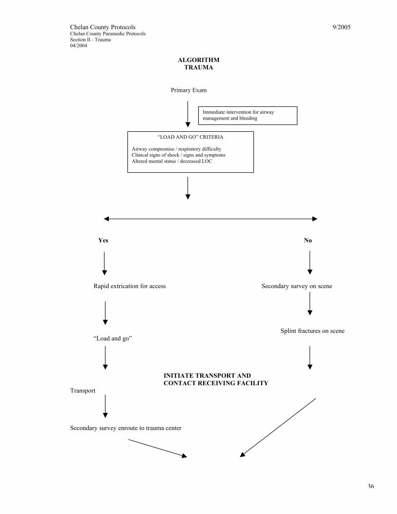
ALGORITHM
TRAUMA
Yes No
Rapid extrication for access Secondary survey on scene
Splint fractures on scene “Load and go”
INITIATE TRANSPORT AND CONTACT RECEIVING FACILITY Transport
Secondary survey enroute to trauma center
Immediate intervention for airway management and bleeding
“LOAD AND GO” CRITERIA Airway compromise / respiratory difficulty Clinical signs of shock / signs and symptoms Altered mental status / decreased LOC
Primary Exam

Chelan County Protocols 9/2005
37
Repeat exam if any change in condition or after any interventions

Chelan County Protocols 9/2005
38
See Appendix A: Washington State Trauma Triage Tool Algorithm
Chelan County Protocols 9/2005
39
Chelan County Paramedic Protocols Section II - Trauma 04/2004
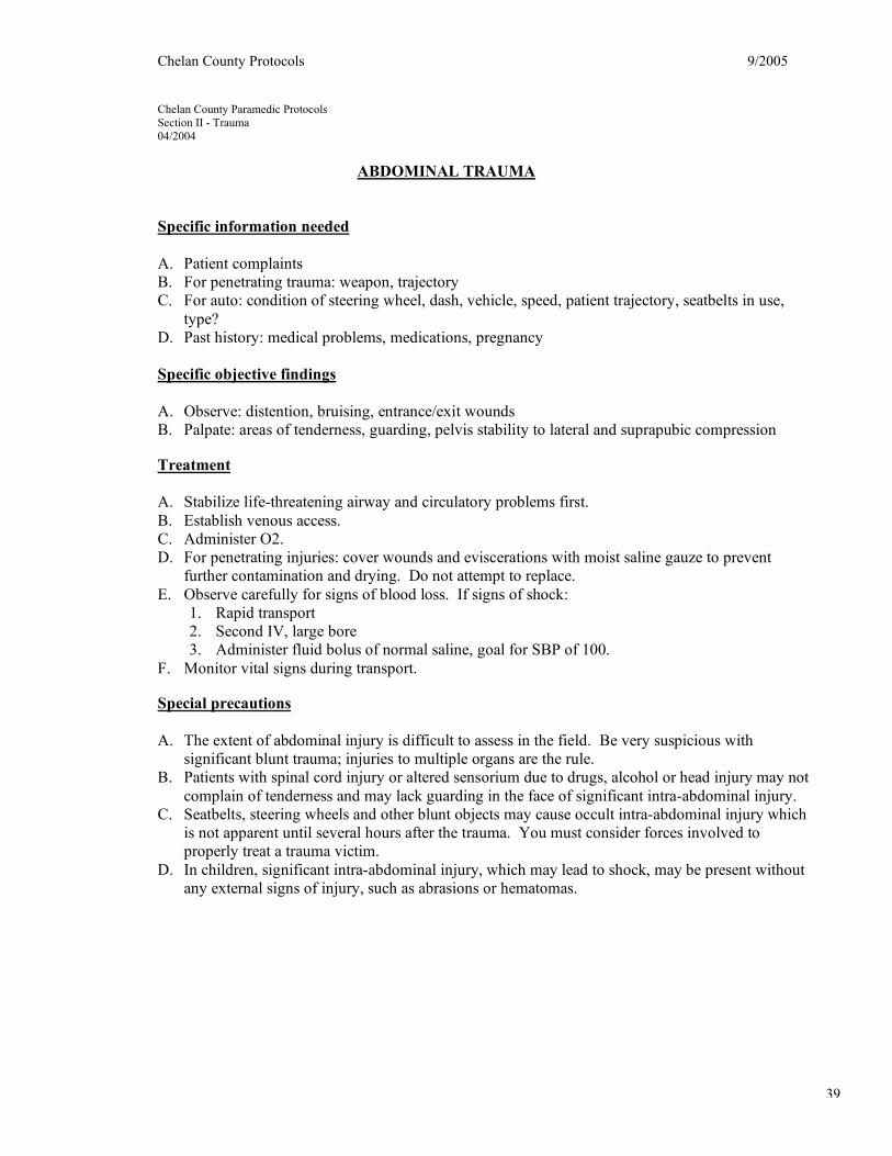
ABDOMINAL TRAUMA
Specific information needed A. Patient complaints B. For penetrating trauma: weapon, trajectory C. For auto: condition of steering wheel, dash, vehicle, speed, patient trajectory, seatbelts in use,
type? D. Past history: medical problems, medications, pregnancy Specific objective findings A. Observe: distention, bruising, entrance/exit wounds B. Palpate: areas of tenderness, guarding, pelvis stability to lateral and suprapubic compression Treatment A. Stabilize life-threatening airway and circulatory problems first. B. Establish venous access. C. Administer O2. D. For penetrating injuries: cover wounds and eviscerations with moist saline gauze to prevent
further contamination and drying. Do not attempt to replace. E. Observe carefully for signs of blood loss. If signs of shock:
1. Rapid transport 2. Second IV, large bore 3. Administer fluid bolus of normal saline, goal for SBP of 100.
F. Monitor vital signs during transport. Special precautions A. The extent of abdominal injury is difficult to assess in the field. Be very suspicious with
significant blunt trauma; injuries to multiple organs are the rule. B. Patients with spinal cord injury or altered sensorium due to drugs, alcohol or head injury may not
complain of tenderness and may lack guarding in the face of significant intra-abdominal injury. C. Seatbelts, steering wheels and other blunt objects may cause occult intra-abdominal injury which
is not apparent until several hours after the trauma. You must consider forces involved to properly treat a trauma victim.
D. In children, significant intra-abdominal injury, which may lead to shock, may be present without any external signs of injury, such as abrasions or hematomas.

Chelan County Protocols 9/2005
40
Chelan County Paramedic Protocols Section II - Trauma 04/2004
AMPUTATIONS
Specific information needed A. History: time and mechanism of amputation, care for severed part prior to rescuer arrival B. Past history: medications, bleeding tendencies, medical problems Specific objective findings A. Vital signs B. Other injuries C. Blood loss at scene D. Structural attachment in partial amputations if identifiable Treatment A. Resuscitate and treat other more urgent injuries. B. Control hemorrhage with direct pressure and elevation. C. If hypotension or signs of shock:
1. Establish venous access 2. Fluid bolus: NS (10-20 cc/kg) 3. Contact base
D. Patient: gently cover stump with sterile dressing, saturate with sterile saline, cover with dry dressing, elevate E. Severed part: wrap in sterile gauze - preserving all amputated material, moisten with sterile saline, place in watertight container (specimen cup, plastic bag, etc…). Place container in cooler with ice (do not freeze). F. Consider morphine sulfate 2-4 mg IV as needed for pain, up to 10 mg (0.05-0.1 mg/kg for
peds). Hold if SBP < 100. G. Contact base for additional morphine. Specific precautions A. Partial amputations should be dressed and splinted in alignment with extremity to ensure optimum blood flow. Avoid torsion in handling and splinting. B. Do not use dry ice to preserve severed part. C. Control all bleeding by direct pressure only to preserve tissues. The most profuse bleeding may occur in partial amputations, where cut vessel ends cannot retract to stop bleeding. Avoid tourniquet if at all possible. Never clamp bleeding vessels. D. Many factors enter into the decision to attempt reimplantation (age, location, condition of tissues, other options). A decision regarding treatment cannot be made until the patient and part have been examined by a physician; this may not be made at the primary care hospital. Try to help the family and patient understand this and do not falsely elevate hopes.

Chelan County Protocols 9/2005
41
Chelan County Paramedic Protocols Section II - Trauma 04/2004
BURNS Specific information needed A. History of injury: time elapsed since burn. Was patient in a closed space with steam or smoke? Electrical contact? Loss of consciousness? Accompanying explosion, toxic fumes, other possible trauma? B. Past history: prior cardiac or pulmonary disease, medications? Specific objective findings A. Vital signs B. Extent of burns: describing or diagram of areas involved C. Depth of burns: superficial-erythema only, partial or full thickness – blistered or charred areas. (Call-in description should include extent of burn only: one patient
palm = 1% burn). D. Evidence of CO poisoning or other toxic inhalation: altered mental state, headache, vomiting, seizure, coma E. Evidence of inhalation burns: respiratory distress, cough, hoarseness, singed nasal or
facial hair, soot or erythema of mouth. F. Entrance and exit wounds for electrical burns. G. Associated trauma Treatment A. Thermal burns
1. Remove clothing which is smoldering or which is non-adherent to the patient. 2. Administer O2 if indications from history or physical of respiratory burns, toxic
inhalation, or significant flame or smoke exposure. 3. Assess and treat for associated trauma (blast or fall). 4. Remove rings, bracelets and other constricting items. 5. If significant burn is moderate-to-severe (over 10% of body surface area), cover
wounds with dry clean dressings to avoid hypothermia. 6. Use cool, wet dressings in smaller burns (less than 10% of body surface area)
for patient comfort. 7. Establish venous access. 8. Consider morphine sulfate for pain relief 2-4 mg IV up to 10 mg (0.05 – 0.1 mg/kg in peds). Hold if SBP < 100. 9. Contact base for additional morphine. 10. Transport, monitor vital signs 11. Watch for airway edema and be prepared to intubate.
B. Inhalation injury
1. Administer O2 by NRB during full time of transport. 2. Be prepared to ventilate or assist if respirations inadequate. 3. Monitor cardiac rhythm.

Chelan County Protocols 9/2005
42
Chelan County Paramedic Protocols Section II - Trauma 04/2004
BURNS CONT…
C. Chemical Burns 1. Protect rescuer from contamination. Wear appropriate gloves and clothing. 2. Remove all clothing and any solid chemical which provide continuing
contamination. 3. Assess and treat for associated injuries. 4. Decontaminate the patient using running water for 15 minutes prior to transport
if patient stable. 5. Check eyes for exposure and rinse with free-flowing water for 15 minutes. 6. Evaluate for systemic symptoms which might be caused by chemical
contamination. Contact base for possible treatment. 7. Remove rings, bracelets and constricting bands. 8. Wrap burned area in clean, dry cloths for transport. Keep patient as warm as
possible after decontamination. 9. Consider morphine sulfate for pain relief 2-4 mg IV up to 10 mg (0.05 – 0.1 mg/kg in peds). Hold if SBP < 100. 10. Contact base for additional morphine 11. Contact base for special treatment or procedures as needed.
D. Electrical Burns 1. Protect rescuers from continued live electric wires or source. 2. Separate victim from electrical source when area safe for rescuers. 3. Initiate CPR as needed. Treat defibrillation as usual. 4. Prolonged respiratory support may be needed. 5. Immobilize cervical spine, assess for other injuries. 6. Monitor patient for possible arrhythmias. Treat as per Arrhythmia protocol. 7. Establish venous access. 8. Consider morphine sulfate for pain relief 2-4 mg IV up to 10 mg (0.05 – 0.1 mg/kg in peds). Hold if SBP < 100. 9. Contact base for additional morphine
Specific precautions A. Leave blisters intact when possible B. Suspect airway burns in any facial burns received in closed places. Edema may
become severe, but not be immediately apparent. Avoid unnecessary trauma to the airway. Humidified O2 is useful, if available.
C. Assume carbon monoxide poisoning in all closed space burns. Treatment is 100% O2 continued for several hours. In addition, other toxic products of combustion are more commonly encountered than realized. D. Consider suicide attempt as cause of burn, and child abuse in pediatric burns. E. Lightening injuries can cause prolonged respiratory arrest. Prompt, continuous
respiratory assistance (sometimes for hours to days) can result in full recovery. F. Notify hospital of chemical contamination immediately to mobilize internal resources. G. EMS personnel should not participate in decontamination unless trained and equipped to do so.

Chelan County Protocols 9/2005
43
Chelan County Paramedic Protocols Section II - Trauma 04/2004
CHEST INJURY
Specific information needed A. Patient complaints: chest pain, type, respiratory distress, neck pain, other areas of injury B. Mechanism: amount of force involved (particularly deceleration), speed of impact, seatbelt use / type C. Penetrating trauma: size of object, caliber of bullet D. Past medical history: medications, prior medical problems Specific objective findings A. Observe: wounds, air leaks, chest wall movement, neck veins B. Palpate: tenderness, crepitus, tracheal position, tenderness on sternal compression, pulse pressure C. Auscultate: breath sounds, heart sounds (quality) D. Surroundings: vehicle, steering wheel condition, compartment intrusion Treatment A. Clear and open airway. Stabilize neck if indicated. B. Assist breathing if patient is apneic or respirations depressed. C. Administer O2. D. If penetrating injury present, transport rapidly with further stabilization enroute. E. For open chest wound with air leak, use Vaseline-type gauze or occlusive dressing taped on three sides only to allow air to escape but not enter the chest. F. Observe chest for paradoxical movements. G. Intubate for respiratory insufficiency. H. Control exsanguinating hemorrhage with direct pressure. I. Obtain baseline vital signs, neurologic assessment (be sure to check lower extremity PMS). J. Morphine sulfate 2-4 mg IV (0.05-0.1 mg/kg peds) up to 10 mg. Hold if SBP <100. K. Evaluate neck veins and blood pressure.
1. If neck veins flat and patient in shock, transport rapidly and treat hypovolemia enroute. a. Establish venous access b. Fluid bolus: IV, NS (10-20 cc/kg) c. Monitor cardiac rhythm d. Contact base.
2. If patient in shock with neck veins distended, also transport rapidly and consider: a. Tension pneumothorax if respiratory and circulatory status markedly
compromised with clinical findings of pneumothorax. 1.) Release occlusive dressings on open chest wounds. 2.) Needle decompression.
b. Pericardial tamponade, if mechanism of injury suspicious (may have distant heart sounds, narrow pulse pressure).
1.) Establish venous access.

Chelan County Protocols 9/2005
44
2.) Fluid bolus: IV, NS (10-20 cc/kg).

Chelan County Protocols 9/2005
45
Chelan County Paramedic Protocols Section II - Trauma 04/2004
CHEST INJURY CONT…
c. Cardiac contusion with typical ischemic chest pain or severe chest wall contusion.
1.) Monitor cardiac rhythm. 2.) Lidocaine IV (1.5 mg/kg) for significant PVC’s (see Chest Pain
Protocol). 3. If patient stable without signs or symptoms of shock:
a. Complete secondary survey. b. If significant injury present:
1.) Establish venous access. 2.) Monitor cardiac rhythm enroute. 3.) Lidocaine IV (1.5 mg/kg) for significant PVC’s (see Chest Pain
Protocol). L. Immobilize impaled objects in place with dressings to prevent movement. M. Monitor vital signs and level of consciousness every five minutes with significant injury. Specific precautions A. Chest trauma is treated with difficulty in the field and prolonged treatment before transport is
not indicated if significant injury is suspected. If patient is critical, transport rapidly and avoid treatment of non-emergent problems at the scene. Penetrating injury particularly should receive immediate transport with minimal interventions in the field.
B. Consider medical causes of respiratory distress such as asthma, pulmonary edema or COPD that have either caused trauma or been aggravated by it.
C. Chest injuries sufficient to cause respiratory distress are commonly associated with significant blood loss. Look for hypovolemia.
D. Myocardial contusion can occur, particularly with anterior chest wall injury, as from a steering wheel. Pain is similar to myocardial infarct pain. Monitor the patient and treat arrhythmias as in a medical patient, but think first of hypoxia and hypovolemia as potential causes of arrhythmias.
E. Do not forget to check the back for injuries, especially the patient in shock, where a cause is not evident (check the entire back, axillary region and base of neck).
F. Significant intrathoracic injuries can exist without any external signs of injury.

Chelan County Protocols 9/2005
46
Chelan County Paramedic Protocols Section II - Trauma 04/2004
EXTREMITY INJURIES
Specific information needed A. Mechanism of injury: direction of forces, if known B. Areas of pain or limited movement C. Treatment prior to arrival: realignment of open or closed fracture, movement of patient D. Past medical history: medications, medical illnesses Specific objective findings A. Vital signs B. Observe: localized swelling, discoloration, angulation, lacerations, exposed bone fragments, loss of function, guarding C. Palpate: tenderness, crepitus, instability, quality of distal pulses, sensation D. Note estimated blood loss at scene. Treatment A. Treat airway, breathing, and hypotension as first priorities (see Multiple Trauma Overview). B. Immobilize cervical spine when appropriate. C. Examine for additional injuries to head, face, chest, and abdomen; treat those problems with higher priority first. D. If patient unstable, transport rapidly, treating life-threatening problems enroute. Splint patient by securing to long back board to minimize fracture movement. E. If patient stable, or isolated extremity injury exists:
1. Check distal pulses and sensation prior to immobilization of injured extremity. 2. Apply sterile dressing to open fractures. Note carefully wounds that appear to communicate with
bone. 3. Splint areas of tenderness or deformity: apply gentle traction throughout treatment and try to
immobilize the joint above and below the injury in the splint. 4. Realign fractures / dislocations by applying gentle axial traction if indicated:
a. To restore circulation distally b. To immobilize adequately (i.e. realign femur fracture)
5. Check distal pulses and sensation after reduction and splinting. 6. Elevate simple extremity injuries. Apply ice if time and extent of injuries allow. 7. Monitor circulation (pulse and skin temperature), sensation and motor function distal to site of injury during transport. 8. Establish venous access. 9. Consider morphine sulfate 2-4mg IV (0.05 – 0.1mg/kg for peds) up to 10mg. Hold if SBP <100. 10. Contact base for additional morphine.

Chelan County Protocols 9/2005
47
Chelan County Paramedic Protocols Section II - Trauma 04/2004
EXTREMITY INJURIES CONT…
Special precautions A. Patients with multiple injuries have a limited capacity to recognize areas which have been injured. A patient with a femur fracture may be unable to recognize that he has other areas of pain. Be
particularly aware of missing injuries proximal to the obvious ones (e.g. hip dislocation with a femur fracture, or a humerus fracture with a forearm fracture).
B. Do not use ice or cold packs directly on skin or under air splints. Pad with towels or leave cooling for hospital setting.
C. Do not attempt to realign dislocations in the field unless circulation is compromised. Splint in the position of comfort. D. Injuries around joints may become more painful and circulation may be lost with attempted realignment. If this occurs, stabilize the limb in the position of most comfort with the best distal
circulation.

Chelan County Protocols 9/2005
48
Chelan County Paramedic Protocols Section II – Trauma 10/2004
EYE INJURIES Specific information needed
A. History of injury: time injury occurred, cause of injury B. If chemical exposure, type of chemical. C. Is there a foreign body sensation? D. Does the patient notice any vision changes or light sensitivity? E. Is there a history of corrective lenses, eye surgery, or glaucoma?
Specific objective findings
A. May have visible foreign body or visible globe laceration B. Assess for pupillary irregularity and reactivity C. Assess gross visual acuity, eg. counting fingers
Treatment
A. Assure ABC’s. Identify and treat any life or limb threatening injuries first. B. If chemical exposure or a non-penetrating foreign body in the eye:
1.) Instill 2 drops of tetracaine (0.5% solution) ophthalmic drops into the affected eye. 2.) Flush immediately with copious amounts of sterile normal saline and continue
flushing enroute. C. If there is an impaled object, stabilize it in place. Cover non-affected eye as well. D. If there is evidence of a penetrating eye injury such as visible globe laceration or fluid draining
from globe, then place a shield over the eye and secure in place. Do not press on the globe, or allow any pressure to be applied to the globe; such as from a dressing.
Specific precautions
A. Do not use tetracaine ophthalmic drops for patients who are allergic to “caine” class of local anesthetics.
B. Warn the patient not to rub the eye while the cornea is anesthetized, since this may cause corneal abrasion and greater discomfort when the anesthesia wears off.
C. If transport time exceeds 20 minutes, a second dose of tetracaine may be given at that time if needed.
D. As a general rule, you should not attempt to remove contact lenses in patients with eye injuries. However, in cases of chemical exposure, contact base to discuss removal of the lenses if patient is unable to do so.

Chelan County Protocols 9/2005
49
Chelan County Paramedic Protocols Section II - Trauma 04/2004
FACE AND NECK TRAUMA Specific information needed A. Mechanism of injury: impact to steering wheel, windshield or other objects, clothesline-type injury to face or neck. B. Management before arrival by bystanders, first responders C. Patient complaints: areas of pain, trouble with vision, hearing, neck pain, abdominal bite, short of breath D. Past medical history: medications, medical illnesses Specific objective findings A. Vital signs B. Airway: jaw or tongue instability, loose teeth, vomitus or blood in airway, other evidence of impairment or obstruction C. Neck: tenderness, crepitus, hoarseness, bruising, swelling, stridor D. Blood or drainage from ears or nose E. Level of consciousness, evidence of head trauma F. Injury to eye: lid laceration, blood anterior to pupil, abnormal pupil, abnormal globe position Treatment A. Control airway with C-spine immobilization if indicated:
1. Open airway using jaw thrust, keeping neck in alignment with in-line cervical immobilization.
2. Use finger sweep to remove oral foreign bodies. 3. Suction blood and other debris. 4. Stabilize tongue and mandible with chin lift. Manual traction of the tongue may be
necessary to keep posterior pharynx open as needed. 5. Note evidence of laryngeal injury and transport immediately if signs present. 6. If intubation cannot be performed due to severe facial injury, attempt to manage with
suctioning and bag-valve mask. 7. If necessary, consider cricothyrotomy.
B. Support breathing as needed. C. Administer O2. D. Stop hemorrhage; check pulse and circulation. E. Establish venous access:
1. TKO if stable 2. With signs of hypovolemia: fluid bolus, further fluids as directed.
F. Obtain vital signs, assess neurologic status. G. Complete secondary survey if no life-threatening injuries present. H. Consider morphine sulfate 2 – 4mg IV up to 10 mg (0.05 – 0.1mg/kg for peds). Hold if SBP <100. I. Contact base for additional morphine. J. Cover injured eyes with protective shield or cup (avoid pressure or direct contact to eye). K. Do not attempt to stop free drainage from ears or nose. Cover lightly with dressing to avoid contamination. L. Bring avulsed teeth with you. Keep moist in saline-soaked gauze. M. Monitor airway closely during transport for development of obstruction or respiratory distress.

Chelan County Protocols 9/2005
50
Suction and treat as needed.

Chelan County Protocols 9/2005
51
Chelan County Paramedic Protocols Section II - Trauma 04/2004
FACE AND NECK TRAUMA CONT…
Specific precautions A. Fracture of the larynx should be suspected in patients with respiratory distress, abnormal voice,
and history of direct blow to neck from steering wheel, rope, fence wire, etc… Both intubation and cricothyrotomy may be unsuccessful in the patient with a fractured larynx, and attempts may precipitate respiratory arrest. Transport rapidly for definitive treatment if you suspect this potentially lethal injury. Do not attempt intubation or cricothyrotomy unless the patient is in severe respiratory distress.
B. Airway obstruction is the primary cause of death in persons sustaining head and face trauma. Meticulous attention to suctioning, and stabilization of tongue and mandible may be the most important treatment rendered.
C. Remember that the apex of the lung extends into the lower neck and may be injured in penetrating injuries of the lower neck, resulting in pneumothorax or hemothorax.
D. Do not be concerned with contact lens removal in the field.

Chelan County Protocols 9/2005
52
Chelan County Paramedic Protocols Section II - Trauma 04/2004
HEAD TRAUMA Specific information needed A. History: mechanism of injury, estimate of force involved; with motorcycle or bicycle, was helmet worn? B. History since injury: loss of consciousness (duration), change in level of consciousness, memory
loss for events before and after trauma, movement (spontaneous or moved by bystanders), seizure activity
C. Past history: medications (esp. insulin), medical problems, seizure history, alcohol or drug use Specific objective findings A. Vital signs B. Neurologic assessment: Glascow Coma Scale C. External evidence of trauma: contusion, abrasions, lacerations, bleeding from nose or ears Treatment A. Assess airway and breathing; treat life-threatening difficulties (see Multiple Trauma Overview). Use
assistant to provide in-line cervical immobilization when indicated while managing respiratory difficulty.
B. Administer O2. C. Control hemorrhage. Stop scalp bleeding with direct pressure. Continued pressure may be needed. D. Transport rapidly and contact base early if patient has multiple injuries or unstable neurologic,
respiratory or circulatory status. E. Obtain initial vital signs and neurologic assessment. F. If unconscious:
1. Assist ventilations. 2. Consider intubation. If time allows, administer Lidocaine IV (1.5 mg/kg) 1 min. prior to
intubation. 3. If patient is combative, consider haloperidol (2.5-5 mg IV).
G. Immobilize cervical, thoracic and lumbosacral spine when indicated. H. If signs of hypovolemic shock are present, initiate treatment enroute:
1. Establish venous access. 2. Fluid bolus: IV, NS (goal SBP = 110). 3. Look carefully for possible sources of bleeding (abdomen, pelvis, chest).
I. If patient stable (respiratory, circulatory, neurologically): 1. Establish venous access. 2. Fluid bolus: IV, NS. 3. Splint fractures and dress wounds if time permits.
J. Monitor airway, vital signs, and level of consciousness repeatedly at scene and during transport. Status changes are important.

Chelan County Protocols 9/2005
53
Chelan County Paramedic Protocols Section II - Trauma 04/2004
HEAD TRAUMA CONT…
Specific precautions A. When head injury patients deteriorate, check first for airway, oxygenation and blood pressure.
These are the most common causes of “neurologic” deterioration. If the patient has tachycardia or hypotension, evaluate for hypovolemia from associated injuries.
B. Hypotension and hypoxia are the “enemies” of significant head injuries – avoid at all cost. C. The most important information you provide for the base physician is level of consciousness and its’
changes. Is the patient stable, deteriorating or improving? D. Restlessness can be a sign of hypoxia. Cerebral anoxia is the most frequent cause of death in head
injury. E. If active airway ventilation is needed, intubate and ventilate at 16-20/min. Hypoventilation
aggravates cerebral edema. If ETCO2 monitor is available, the goal is 30-35. F. If patient is combative from head injury, consider sedation. Contact base for orders. The airway and
C-spine can be more appropriately managed with a relaxed patient and the effects can be reversed at the receiving facility if desired.
G. Scalp lacerations can cause profuse bleeding and are difficult to define and control in the field. If direct local pressure is insufficient to control the bleeding, evacuate any large clots from flaps and large lacerations with sterile gauze, and use direct hand pressure to provide hemostasis. If the underlying skull is unstable, pressure should be applied to the periphery of the laceration over the intact bone.
H. In children, because of dura that are tightly adherent to the skull, critical intracranial bleeding may present for care as late as one week after the cranial injury.
I. If signs of potential herniation (e.g. “blown” pupil or Cushing’s response), hyperventilate.

Chelan County Protocols 9/2005
54
Chelan County Paramedic Protocols Section II - Trauma 04/2004
SPINAL TRAUMA
Specific information needed A. Mechanism of injury and forces involved: be suspicious with falls, decelerations, diving
accidents and motor vehicle accidents. B. Past medical problems and medications Specific objective findings A. Vital signs, including neurologic assessment B. Level of sensory and motor deficit; presence of any evidence of neurologic function below level of
injury C. Physical exam, with careful attention to organs or limbs which may not have sensation Treatment A. Assess airway and breathing; treat life-threatening difficulties; use controlled ventilations for
high cervical cord injury associated with abdominal breathing. Use assistant to provide in-line cervical immobilization while managing ABC’s.
B. Administer O2. C. Control hemorrhage. D. Immobilize cervical, thoracic and lumbosacral spine. Use firm cervical collar, foam
blocks/towel rolls, tape and long spine board. E. Obtain initial vital signs and neurologic assessment. F. Establish venous access. If signs of hypovolemia: fluid bolus, NS, further fluids as directed. G. Consider morphine sulfate 2 – 4mg IV, up to 10mg (0.05 – 0.1mg/kg for peds). Hold for SBP
<100. H. Contact base for additional morphine. I. Monitor airway, vital signs and neurologic status frequently at scene and during transport. Specific precautions A. Be prepared to turn entire board on side if patient vomits (patient must be secured to spine board or scoop stretcher). B. Neurogenic shock is likely with significant spinal cord injury. Raise the foot of the spine board.
Be sure respirations remain adequate. C. If hypotension is unresponsive to simple measures, it is likely due to other injuries. Neurologic
deficits make these other injuries hard to evaluate. Cord injury above the level of T-8 removes tenderness, rigidity and guarding as clues to abdominal injury.
D. Spinal immobilization in patients with penetrating trauma should be accomplished only when neurologic deficit or impaled foreign body is present.
E. The spine can be cleared if: 1. No spine pain 2. No spine tenderness 3. No neurologic deficit 4. No altered mental status or “mind-altering” substances

Chelan County Protocols 9/2005
55
5. No distracting injuries

Chelan County Protocols 9/2005
56
Chelan County Paramedic Protocols Section II - Trauma 04/2004
TRAUMA ARREST
Specific information needed A. Time of arrest B. Mechanism: blunt vs. penetrating C. Signs of irreversible death (decapitation, dependent lividity, etc…) Specific objective findings
A. Vital signs B. Evidence of massive external blood loss C. Evidence of massive blunt head, thorax or abdominal trauma Treatment A. Blunt trauma arrest
1. Initiate basic life support. 2. Manage airway. 3. Establish venous access: fluid bolus, IV, NS (20 cc/kg). 4. If cardiac activity returns with above treatment, treat arrhythmias per protocols. 5. Contact base to discuss field pronouncement of death for the following:
a. Signs of irreversible death b. No vital signs or signs of life ever noted or observed by EMS or bystanders. c. Failure to obtain return of spontaneous circulation with above measures.
B. Penetrating traumatic arrest 1. Initiate basic life support 2. Manage airway 3. Establish venous access: fluid bolus, IV NS (20cc/kg) 4. If cardiac activity returns with above treatment, treat arrhythmias per protocols. 5. If vital signs were noted by bystanders or first responders (but subsequently lost), and transport
time is less than 15 minutes, “load and go”. 6. If vital signs were never noted by bystanders or first responders, contact base to discuss field
pronouncement. Specific precautions A. Victims of blunt trauma arrest without vital signs at the scene after initiation of ALS have a
mortality rate of 100%. B. Trauma arrests secondary to penetrating truncal injuries have, on rare occasions, been successfully
resuscitated. There is a higher rate of survival in victims of low velocity penetrating injuries versus victims of high velocity injuries.

Chelan County Protocols 9/2005
57
Section III Treatment Protocols: Pediatrics
TABLE OF CONTENTS III Treatment Protocols: Pediatrics Page Number General Guidelines for Pediatrics 58 Pediatric Trauma 59 Pediatric Resuscitation 60 Pediatric Dysrythmias Pediatric Bradycardia 62 Pediatric VF / Pulseless VT 63 Pediatric Wide Complex Tachycardia w/ pulse 64 Pediatric Narrow Complex Tachycardia 65 Pediatric PEA / Asystole 66 Pediatric Respiratory Distress 67 Pediatric Seizures 69 Sudden Infant Death Syndrome (SIDS) 70 Pediatric Altered Mental Status 71 Child Abuse 72

Chelan County Protocols 9/2005
58
Pediatric Treatment
GENERAL GUIDELINES FOR PEDIATRICS Pediatric patients, for the purpose of the protocols, defined as age £12 years, have unique anatomy, physiology, and developmental needs that affect pre-hospital care as well as hospital care. Because children make up a small percentage of total calls, and few pediatric calls are critically ill or injured, it is important to stay attuned to these differences to provide good care. Therefore, contact base for guidance if there is any doubt in how to proceed. If recognition of compromise occurs early, and intervention is swift and effective, the child will often be restored to full health. The following should be kept in mind during the care of children in the pre-hospital setting: 1. Airways are smaller, softer, and easier to obstruct or collapse. 2. Respiratory reserves are small. A minor insult like improper position, vomiting, or airway narrowing can result in major deficits in ventilation and oxygenation. 3. Circulatory reserves are also small. The loss of as little as one unit of blood can produce severe
shock in an infant. Conversely, it is difficult to fluid overload children. 4. Listen to the parent’s assessment of the patient’s problem. They often can detect small changes
in their child’s condition. This is particularly true if the patient has chronic disease. 5. The proper equipment is very important when dealing with the pediatric patient. A compete
selection of pediatric airway management equipment, IV catheters, cervical collars, and drugs has been mandated by the state. This equipment should be stored separately to minimize confusion.
6. Use Broselow’s Pediatric Emergency tape for critically ill or injured pediatric patients (less than 34kg) requiring medications or procedures.
7. In the critically ill patient requiring fluid or medications, if an IV cannot be established in 60 seconds, or 2 attempts, then an intraosseous line should be placed.
8. When following these protocols, the age groups used are: INFANTS: birth to one year TODDLERS: one through five years SCHOOL AGE: six through fourteen years
NORMAL VITAL SIGNS IN THE PEDIATRIC AGE GROUP
AGE PULSE (average/min.)
RESPIRATIONS (per minute)
BLOOD PRESSURE (Systolic)
Newborn 150 40-60 60-80 6 months 140 25-40 65-105 1 year 135 20-30 70-110 3 years 110 20-30 76-116 5 years 100 20-30 80-120 8 years 90 12-25 86-126 12 years 80 12-25 95-120

Chelan County Protocols 9/2005
59
Pediatric Treatment
Pediatric Trauma In general, except for fluid volumes and medication doses, use of the Trauma Protocols will provide adequate guidance in pediatric trauma. Special Precautions 1. Realize that a preverbal or scared child will not be able to provide an adequate history, and therefore you may not know they have abdominal pain. 2. Pediatric patients can often maintain relatively normal vital signs and then precipitously decline. 3. With the above two points in mind, don’t let yourself become complacent with the child who has
normal vital signs.

Chelan County Protocols 9/2005
60
Pediatric Treatment
PEDIATRIC RESUSCITATION: GENERAL Specific information needed
A. Present symptoms: sudden or gradual onset; elapsed time since the child was last seen in good
health; any recent illness B. Associated symptoms: fainting, trouble breathing, choking, fever C. Prior history: congenital heart disease D. Current medications (including OTC meds); any potential medication ingestion E. Make careful documentation of the history (non-accidental trauma or SIDS) Specific objective finding
A. General appearance: LOC, muscle tone, color B. Airway: obstruction, stridor, inspiratory phase work, drooling, cough C. Breathing: respiratory rate, skin color (cyanosis late sign), chest wall movement and symmetry,
work of breathing (grunting, nasal flaring, retractions), wheezing D. Circulation: heart rate, pulse, capillary filling time, skin color, extremity skin temperature E. Level of consciousness, mentation F. Associated injuries F. Note the position and location of patient Advanced treatment, general
A. Airway/Breathing: 1. Manage airway
2. Administer oxygen via blow-by, non-rebreather mask, or an age appropriate bag mask ventilation system
3. If apneic, ventilate with a BVM, intubate as indicated. Ventilation rate should be 20 breaths/minute. Ensure adequate chest rise and fall (tidal volumes should be about 10ml/kg)
4. Note the drugs that are appropriate for endotracheal administration (naloxone, epinephrine, atropine, lidocaine). B. Circulation: 1. Initiate CPR 2. Monitor cardiac rhythm. 3. Establish a peripheral venous access.
4. If unable to establish a peripheral IV after 60 seconds (1 or 2 attempts), establish an intraosseous infusion.
5. If any question of volume depletion, infuse a 20cc/kg NS fluid bolus. Repeat if necessary. C. Medications:

Chelan County Protocols 9/2005
61
1. Stabilizing the airway and supporting respiration are the mainstays of treatment. Specific treatment should be focused on the etiology of the arrest.
2. For cardiac dysrhythmias, see the individual pediatric protocols.

Chelan County Protocols 9/2005
62
Specific precautions
A. Pediatric arrests are most likely to be primary respiratory events. The rescuer’s primary attention must be directed to securing the airway and providing good ventilation before specific treatment of cardiac rhythm. Any cardiac rhythm can spontaneously convert to sinus rhythm in a well ventilated child.
B. Oxygen and epinephrine are the mainstays of pediatric resuscitations. Atropine and sodium
bicarbonate are much less likely to be effective. C. Cardiopulmonary arrest from trauma is treated with rapid transport, CPR en route, airway management, and fluid administration. D. The most successful pediatric resuscitations occur before a full cardiopulmonary arrest. Assess
pediatric patients carefully and assist with airway, breathing, and circulatory problems before the arrest occurs, to improve the outcome in pediatric patients.
E. The current American Heart Association recommendations for obstructed airway are for
abdominal thrusts over the age of one year. Infants less than one year old should be treated with back blows and chest thrusts. Early laryngoscopy should be used in an attempt to visualize and remove upper airway obstructions.

Chelan County Protocols 9/2005
63
Pediatric Treatment
PEDIATRIC BRADYCARDIA
INITIATE SUPPORTIVE MEASURES: - ABCs - Initiate oxygen therapy - Airway management as indicated - Establish venous access (fluid bolus 20-40 cc/kg NS)
Ø
Is the patient conscious, alert without signs of poor perfusion? ___________Ø____________
YES NO Ø Ø INITIATE TRANSPORT CPR for HR<80 infants and AND CONTACT BASE HR<60 children <10 years Ø EPINEPHRINE ● IV/IO: 0.01 mg/kg (1:10,000) ● ET: 0.1 mg/kg (1:1,000) ● Repeat every 3-5 minutes Ø
NO RESPONSE Ø ATROPINE 0.02 mg/kg ● minimum dose of 0.1mg ● maximum dose 1mg ● may repeat once Ø
NO RESPONSE Ø
Re-assess rhythm and treat appropriately ______________________________________________________________________________ Special notes
A. By far, the most common reason for bradycardia in the pediatric population is hypoxia. If the patient appears sick, aggressive airway management is in order. Intubation will likely be needed. Often, with adequate airway management, drugs will not be needed.
B. The second most common reason is shock. Note the need for fluid bolus early on. C. If airway management and fluid resuscitation is not successful, ask about any possible drug
ingestion. Are there any anti-hypertensive medications, such as beta-blockers, in the house?

Chelan County Protocols 9/2005
64
Pediatric Treatment VENTRICULAR FIBRILLATION/
PULSELESS VENTRICULAR TACHYCARDIA
INITIATE SUPPORTIVE MEASURES: ABCs CPR until cardiac monitor attached
Ø VF/VT PRESENT - Defibrillate up to 3 times if needed (2 joules/kg then 4 joules/kg x2: if biphasic unit, follow manufacturer recommendation.)
RHYTHM AFTER FIRST 3 COUNTERSHOCKS? Ø ____________________________________________________________________
Ø Pulseless VT/VF Ø CPR Endotracheal intubation Hyperventilate 100% 02 Establish venous or IO access Ø EPINEPHRINE (1:10,000) 0.01mg/kg IV/IO push repeat every 3-5 min.
ET: 0.1mg/kg (1:1,000) Ø DEFIBRILLATE 4 J/kg (unless biphasic)
Ø LIDOCAINE 1 mg/kg IV/IO push May repeat x1 @ 0.5mg/kg Ø
DEFIBRILLATE 4 J/kg (unless biphasic) Ø REPEAT CYCLE AS INDICATED. INITIATE TRANSPORT AND CONTACT BASE Special Notes: A. High dose epinephrine, in general, is no longer indicated. B. Prolonged QT syndrome may cause VT. Consider Magnesium Sulfate 25-50mg/kg IV if rhythm suspicious
for torsades de pointes.
Ø NSR
Ø Other Dysrhythmias
Ø Reassess Support ABCs
Ø Support ABCs Treat per protocol
Ø INITIATE TRANSPORT AND CONTACT BASE Ø
LIDOCAINE 1 mg/kg IV/IO push
Ø Consider lidocaine gtt @ 0.02 – 0.05mg/min if >10 min from receiving facility or requires 2nd bolus
Ø INITIATE TRANSPORT AND CONTACT BASE

Chelan County Protocols 9/2005
65
Pediatric Treatment WIDE COMPLEX TACHYCARDIA WITH PULSE INITIATE SUPPORTIVE MEASURES:
- ABCs - Airway management as indicated - Initiate oxygen therapy - Establish venous or intraosseous access
Ø
Is the patient conscious, alert, without signs of poor perfusion?
Ø
________________________________________________________
YES NO Ø Ø LIDOCAINE 1 mg/kg IV push SYNCHRONIZED CARDIOVERSION
Starting at 0.5 J/kg Ø Increase as needed to 2 J/kg
Rhythm persists Ø Ø LIDOCAINE CONSIDER MAGNESIUM SULFATE 1 mg/kg IV push 25 – 50 mg/kg IV over 5-10 minutes consider lidocaine gtt 0.02-0.05 (If polymorphic wave form) mg/min for transport > 10 min.
Ø INITIATE TRANSPORT AND Ø CONTACT BASE Rhythm persists Ø INITIATE TRANSPORT AND CONTACT BASE
Repeat Lidocaine 0.5 mg/kg IV push Special notes
A. Though rare, cases of primary VT do occur in the pediatric population. A wide complex tachycardia in the pediatric patient should always be considered ventricular in origin since abberancy is almost unheard of in this population.
B. A wide QRS complex is considered 0.12 seconds or more. C. For synchronized cardioversion, consider versed 0.5 to 0.1 mg/kg.

Chelan County Protocols 9/2005
66
Pediatric Treatment NARROW COMPLEX TACHYCARDIA
INITIATE SUPPORTIVE MEASURES: - ABCs - Airway management as indicated - Initiate oxygen therapy - Establish venous or intraosseous access - Decide if sinus tachycardia vs. SVT (see below)
Ø
Is the patient conscious, alert, without signs of poor perfusion?
Ø ______________________________________________________
Ø YES Ø OBTAIN 12 LEAD EKG IF AVAILABLE Ø may attempt Valsalva maneuver Ø ADENOSINE 0.1 mg/kg IV May repeat at 0.3 mg/kg IV Ø INITIATE TRANSPORT AND CONTACT BASE:
Special notes
A. A narrow QRS complex is less than .12 seconds in duration. B. Differentiating sinus tachycardia versus SVT in the pediatric patient can be very difficult.
The response to shock may drive the heart rate over 200 beats/min. In general rates over 230 are generally not sinus tachycardia. One should at least consider SVT when rates are over 200, though rates less than 200 can also be SVT. An early fluid bolus may bring down the heart rate, thus assisting in determining the origin of the tachycardia.
Ø NO Ø
Ø Patient UNRESPONSIVE
Ø Patient AWAKE
Ø Ø
IMMEDIATE SYNCHRONIZED CARDIOVERSION Starting at 0.5 J/kg Increase to 2 J/kg if needed Ø INITIATE TRANSPORT AND CONTACT BASE
ADENOSINE 0.1 mg/kg IV rapid push May repeat at 0.3 mg/kg Ø INITIATE TRANSPORT AND CONTACT BASE

Chelan County Protocols 9/2005
67
Pediatric Treatment ASYSTOLE / PULSELESS ELECTRICAL ACTIVITY (PEA)
INITIATE SUPPORTIVE MEASURES: - ABCs - CPR - Endotracheal intubation - Establish venous or intraosseous access
CONSIDER POSSIBLE CAUSES: -Hypovolemia -------------IV fluid bolus
(20 ml/kg NS) -Tension pneumothorax ---Chest decompression
(per protocol) -Hypoxia -----------------Check tube placement -Acidosis ----------------- Ventilation -Hyperkalemia (Renal Failure)---Calcium Chloride 10ml of 10% sol’n -Cardiac tamponade -Hypothermia -Pulmonary embolism -Myocardial infarction -Drug overdose Ø EPINEPHRINE IV/IO: 0.01 mg/kg (1:10,000; 0.1 ml/kg) ET: 0.1 mg/kg (1:1,000; 0.1 ml/kg) Ø If HR < 60, Atropine 0.01 mg/kg IV q 3-5min, up to 0.04 mg/kg
Ø
INITIATE TRANSPORT AND CONTACT BASE
Confirm rhythm in at least two leads Ø EPINEPHRINE IV/IO: 0.01 mg/kg (1:10,000; 0.1ml/kg) ET: 0.1 mg/kg (1:1,000; 0.1ml/kg) Ø Continue CPR Ø Repeat EPINEPHRINE Ø If continued asystole, re-confirm in at least two leads Ø Contact Base to consider termination of efforts
PEA Asystole

Chelan County Protocols 9/2005
68
Pediatric Treatment
PEDIATRIC RESPIRATORY DISTRESS Specific Information needed: A. History: sudden or gradual onset of symptoms, cough, fever, sore throat, hoarseness, duration B. History of potential foreign body aspiration or trauma C. Past medical history D. Current medications Specific objective findings: A. Airway: look for respiratory distress, listen for abnormal breath sounds, feel for air
movement, crepitus, tracheal deviation. B. Breathing: respiratory rate and effort, chest wall movement/adequacy of tidal volume, use of accessory muscles, retractions, nasal flaring, head bobbing, or grunting. C. Audible respiratory noises: stridor, cough (croupy?), wheezing D. Respiratory sounds by auscultation of chest: wheezing, rales, decreased (unilateral?), clear, prolonged inspiratory (croup) or expiratory (wheezing) phases. E. Mental status: AVPU F. General appearance: leaning forward or drooling (suggesting epiglottitis), skin color and temperature, muscle tone. Treatment: A. As long as the child is adequately ventilating and has adequate mentation, avoid agitating the patient. Keep the patient in the position of comfort. B. Administer high-flow oxygen by blow-by or non-rebreather mask. C. If the child in not ventilating adequately, assist with a BVM. D. If there is concern of foreign body obstruction: 1) <1 year old: backblow, chest thrust 2) >1 year old: abdominal thrusts E. If the child cannot be ventilated with a BVM: 1) Reposition airway. Consider oral airway if patient unconscious. 2) If still unable to ventilate, visualize the airway with a laryngyscope. Remove any foreign object with McGill forceps. 3) If nothing is seen, orally intubate the patient. F. Consider intubation if unable to provide ventilatory support with a BVM and oral airway. G. Assess and consider treatment for the following problems if respiratory distress is severe and
the patient does not respond to proper positioning and administration of high flow oxygen. 1) Croup or epiglottitis: A) Allow patient to remain in position of comfort if alert. B) For severe distress, consider administration of epinephrine via nebulizer. 1ml/kg (up to 5ml) Epinephrine 1:1,000 nebulized with NS 2) Wheezing: A) Consider albuterol sulfate via nebulizer. 1) <2 years old: 1.25mg nebulized 2) 2-6 years old: 2.5 mg nebulized 3) >6 years old: 2.5mg to 5 mg nebulized B) Consider epinephrine, SQ. CONTACT BASE H. If diagnosis is unclear, transport patient with 100% oxygen, reassess frequently and be
prepared to manage the patient’s airway.

Chelan County Protocols 9/2005
69
Specific Precautions A. Children with croup, epiglottitis, or laryngeal edema usually have respiratory arrest due to exhaustion. Most children can still be ventilated with a BVM B. Children with severe asthma may not exhibit wheezing. The patients will have prolonged expiratory phases and may appear listless, agitated, or unresponsive. C. Respiratory distress is a critical situation that can be made worse with prolonged scene times. D. Intubation of the infant is most easily accomplished with an infant-sized straight
laryngoscope blade. E. Do not intubate unless you can visualize the ETT going through the cords. If you are unable
to intubate the trachea quickly, withdraw, re-oxygenate with BVM, and try again. No harm will result to the child if you keep the patient well oxygenated and don’t traumatize the airway with intubation attempts.
F. Any child with a witnessed or suspected apnea episode should be transported. G. Do not use nebulized epinephrine routinely for croup. Once given, patients will need to be
observed for 2-4 hours to ensure there is no rebound of croup symptoms. Croup patients will often have some stridor when agitated/crying. Once they can be calmed down, the stridor often resolves. When it doesn’t, this is referred to as resting stridor. The child with ‘resting stridor’, who appears in distress should be treated with nebulized epinephrine.

Chelan County Protocols 9/2005
70
Pediatric Treatment PEDIATRIC SEIZURES
Specific Information Needed A. History: preceding activity level, onset and duration of seizure, description of seizure activity, fever, color change, recent illness, head trauma, possibility of ingestion. B. Past history: previous seizures, current medications, chronic illness. Specific objective findings A. Airway: look for respiratory distress, listen for abnormal breathing sounds, feel for air
movement, crepitus, tracheal deviation. B. Breathing: respiratory rate and effort, chest wall movement (adequacy of tidal volume), use of accessory muscles, retractions. C. Circulation: heart rate, pulse, capillary filling time, skin color, blood pressure D. Neurologic: mental status, focal findings, postictal period, incontinence. Note improvement or deteriorations in mental status with time. E. Musculoskeletal: note any associated injuries. Treatment A. Airway: manage as indicated; suction as needed B. Breathing: administer 100% oxygen C. If child is in status seizure:
1. Attempt peripheral venous access x 1. Administer IV diazepam 0.2 mg/kg IV push (max 10mg/dose). May repeat in 3-4 minutes if seizures continues.
2. If unable to start peripheal IV, then administer diazepam rectally 0.5 mg/kg 3. Draw appropriate blood tubes if possible, and test blood glucose. 4. If seizures continue, then give IV dextrose A. < 3 months, give D10 W 2-4 cc/kg IV bolus B. > 3 months, give D25W 2cc/kg IV bolus 5. If seizures continue, contact base. D. If the child has stopped seizing and is postictal, transport while continuing to monitor vital
signs and neurologic condition. E. If child is febrile initiate cooling measures. Specific Precautions A. Febrile seizures occur in normal children between 5 months and 5 years. Such seizures are
usually short, lasting less than 5 minutes, and usually do not require anti-seizure drug therapy.
B. Do not force anything between the teeth. C. Consider hypoglycemia as a cause for non-traumatic seizure. D. Breath-holding spells in toddlers can result in seizures. E. Most children’s airways can be managed with BLS measures. Intubation is only necessary if
there is apnea from diazepam or from the seizure activity itself.

Chelan County Protocols 9/2005
71
Pediatric Treatment
SUDDEN INFANT DEATH SYNDROME (SIDS) Specific information needed A. History: position in which the child was found, condition of the bed, last time the child was
seen well, seizure activity, trauma, possibility of ingestion. B. Associated symptoms: history of fever, respiratory symptoms, infection, vomiting, diarrhea,
other signs of infections. C. Past medical history: prematurely, development, nutrition. Specific objective findings A. Airway: look for respiratory distress, listen for abnormal breathing sounds, feel for air
movement, crepitus, tracheal deviation. B. Breathing: respiratory effort, skin color C. Circulatory: heart rate, pulses D. Neurologic: level of consciousness, responsiveness, muscle activity and tone E. Signs of trauma? F. Check for presence of froth or blood-tinged sputum at mouth or nose. G. Dependent lividity or early rigor mortis. Body temperature. H. Note behavior of all adults present. Treatment A. Airway: manage as indicated. B. Breathing: ventilate with 100% oxygen; suction as needed. C. Circulatory: support cardiac output as indicated by: 1. External chest compressions 2. Establish venous access 3. Pediatric ALS as indicated 4. Monitor cardiac rhythm D. Contact Base for field pronouncement if appropriate. E. Support the family. Special considerations A. Initiate or continue resuscitation based on field pronouncement protocol. B. Activate appropriate support for the family if the patient is pronounced dead in the field.
Avoid premature assessments. Rarely are “SIDS” cases homicides. C. Avoid questions or comments that would suggest blame. D. SIDS cause is unknown. Cases occur between one month and one year of age. All cases are mandatory coroner cases. E. Evaluate surroundings for potential NAT (non accidental trauma)

Chelan County Protocols 9/2005
72
Pediatric Treatment PEDIATRIC ALTERED MENTAL STATUS
Specific Information Needed A. Duration of AMS; and recent trauma B. Recent illness: Fever, cough, vomiting, diarrhea, urine output, oral intake. C. Any tonic/clonic activity or history of seizures. D. Household medications (possible ingestion?) Specific Objective Findings: A. General appearance, muscle tone, LOC, color. B. Assess patent airway and adequate ventilation. C. Assess for signs of poor perfusion: weak pulses, rapid pulse, delayed capillary refill, skin
color. D. Any evidence of trauma (especially head trauma) E. If possible, examine scene for pill bottles, etc. Treatments A. Ensure patent airway and secure as needed B. Administer high flow oxygen C. Assist ventilations as needed; consider endotracheal intubation if appropriate. D. Establish venous access. If indicated, 20 cc/kg NS fluid bolus. E. Monitor cardiac rhythm F. Obtain blood sample, and if blood glucose <60 mg/dl, administer glucose: A. if patient <3 months: 2-4 cc/kg D10W IV or IO B. if patient >3 months: 2cc/kg D25W IV or IO G. If blood glucose <60 mg/dl and unable to establish IV access: A. give Glucagon 0.1 mg/kg up to 1mg IM H. Administer Narcan 0.1 mg/kg IV/IO up to maximum of 2 mg. I. Monitor vitals and affects of treatment en route. Specific Precautions A. Be particularly attentive to the airway. Always consider obstruction and hypoventilation as
causes of AMS. B. These situations are among the most stressful encountered. Remember the critical actions. C. Remember possible causes include trauma, shock, infection, ingestion, metabolic disorders, hypo/hyperglycemia and seizures (postictal).

Chelan County Protocols 9/2005
73
Pediatric Treatment
Child Abuse Specific Information Needed A. Explanation of injuries / conditions present. B. When did the injury occur? C. Past history of injuries D. Child’s explanation, if possible and alone. Specific Objective Findings A. Examine and document all injuries B. The general surrounding environment C. Reaction of all adults D. Patients clothing: Is there evidence of urine/feces on clothing / sleeping area? Bring these in. Treatment A. Evaluate and treat the ABC’s as usual. B. Treat all traumatic injuries per trauma protocols C. Support and reassure the child Specific Precautions A. Be non-judgmental and supportive to family concerns. Leave the decision as to who is guilty
up to the judicial system. B. Encourage family/caretaker to allow transport of child to hospital for medical evaluation. If
they refuse, contact law enforcement and/or Child Protective Services. C. Make special note of discrepancies in different stories, blame being placed upon others,
explanations that don’t fit mechanism, and procrastination in seeking medical care.

Chelan County Protocols 9/2005
74
Section IV Treatment Protocols: Guidelines For Patient Care
TABLE OF CONTENTS IV Treatment Protocols: Guidelines for Patient Care Page Number Non-Transport of Patients 75 Patient Refusals 76 Relationship between EMS and Private Physician 77 Rapid Sequence Intubation 78 Needle Thoracostomy 79 Pediatric Intraosseous Infusion 80 Adult Intraosseous Infusion 81 Transcutaneous Pacing 82

Chelan County Protocols 9/2005
75
NON-TRANSPORT OF PATIENTS The decision to seek emergency medical care is most often made by the patient, family or legal custodian. Similarly, the decision to transport or not transport should reside with the patient, family or legal custodian (please see patient refusal guidelines). In general, the only reasons for a non-transport are:
1) Signed refusal for transport completed by competent patient, family, or custodian. 2) No patient (DOA, termination of resuscitation efforts, etc,)
The emergency care provider may be of the judgment that the patient need not be transported by ambulance, but unless the patient and/or custodian agrees with this judgement , transport will be done.

Chelan County Protocols 9/2005
76
PATIENT REFUSALS
The function of emergency medical services is to provide, within their scope of practice, medical evaluation, treatment and transport of patients to a medical facility. When any, or all, of these functions are refused by the patient, the EMS agencies and their personnel are placed in a position of significant medical-legal risk. Careful documentation as well as adherence to medical release principles will minimize liability and maximize patient care. Medical Release Principles The foundation principle for medical release is informed consent by the patient. The patient cannot be held to have refused treatment or care unless and until (1) the patient has been fully informed of their condition and (2) the patient understands the information provided on their condition and the potential consequences of refusing the treatment or care and (3) a medical release form has been read to, understood by and signed by the patient. In order to ensure that these principles are adhered to, the following guidelines must be met when encountering a refusal for medical evaluation, treatment and/or transport to a medical facility.
1) All patient encounters will have complete documentation of the patient history and physical exam, including vital signs. If the patient refuses to cooperate then document refusal by patient.
2) Assessment and documentation that the patient is not disabled by alcohol or drugs, head injury or altered mental status.
3) Patient refusals may not be accepted from minors without the consent of a parent or legal guardian.
4) Provide and document informed consent based on the principles outlined above. Make sure the patient understands the risks of refusal of care.
5) Contact medical control while at the scene. If out of radio/phone contact, then contact must be made as soon as possible.
6) Have the patient sign the medical release form. If informed consent cannot be obtained for any reason (e.g. impaired mental status, minor etc), then make all attempts to convince the patient of their need for transport to a medical facility, including the use of medical control. If this fails, contact law enforcement for assistance.
Thank you for your offer of assistance
The EMS team is operating under Washington State Law and EMS policy approved by the Chelan/Douglas County Emergency Medical Service Council. The EMS team is functioning under standing orders from the Medical Program Director of Chelan/Douglas County and is in direct radio contact with an authorized Medical Control Officer at their designated base hospital. If you, as a physician wish to assist, please see the following page for your options.

Chelan County Protocols 9/2005
77
The relationship between the EMS team and Private Physician
In general, the physician who has the most expertise in management of the emergency should take control. This is usually the base hospital physician (on-line medical control). You may:
1. Request to speak directly to the base hospital physician to offer your advice and assistance. 2. Offer your assistance to the EMS team with another pair of eyes, hands, or suggestions, but allow
the EMS team to remain under the medical control of the base hospital physician. 3. If you have an area of special expertise for the patient’s problem, you may take total
responsibility, if delegated by the base hospital physician, and you will accompany the patient to the hospital.
If during transport, the patient’s condition should warrant treatment other than that requested by the assisting physician, the supervising physician will be contacted via radio for information and concurrence with any treatment, except in cases of cardiopulmonary arrest. The above criteria will also apply to cases where a physician may happen upon the scene of a medical or trauma emergency and interact with the EMS team. Show the physician on scene the “Thank you for your offer of assistance” card provided. (See previous page for example).

Chelan County Protocols 9/2005
78
RAPID SEQUENCE INTUBATION
Indications
1) optimizing conditions for a successful intubation 2) minimizing risk of aspiration 3) NOT to be used in a “crash” intubation where the patient is not breathing and without a gag reflex.
Procedure
1) Prepare all necessary items, including drugs, suction, tube, laryngoscope and proper positioning of the patient. Insure adequate IV access.
2) Preoxygenate with 100% oxygen as soon as possible 3) Pretreatment with atropine 0.02 mg/kg in children less than 10 years 4) Paralysis with induction. Etomidate 0.3 mg/kg IV push, followed immediately by succinylcholine
1.5 mg/kg IV push. Crycoid pressure should be applied in patients who are unconscious or otherwise unable to protect their airway. This includes patients receiving BVM positive pressure ventilation, as well as the chemically paralyzed patient.
5) Place the endotracheal tube with direct visualization. Remember optimal patient positioning, and appropriate visual axis, which may require laying on the ground at the patient’s head. Maintain inline stabilization of C-spine when indicated.
6) Post-intubation management requires: a. securing the tube b. use of CO2 detector or end tidal CO2 monitor to confirm tube placement. Avoid
hyperventilation; goal pCO2 is 35-40. c. auscultate breathsounds d. provide sedation AND analgesia as needed:
i. midazolam 2-4 mg IV q5 minutes prn agitation AND SBP > 95; call medical control if more than 10 mg required.
ii. morphine 4-6 mg IV q10 minutes prn agitation AND SBP > 95; call medical control if more than 10 mg required.
e. except for transports greater than 30 minutes, avoid the use of long acting paralytics that will hamper evaluation by the receiving physician.
Specific Precautions
1) Hypoxia is the patients’ enemy, especially in head injured patients. If the patient is hypoxic and has a decreased level of consciousness, use BVM to raise the pulse oximetry >95% prior to intubation. Obviously, in the cardiac arrest patient, immediate intubation is the priority.
2) Do NOT hyperventilate the head injured patient; goal is 10-12 bpm. 3) Make sure O2 is connected and at high flow.

Chelan County Protocols 9/2005
79
NEEDLE THORACOSTOMY Indications:
A. Tension pneumothorax is the ONLY indication for needle decompression of the pleural space in the field.
B. Unrecognized tension pneumothorax may be rapidly fatal without aggressive intervention to reduce or eliminate the increasing pressure.
Specific objective findings:
A. Assure a patent airway. B. Evaluate for signs of tension pneumothorax: hypotension, tachycardia, jugular venous distention,
tachypnea or progressive shortness of breath, tracheal deviation, unilateral diminished or absent breath sounds, cyanosis.
C. Hypotension is the key objective finding. Needle decompression should not be done on a normotensive patient.
D. Chest injury: subcutaneous air, boney crepitus, absent/diminished lung sounds. E. Level of consciousness.
Equipment needed:
A. Gloves. B. Surgical skin cleanser such as Betadine. C. 14 Gauge angiocath needle or commercially acquired equivalent. D. One way valve (either from adapted equipment or commercially acquired).
Procedure:
A. Identify 2nd intercostal space at the midclavicular line B. Prep area with Betadine or other appropriate surgical skin cleanser. C. Use 14 gauge over-the-needle angiocath with 10 cc syringe attached D. Insert catheter in a vertical fashion over the superior border of the third rib into the pleural space. E. Remove the syringe and needle, leaving the catheter in place. F. Attach an improvised or commercial flutter valve, if available. G. Reassess the patient and catheter frequently after the procedure.
Complications:
A. Failure to enter the pleural space (most common complication). B. Creation of pneumothorax secondary to misdiagnosis of tension pneumothorax. C. Recurrence of tension pneumothorax. D. Iatrogenic vascular injury resulting in hemothorax. E. Lung laceration. F. Infection. G. Puncture/laceration of the heart, liver, diaphragm or spleen.

Chelan County Protocols 9/2005
80
PEDIATRIC INTRAOSSEOUS INFUSION Indications: To obtain emergency intraosseous access in the critically unstable infant or child age 6 or under, when peripheral IV’s cannot be established within 90 seconds. DO NOT delay transport of critical pediatric patients by prolonged attempts with this procedure. Equipment needed:
A. 15 gauge bone marrow needle (IO needle) B. Surgical skin cleanser, such as Betadine C. Gloves D. 2x2 gauze E. Tape F. T-piece adapter G. 3-way stopcock H. 10 or 20 cc syringe I. IV fluid (normal saline)
Procedure:
A. Position patient in supine position, reposition limb as necessary B. Cleanse skin with povidone-iodine or other surgical skin cleanser C. Locate site approximately 1.5 to 3 cm below and slightly medial to tibial tuberosity over the flat
edge of the bone D. Using aseptic technique, direct the needle perpendicular or slightly inferior into bone marrow,
avoiding epiphyseal (growth) plate E. Advance the needle through the bone cortex using a rotating motion F. Needle is in the correct position when all of the following conditions are present:
a. Decrease in resistance after pushing through the bone cortex b. Needle is firmly in position and stands upright without support c. Syringe aspiration yields bone marrow d. Free flow of fluids with no significant SQ infiltration
G. Connect T-piece adapter and stopcock to needle H. Attach stopcock to appropriate IV infusion solution. Bolus with 20-40 cc/kg of normal saline. In
the critical, hypotensive patient, the bolus should be pushed using a syringe, watching closely for extavasation of fluid in the surrounding tissues.
I. Stabilize needle on both sides with gauze. Secure with tape, minimizing direct tension on needle. Complications:
A. Incorrect placement with SQ infiltration B. Bone fracture C. Osteomyelitis D. Cellulites E. Skin necrosis F. Compartment syndrome G. Leakage from original puncture site if the same site is repeatedly punctured. H. Sepsis I. Fat embolism

Chelan County Protocols 9/2005
81
ADULT INTRAOSSEOUS INFUSION* Indications:
A. Patients in critical need of vascular access for medication administration or volume resuscitation, in whom peripheral intravenous attempts have failed, and
B. Patients 15 years of age and older, and greater than 50 kg. C. All medications that are given IV, may be given IO through the F.A.S.T. 1 system. D. May only be used by paramedics who have undergone specific training regarding the use of the
F.A.S.T 1 system. Equipment needed:
A. F.A.S.T. 1 sternal IO kit B. Betadine or other approved surgical skin cleanser C. Gloves
Procedure:
A. Assemble and prepare all equipment, including bag of normal saline with IV line flushed. B. Prep insertion site with betadine or other approved surgical skin cleanser. C. Locate the sternal notch with finger and apply patch. D. Verify the target zone is on midline over manubrium. E. Remove sharp protector and position introducer in the target zone perpendicular to the skin. F. Push introducer with gradually increasing force until introducer release is heard and felt. G. Remove the introducer and replace the sharp protector. H. Connect the infusion tube to the male connector on the patch. I. Aspirate with a syringe for free flow of marrow. J. Attach the female connector to purged fluid tubing and begin to run fluids. If drip rate is slow, flush
with 10 cc normal saline. Utilize pressure infusion bag. K. Check for infiltration. L. Apply the protector dome to the site. M. Ensure that the remover package remains with the patient (unopened) and is forwarded to emergency
department personnel along with removal instructions. Advice hospital staff that device should be removed within 24 hours.
Complications:
A. Osteomyelitis B. Cellulites C. Placement failure D. Possible sternal fracture
Contraindications:
A. Previous midline sternotomy may reduce efficacy B. Suspected fractured sternum or significant tissue damage (burns, infection, or trauma) at insertion site C. Obvious congenital sternal malformations D. Severe osteoporosis or bone softening condition
* Based on the Pyng Medical Corp. F.A.S.T. 1 Adult IO System

Chelan County Protocols 9/2005
82
TRANSCUTANEOUS PACING
Indications
1) For patients with serious signs or symptoms secondary to a bradycardic rhythm not responsive to atropine.
Procedure
1) Place anterior electrode just left of sternal border at point of maximum cardiac impulse 2) Place posterior electrode on back directly behind anterior electrode 3) Sedation: Versed 2-4mg IV q 5 minutes to maximum 10mg. SBP maintained >95. 4) Set rate, usually 80 bpm 5) Increase voltage until electrical capture is achieved. Capture is usually achieved with less than 70
mAmps. 6) Electrical capture is characterized by a widened QRS with a broad T-wave opposite the polarity of the
QRS 7) Verify mechanical capture at the Right carotid, brachial, or femoral to avoid misinterpreting “jerking”
muscles as mechanical capture. 8) Assess hemodynamic response to pacing, HR, BP, SpO2, LOC
Specific Precautions
1) Hemodynamically stable patients with a Mobitz II second-degree AV block or third degree heart block should have pacing pads placed, ready to use, since this patients may quickly deteriorate.
2) Document with rhythm strips initial rhythm and response to pacing 3) Consider pacing in children only for toxicological or electrolyte induced bradydysrythmias, those with
primary congenital defects, or post open-heart surgery. 4) Do not use in the asystolic patient. An exception would be the patient who has just gone into asystole
after having a witnessed perfusing rhythm.