characterization of smooth muscle cells and urothelial
TRANSCRIPT
Clemson UniversityTigerPrints
All Theses Theses
5-2016
Characterization of Smooth Muscle Cells andUrothelial Cells Differentiated from HumanAdipose Derived Stem Cells for Seeding a BladderTissue Biomimetic PatchCaitlyn AmbroseClemson University, [email protected]
Follow this and additional works at: https://tigerprints.clemson.edu/all_theses
This Thesis is brought to you for free and open access by the Theses at TigerPrints. It has been accepted for inclusion in All Theses by an authorizedadministrator of TigerPrints. For more information, please contact [email protected].
Recommended CitationAmbrose, Caitlyn, "Characterization of Smooth Muscle Cells and Urothelial Cells Differentiated from Human Adipose Derived StemCells for Seeding a Bladder Tissue Biomimetic Patch" (2016). All Theses. 2332.https://tigerprints.clemson.edu/all_theses/2332
CHARACTERIZATION OF SMOOTH MUSCLE CELLS AND UROTHELIAL CELLS DIFFERENTIATED FROM HUMAN ADIPOSE DERIVED STEM CELLS
FOR SEEDING A BLADDER TISSUE BIOMIMETIC PATCH
A Thesis Presented to
the Graduate School of Clemson University
In Partial Fulfillment of the Requirements for the Degree
Master of Science Bioengineering
by Caitlyn Ambrose
May 2016
Accepted by: Dr. Jiro Nagatomi, Committee Chair
Dr. Dan Simionescu Dr. Ken Webb
ii
ABSTRACT
Approximately 400 million people worldwide suffer from bladder disease, which
can lead to the development of high pressure and low-compliance bladders. Current
surgical solutions include replacing diseased bladder tissue with a segment of
gastrointestinal (GI) tissue, referred to as a bladder augmentation. However, GI segments
are known to cause a myriad of issues when implanted within the bladder, including
urinary tract infection, metabolic abnormalities and abnormal drug kinetics. The objective
of the present study is to investigate hADSCs as a cell source to seed a biomimetic
bladder tissue patch for the purpose of bladder tissue remodeling and regeneration.
hADSCs were exposed to smooth muscle inductive medium (SMIM) to induce smooth
muscle cell (SMC) differentiation. Differentiated SMCs and urothelial cells were also
cultured in non-inductive growth media to determine the level of differentiation. Changes
at the morphological, mRNA and protein level were observed through phase-contrast
imaging, RT-PCR and immunofluorescence. Differentiated SMCs and urothelial cells
were co-cultured in varying media conditions to determine the effect of each cell type on
the retention of the other’s differentiated traits, and changes at the protein level were
observed. Differentiated SMCs were also cultured on fibronectin-gelatin coated PCUU
scaffolds to determine the SMCs ability to infiltrate the scaffold. After culture in SMIM
for 10 days, ADSCs exhibited increased mRNA expression of αSMA, SM-22α and SM-
MHC and decreased expression of CD90 and CD105. Protein expression of αSMA, SM-
22α and SM-MHC was also exhibited after incubation in SMIM. Culture in non-
iii
inductive media led to the decrease in mRNA expression of α-SMA, SM-22α and SM-
MHC and UP1b in SMCs and urothelial cells, respectively, while CK20 remained
unchanged. mRNA expression of CD90 and CD105 was increased after incubation in
non-inductive media in both dedifferentiated SMCs and urothelial cells. At the protein
level, expression of SM-MHC and UP1b was lost in dedifferentiated SMCs and urothelial
cells, respectively, while expression of α-SMA, SM-22α and CK20 was retained. After
incubation in a co-culture, protein expression of SM-MHC and UP1b was lost in SMCs
and urothelial cells, respectively, while expression of α-SMA, SM-22α and CK20 was
retained. Differentiated SMCs seeded onto a coated PCUU scaffold infiltrated the
scaffold to a greater degree than SMCs seeded onto an uncoated scaffold. Results of the
present study indicate that soluble factors present in the cellular microenvironment have a
pronounced effect on differentiation. In the present study, culturing ADSCs in SMIM was
proven to be an effective method in differentiating toward a SMC lineage. However,
differentiated SMCs and urothelial cells dedifferentiated once factors important to
differentiation were removed. Co-culturing of differentiated SMCs and urothelial cells
did not support retention of differentiated traits either. Taken together, these in vitro
results indicate that cells differentiated via soluble factors within the media only achieve
partial differentiation. When the differentiated SMCs were seeded on a PCUU scaffold,
layer-by-layer coating with fibronectin and gelatin enhanced the infiltration into the
scaffold. This is likely due to the adsorption of fibronectin to the scaffold and subsequent
interaction with gelatin provides cellular adhesion sites both within and on the surface of
the scaffold.

iv
DEDICATION
I would like to dedicate this thesis to my family and friends. I have been blessed
with two supportive and loving parents who have always believed in me. For their
guidance and love, I am eternally grateful. Throughout the duration of my academic
career, we have been hundreds of miles apart, but I have always felt my family’s
encouragement. My friends, both old and new, have wonderfully enriched the past six
years and I am forever thankful for the ability to share my life with them. This work
would not have been possible without their unfailing love and support.
v
ACKNOWLEDGMENTS
I cannot thank my advisor Dr. Jiro Nagatomi enough for his guidance and support
throughout my graduate career. I cannot imagine another advisor from whom I could
have learned so much.
This research would not have been possible without help from the members of the
Cell Mechanics and Mechanobiology Laboratory. I could not have completed all of my
experiments and analysis without the assistance of Dr. James Turner. For his support and
advice I will be eternally grateful.
I would also like to thank my committee members: Dr. Dan Simionescu and Dr.
Ken Webb. The accomplishment of this project would not have been possible without
their knowledgeable assistance.
Thank you to Clemson University and the Bioengineering Department for the
opportunity to conduct research.
vi
TABLE OF CONTENTS Page
TITLE PAGE ................................................................................................................. i ABSTRACT .................................................................................................................. ii
DEDICATION ............................................................................................................. iv ACKNOWLEDGMENTS .............................................................................................v
LIST OF FIGURES ................................................................................................... viii LIST OF TABLES ....................................................................................................... ix
CHAPTER 1: INTRODUCTION AND BACKGROUND ..................................... ix 1.1 Urinary Bladder .......................................................................................................... 1
1.1.1 Bladder Anatomy and Physiology ............................................................................. 1 1.1.2 Bladder Pathology ..................................................................................................... 4 1.1.2.1 Congenital Malformations .................................................................................. 4 1.1.2.2 Bladder Cancer ................................................................................................... 5 1.1.2.3 Voiding Dysfunction ........................................................................................... 5 1.1.3 Current Treatments for Bladder Pathologies ............................................................. 6 1.1.4 Market Pull for Tissue Engineering of the Human Bladder ...................................... 7
1.2 Bladder Tissue Engineering Scaffolds ...................................................................... 8 1.2.1 Naturally Derived Acellular Scaffolds ...................................................................... 8 1.2.2 Acellular Synthetic Scaffolds .................................................................................... 9 1.2.3 Cell Seeded Scaffolds .............................................................................................. 10
1.3 Cell Types Used in Tissue Engineering Applications ............................................ 10 1.3.1 Differentiated Autologous Stem Cells ..................................................................... 11 1.3.2 Stem Cells ................................................................................................................ 11 1.3.2.1 Induced Pluripotent Stem Cells ........................................................................ 11 1.3.2.2 Embryonic Stem Cells ...................................................................................... 12 1.3.2.3 Amniotic Fluid of Placental Stem Cells ........................................................... 12 1.3.2.4 Adult Stem Cells ............................................................................................... 13
1.4 Differentiation Techniques ....................................................................................... 14 1.4.1 Conditioned Media .................................................................................................. 14 1.4.2 Soluble Factors ........................................................................................................ 15 1.4.3 Indirect and Direct Co-Culture of Stem Cells and Diffeentiated Cells ................... 16
1.5 Cell Seeding Techniques for Tissue Engineered Scaffolds .................................... 18 1.5.1 Two Dimensional Cell Seeding ............................................................................... 18 1.5.2 Three Dimensional Cell Seeding/ Layer-by-Layer Assembly ................................ 19
1.6 Cellular Infiltration of Scaffolds ............................................................................. 20 1.7 Dedifferentiation of Fully and Partially Differentiated Cells ............................... 20
1.7.1 In Vivo Dedifferentiation ......................................................................................... 22 1.7.2 In Vitro Dedifferentiation ........................................................................................ 23
CHAPTER 2: Research Rationale ........................................................................... 25
CHAPTER 3: Materials and Methods .................................................................... 28 3.1 Cell Culture ............................................................................................................... 28
vii
Table of Contents (Continued)
Page3.2 Immunofluorescence ................................................................................................. 28 3.3 Reverse Transcription Polymerase Chain Reaction .............................................. 29 3.4 Co-Culture of Differentiated Smooth Muscle Cells and Differentiated Urothelail Cells .................................................................................................................... 31
3.4.1 Hematoxylin and Eosin Staining and Immunofluorescence ................................... 32 3.5 Infiltration of Differentiated SMCs into PCUU Scaffold ...................................... 33
CHAPTER 4: Results ................................................................................................ 35 4.1 Smooth Muscle Cell Differentiation ........................................................................ 35 4.2 Smooth Muscle Cell and Urothelial Cell Dedifferentiation .................................. 37 4.3 Co-Culture of Differentiated Smooth Muscle Cells and Urothelial Cells ............ 39 4.4 Infitration of Differentiated Smooth Muscle Cells into PCUU Scaffold .............. 41
CHAPTER 5: Discussion .......................................................................................... 49 5.1 Smooth Muscle Cell Differentiation ........................................................................ 49 5.2 Smooth Muscle Cell and Urothelial Cell Dedifferentiation .................................. 51 5.3 Co-Culture of Differentiated Smooth Muscle Cells and Differentiated Urothelial Cells .................................................................................................................... 52 5.4 Infiltration of Differentiated Smooth Muscle Cells into a PCUU Scaffold ......... 54
CHAPTER 6: Conclusions and Recomendations ................................................... 56
REFERENCES ........................................................................................................... 59
viii
LIST OF FIGURES
Figure Page
Figure 1: Anatomy of the Bladder Wall .............................................................................. 4
Figure 2: Bladder Reconstruction ........................................................................................ 8
Figure 3: Methods of Differentiation ................................................................................ 17
Figure 4: PCUU Cell Culture Insert .................................................................................. 33
Figure 5: Phase-Contrast Images of ADSCs and Differentiated SMCs ............................ 35
Figure 6: Relative Expression Ratios of mRNA Markers in Differentiated SMCs .......... 36
Figure 7: Immunofluorescence Images of ADSCs, Differentiated SMCs and Dedifferentiated SMCs ..................................................................................... 37
Figure 8:Phase-Contrast Images of ADSCs Under Media Conditions ............................. 38
Figure 9: Relative Expression Ratios of mRNA Markers in Dedifferentiated SMCs and Dedifferentiated Urothelial Cells ...................................................................... 39
Figure 10: Immunofluorescence Images of ADSCs, Differentiated Urothelial Cells and Dedifferentiated Urothelial Cells ...................................................................... 40
Figure 11: Gross Morphology of Urothelial Cells ........................................................... 41
Figure 12: H&E Images of Co-Cultured Differentiated SMCs and Urothelial Cells ........ 42
Figure 13: Immunofluorescence of Co-Cultured Differentiated SMCs and Urothelial Cells Incubated in Growth Medium .................................................................. 43
Figure 14: Immunofluorescence of Co-Cultured Differentiated SMCs and Urothelial Cells in 1/2 SMIM - 1/2 KSFM Media ............................................................................. 44
Figure 15: H&E Images of SMCs Cultured on PCUU Scaffolds 100x ............................ 45
Figure 16: Distribution of Differentiated SMC Infiltration into Uncoated and Coated PCUU Scaffolds ................................................................................................................ 47
Figure 17: H&E Images of Differentiated SMCs Cultured on PCUU Scaffolds 400x ..... 48
ix
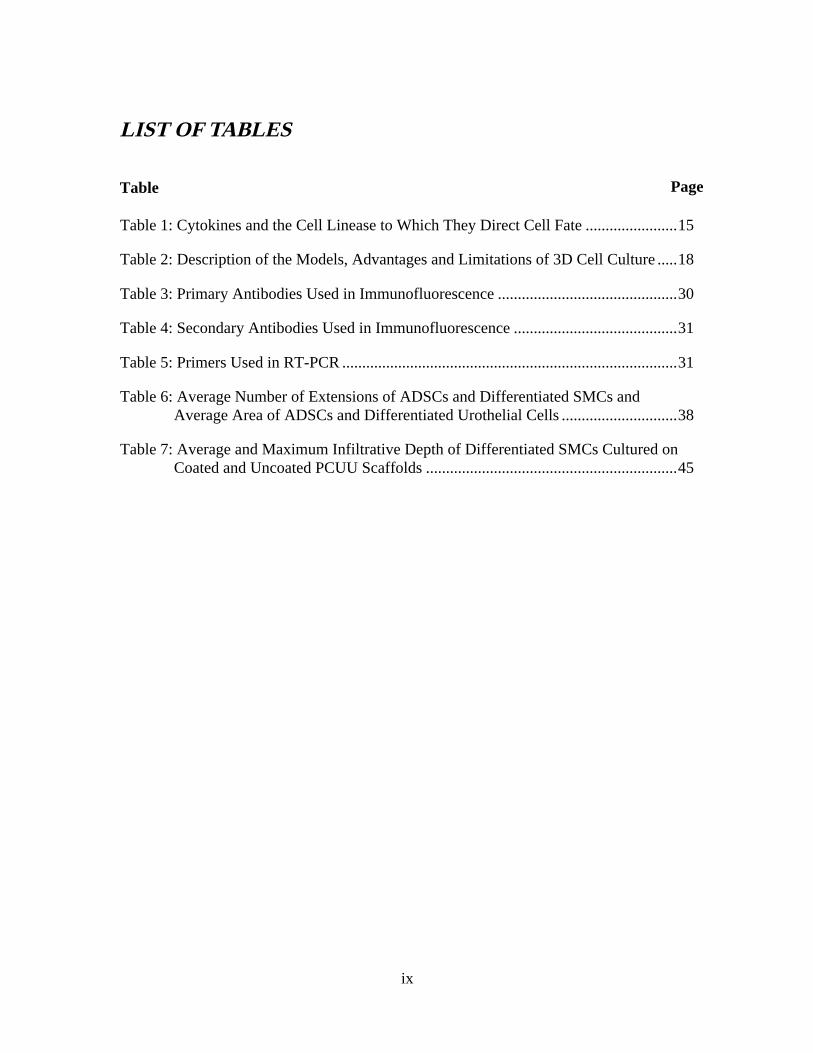
LIST OF TABLES
Table Page
Table 1: Cytokines and the Cell Linease to Which They Direct Cell Fate ....................... 15
Table 2: Description of the Models, Advantages and Limitations of 3D Cell Culture ..... 18
Table 3: Primary Antibodies Used in Immunofluorescence ............................................. 30
Table 4: Secondary Antibodies Used in Immunofluorescence ......................................... 31
Table 5: Primers Used in RT-PCR .................................................................................... 31
Table 6: Average Number of Extensions of ADSCs and Differentiated SMCs and Average Area of ADSCs and Differentiated Urothelial Cells ............................. 38
Table 7: Average and Maximum Infiltrative Depth of Differentiated SMCs Cultured on Coated and Uncoated PCUU Scaffolds ............................................................... 45
1
Chapter 1: Introduction and Background
1.1 Urinary Bladder
The urinary bladder is a visceral organ responsible for the storage and voiding of
urine. The organ can become diseased and undergo damage from a variety of medical
conditions, which can reduce its functionality. Tissue engineering of the urinary bladder
aims to regenerate a diseased urinary bladder to full functionality through the use of
tissue scaffolds and cellular material. Currently, researchers are developing novel
methods of bladder tissue regeneration using scaffolds seeded with cells to aid and
accelerate the tissue healing process.
1.1.1 Bladder Anatomy and Physiology
The urinary bladder is a hollow organ that is composed of two main parts: the
body and the neck. The overall function of the bladder is the temporary storage and
periodic voiding of urine. To properly perform these functions, the bladder must be able
to expand and contract, while maintaining a low internal pressure. The capacity of a
normal bladder is 400-500 mL and the normal range of bladder intravesical pressure is 5-
50 cm H2O, depending on the position of the body (Tanango and McAninch, 2008). The
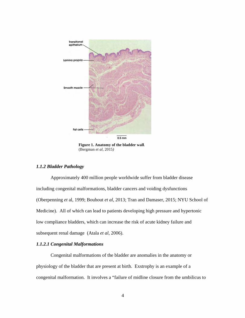
bladder is primarily made up of three layers: detrusor (smooth muscle), stroma, also
known as the lamina propria, and transitional epithelium and is illustrated in Figure 1.
The transitional epithelium, or urothelium, is composed of urothelial cells lining
the inside of the bladder, which are joined by tight junctions. The main role of the
urothelium is to contain the urine within the bladder, and disallow the interaction of urine
with other parts of the body. These cells are characterized by the protein groups
2
uroplakins and cytokeratins, specifically uroplakin-1b (UP1b), and cytokeratin-20
(CK20). Other examples of uroplakins and cytokeratins include UP1a, UPII, UPIII,
cytokeratin-5, -7, -8,- 9, -14, -17, -18 and -19 (Khandelwal, 2009). Cytokeratins are
keratin containing intermediate filaments found in the intracytoplasmic cytoskeleton of
epithelial tissue (Veranic et al, 2006). They contribute to urothelial cells’ large elastic
modulus and to the protection of the bladder against mechanical stress. Different types of
cytokeratins are expressed in the different layers of urothelium. For example,
cytokeratin-13 is expressed by intermediate and basal urothelial cells, CK-5, -14 and -17
are expressed by only basal cells and CK-20 is solely expressed in superficial, umbrella
cells (Veranic, 2006; Khandelwal, 2009). Uroplakins are located mainly on the
superficial and upper intermediate cell surface. They are categorized as an asymmetric
unit membrane (AUM) particle, and further grouped as transmembrane proteins. These
types of proteins contribute to the impermeability of the urothelium (Khandelwal, 2009)
The stroma is made up of myofibroblasts, collagen and elastin. Collagen gives
the bladder structural support while elastin allows for it to be highly compliant and gives
it the ability to expand to great volumes. The myofibroblasts within the stroma have been
reported to, under certain conditions, differentiate into new smooth muscle cells to aid in
bladder repair (DeLancey, 2002). The main function of the stroma is to provide support
and compliance for the other layers of the bladder.
The detrusor layer is made up of smooth muscle cells (SMCs) and functions in
much the same way as other muscle groups within the body; contraction is caused by an
interaction between thin and thick filaments. However, contractions of the detrusor
3
muscle are more phasic in nature when compared to any other smooth muscles
(DeLancey, 2002). The main role of the detrusor muscle is to “maintain the integrity of
the bladder without generating significant intravesical pressure during filling, and to
contract synchronously to elevate intravesical pressure sufficiently and for long enough
to empty the bladder” (DeLancey, 2002). The SMCs that make up the detrusor muscle of
the bladder are characterized by the proteins α-smooth muscle actin (α-SMA), smooth
muscle-22α (SM-22α) and smooth muscle-myosin heavy chain (SM-MHC), among
others. α-SMA and SM-22α are contractile proteins found in SMCs (Wang et al, 2010;
Harris et al, 2011). However, α-SMA is a widely conserved protein within many cell
types and is not specific to a smooth muscle lineage. SM-22α is also a conserved protein
shown to be specifically transcribed in two cell types: SMCs and pluripotent stem cells,
specifically adipose derived stem cells (Harris et al, 2011). SM-MHC is also a
contraction protein found in SMCs and is highly restricted to differentiated smooth
muscle and is indicative of a fully differentiated SMC (de Villiers et al, 2009).
All the layers of the bladder work together to store and release urine during a
process known as micturition. The micturition process has two main phases, the storage
phase and the voiding phase. During storage, urine fills the bladder cavity and the organ
expands to keep the intravesical pressure low. During voiding, a voluntary signal is sent
from the brain to nerves within the bladder that stimulates the detrusor muscle to
contract. This increases the pressure within the bladder and leads to urine flowing
through the urethra and exiting the bladder.
4
1.1.2 Bladder Pathology
Approximately 400 million people worldwide suffer from bladder disease
including congenital malformations, bladder cancers and voiding dysfunctions
(Oberpenning et al, 1999; Bouhout et al, 2013; Tran and Damaser, 2015; NYU School of
Medicine). All of which can lead to patients developing high pressure and hypertonic
low compliance bladders, which can increase the risk of acute kidney failure and
subsequent renal damage (Atala et al, 2006).
1.1.2.1 Congenital Malformations
Congenital malformations of the bladder are anomalies in the anatomy or
physiology of the bladder that are present at birth. Exstrophy is an example of a
congenital malformation. It involves a “failure of midline closure from the umbilicus to
Figure 1. Anatomy of the bladder wall. (Bergman et al, 2015)
5
the perineum, resulting in bladder mucosa continuity with the abdominal skin” (Merck
Manuals). This results in urine dripping from the open bladder, rather than flowing
through the urethra.
1.1.2.2 Bladder Cancer
About 74,000 people are diagnosed with bladder cancer each year and the most
common type is transitional cell carcinoma (American Cancer Society). This type of
cancer starts within the urothelium of the bladder, as most bladder cancers do.
Transitional cell carcinoma results in tumor growth and weakening of the bladder wall.
1.1.2.3 Voiding Dysfunction
Voiding dysfunction is an abnormality of the filling and/or emptying of the
bladder. It can be caused by inappropriate muscle activity of the bladder wall or pelvic
floor that may deter the starting and stoppage of urine flow (NYU School of Medicine).
Neurological damage may also contribute to voiding dysfunction as well as some
medications. This medical condition encompasses neurogenic bladder, stress urinary
incontinence and overactive bladder.
Neurogenic bladder is caused by a number of conditions including trauma,
Parkinson’s disease and multiple sclerosis (Cleveland Clinic). These injuries and diseases
result in an interruption in communication between the nerves in the spinal cord that
control bladder function (Mayoclinic). This results in urgency, an inability to urinate,
and frequent or painful urination.
Stress urinary incontinence (SUI) is the involuntary leakage of urine during
events that cause increased abdominal pressure in the absence of bladder contraction
6
(Tran & Damaser, 2015). This is a prevalent condition affecting almost one in five adult
women and half of all incontinent women (Nikolopoulos et al, 2015). SUI results from
an anatomical defect in the endopelvic fascial layer weakening of pelvic floor muscles
and failure of the urethral sphincter (Pate et al, 2007; Wood and Anger, 2014; Tran and
Damaser, 2015).
Overactive bladder (OAB) affects up to 16% of men and women in the United
States (Stewart et al, 2003). OAB is characterized by urgency usually accompanied with
frequency of urination and nocturia in the absence of infection or other pathology, and
may be associated with detrusor over activity (Tran and Damaser, 2015). In OAB,
detrusor over activity may result from “increased cellular excitability of the detrusor
muscle and/or abnormal neural propagation locally as well as altered peripheral afferent
nerve and central nervous system function” (Tran & Damaser, 2015).
1.1.3 Current Treatment for Bladder Pathologies
Bladder pathologies, such as neurogenic bladder, congenital malformations and
bladder cancer are often treated by replacing diseased tissue with gastrointestinal (GI)
segments for tissue repair and replacement, technically referred to as a bladder
augmentation. However, GI segments are known to cause a myriad of issues when
implanted within the bladder including: urinary tract infection, metabolic abnormalities,
abnormal drug kinetics, secondary malignancies, impaired renal function and donor site
morbidity (Tu et al, 2013; Vasdev et al, 2013). These problems are often due to the fact
that GI tissues naturally absorb specific solutes that bladder tissue is designed to excrete
(Atala, 2011).
7
Experiments involving cell-seeded tissue-engineered bladder composites have
been underway since 1992, and traditionally involve a surgical harvest of a portion of a
patient’s bladder to obtain primary bladder SMC’s (Jack et al, 2009). These primary
cells were then cultured for 6-8 weeks in a laboratory setting to obtain a cell mass large
enough to seed a scaffold. However, there is a lack of viable, accessible cells within
malignant and pathological bladders. There are still concerns when harvesting primary
cells from non-pathologic bladders; investigators have found transmission of neuropathic
cells from neuropathic bladders into the tissue-engineered bladder (Jack et al, 2009).
Given the lack of healthy primary cells to infiltrate a tissue-engineered scaffold,
other cell sources must be researched. Alternative cell types can include induced
pluripotent stem cells, embryonic stem cells, placental stem cells and adult stem cells
such as adipose derived stem cells and bone marrow mesenchymal stem cells. These
types of cells are guaranteed to be healthy and viable, and can differentiate into a
multitude of cell lines, which can then be used for a number of tissue engineering
applications.
1.1.4 Market Pull for Tissue Engineering of Human Bladder
On average, 20,000 bladder augmentations are performed each year, and each
procedure costs about $15,000 USD (BioPlan Associates, 2001; Sahai, 2014). Therefore,
the overall market for bladder augmentations is 300 million dollars per year. Due to GI
tissues’ inability to create a fully functional and integratable organ substitute, there exists
a need for an innovative replacement tissue that performs the same function as urinary
bladder tissue but does not generate an array of medical complications. Once this novel
8
product has been developed, it will have the opportunity to revolutionize the bladder
reconstruction industry and envelope the market for this procedure.
1.2 Bladder Tissue Engineering Scaffolds
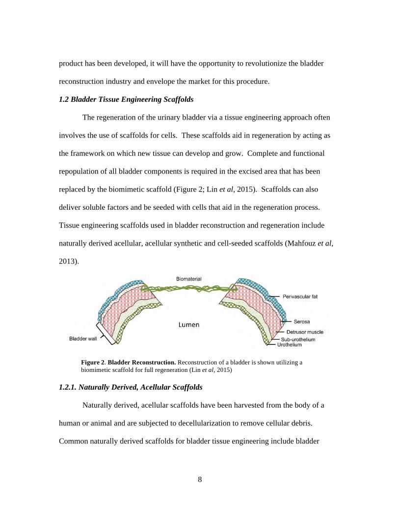
The regeneration of the urinary bladder via a tissue engineering approach often
involves the use of scaffolds for cells. These scaffolds aid in regeneration by acting as
the framework on which new tissue can develop and grow. Complete and functional
repopulation of all bladder components is required in the excised area that has been
replaced by the biomimetic scaffold (Figure 2; Lin et al, 2015). Scaffolds can also
deliver soluble factors and be seeded with cells that aid in the regeneration process.
Tissue engineering scaffolds used in bladder reconstruction and regeneration include
naturally derived acellular, acellular synthetic and cell-seeded scaffolds (Mahfouz et al,
2013).
1.2.1. Naturally Derived, Acellular Scaffolds
Naturally derived, acellular scaffolds have been harvested from the body of a
human or animal and are subjected to decellularization to remove cellular debris.
Common naturally derived scaffolds for bladder tissue engineering include bladder
Figure 2. Bladder Reconstruction. Reconstruction of a bladder is shown utilizing a biomimetic scaffold for full regeneration (Lin et al, 2015)
9
allografts, dura, placenta, pericardium and small intestine submucosa (Pattison et al,
2004). These scaffolds are advantageous because they offer the ideal mechanical and
biomechanical environment for tissue regeneration (Bouhout et al, 2013). Bladder
acellular matrices (BAMs) are especially advantageous because they resist graft
shrinkage and have shown positive results in respect to cellular repopulation (Brown et
al, 2002). Specifically, BAMs have been shown to promote the ingrowth of urothelium,
smooth muscle and blood vessels (Brown et al, 2002). Small intestine submucosa (SIS)
has also been shown to regenerate a variety of host tissues including blood vessels,
urothelium and neophyte muscle tissue (Yoo et al, 2011). However, SIS has also shown
a significant decrease in the muscle to collagen ratio, and a significant decrease in
maximum contraction once implanted (Yoo et al, 2011). The decellularization process of
these naturally derived scaffolds can denature the extracellular matrix proteins and alter
the physiological environment resulting in mechanical failure and poor biocompatibility
properties. Other drawbacks to naturally occurring scaffolds include limited availability
of donor tissue, ethical issues and the possible transfer of diseases from donor tissues
(Pattison et al, 2004). Therefore, the use of synthetic materials for bladder regeneration
and reconstruction has become the focus and material of choice for recent studies.
1.2.2 Acellular Synthetic Scaffolds
Acellular synthetic scaffolds are created through the use of polymers such as
poly-lactic-glycolic acid (PLGA), polyglycolic acid (PGA) and silk fibroin (Tu et al,
2013; Atala et al, 2006; Lai et al, 2005). These synthetic polymers, and their degradation
products, have been shown to induce a minimal inflammatory response. Synthetic
10
scaffolds are advantageous due to the ability to finely tune the physical properties of the
polymers such as degradation, mechanical strength and pore size. However, these
scaffolds often result in poor vascularization and cellular infiltration, calcification and
urinary stone formation once implanted within the body (Bouhout et al, 2013; Pattison et
al, 2004). Although there are some drawbacks to the use of synthetic scaffolds, polymers
are considered by many to be the future of urologic replacement materials due to their
numerous advantages, such as superior biocompatibility, biodegrability, ease of
procurement and ease of modification over naturally occurring scaffolds (Pattison et al,
2004). The most common types of synthetic scaffolds used in bladder tissue engineering
are those that are produced through electrospinning.
1.2.3 Cell Seeded Scaffolds
Both naturally derived and synthetic scaffolds may be seeded with cells to aid in
the regeneration of bladder tissue. Cells can be harvested from many different sources
and used in tissue engineering for tissue regeneration and repair. The two main cell
sources used in bladder tissue engineering include differentiated autologous cells and
stem cells.
1.3 Cell Types Used in Tissue Engineering Applications
Cells used in tissue engineering applications are present to enhance the efficacy
and rate in the regeneration of tissue. The cells used to infiltrate a scaffold can be either
autologous differentiated cells, or autologous stem cells. Autologous differentiated cells
are harvested from the bladder, and autologous stem cells can be harvested from multiple
areas including adipose tissue, amniotic fluid and bone marrow.
11
1.3.1 Differentiated Autologous Cells
Primarily, differentiated autologous cells are unipotent, urothelial cells and
smooth muscle cells that have been harvested from the patient to be used for scaffold-
seeding purposes. However, major concerns have become apparent from using
autologous cells. If the cells are to be harvested from a diseased organ, there may not be
enough normal and healthy cells in the organ to be obtained for expansion (Mahfouz et
al, 2013). In addition, it is difficult to conclude if the underlying disease has altered
healthy cells within the organ. The process of harvesting autologous cells and expanding
them to a population large enough to seed a scaffold is also often extremely lengthy. It
can require up to six weeks to expand the autologous cells to acquire the necessary
number to seed the scaffold.
1.3.2 Stem Cells
Unlike differentiated cells, stem cells have the following set of distinct
characteristics: unlimited self-renewal, replication in an undifferentiated state, the ability
to differentiate into other cell types, specialization of cells and acquisition of function.
Stem cells can be categorized into 5 subsets depending on their function. Totipotent stem
cells can give to any type of cell within the human body. Pluripotent stem cells can give
rise to any type of cell in the human body except for gametes. Multipotent stem cells can
differentiate into most cell types within the body, but not all. Oligopotent stem cells can
give rise to a few cell types in the body. Finally, unipotent stem cells can only be
differentiated into one cell type. Stem cells are especially useful for researchers because
they can be infinitely expanded in the laboratory while still maintaining their
12
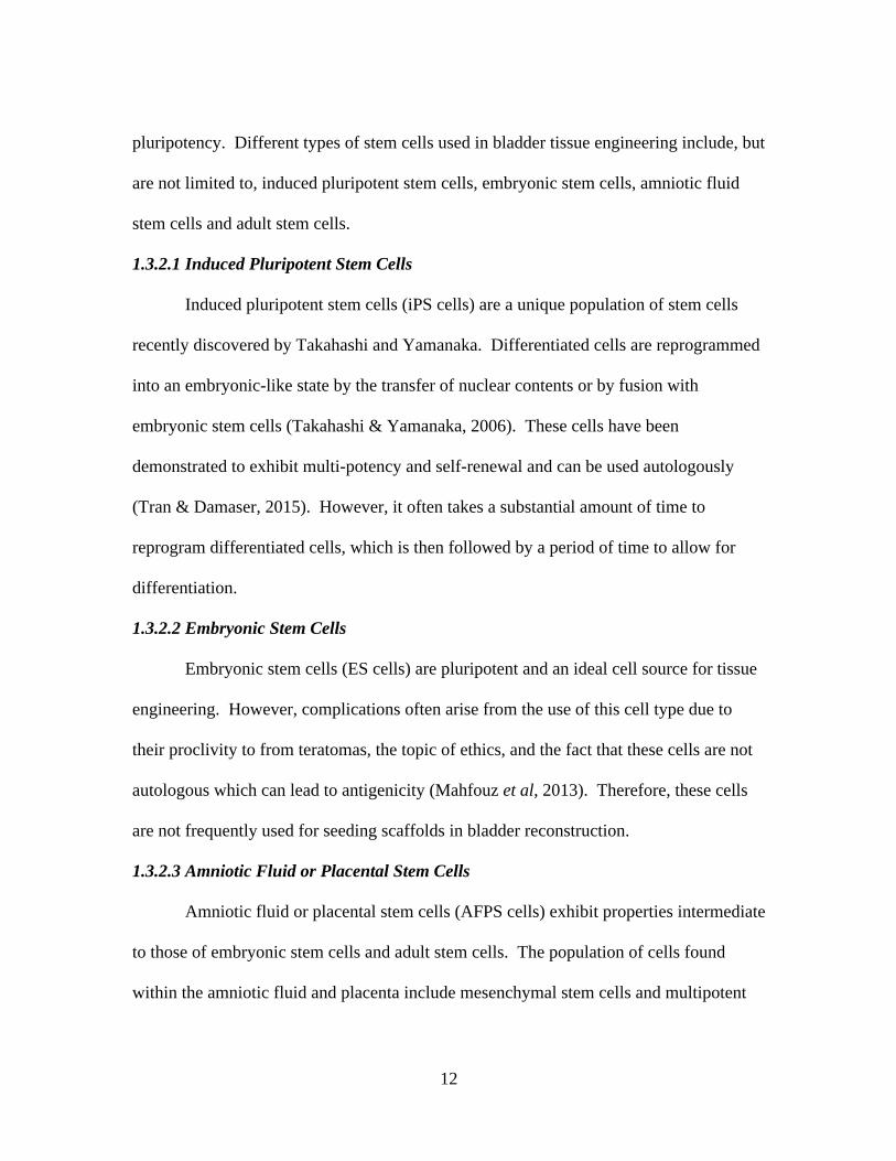
pluripotency. Different types of stem cells used in bladder tissue engineering include, but
are not limited to, induced pluripotent stem cells, embryonic stem cells, amniotic fluid
stem cells and adult stem cells.
1.3.2.1 Induced Pluripotent Stem Cells
Induced pluripotent stem cells (iPS cells) are a unique population of stem cells
recently discovered by Takahashi and Yamanaka. Differentiated cells are reprogrammed
into an embryonic-like state by the transfer of nuclear contents or by fusion with
embryonic stem cells (Takahashi & Yamanaka, 2006). These cells have been
demonstrated to exhibit multi-potency and self-renewal and can be used autologously
(Tran & Damaser, 2015). However, it often takes a substantial amount of time to
reprogram differentiated cells, which is then followed by a period of time to allow for
differentiation.
1.3.2.2 Embryonic Stem Cells
Embryonic stem cells (ES cells) are pluripotent and an ideal cell source for tissue
engineering. However, complications often arise from the use of this cell type due to
their proclivity to from teratomas, the topic of ethics, and the fact that these cells are not
autologous which can lead to antigenicity (Mahfouz et al, 2013). Therefore, these cells
are not frequently used for seeding scaffolds in bladder reconstruction.
1.3.2.3 Amniotic Fluid or Placental Stem Cells
Amniotic fluid or placental stem cells (AFPS cells) exhibit properties intermediate
to those of embryonic stem cells and adult stem cells. The population of cells found
within the amniotic fluid and placenta include mesenchymal stem cells and multipotent
13
AFPS cells (Tran & Damaser, 2015). AFPS cells can be induced to differentiate into
cells of all three germ layers including cells of adipogenic, osteogenic, myogenic,
endothelial, neural, and hepatic lineages (Tran and Damaser, 2015).
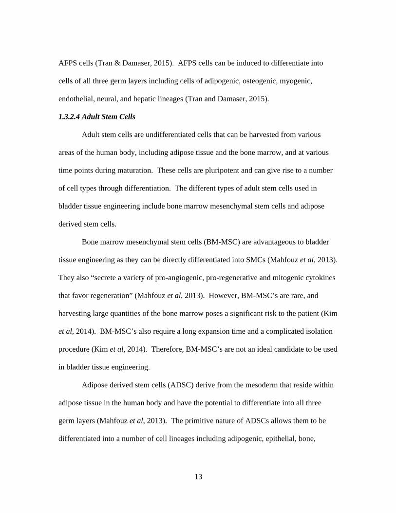
1.3.2.4 Adult Stem Cells
Adult stem cells are undifferentiated cells that can be harvested from various
areas of the human body, including adipose tissue and the bone marrow, and at various
time points during maturation. These cells are pluripotent and can give rise to a number
of cell types through differentiation. The different types of adult stem cells used in
bladder tissue engineering include bone marrow mesenchymal stem cells and adipose
derived stem cells.
Bone marrow mesenchymal stem cells (BM-MSC) are advantageous to bladder
tissue engineering as they can be directly differentiated into SMCs (Mahfouz et al, 2013).
They also “secrete a variety of pro-angiogenic, pro-regenerative and mitogenic cytokines
that favor regeneration” (Mahfouz et al, 2013). However, BM-MSC’s are rare, and
harvesting large quantities of the bone marrow poses a significant risk to the patient (Kim
et al, 2014). BM-MSC’s also require a long expansion time and a complicated isolation
procedure (Kim et al, 2014). Therefore, BM-MSC’s are not an ideal candidate to be used
in bladder tissue engineering.
Adipose derived stem cells (ADSC) derive from the mesoderm that reside within
adipose tissue in the human body and have the potential to differentiate into all three
germ layers (Mahfouz et al, 2013). The primitive nature of ADSCs allows them to be
differentiated into a number of cell lineages including adipogenic, epithelial, bone,
14
muscle, cartilage and neuronal (de Villiers et al, 2009). Unlike bone marrow
mesenchymal stem cells, adipose tissues are easily harvested from the body during
liposuction and ADSCs are then easily isolated (Jack et al, 2009). ADSCs are easy to
handle and exhibit great proliferative capacity (de Villiers et al, 2009). Donated
allogeneic ADSCs also show a very low antigenicity, with no expression of
immunologically relevant surface antigens both in a non-differentiated and differentiated
state (Niemeyer et al, 2007). Therefore, autologous ADSC’s are an ideal candidate for
regenerative medicine, specifically bladder regeneration and reconstruction.
1.4 Differentiation Techniques
Stem cells may be differentiated into a terminal cell lineage prior to seeding on a
scaffold for tissue regeneration purposes. Timely cell proliferation and native tissue
development from stem cells will facilitate overall regeneration and recovery time. The
differentiation technique varies depending on cell lineages of interest and investigators.
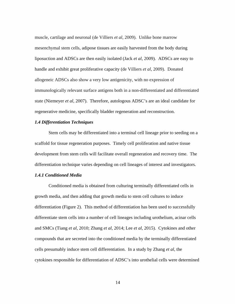
1.4.1 Conditioned Media
Conditioned media is obtained from culturing terminally differentiated cells in
growth media, and then adding that growth media to stem cell cultures to induce
differentiation (Figure 2). This method of differentiation has been used to successfully
differentiate stem cells into a number of cell lineages including urothelium, acinar cells
and SMCs (Tiang et al, 2010; Zhang et al, 2014; Lee et al, 2015). Cytokines and other
compounds that are secreted into the conditioned media by the terminally differentiated
cells presumably induce stem cell differentiation. In a study by Zhang et al, the
cytokines responsible for differentiation of ADSC’s into urothelial cells were determined
15
to include TGF-α, PDGF-BB, VEGF, IGF-I and EGF among others (Zhang et al, 2014).
However, one drawback to this methodology is the requirement of a terminally
differentiated cell population. To apply this to bladder regeneration, autologous
urothelial and smooth muscle cells would be needed. Healthy autologous cells would be
few in the patient’s diseased bladder, rendering this option less attractive.
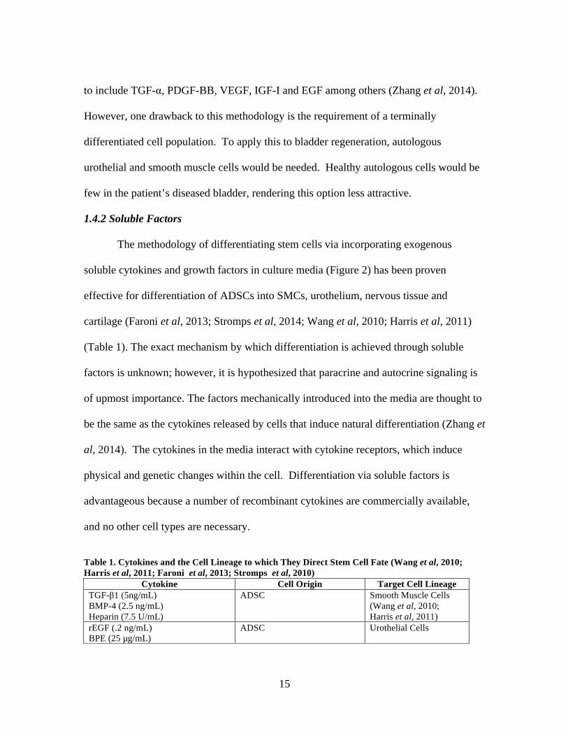
1.4.2 Soluble Factors
The methodology of differentiating stem cells via incorporating exogenous
soluble cytokines and growth factors in culture media (Figure 2) has been proven
effective for differentiation of ADSCs into SMCs, urothelium, nervous tissue and
cartilage (Faroni et al, 2013; Stromps et al, 2014; Wang et al, 2010; Harris et al, 2011)
(Table 1). The exact mechanism by which differentiation is achieved through soluble
factors is unknown; however, it is hypothesized that paracrine and autocrine signaling is
of upmost importance. The factors mechanically introduced into the media are thought to
be the same as the cytokines released by cells that induce natural differentiation (Zhang et
al, 2014). The cytokines in the media interact with cytokine receptors, which induce
physical and genetic changes within the cell. Differentiation via soluble factors is
advantageous because a number of recombinant cytokines are commercially available,
and no other cell types are necessary.
Table 1. Cytokines and the Cell Lineage to which They Direct Stem Cell Fate (Wang et al, 2010; Harris et al, 2011; Faroni et al, 2013; Stromps et al, 2010)
Cytokine Cell Origin Target Cell Lineage TGF-β1 (5ng/mL) BMP-4 (2.5 ng/mL) Heparin (7.5 U/mL)
ADSC Smooth Muscle Cells (Wang et al, 2010; Harris et al, 2011)
rEGF (.2 ng/mL) BPE (25 µg/mL)
ADSC Urothelial Cells
16
1.4.3 Indirect and Direct Co-Culture of Stem Cells and Differentiated Cells
An indirect co-culture of stem cells with differentiated cells utilizing a trans-well
insert to separate the two cell types (Figure 3) has been proven effective in differentiating
stem cells toward bone, cartilage, adipose, urothelial and muscle lineages (Zhao, 2012;
Zhang et al, 2014). Direct co-cultures involve culturing stem cells in direct contact with
differentiated cells. This methodology has been proven to effectively differentiate stem
cells toward neuronal, smooth muscle and urothelial lineages (Zhao, 2015; Merfeld-
Clauss, 2014; Liu, 2009).
The exact differentiation mechanism of indirect co-cultures is unknown, but it has
been demonstrated that paracrine signaling is a large factor in the differentiation of the
stem cells. Cytokines released from the differentiated cells interact with cytokine
receptors on the stem cells that induce genetic and physical changes within the stem cell
population. While this approach efficiently directs stem cell fate toward a specific cell
lineage, the use of differentiated cells causes concerns regarding the attainability of
healthy autologous cells, and the immunogenicity of stem cells differentiated with
allogeneic cells.
γ-aminobutyric acid adenosine 5′-triphosphate
ADSC Schwann Cells (Faroni et al, 2013)
TGF-β3 (10 ng/mL) Insulin (6.25 ng/mL)
ADSC Chondrocytes (Stromps et al, 2010)
17
Like indirect co-culture, the exact mechanism of differentiation is unknown for
direct co-cultures, but it has been shown that cell signaling and cell-contact play a role in
the differentiation of stem cells. In a study by Merfeld-Clauss et al, ADSCs directly co-
cultured with endothelial cells differentiated toward a SMC lineage. The ADSCs in
direct contact with the endothelial cells differentiated more quickly, while those further
away differentiated to SMCs over a longer period of time. They concluded that direct
contact with endothelial cells will differentiate ADSCs toward a SMC lineage, and
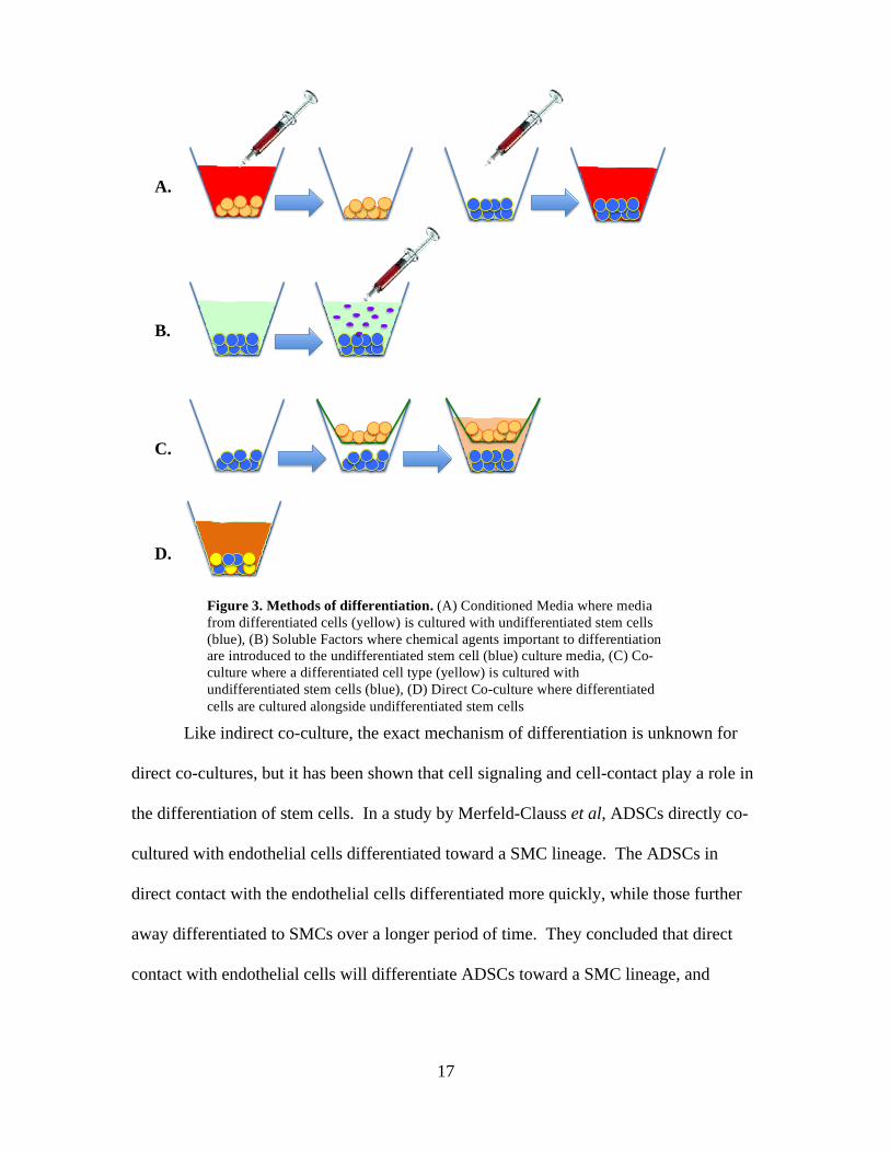
Figure 3. Methods of differentiation. (A) Conditioned Media where media from differentiated cells (yellow) is cultured with undifferentiated stem cells (blue), (B) Soluble Factors where chemical agents important to differentiation are introduced to the undifferentiated stem cell (blue) culture media, (C) Co-culture where a differentiated cell type (yellow) is cultured with undifferentiated stem cells (blue), (D) Direct Co-culture where differentiated cells are cultured alongside undifferentiated stem cells
A.
B.
C.
D.
18
factors secreted by endothelial cells will differentiate ADSCs toward a SMC lineage. In
another study by Liu et al, ADSCs were directly and indirectly co-cultured with
urothelial cells and cultured in conditioned media from urothelial cells. They found that
only the ADSCs directly co-cultured with urothelial cells were differentiated toward a
urothelial lineage. Therefore, they concluded that direct cell-to-cell contact was
responsible for the differentiation.
1.5 Cell Seeding Techniques for Tissue Engineering Scaffolds
Cells can be seeded onto naturally derived and acellular scaffolds in either a two-
dimensions or a three-dimensional manner. Two dimensional cell seeding is a simpler
methodology that results in a single layer of cells. Three-dimensional cell seeding is a
more intricate method and results in multiple layers of cells.
1.5.1 Two Dimensional Cell Seeding
Two dimensional cell seeding involves placing cells on top of a scaffold or flat
surface and waiting for the cells to naturally attach to the surface. The cells seeded in this
manner are not able to pile on top of one another, which results in a monolayer
morphology which is not natural for all cell types (Antoni et al, 2015). In addition, due
to this monolayer structure, the cells are only in contact with cells at their periphery, and
can only communicate and interact with those cells as well. Therefore, this cell culture
model does not accurately model the in vivo state.
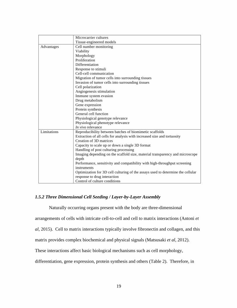
Table 2. Description of the Models, Advantages and Limitations of 3D Cell Culture (Antoni et al, 2015)
Characteristics Properties 3D Culture Models Whole animals and organotypic explant cultures
Cell spheroids cultures Polarized epithelial cell cultures
19
Microcarrier cultures Tissue-engineered models
Advantages Cell number monitoring Viability Morphology Proliferation Differentiation Response to stimuli Cell-cell communication Migration of tumor cells into surrounding tissues Invasion of tumor cells into surrounding tissues Cell polarization Angiogenesis stimulation Immune system evasion Drug metabolism Gene expression Protein synthesis General cell function Physiological genotype relevance Physiological phenotype relevance In vivo relevance
Limitations Reproducibility between batches of biomimetic scaffolds Extraction of all cells for analysis with increased size and tortuosity Creation of 3D matrices Capacity to scale up or down a single 3D format Handling of post culturing processing Imaging depending on the scaffold size, material transparency and microscope depth Performance, sensitivity and compatibility with high-throughput screening instruments Optimization for 3D cell culturing of the assays used to determine the cellular response to drug interaction Control of culture conditions
1.5.2 Three Dimensional Cell Seeding / Layer-by-Layer Assembly
Naturally occurring organs present with the body are three-dimensional
arrangements of cells with intricate cell-to-cell and cell to matrix interactions (Antoni et
al, 2015). Cell to matrix interactions typically involve fibronectin and collagen, and this
matrix provides complex biochemical and physical signals (Matsusaki et al, 2012).
These interactions affect basic biological mechanisms such as cell morphology,
differentiation, gene expression, protein synthesis and others (Table 2). Therefore, in
20
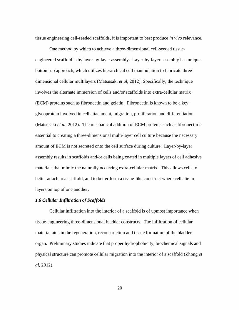
tissue engineering cell-seeded scaffolds, it is important to best produce in vivo relevance.
One method by which to achieve a three-dimensional cell-seeded tissue-
engineered scaffold is by layer-by-layer assembly. Layer-by-layer assembly is a unique
bottom-up approach, which utilizes hierarchical cell manipulation to fabricate three-
dimensional cellular multilayers (Matsusaki et al, 2012). Specifically, the technique
involves the alternate immersion of cells and/or scaffolds into extra-cellular matrix
(ECM) proteins such as fibronectin and gelatin. Fibronectin is known to be a key
glycoprotein involved in cell attachment, migration, proliferation and differentiation
(Matsusaki et al, 2012). The mechanical addition of ECM proteins such as fibronectin is
essential to creating a three-dimensional multi-layer cell culture because the necessary
amount of ECM is not secreted onto the cell surface during culture. Layer-by-layer
assembly results in scaffolds and/or cells being coated in multiple layers of cell adhesive
materials that mimic the naturally occurring extra-cellular matrix. This allows cells to
better attach to a scaffold, and to better form a tissue-like construct where cells lie in
layers on top of one another.
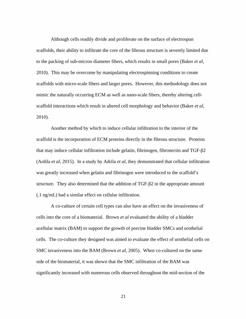
1.6 Cellular Infiltration of Scaffolds
Cellular infiltration into the interior of a scaffold is of upmost importance when
tissue-engineering three-dimensional bladder constructs. The infiltration of cellular
material aids in the regeneration, reconstruction and tissue formation of the bladder
organ. Preliminary studies indicate that proper hydrophobicity, biochemical signals and
physical structure can promote cellular migration into the interior of a scaffold (Zhong et
al, 2012).
21
Although cells readily divide and proliferate on the surface of electrospun
scaffolds, their ability to infiltrate the core of the fibrous structure is severely limited due
to the packing of sub-micron diameter fibers, which results in small pores (Baker et al,
2010). This may be overcome by manipulating electrospinning conditions to create
scaffolds with micro-scale fibers and larger pores. However, this methodology does not
mimic the naturally occurring ECM as well as nano-scale fibers, thereby altering cell-
scaffold interactions which result in altered cell morphology and behavior (Baker et al,
2010).
Another method by which to induce cellular infiltration to the interior of the
scaffold is the incorporation of ECM proteins directly in the fibrous structure. Proteins
that may induce cellular infiltration include gelatin, fibrinogen, fibronectin and TGF-β2
(Ardila et al, 2015). In a study by Adrila et al, they demonstrated that cellular infiltration
was greatly increased when gelatin and fibrinogen were introduced to the scaffold’s
structure. They also determined that the addition of TGF-β2 in the appropriate amount
(.1 ng/mL) had a similar effect on cellular infiltration.
A co-culture of certain cell types can also have an effect on the invasiveness of
cells into the core of a biomaterial. Brown et al evaluated the ability of a bladder
acellular matrix (BAM) to support the growth of porcine bladder SMCs and urothelial
cells. The co-culture they designed was aimed to evaluate the effect of urothelial cells on
SMC invasiveness into the BAM (Brown et al, 2005). When co-cultured on the same
side of the biomaterial, it was shown that the SMC infiltration of the BAM was
significantly increased with numerous cells observed throughout the mid-section of the
22
matrix (Brown et al, 2005). Therefore, it can be concluded that cellular infiltration is
significantly affected by cell-to-cell interactions.
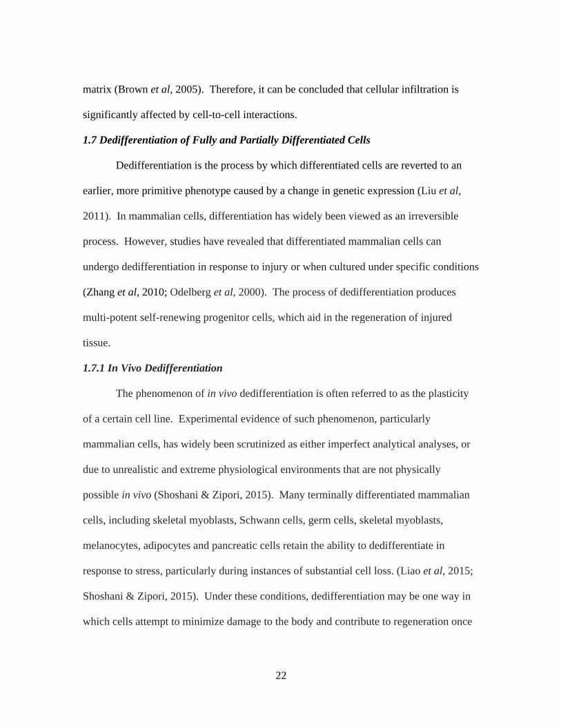
1.7 Dedifferentiation of Fully and Partially Differentiated Cells
Dedifferentiation is the process by which differentiated cells are reverted to an
earlier, more primitive phenotype caused by a change in genetic expression (Liu et al,
2011). In mammalian cells, differentiation has widely been viewed as an irreversible
process. However, studies have revealed that differentiated mammalian cells can
undergo dedifferentiation in response to injury or when cultured under specific conditions
(Zhang et al, 2010; Odelberg et al, 2000). The process of dedifferentiation produces
multi-potent self-renewing progenitor cells, which aid in the regeneration of injured
tissue.
1.7.1 In Vivo Dedifferentiation
The phenomenon of in vivo dedifferentiation is often referred to as the plasticity
of a certain cell line. Experimental evidence of such phenomenon, particularly
mammalian cells, has widely been scrutinized as either imperfect analytical analyses, or
due to unrealistic and extreme physiological environments that are not physically
possible in vivo (Shoshani & Zipori, 2015). Many terminally differentiated mammalian
cells, including skeletal myoblasts, Schwann cells, germ cells, skeletal myoblasts,
melanocytes, adipocytes and pancreatic cells retain the ability to dedifferentiate in
response to stress, particularly during instances of substantial cell loss. (Liao et al, 2015;
Shoshani & Zipori, 2015). Under these conditions, dedifferentiation may be one way in
which cells attempt to minimize damage to the body and contribute to regeneration once
23
the risk has been diminished (Odelberg et al, 2000; Zhang et al, 2010; Liu et al, 2011;
Shoshani & Zipori, 2015).
Based on observations of dedifferentiation, it has been proposed that all
mammalian cells retain specific molecular machinery that dictates dedifferentiation
(Shoshani & Zipori, 2015). This molecular machinery is referred to as “return to stem
state” (RtSS) and functions by sensing the cells’ external environment. If these senses
predict cell damage, the RtSS will “turn on” and push the cell “backwards down the
differentiation cascade, either partially or all the way back to pluripotency” (Shoshani &
Zipori, 2015). Dedifferentiation may also be a mechanism, which enhances and provides
an alternative to stem cell self-renewal (Shoshani & Zipori, 2015).
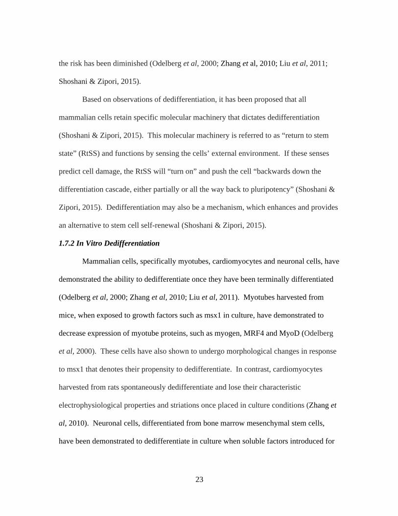
1.7.2 In Vitro Dedifferentiation
Mammalian cells, specifically myotubes, cardiomyocytes and neuronal cells, have
demonstrated the ability to dedifferentiate once they have been terminally differentiated
(Odelberg et al, 2000; Zhang et al, 2010; Liu et al, 2011). Myotubes harvested from
mice, when exposed to growth factors such as msx1 in culture, have demonstrated to
decrease expression of myotube proteins, such as myogen, MRF4 and MyoD (Odelberg
et al, 2000). These cells have also shown to undergo morphological changes in response
to msx1 that denotes their propensity to dedifferentiate. In contrast, cardiomyocytes
harvested from rats spontaneously dedifferentiate and lose their characteristic
electrophysiological properties and striations once placed in culture conditions (Zhang et
al, 2010). Neuronal cells, differentiated from bone marrow mesenchymal stem cells,
have been demonstrated to dedifferentiate in culture when soluble factors introduced for
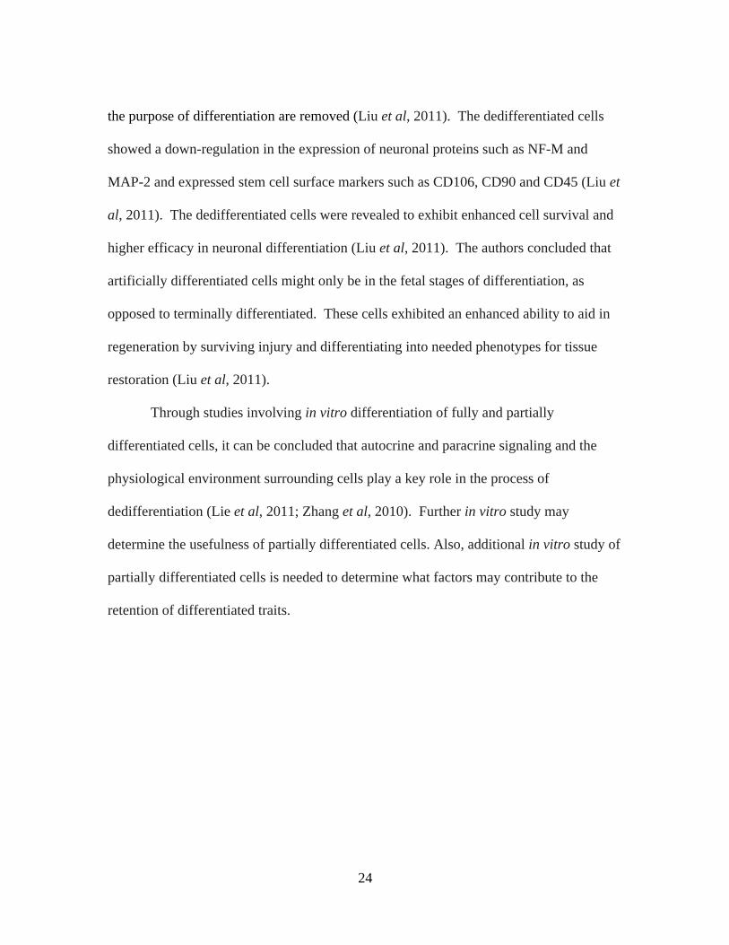
24
the purpose of differentiation are removed (Liu et al, 2011). The dedifferentiated cells
showed a down-regulation in the expression of neuronal proteins such as NF-M and
MAP-2 and expressed stem cell surface markers such as CD106, CD90 and CD45 (Liu et
al, 2011). The dedifferentiated cells were revealed to exhibit enhanced cell survival and
higher efficacy in neuronal differentiation (Liu et al, 2011). The authors concluded that
artificially differentiated cells might only be in the fetal stages of differentiation, as
opposed to terminally differentiated. These cells exhibited an enhanced ability to aid in
regeneration by surviving injury and differentiating into needed phenotypes for tissue
restoration (Liu et al, 2011).
Through studies involving in vitro differentiation of fully and partially
differentiated cells, it can be concluded that autocrine and paracrine signaling and the
physiological environment surrounding cells play a key role in the process of
dedifferentiation (Lie et al, 2011; Zhang et al, 2010). Further in vitro study may
determine the usefulness of partially differentiated cells. Also, additional in vitro study of
partially differentiated cells is needed to determine what factors may contribute to the
retention of differentiated traits.
25
Chapter 2: Research Rationale
Over 400 million people worldwide suffer from bladder pathology including
congenital malformations, bladder cancer and voiding dysfunctions and as many as
50,000 people in the United States can benefit from bladder augmentation surgery
(Adamowicz, 2013; Oberpenning et al, 1999; Bouhout et al, 2013; Tran & Damaser,
2015; NYU School of Medicine; BioPlan Associates, 2001). The current gold standard
approach for bladder augmentation replaces diseased bladder tissue with gastrointestinal
(GI) segments, which are known to cause numerous complications including urinary tract
infection, metabolic abnormalities, abnormal drug kinetics, secondary malignancies,
impaired renal function and donor site morbidity (Tu et al, 2013; Vasdev et al, 2013).
The cause of these complications is due to the fact that GI tissues naturally absorb
specific solutes that bladder tissue is designed to excrete (Atala, 2011).
Previous studies demonstrated that a tissue-engineered construct may be used as a
bladder tissue replacement. In a study by Atala et al, a PGA+collagen scaffold seeded
with autologous smooth muscle and urothelial cells provided relief for high pressure or
poorly compliant bladders (Atala et al, 2006). The tissue engineered bladder biopsies
also showed an adequate structural architecture and phenotype and no metabolic
consequences were recorded (Atala et al, 2006). However, in a Phase II clinical trial
study using the same methodology to treat neurogenic bladders, adverse events were
recorded in all patients and the results did not support the prior effectiveness of the
tissue-engineered bladder (Joseph et al, 2014).
26
The exact reason for the failure in clinical trials is unknown. However flaws in
scaffold design and the use of autologous cells from diseased bladders may be a
contributing factor to the adverse outcomes. This project aimed to establish human
adipose derived stem cells (hADSCs) as a potential cell source, as opposed to autologous
cells, to seed a tissue-engineered biomimetic patch to replace GI segments in bladder
augmentations. To explore hADSCs as a possible cell source to infiltrate a biomimetic
bladder tissue scaffold, the present master’s thesis research consisted of the following
four aims:
Aim1: To Analyze the Effectiveness of SMIM in Differentiating ADSCs to SMCs
Rationale: Previous studies demonstrated that hADSCs have the ability to differentiate
into SMCs (Wang et al, 2010; Harris et al, 2011; Merfeld-Clauss et al, 2014).
Approach: The cell morphology, mRNA expression and protein expression of hADSCs
exposed to smooth muscle inductive media (SMIM) for 10 days were examined through
phase-contrast microscopy, RT-PCR and immunofluorescence.
Aim 2: To Determine the Reversibility of Differentiated SMCs and Urothelial Cells
Rationale: Dedifferentiation has been observed in both fully and partially differentiated
cells both in vitro and in vivo (Zhang et al, 2010; Odelberg et al, 2000; Liao et al, 2015;
Shoshani & Zipori, 2015; Liu et al, 2011).
Approach: The cell morphology, mRNA expression and protein expression of
differentiated SMCs and urothelial cells exposed to growth media for 7 days were
examined through phase-contrast microscopy, RT-PCR and immunofluorescence.
27
Aim 3: To Determine the Retention of Differentiated Traits of SMCs and Urothelial
Cells in a Co-Culture
Rationale: Previous studies have demonstrated that a co-culture of cell types can induce
differentiation, and help retain differentiation (Merfeld-Clauss et al, 2014; Zhao et al,
2012; Zhang et al, 2014; Zhao et al, 2015; Liu et al, 2009).
Approach: The cell morphology and protein expression of SMCs and urothelial cells
incubated in a co-culture in various types of media (Growth media, SMIM, KSFM, ½
SMIM – ½ KSFM) for 7 days were examined histologically.
Aim 4: To Evaluate the Infiltration of Differentiated SMCs into a PCUU Scaffold
Rationale: Cellular infiltration into the interior of a scaffold is of upmost importance in
tissue-engineered bladder constructs in the regeneration of 3D tissue structure.
Approach: The degree of infiltration of SMCs incubated on fibronectin and gelatin coated
PCUU scaffolds and uncoated PCUU scaffolds for 7 days were evaluated semi-
quantitatively on histological sections.
28
Chapter 3: Materials and Methods
3.1 Cell Culture
Human adipose derived stem cells (hADSCs, Lonza, Walkersville, MD) were
cultured in growth medium (DMEM supplemented with 10% FBS and 1% Glutamax,
Thermo Fisher Scientific) under standard cell culture conditions, that is, a humidified
atmosphere at 95% air and 5% CO2 at 37°C. Upon confluency, hADSCs were passaged
using a trypsin solution (.25% trypsin, 2.21 mM EDTA and sodium bicarbonate solution,
Corning). Cells under passage number 9 were used for experimentation.
To induce differentiation into SMCs, hADSCs were seeded at a density of 2*103
cells / cm2 and cultured in growth medium for 24 hours and the medium was switched to
smooth muscle inductive medium (SMIM: DMEM supplemented with 2.5 ng/mL TGF-
β1 (ProSci), 5 ng/mL PDGF-BB (ProSci)) for the next 10 days with a media change
every 2 days. Induction of hADSCs differentiation toward a urothelial lineage was
achieved by following a protocol previously established in the Nagatomi Lab (Turner,
2015). Briefly, cells were cultured in growth medium until confluent and then cultured in
defined keratinocyte serum free media (KSFM, Thermo Fisher Scientific) for the next 14
days with a media change every 2-3 days. To test for dedifferentiation, the differentiation
media (SMIM, KSFM) were switched back to growth medium.
3.2 Immunofluorescence
Cells cultured on chamber slides were washed with PBS and fixed using 10%
neutral buffered formalin (NBF) for 30 minutes. The cells were then subjected to
29
immunofluorescence staining using the routine protocol. Briefly, the cells were
incubated in a blocking solution, which minimized unspecific binding of the primary
antibody, consisting of 2% non-fat dried milk in 50mM Tris-HCl for 30 minutes. For
intracellular proteins of interest, .1% Triton was added to the blocking solution to
permeablize the membrane. Cells were incubated with the primary antibody (Table 3)
either overnight at 4°C or at room temperature for 2 hours. After incubation with the
primary antibody, the cells were incubated with the fluorescently labeled secondary
antibody (Table 4) for 2 hours at room temperature, shielded from light. To counterstain
the nuclei, cells were incubated with 300 nM DAPI for 5 minutes. The cells were then
dried in increasing concentrations of ethanol (50%, 75%, 90%, 100%) for 2 minute each,
and mounted on a cover slip.
3.3 Reverse Transcription Polymerase Chain Reaction
Total RNA was isolated from cells (SMCs: 450,000; Urothelial cells: 2*106;
hADSCs: 2*106) using the RNeasy kit (VENDOR) following the manufacturer’s
instructions. Briefly, cells were removed from each flask using a trypsin solution (.25%
trypsin, 2.21 mM EDTA and sodium bicarbonate solution, Corning) and then centrifuged
(400 rcf) to attain a cell pellet for each sample. The cell pellets were homogenized,
transferred to RNeasy spin columns and centrifuged (400 rcf). Following wash steps,
RNase free water is added to the spin column and centrifuged to elute the RNA samples.
The nucleic acid concentration was quantified (ng/ µL) using a nanodrop
spectrophotometer (Thermo Fisher Scientific) and samples were stored at -20°C until use
in subsequent experiments.
30
Reverse transcription was carried out with 1 µg of RNA from each sample using the
RETROscript kit (Thermo Fisher Scientific) and following the manufacturer’s
instructions. Briefly, each sample was mixed with Oligo(dT) and was incubated at 85°C
for 3 minutes for the purpose of purifying the mRNA and adding a tail of 100-300
adenine residues. After the cycle was completed, the samples were mixed with 10x RT
buffer, dNTP mix, and MMLV-RT enzyme (Thermo Fisher Scientific). The samples
were then incubated at 44°C for 1 hour and 92°C for 10 minutes to synthesize cDNA.
Polymerase chain reaction was performed using the QuantiTech SYBR Green PCR
kit and the manufacturer’s instructions (Qiagen). Each sample of cDNA (.05 µg) was
mixed with the forward and reverse primer of interest (2 µL; 5µM; Table 5), and 2X
SYBR Green master mix, which contains DNA polymerase, dNTPs and dUTPs. A PCR
protocol was used that had been previously established in the Nagatomi Lab (Turner,
2015). Each cycle consisted of the DNA denaturation step at 95°C followed by primer
annealing at 55°C and primer extension at 72°C , all of which(?) were repeated for 40
cycles. Gene expression levels of differentiated SMCs, dedifferentiated SMCs and
dedifferentiated urothelial cells were analyzed and compared to control hADSCs,
differentiated SMCs and differentiated urothelial cells, respectively, using the ΔΔCt
method.
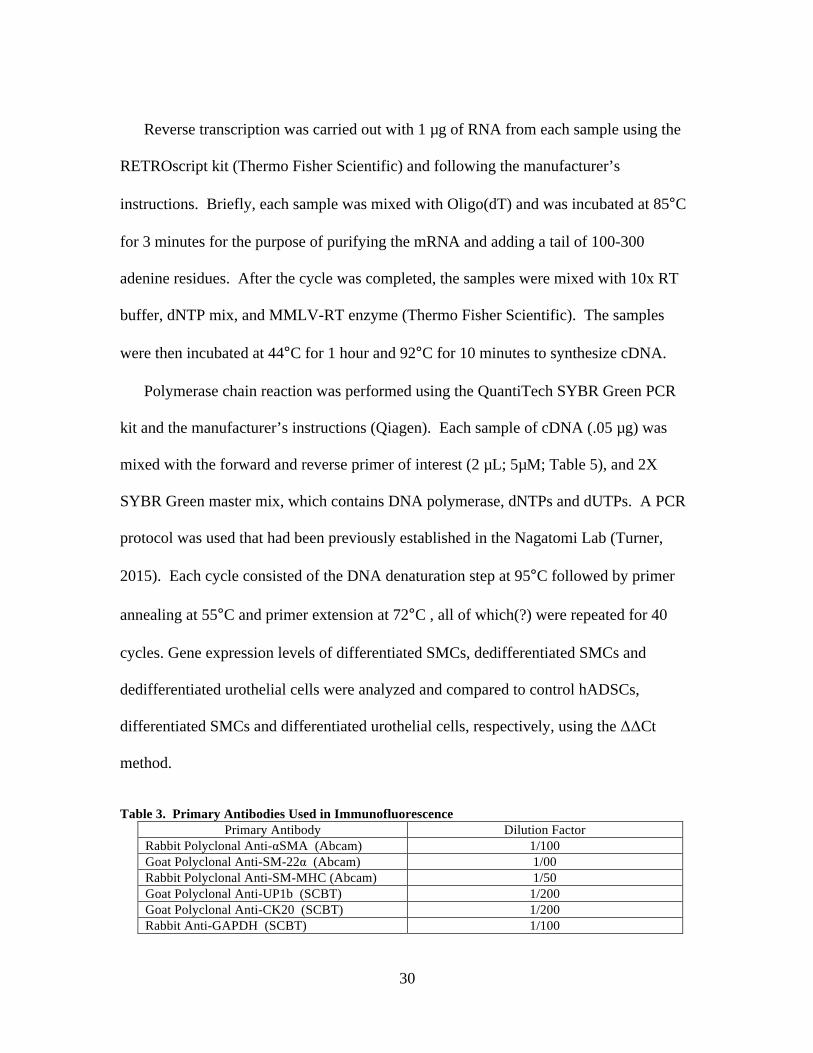
Table 3. Primary Antibodies Used in Immunofluorescence Primary Antibody Dilution Factor
Rabbit Polyclonal Anti-αSMA (Abcam) 1/100 Goat Polyclonal Anti-SM-22α (Abcam) 1/00 Rabbit Polyclonal Anti-SM-MHC (Abcam) 1/50 Goat Polyclonal Anti-UP1b (SCBT) 1/200 Goat Polyclonal Anti-CK20 (SCBT) 1/200 Rabbit Anti-GAPDH (SCBT) 1/100
31
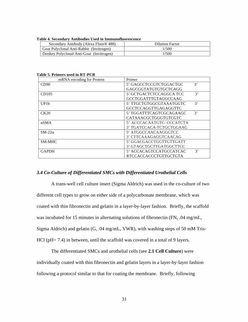
Table 4. Secondary Antibodies Used in Immunofluorescence Secondary Antibody (Alexa Fluor® 488) Dilution Factor
Goat Polyclonal Anti-Rabbit (Invitrogen) 1/500 Donkey Polyclonal Anti-Goat (Invitrogen) 1/500
Table 5. Primers used in RT-PCR mRNA encoding for Protein Primer
CD90 5’ GAGCCTCCGTCTGGACTGC 3’ GAGCGGTATGTGTGCTCAGG
CD105 5’ GCTGACTCTCCAGGCA TCC 3’ GCCTGGATTTGTAGGCCAAG
UP1b 5’ TTGCTGTGGCGTAAATGGTC 3’ GCCTCCAGGTTGAGAGGTTC
CK20 5’ TGGATTTCAGTCGCAGAAGC 3’ CATAAACGCTGGGTGTCGTC
αSMA 5’ ACCCACAATGTC- CCCATCTA 3’ TGATCCACA-TCTGCTGGAAG
SM-22α 5’ ATGGCCAACAAGGGTCC 3’ CTTCAAAGAGGTCAACAG
SM-MHC 5’ GGACGACCTGGTTGTTGATT 3’ GTAGCTGCTTGATGGCTTCC
GAPDH 5’ ACCACAGTCCATGCCATCAC 3’ RTCCACCACCCTGTTGCTGTA
3.4 Co-Culture of Differentiated SMCs with Differentiated Urothelial Cells
A trans-well cell culture insert (Sigma Aldrich) was used in the co-culture of two
different cell types to grow on either side of a polycarbonate membrane, which was
coated with thin fibronectin and gelatin in a layer-by-layer fashion. Briefly, the scaffold
was incubated for 15 minutes in alternating solutions of fibronectin (FN, .04 mg/mL,
Sigma Aldrich) and gelatin (G, .04 mg/mL, VWR), with washing steps of 50 mM Tris-
HCl (pH= 7.4) in between, until the scaffold was covered in a total of 9 layers.
The differentiated SMCs and urothelial cells (see 2.1 Cell Culture) were
individually coated with thin fibronectin and gelatin layers in a layer-by-layer fashion
following a protocol similar to that for coating the membrane. Briefly, following
32
centrifugation (400 rcf) the cell pellet was re-suspended and incubated in the FN solution
for 1 minute, centrifuged (400 rcf) for 2 minutes and the supernatant was discarded. This
process was repeated with Tris-HCl to wash the cells and then a G coating was applied
until a total of 9 layers were present on the surface of the cells.
The coated SMCs were suspended in SMIM and 200,000 cells were seeded onto
the bottom of each insert and were incubated upside-down for 2 hours. The inserts were
placed right-side up in the well-plate and immersed in SMIM for 24 hours. After the 24
hours incubation period, the urothelial cells (200,000) were then seeded on the top in a
specific media type: Growth media, SMIM, KSFM and ½ SMIM – ½ KSFM. Each co-
culture group was incubated for 7 days with a media change every 2-3 days.
3.4.1 Hematoxylin and Eosin Staining and Immunofluorescence
At the end of the prescribed time periods, the cells on the inserts were fixed in
10% NBF for 1 hour and then incubated in 30% sucrose at 4°C overnight. The
membranes carrying the cells were then embedded in OCT (Optimal Cutting
Temperature), cryosectioned and stained. For H&E staining, after immersing in distilled
water the sections were stained with hematoxylin for 1 minute and excess was rinsed off
with distilled water. The samples were then dipped in a bluing reagent and washed in
distilled water. This was followed by immersion in eosin for 20 seconds. The sections
were then dried in different solutions of alcohol (95%, 100%) and cover slipped for
preservation. Immunostaining of the co-culture scaffolds that had been cryosectioned
were performed according to a protocol similar to that for cells cultured on glass slides
(See 2.2 Immunofluorescence) with appropriate primary and secondary antibodies
33
(Tables 3 and 4).
3.5 Infiltration of Differentiated SMCs into PCUU Scaffold
PCUU scaffolds were donated to the Nagatomi Lab by Dr. William Wagner
(McGowan Institute of Regenerative Medicine, University of Pittsburg) and were
prepared using conventional electrospinning methods. Briefly, PCUU dissolved in HFIP
(12 wt%) and cell culture media (DMEM, 10% FBS, 5% penicillin/streptomyocin) was
fed at .98-1.2 mL/hr using two syringe pumps (Havard Apparatus PhD) located 17 and
4.5 cm from the target mandrel (Sivaraman, 2015). High voltage was utilized to charge
the PCUU solution at 12 kV and the target at -7 kV). Electrospinning was performed at
~200 rpm with a 5 cm motor translation pattern for 2 hours.
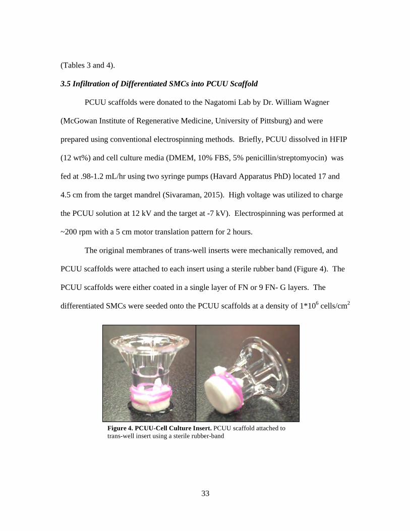
The original membranes of trans-well inserts were mechanically removed, and
PCUU scaffolds were attached to each insert using a sterile rubber band (Figure 4). The
PCUU scaffolds were either coated in a single layer of FN or 9 FN- G layers. The
differentiated SMCs were seeded onto the PCUU scaffolds at a density of 1*106 cells/cm2
Figure 4. PCUU-Cell Culture Insert. PCUU scaffold attached to trans-well insert using a sterile rubber-band
34
and cultured in SMIM for up to 7 days. After 1, 3, and 7 days, the cells were fixed and
stained with hematoxylin and eosin (See 2.4.1 Hematoxylin and Eosin Staining and
Immunofluorescence). Semi-quantitative analysis was performed which included
determining the maximum infiltration depth, average infiltration depth and the
distribution of cellular infiltration.
35
Chapter 4: Results
4.1 SMC Differentiation
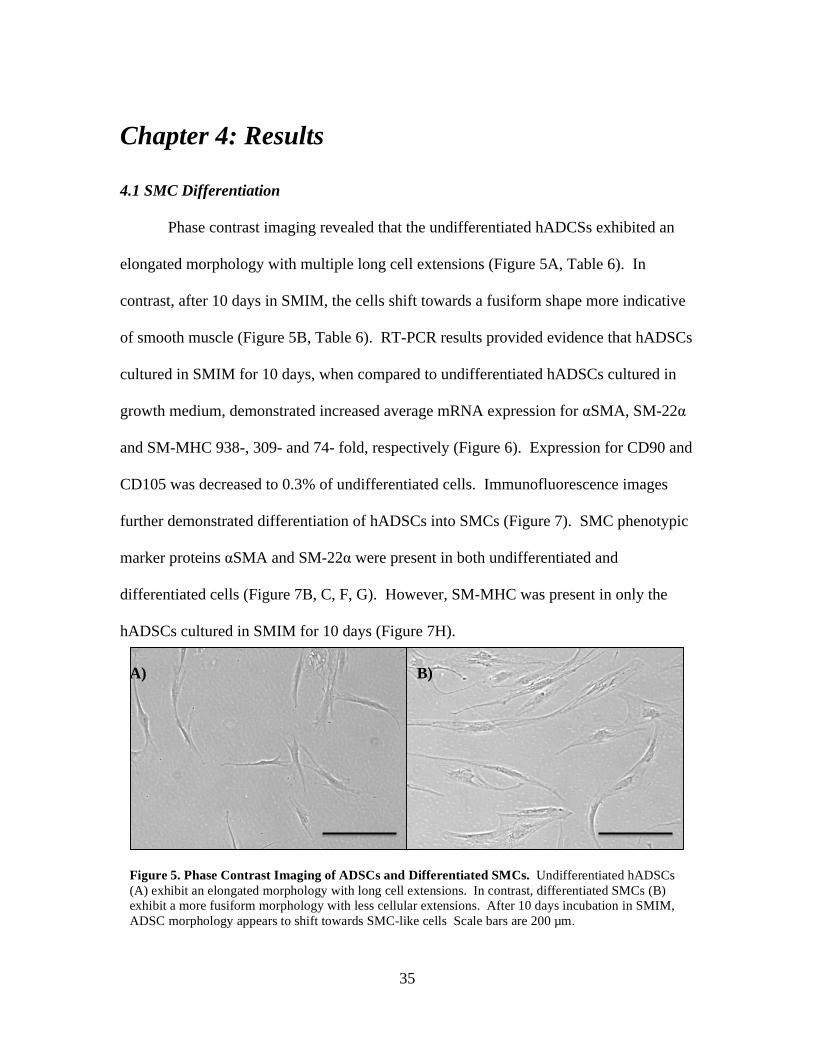
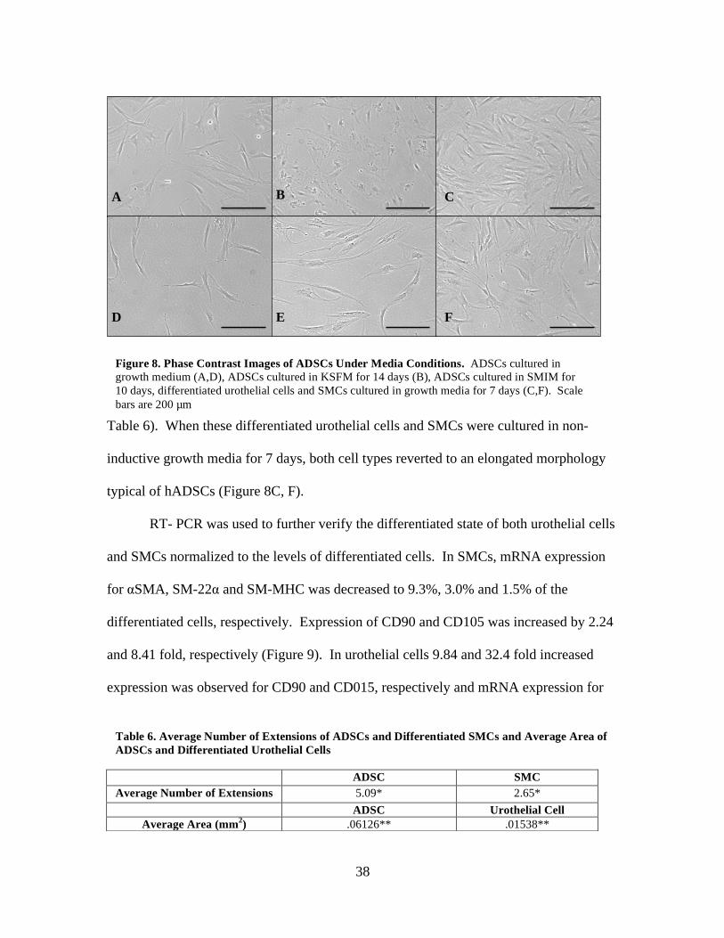
Phase contrast imaging revealed that the undifferentiated hADCSs exhibited an
elongated morphology with multiple long cell extensions (Figure 5A, Table 6). In
contrast, after 10 days in SMIM, the cells shift towards a fusiform shape more indicative
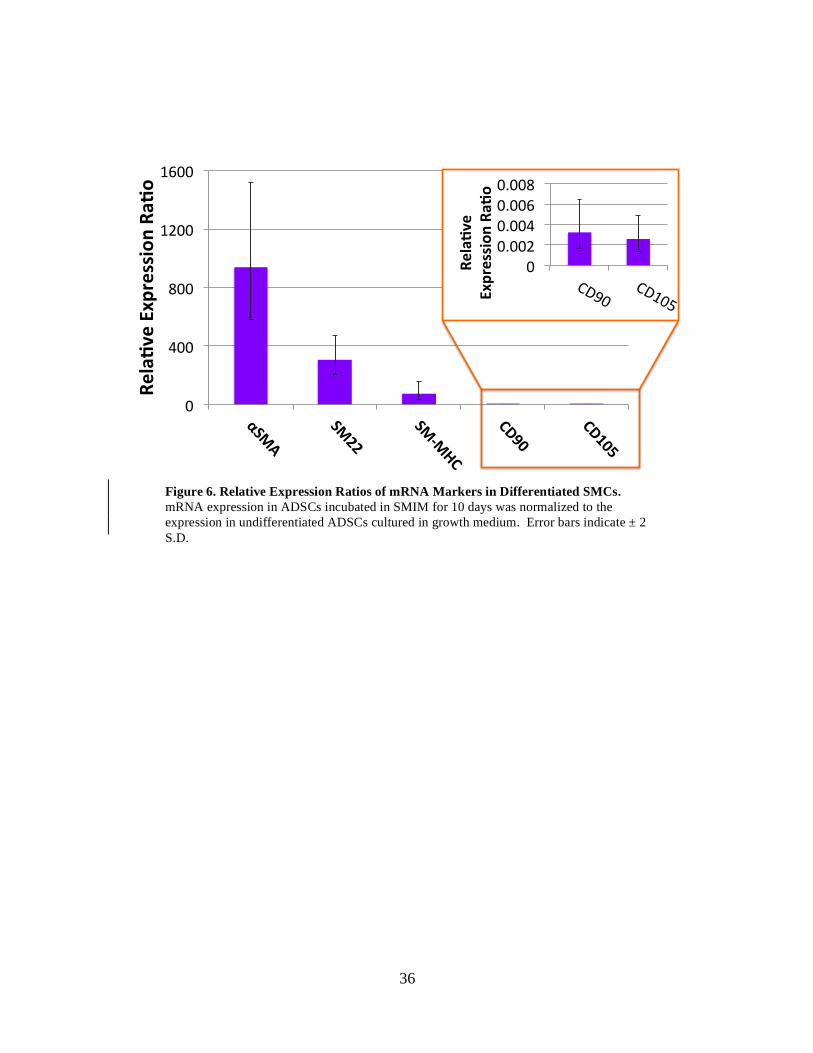
of smooth muscle (Figure 5B, Table 6). RT-PCR results provided evidence that hADSCs
cultured in SMIM for 10 days, when compared to undifferentiated hADSCs cultured in
growth medium, demonstrated increased average mRNA expression for αSMA, SM-22α
and SM-MHC 938-, 309- and 74- fold, respectively (Figure 6). Expression for CD90 and
CD105 was decreased to 0.3% of undifferentiated cells. Immunofluorescence images
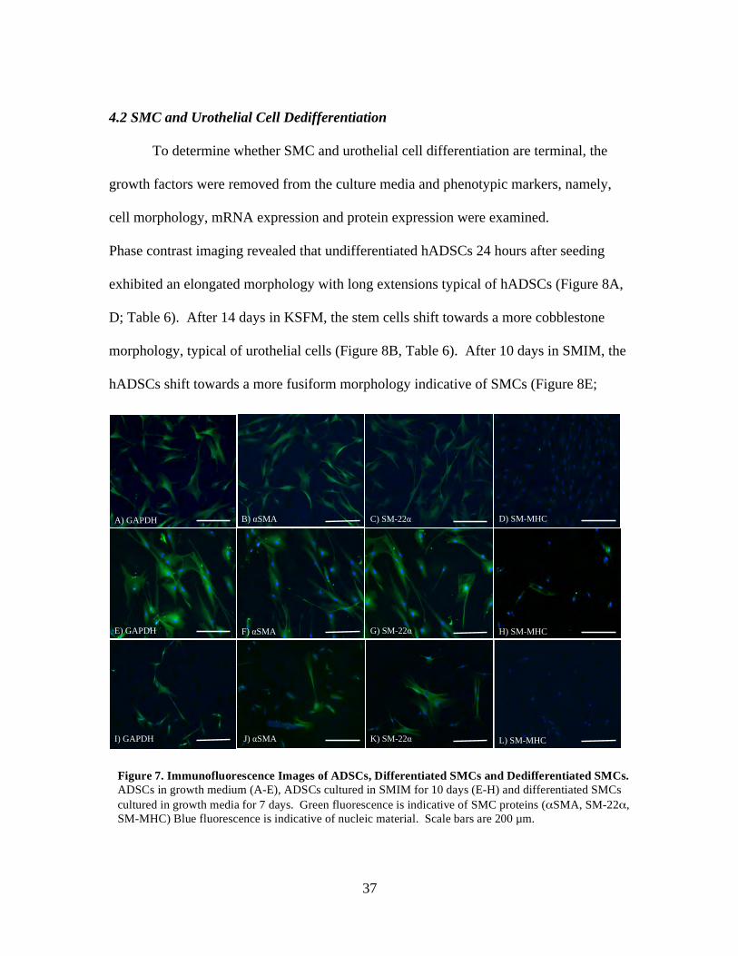
further demonstrated differentiation of hADSCs into SMCs (Figure 7). SMC phenotypic
marker proteins αSMA and SM-22α were present in both undifferentiated and
differentiated cells (Figure 7B, C, F, G). However, SM-MHC was present in only the
hADSCs cultured in SMIM for 10 days (Figure 7H).
Figure 5. Phase Contrast Imaging of ADSCs and Differentiated SMCs. Undifferentiated hADSCs (A) exhibit an elongated morphology with long cell extensions. In contrast, differentiated SMCs (B)exhibit a more fusiform morphology with less cellular extensions. After 10 days incubation in SMIM,ADSC morphology appears to shift towards SMC-like cells Scale bars are 200 µm.
A) B)
36
0"
400"
800"
1200"
1600"
αSMA%
SM22%
SM'MHC%
CD90%
CD105%
Rela3v
e%Expression
%Ra3
o%
0"0.002"0.004"0.006"0.008"
CD90"CD105"
Rela3v
e%Expression
%Ra3
o%Figure 6. Relative Expression Ratios of mRNA Markers in Differentiated SMCs. mRNA expression in ADSCs incubated in SMIM for 10 days was normalized to the expression in undifferentiated ADSCs cultured in growth medium. Error bars indicate ± 2 S.D.
37
4.2 SMC and Urothelial Cell Dedifferentiation
To determine whether SMC and urothelial cell differentiation are terminal, the
growth factors were removed from the culture media and phenotypic markers, namely,
cell morphology, mRNA expression and protein expression were examined.
Phase contrast imaging revealed that undifferentiated hADSCs 24 hours after seeding
exhibited an elongated morphology with long extensions typical of hADSCs (Figure 8A,
D; Table 6). After 14 days in KSFM, the stem cells shift towards a more cobblestone
morphology, typical of urothelial cells (Figure 8B, Table 6). After 10 days in SMIM, the
hADSCs shift towards a more fusiform morphology indicative of SMCs (Figure 8E;
A) GAPDH B) αSMA C) SM-22α D) SM-MHC
E) GAPDH F) αSMA G) SM-22α H) SM-MHC
I) GAPDH J) αSMA K) SM-22α L) SM-MHC
Figure 7. Immunofluorescence Images of ADSCs, Differentiated SMCs and Dedifferentiated SMCs. ADSCs in growth medium (A-E), ADSCs cultured in SMIM for 10 days (E-H) and differentiated SMCs cultured in growth media for 7 days. Green fluorescence is indicative of SMC proteins (αSMA, SM-22α, SM-MHC) Blue fluorescence is indicative of nucleic material. Scale bars are 200 µm.
38
Table 6). When these differentiated urothelial cells and SMCs were cultured in non-
inductive growth media for 7 days, both cell types reverted to an elongated morphology
typical of hADSCs (Figure 8C, F).
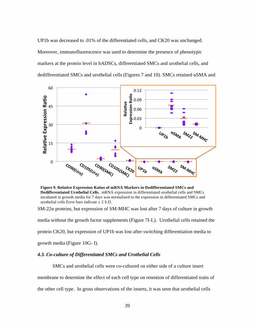
RT- PCR was used to further verify the differentiated state of both urothelial cells
and SMCs normalized to the levels of differentiated cells. In SMCs, mRNA expression
for αSMA, SM-22α and SM-MHC was decreased to 9.3%, 3.0% and 1.5% of the
differentiated cells, respectively. Expression of CD90 and CD105 was increased by 2.24
and 8.41 fold, respectively (Figure 9). In urothelial cells 9.84 and 32.4 fold increased
expression was observed for CD90 and CD015, respectively and mRNA expression for
ADSC SMC Average Number of Extensions 5.09* 2.65*
ADSC Urothelial Cell Average Area (mm2) .06126** .01538**
A B C
D E F
Figure 8. Phase Contrast Images of ADSCs Under Media Conditions. ADSCs cultured in growth medium (A,D), ADSCs cultured in KSFM for 14 days (B), ADSCs cultured in SMIM for 10 days, differentiated urothelial cells and SMCs cultured in growth media for 7 days (C,F). Scale bars are 200 µm
Table 6. Average Number of Extensions of ADSCs and Differentiated SMCs and Average Area of ADSCs and Differentiated Urothelial Cells
39
UP1b was decreased to .01% of the differentiated cells, and CK20 was unchanged.
Moreover, immunofluorescence was used to determine the presence of phenotypic
markers at the protein level in hADSCs, differentiated SMCs and urothelial cells, and
dedifferentiated SMCs and urothelial cells (Figures 7 and 10). SMCs retained αSMA and
SM-22α proteins, but expression of SM-MHC was lost after 7 days of culture in growth
media without the growth factor supplements (Figure 7I-L). Urothelial cells retained the
protein CK20, but expression of UP1b was lost after switching differentiation media to
growth media (Figure 10G- I).
4.3. Co-culture of Differentiated SMCs and Urothelial Cells
SMCs and urothelial cells were co-cultured on either side of a culture insert
membrane to determine the effect of each cell type on retention of differentiated traits of
the other cell type. In gross observations of the inserts, it was seen that urothelial cells
0
15
30
45
60
Rela%v
eExpression
Ra%
o
CD90(Uro)
CD105(Uro)
CD90(SMC)
CD105(SMC)
CK20UP1b
αSMASM22
SM-MHC
UP1bαSMA
SM22SM-MHC
0
0.03
0.06
0.09
0.12
Rela%v
eExpression
Ra%
o
Figure 9. Relative Expression Ratios of mRNA Markers in Dedifferentiated SMCs and Dedifferentiated Urothelial Cells. mRNA expression in differentiated urothelial cells and SMCs incubated in growth media for 7 days was normalized to the expression in differentiated SMCs and urothelial cells Error bars indicate ± 2 S.D.

40
Figure 10. Immunofluorescence Images of ADSCs Differentiated Urothelial Cells and Dedifferentiated Urothelial Cells. Undifferentiated ADSCs cultured in growth medium (A-C), ADSCs cultured in KSFM for 14 days (D-F), differentiated urothelial cells cultured in growth media for 7 days (G-I). Green fluorescence is indicative of urothelial proteins (UP1b, CK20). Blue fluorescence is indicative of nucleic material. Scale bars are 200 µm.
A) GAPDH B) CK20 C) UP1b
E) CK20
H) CK20 I) UP1bG) GAPDH
D) GAPDH F) UP1b
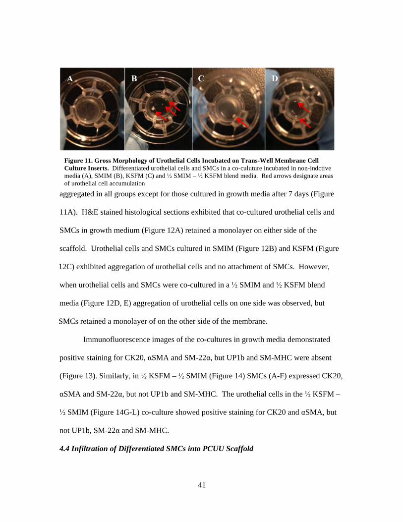
41
aggregated in all groups except for those cultured in growth media after 7 days (Figure
11A). H&E stained histological sections exhibited that co-cultured urothelial cells and
SMCs in growth medium (Figure 12A) retained a monolayer on either side of the
scaffold. Urothelial cells and SMCs cultured in SMIM (Figure 12B) and KSFM (Figure
12C) exhibited aggregation of urothelial cells and no attachment of SMCs. However,
when urothelial cells and SMCs were co-cultured in a ½ SMIM and ½ KSFM blend
media (Figure 12D, E) aggregation of urothelial cells on one side was observed, but
SMCs retained a monolayer of on the other side of the membrane.
Immunofluorescence images of the co-cultures in growth media demonstrated
positive staining for CK20, αSMA and SM-22α, but UP1b and SM-MHC were absent
(Figure 13). Similarly, in ½ KSFM – ½ SMIM (Figure 14) SMCs (A-F) expressed CK20,
αSMA and SM-22α, but not UP1b and SM-MHC. The urothelial cells in the ½ KSFM –
½ SMIM (Figure 14G-L) co-culture showed positive staining for CK20 and αSMA, but
not UP1b, SM-22α and SM-MHC.
4.4 Infiltration of Differentiated SMCs into PCUU Scaffold
Figure 11. Gross Morphology of Urothelial Cells Incubated on Trans-Well Membrane Cell Culture Inserts. Differentiated urothelial cells and SMCs in a co-culuture incubated in non-indctive media (A), SMIM (B), KSFM (C) and ½ SMIM – ½ KSFM blend media. Red arrows designate areas of urothelial cell accumulation
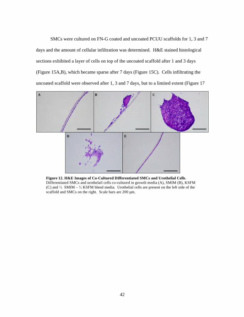
42
SMCs were cultured on FN-G coated and uncoated PCUU scaffolds for 1, 3 and 7
days and the amount of cellular infiltration was determined. H&E stained histological
sections exhibited a layer of cells on top of the uncoated scaffold after 1 and 3 days
(Figure 15A,B), which became sparse after 7 days (Figure 15C). Cells infiltrating the
uncoated scaffold were observed after 1, 3 and 7 days, but to a limited extent (Figure 17
A B C
D E
Figure 12. H&E Images of Co-Cultured Differentiated SMCs and Urothelial Cells. Differentiated SMCs and urothelail cells co-cultured in growth media (A), SMIM (B), KSFM (C) and ½ SMIM – ½ KSFM blend media. Urothelial cells are present on the left side of thescaffold and SMCs on the right. Scale bars are 200 µm.
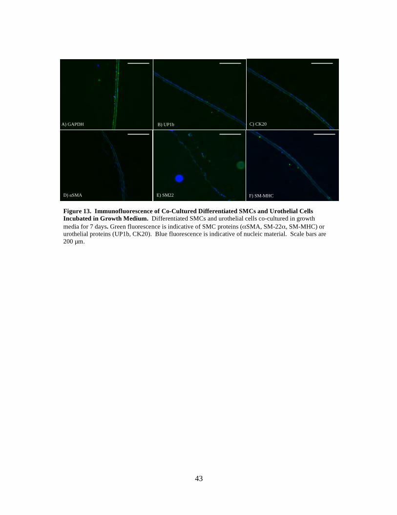
43
C) CK20 A) GAPDH
F) SM-MHC
B) UP1b
D) αSMA E) SM22
Figure 13. Immunofluorescence of Co-Cultured Differentiated SMCs and Urothelial Cells Incubated in Growth Medium. Differentiated SMCs and urothelial cells co-cultured in growth media for 7 days. Green fluorescence is indicative of SMC proteins (αSMA, SM-22α, SM-MHC) or urothelial proteins (UP1b, CK20). Blue fluorescence is indicative of nucleic material. Scale bars are 200 µm.
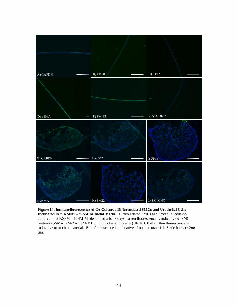
44
Figure 14. Immunofluorescence of Co-Cultured Differentiated SMCs and Urothelial Cells Incubated in ½ KSFM – ½ SMIM Blend Media. Differentiated SMCs and urothelial cells co-cultured in ½ KSFM – ½ SMIM blend media for 7 days. Green fluorescence is indicative of SMC proteins (αSMA, SM-22α, SM-MHC) or urothelial proteins (UP1b, CK20). Blue fluorescence is indicative of nucleic material. Blue fluorescence is indicative of nucleic material. Scale bars are 200 µm.
D) αSMA
B) CK20A) GAPDH
F) SM-MHCE) SM-22
C) UP1b
J) αSMA
H) CK20G) GAPDH
L) SM-MHCK) SM22
I) UP1b
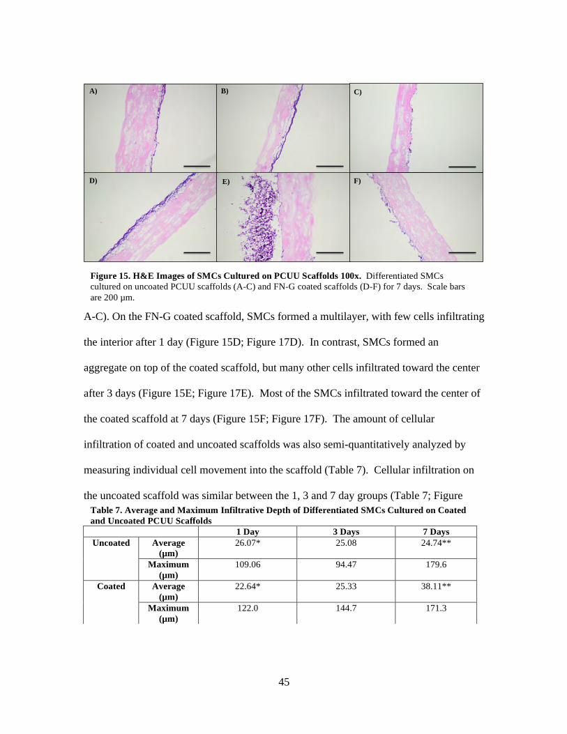
45
A-C). On the FN-G coated scaffold, SMCs formed a multilayer, with few cells infiltrating
the interior after 1 day (Figure 15D; Figure 17D). In contrast, SMCs formed an
aggregate on top of the coated scaffold, but many other cells infiltrated toward the center
after 3 days (Figure 15E; Figure 17E). Most of the SMCs infiltrated toward the center of
the coated scaffold at 7 days (Figure 15F; Figure 17F). The amount of cellular
infiltration of coated and uncoated scaffolds was also semi-quantitatively analyzed by
measuring individual cell movement into the scaffold (Table 7). Cellular infiltration on
the uncoated scaffold was similar between the 1, 3 and 7 day groups (Table 7; Figure
A) B) C)
D) E) F)
1 Day 3 Days 7 Days Uncoated Average
(µm) 26.07* 25.08 24.74**
Maximum (µm)
109.06 94.47 179.6
Coated Average (µm)
22.64* 25.33 38.11**
Maximum (µm)
122.0 144.7 171.3
Figure 15. H&E Images of SMCs Cultured on PCUU Scaffolds 100x. Differentiated SMCs cultured on uncoated PCUU scaffolds (A-C) and FN-G coated scaffolds (D-F) for 7 days. Scale bars are 200 µm.
Table 7. Average and Maximum Infiltrative Depth of Differentiated SMCs Cultured on Coated and Uncoated PCUU Scaffolds
46
16A). When compared to the coated scaffold, SMC infiltration of the uncoated scaffold
was significantly greater after 1 day (Table 7). The amount of cellular infiltration
between coated and uncoated scaffolds was similar after 3 days. However, cellular
infiltration of the coated scaffold was significantly greater than that of the uncoated
scaffold after 7 days (Table 7). In addition, after 7 days, SMCs infiltrated to a greater
degree when compared to 1 and 3 days, and to all uncoated groups (Figure 16).
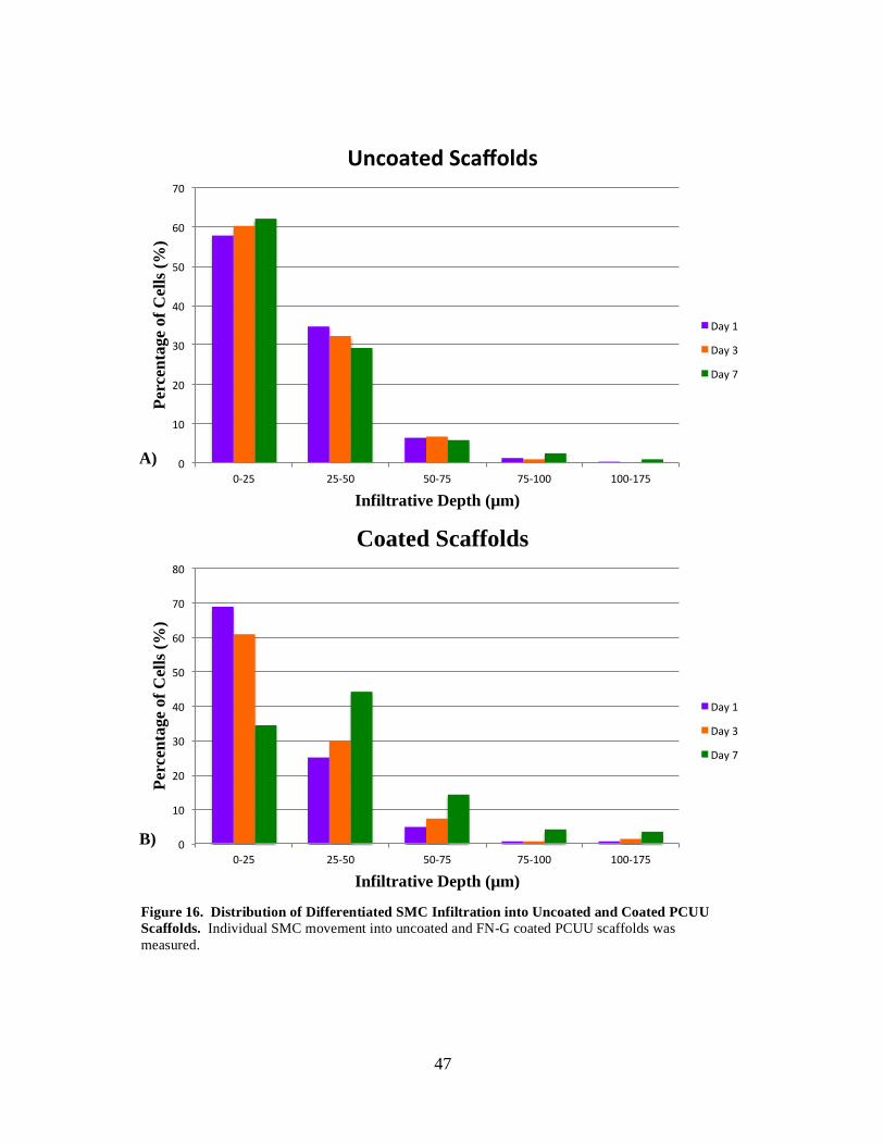
47
0
10
20
30
40
50
60
70
0-25 25-50 50-75 75-100 100-175
Perc
enta
ge o
f Cel
ls (%
)
Infiltrative Depth (µm)
UncoatedScaffolds
Day1
Day3
Day7
A)
0
10
20
30
40
50
60
70
80
0-25 25-50 50-75 75-100 100-175
Perc
enta
ge o
f Cel
ls (%
)
Infiltrative Depth (µm)
Coated Scaffolds
Day1
Day3
Day7
B)
Figure 16. Distribution of Differentiated SMC Infiltration into Uncoated and Coated PCUU Scaffolds. Individual SMC movement into uncoated and FN-G coated PCUU scaffolds was measured.
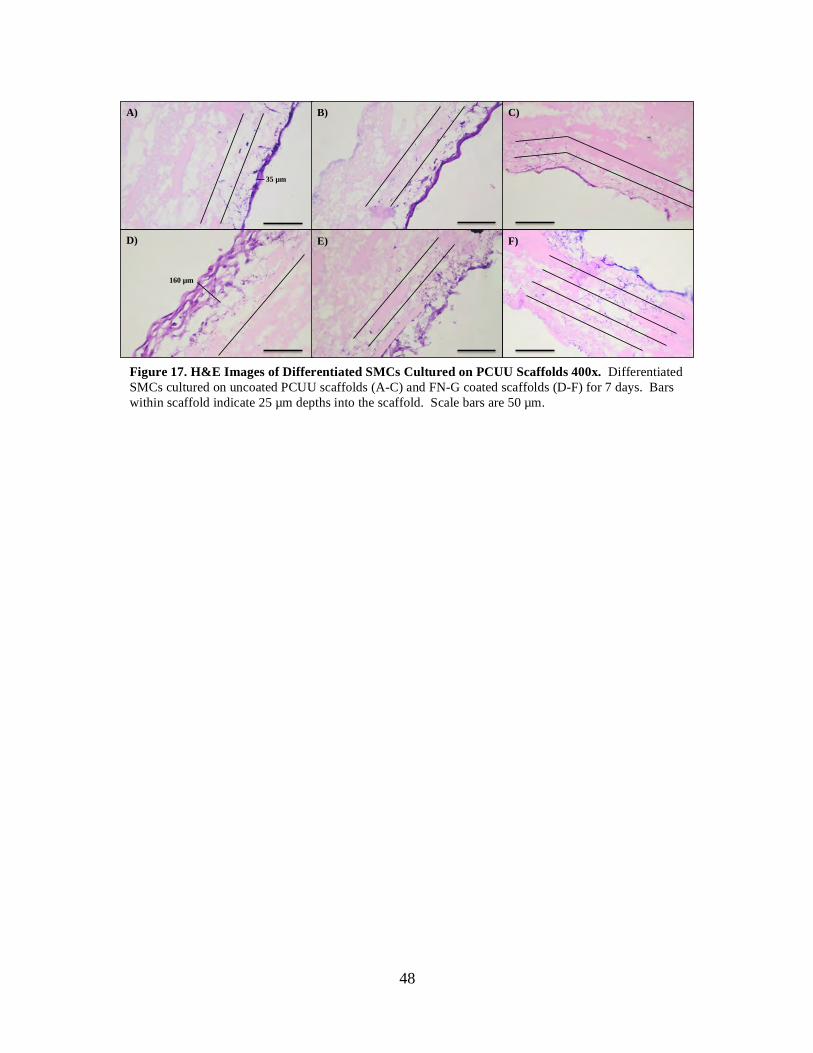
48
35 µm
160 µm
A) B) C)
D) E) F)
Figure 17. H&E Images of Differentiated SMCs Cultured on PCUU Scaffolds 400x. Differentiated SMCs cultured on uncoated PCUU scaffolds (A-C) and FN-G coated scaffolds (D-F) for 7 days. Bars within scaffold indicate 25 µm depths into the scaffold. Scale bars are 50 µm.
49
Chapter 5: Discussion
A number of previous in vitro studies have shown that different media conditions
impact cell morphology, genetic transcription and protein expression of ADSCs (Tiang et
al, 2010; Zhang et al, 2014; Lee et al, 2015; Faroni et al, 2013; Stromps et al, 2014;
Wang et al, 2014; Harris et al, 2011; Zhao, 2015). Other studies have shown that, along
with media conditions, biomechanical signaling from cell-to-cell contact also influences
stem cell differentiation (Merfeld-Clauss et al, 2014; Liu, 2009). Based these studies, it
was hypothesized that factors within the cellular environment, such as soluble factors and
cell-to-cell contact, are important stimuli for attaining and retaining differentiated traits.
For this reason, ADSCs, differentiated SMCs and differentiated urothelial cells were
cultured in varying media conditions. Moreover, differentiated SMCs and differentiated
urothelial cells were co-cultured, and the cellular responses of each cell type were
observed.
5.1 SMC Differentiation
Results of the present study demonstrate changes indicative of a shift towards a
contractile SMC phenotype in cellular morphology when ADSCs were cultured in SMIM
for 10 days. Specifically, about 90% of the differentiated SMCs exhibited a more
spindle-like morphology with less cellular extensions compared to ADSCs. This was
consistent with previous reports of a phenotypic shift from ADSCs toward a contractile
SMC phenotype (Wang et al, 2010; Harris et al, 2011). In addition, SMC mRNA
expression of αSMA, SM-22α and SM-MHC was greater in the differentiated cells than
ADSCs and exhibited a more quantitative indication that SMIM stimulated SMC
50
differentiation (Figure 6). SMC positive protein expression of αSMA, SM-22α and SM-
MHC after differentiation also indicated differentiation toward a contractile SMC
phenotype. Out of the phenotypic markers examined, αSMA is the most highly conserved
among various cell types, including pluripotent stem cells and fibroblasts, and is only an
early indication of SMC differentiation (Harris et al, 2009; Lao et al, 2016). In contrast,
SM-22α is only conserved amongst pluripotent stem cells, including ADSCs, and SMCs
and is therefore an intermediate indicator of SMC differentiation (Harris et al, 2009).
Finally, SM-MHC is only expressed in contractile SMCs, and is therefore a specific
indicator of advanced SMC differentiation (Harris et al, 2009). The results of the present
study are in agreement with previous studies that exposed human ADSCs to soluble
factors in media to induce differentiation into a SMC phenotype. For example, Wang et
al. reported that culturing ADSCs cultured with TGF-β1 (5 ng/mL) and BMP-4 (2.5
ng/mL) resulted in a shift towards a SMC genotype and phenotype (Wang et al, 2010).
However, Wang et al used twice as much TGF-β1 than was used in the present study and
successfully differentiated ADSCs toward a contractile SMC phenotype in 7 days. In a
separate study by Harris et al, ADSCs were differentiated into SMCs by culturing with
angiotensin II (1µM), sphingosylphosphorylcholine (2 µM) or TGF-β1 (2 ng/mL) (Harris
et al, 2011). Harris et al used less TGF- β1, but it took 3 weeks to successfully
differentiate ADSCs into SMCs. The results of the present study indicate that ADSC
differentiation into SMCs can be achieved using a lesser quantity of soluble factors and in
a shorter time period when compared to other reports, which reduces overall costs of cell
preparation.
51
Although there are a multitude of methodologies to obtain cells needed for tissue
engineering (Mahfouz et al, 2013; Tran & Damaser, 2015; Takahashi & Yamanaka,
2006; Kim et al, 2014; de Villiers et al, 2009), due to the lack of healthy autologous cells
in a diseased bladder, differentiation techniques requiring co-cultures and conditioned
media with autologous cells is not a plausible approach (Jack et al, 2009, Mahfouz et al,
2013). Exposing stem cells to soluble factors, however, is a faster approach to
differentiate ADSCs into SMCs. While ADSCs can be differentiated into SMCs under
SMIM in less than 3 weeks, it can require up to 6 weeks to expand autologous cells to
acquire the necessary amount of cells for seeding a scaffold (Mahfouz et al, 2013).
Harvesting smooth muscle and urothelial tissue from the body also includes an invasive
surgical procedure, which introduces risk to the patient. Differentiation using soluble
factors, however, has been shown to only partially differentiate cells, and once the
soluble factors are removed the cells revert back to a more primitive phenotype (Liu et al,
2011).
5.2 SMC and Urothelial Cell Dedifferentiation
After ADSCs were differentiated into SMCs and urothelial cells in the induction
media (SMIM and KSFM, respectively), their ability to retain their phenotypic traits was
inspected. Results of the present study indicate that ADSC differentiation toward SMC
and urothelial lineages were partial and they reverted to a stem cell-like cell once the
soluble factors were removed. There was a shift in cell morphology toward that of an
ADSC in response to the removal of soluble factors (Figure 8). Expression of markers at
both the mRNA and protein level also suggests a shift away from the differentiated
52
phenotype (Figure 9, 10). These results were similar to those of previous studies that also
reported dedifferentiation when soluble factors were removed (Liu et al, 2011). In a
study by Liu et al, bone marrow stem cells were differentiated toward a neuronal lineage
under a neuronal induction medium containing DMEM and soluble factors (2% dimethyl
sulfoxide, 200 µM butylated hydroxyanisole, 25 mM potassium chloride, 2mM valporic
acid, 10 µM forskolin, 1 µM hydrocortisone and 5 µg/ mL insulin), and subsequently
dedifferentiated by culturing in media without the soluble factors. Although these
dedifferentiated cells expressed stem cell characteristics, they exhibited a reprogrammed
phenotype different from the original stem cell population (Liu et al, 2011). Moreover,
after dedifferentiation the cells demonstrated enhanced cell survival and higher efficacy
in neuronal differentiation when compared to unmanipulated stem cells. (Liu et al, 2011).
Together with the previous findings of Liu et al, it can be speculated that the
dedifferentiated cells in the present study are distinctly different from their original
ADSC population and that they may also exhibit a higher efficacy in differentiating
toward SMC and urothelial cells. This is further supported by the retention of CK20 in
dedifferentiated urothelial cells (Figure 9,10). Finally, these dedifferentiated cells might
also exhibit increased cell survival, which may be useful for enduring the body’s
inflammatory response when implanted for tissue-engineering purposes.
5.3 Co-culture of Differentiated SMCs and Urothelial Cells
The present study also aimed to determine the ability of differentiated SMCs and
urothelial cells to retain their phenotypic traits in co-cultures with various media
conditions. After incubation for 7 days, aggregation of urothelial cells was seen in all
53
induction media groups (SMIM, KSFM, ½ SMIM- ½ KSFM) but not in growth medium
(DMEM, 10% FBS, 1% Glutamax). The results of the present study were similar to
those of a previous study in our laboratory. Briefly, differentiated urothelial cells (8*106
cells/ well) in KSFM were seeded onto trans-well cell culture inserts to explore the
feasibility of developing a multilayer urothelial tissue (Turner, 2015). After 24 hours,
however, all of the urothelial cells aggregated, which was visible under gross
examination and H&E staining (Turner, 2015). One of the possible reasons for the
aggregation of the urothelial cells is that the induction media (SMIM, KSFM, ½ SMIM –
½ KSFM) lacked FBS, which contains multiple soluble factors that enhance cell
adhesion, including vitronectin. In a study by Hayman et al, vitronectin was shown to
increase adhesion of bovine corneal endothelial cell adhesion by 8-16 fold when
compared to fibronectin (Hayman et al, 1985; Underwood & Bennett, 1989). The
presence of vitronectin and other factors in FBS could explain the absence of urothelial
cell aggregation in the group cultured in serum-containing growth medium.
In previous studies, indirect co-cultures of autologous cells (adipocytes,
chondrocytes, osteoblasts and urothelial cells) and stem cells have been proven to be an
effective method in differentiating stem cells toward a certain lineage (Zhao et al, 2015;
Zhang et al, 2014). It is widely believed that autocrine and paracrine signaling are
responsible for this differentiation. Therefore, it was hypothesized that two types of
differentiated cells (SMCs and urothelial cells) would release cytokines and aid in the
retention of phenotypic traits. However, after incubation in an indirect co-culture for 7
days, only protein expression of αSMA, SM-22α and CK20 were retained and expression
54
SM-MHC and UP1b were lost. These results are identical to SMCs and urothelial cells
incubated in growth medium separately, meaning that the co-culture did not support the
retention of differentiated traits. The cells in the present study were only partially
differentiated; therefore, it can be hypothesized that they do not release the same
biochemical signals as fully differentiated SMCs and urothelial cells, which would lead
to a difference in cellular response. Moreover, it can be speculated that cell-to-cell
contact is an important factor, although chemical signaling does play a role in
differentiation and retention of differentiated traits. Previously, it was found that only
ADSCs co-cultured directly with urothelial cells differentiated into urothelial cells, but
not ADSCs co-cultured indirectly (Liu et al, 2009). In another study, ADSCs closest to
endothelial cells in a direct co-culture differentiated toward a SMC lineage more quickly
than those further away (Merfeld-Clauss et al, 2014). Therefore, it can be concluded that
both soluble factors and cell-to-cell contact in the cellular microenvironment are of
upmost importance when determining differentiation and retention of differentiated traits.
5.4 Infiltration of Differentiated SMCs into PCUU scaffold
The present study examined the effects of layer-by-layer fibronectin (FN) and
gelatin (G) coating of a PCUU scaffold on SMC infiltration. The results indicate that the
presence of the ECM proteins on a PCUU scaffold increases the effectiveness of SMC
infiltration into a PCUU scaffold. These results are in agreement with previous studies
that have demonstrated the effect of proteins on cellular infiltration into a scaffold. A
study by Ardila et al demonstrated that the presence of TGF-β2 (.1 ng/mL) greatly
increased the amount of infiltration of autologous SMCs into an electrospun membrane
55
when compared to cells cultured on the electrospun membrane without TGF-β2 (Ardila et
al, 2015). Additionally, in a study by Uchida et al, PCUU scaffolds were alternately
immersed in FN and G to create thickness-controlled nanolayers (Uchida et al, 2015).
This nano-layer formation is based on non-specific physical adsorption of FN onto the
polymer and its subsequent interactions with G (Uchida et al, 2015). The presence of G
allows FN to infiltrate the interior space of the fibrous PCUU, without G it was
discovered that FN accumulated on the surface of the scaffold (Uchida et al, 2015). The
presence of FN and G was also shown to aid in cellular adhesion to the scaffold, and in
cellular infiltration into the interior of the scaffold (Uchida et al, 2015). This was likely
due to the cell-binding domain present on FN, which aids in cellular attachment
(Matusaki et al, 2012).
The greater degree of SMC adhesion on the coated scaffolds when compared to
the uncoated scaffolds after 1 day was likely due to the presence of FN and G multilayers
(Figure 16; Figure 17). FN provided cellular adhesion sites both on the surface and
within the fibrous interior of the scaffold, which likely supported SMC adhesion.
Additionally, Uchida et al determined that cells actively migrate toward a scaffold with
FN-G coating (Uchida et al, 2015). The increased infiltration of SMCs into the coated
scaffold when compared to the uncoated scaffold after 7 days was likely attributed to
SMCs affinity for the FN-G coating present within the interior of the scaffold.
56
Chapter 6: Conclusions and Recommendations
In the present study, culturing ADSCs in SMIM was proven to be an effective
method in differentiating toward a SMC lineage. However, differentiated SMCs and
urothelial cells dedifferentiated once factors important to differentiation were removed.
Co-culturing of differentiated SMCs and urothelial cells did not support retention of
differentiated traits either. Taken together, these in vitro results indicate that cells
differentiated via soluble factors within the media only achieve partial differentiation.
When the differentiated SMCs were seeded on a PCUU scaffold, layer-by-layer coating
with fibronectin and gelatin enhanced the infiltration into the scaffold. This is likely due
to the adsorption of fibronectin to the scaffold and subsequent interaction with gelatin
provides cellular adhesion sites both within and on the surface of the scaffold. Future
study recommendations include:
• To explore the effect of a direct co-culture of differentiated SMCs and
urothelial cells on the retention of differentiated traits.
o Rationale: Biomechanical signaling through cell-to-cell contact is
an important factor in the process of differentiation, and may lead
to the retention of differentiated traits.
• To investigate the effect of layer-by-layer coating of both a PCUU
scaffold and SMCs on cellular infiltration into the scaffold.
57
o Rationale: Layer-by-layer coating of the cells will increase cell-to-
matrix interactions and possibly eliminate the accumulation of
SMCs on the PCUU scaffold.
• To explore the effects of soluble factors incorporated into the PCUU
electrospun membrane.
o Rationale: The presence of soluble factors has a direct effect on
the retention of differentiated traits and incorporation of these
factors would lead to paracrine signaling after the inductive media
has been removed.
• To investigate the cellular changes in differentiated SMCs and urothelial
cells after dedifferentiation in regards to cell survival and efficacy in
differentiation.
o Rationale: In the present study, only the process of
dedifferentiation is explored, and not the positive effect
dedifferentiation may have on the cells.
• To explore the effects of cyclic strain on the effectiveness of SMC
infiltration into a PCUU scaffold and on the retention of differentiated
traits.
o Rationale: The present study only investigates static scaffolds
seeded with SMCs and cyclic strain will intermittently increase
porosity of the scaffold. SMCs within the body also undergo
58
constant cyclic strain, which is an environmental condition that can
affect the retention of differentiated traits.
• To investigate the effects of layer-by-layer coating a PCUU electrospun
scaffold with alternating layers of fibronectin, vitronectin and gelatin on
SMC infiltration
o Rationale: The present study only investigates the effects of
fibronectin on cellular adhesion. Vitronectin also greatly enhances
cellular adhesion and may aid in cellular infiltration as well.
59
REFERENCES
1. Tanagho, Emil A., and Jack W. McAninch, eds. Smith's General Urology. 17thed. New York: McGraw Hill, 2008.
2. DeLancey, J. Gross Anatomy and Cell Biology of the Lower Urinary Tract. In: PAmbrams, L Cardozo, S Khoury, A Wein, eds. Incontinence: Third InternationalConsultation on Incontinence, second ed. Plymouth, UK: Health PublicationsLtd.; 2002: 19-82.
3. Bergman, Ronald A., et al. "Urinary Bladder." Anatomy Atlases. N.p., 2015. Web.22 July 2015. <http://www.anatomyatlases.org/>.
4. Oberpenning, Frank, et al. "De novo reconstitution of a functional mammalianurinary bladder by tissue engineering." Nature Biotechnology 17 (1999): 149-55.
5. Bouhout, Sara, et al. "Potential of Different Tissue Engineering Strategies in theBladder Reconstruction." INTECH (2013): 573-97.
6. Atala, Anthony, et al. "Tissue-engineered autologous bladders for patientsneeding cystoplasty." The Lancet 367 (2006): 1241-46.
7. Cleveland Clinic (2012). “Neurogenic Bladder.”http://my.clevelandclinic.org/health/diseases_conditions/hic-neurogenic-bladder
8. Mayoclinic (2014). “Neurogenic Bladder and Bowel Management.”http://www.mayoclinic.org/tests-procedures/neurogenic-bladder-bowel-management/basics/definition/prc-20013781
9. Merck Manual (2014). “Bladder Anomalies.” Merck Sharp and Dohme Corp.http://www.merckmanuals.com/professional/pediatrics/congenital-renal-and-genitourinary-anomalies/bladder-anomalies
10. American Cancer Society (2015). “What Is Bladder Cancer?”http://www.cancer.org/cancer/bladdercancer/detailedguide/bladder-cancer-what-is-bladder-cancer
11. Tu, Duong D., et al. "Bladder tissue regeneration using acellular bi-layer silkscaffolds in a large animal model of augmentation cystoplasty." Biomaterials 34(2013): 8681-89.
12. Vasdev, Nikhil, et al. "Metabolic Complications of Urinary Intestinal Diversion."Indian Journal of Urology 29.4 (2013): 310-15.
60
13. Atala, Anthony. "Tissue Engineering of Human Bladder." British MedicalBulletin 97.1 (2011): 81-104.
14. Silberstein, Jonathon L., et al. "Urinary Diversion Practice Patterns AmongCertifying American Urologists." Journal of Urology 189.3 (2013): 1042-47.
15. Pruthi, Raj S., et al. "Recent Trends in the Urology Workforce in the UnitedStates." Urology 82.5 (2013): 987-94.
16. National Clinical Guideline Center. “Urinary Incontinence in NeurologicalDisease: Management of Lower Urinary Tract Dysfunction in NeurologicalDisease.” National Institute of Health (2012).
17. Mahfouz, W., et al. "Fundamentals of bladder tissue engineering." AfricanJournal of Urology 19 (2013): 51-57.
18. Lai, Jin-Yao, et al. "Bladder autoaugmentation using various biodegradablescaffolds seeded with autologous smooth muscle cells in a rabbit model." Journalof Pediatric Surgery 40 (2005): 1869-73.
19. Kim, Jae Heon, et al. "Treatment of Bladder Dysfunction Using Stem Cell orTissue Engineering Technique." Korean Journal of Urology 55 (2014): 228-38.
20. De Villiers, Jennifer Anne, et al. "Adipose Derived Stem Cells and SmoothMuscle Cells: Implications for Regenerative Medicine." Stem Cell Reviews andReports 5 (2009): 256-65.
21. Niemeyer, Philipp, et al. "Comparison of Immunological Properties of BoneMarrow Stromal Cells and Adipose Tissue–Derived Stem Cells Before and AfterOsteogenic Differentiation In Vitro." Tissue Engineering 13.1 (2007): 111-21.
22. Tian, Hong, et al. "Differentiation of Human Bone Marrow Mesenchymal StemCells into Bladder Cells: Potential for Urological Tissue Engineering." TissueEngineering 16.5 (2010): 1769-79.
23. Zhang, Ming, et al. "The Differentiation of Human Adipose-Derived Stem Cellstowards a Urothelium-Like Phenotype In Vitro and the Dynamic TemporalChanges of Related Cytokines by Both Paracrine and Autocrine SignalRegulation." PLOS ONE 9.4 (2014): 1-9.
24. Lee, Jingu, et al. "Transdifferentiation of mouse adipose-derived stromal cellsinto acinar cells of the submandibular gland using a co-culture system."Experimental Cell Research 334 (2015): 160-72.
61
25. Faroni, A., et al. "Differentiation of adipose-derived stem cells into Schwann cellphenotype induces expression of P2X receptors that control cell death." CellDeath and Disease 4 (2013): 1-11.
26. Zhao, Zhankui, et al. "Differentiation of adipose-derived stem cells promotesregeneration of smooth muscle for ureteral tissue engineering." Journal ofSurgical Research 178 (2012): 55-62.
27. Stromps, Jan-Philipp, et al. "Chondrogenic Differentiation of Human Adipose-Derived Stem Cells: A New Path in Articular Cartilage Defect Management?"BioMed Research International (2014): 1-7.
28. Wang, Chen, et al. "Differentiation of Adipose-Derived Stem Cells intoContractile Smooth Muscle Cells Induced by Transforming Growth Factor-b1 andBone Morphogenetic Protein-4." Tissue Engineering: Part A 16.4 (2010): 1201-13.
29. Harris, Lisa J., et al. "Differentiation of Adult Stem Cells into Smooth Muscle forVascular Tissue Engineering." Journal of Surgical Research 168 (2011): 306-14.
30. Zhao, Yimu, et al. "Multilineage co-culture of adipose-derived stem cells fortissue engineering." Journal of Tissue Engineering and Regenerative Medicine(2012): 1-12.
31. Liu, Yang, et al. "Dedifferentiation-Reprogrammed Mesenchymal Stem Cellswith Improved Therapeutic Potential." Stem Cells 29 (2011): 2077-89.
32. Zhang, Yiqiang, et al. "Dedifferentiation and Proliferation of MammalianCardiomyocytes." PLOS ONE 5.9 (2010): 1-13.
33. Odelberg, Shannon J., et al. "Dedifferentiation of Mammalian Myotubes Inducedby msx1." Cell 103 (2000): 1099-109.
34. Tran, Christine, and Margot S. Damaser. "The potential role of stem cells in thetreatment of urinary incontinence." Therapeutic Advances in Urology 7.1 (2015):22-40.
35. Takahashi, Kazutoshi, and Shinya Yamanaka. "Induction of Pluripotent StemCells from Mouse Embryonic and Adult Fibroblast Cultures by Defined Factors."Cell 126.4 (2006): 663-76.
36. Patal, Payal D, et al. "Pelvic organ prolapse and stress urinary incontinence: Areview of etiological factors." Indian Journal of Urology 23.2 (2007): 135-41.
62
37. Wood, Lauren N., and Jennifer T. Anger. "Urinary incontinence in women." Thebmj (2014): 1-11.
38. Stewart, WF. "Prevalence and burden of overactive bladder in the United States."World Journal of Urology 20.6 (2003): 327-36.
39. Liao, Yunjun, et al. "In Vivo Dedifferentiation of Adult Adipose Cells." PLOSOne (2015): 1-13.
40. Shoshani, Ofer, and Dov Zipori. "Stress as a fundamental theme in cell plasticity."Biochimica et Biophysica Acta (BBA) - Gene Regulatory Mechanisms 1849.4(2015): 371-77.
41. Veranic, Peter, and Kristijan Jezernik. "The Cytokeratins of Urinary BladderEpithelial Cells." Asian Journal of Cell Biology 1 (2006): 1-8.
42. Khandelwal, P., SN Abraham, and G. Apodaca. "Cell biology and physiology ofthe uroepithelium." American Journal of Physiology- Renal Physiology 297.6(2009): 1477-501.
43. Antoni, Delphine, et al. "Three-Dimensional Cell Culture: A Breakthrough inVivo." International Journal of Molecular Sciences 16.3 (2015): 5517-27.
44. Jack, Gregory S, et al. "Urinary bladder smooth muscle engineered from adiposestem cells and a three dimensional synthetic composite." Biomaterials 30.19(2009): 3259-70.
45. Kelly, Christopherl. "Bladder Dysfunction." NYU School of Medicine. N.p., n.d.Web. 24 Jan. 2016. <http://www.med.nyu.edu/urology/patient-care-information/conditions-we-treat/ bladder-dysfunction#definition-causes>.
46. Lin, Hsueh-Kung, et al. "Biomatrices for bladder reconstruction." Advanced DrugDelivery Reviews 82 (2015): 47-63.
47. Nikolopoulos, Kostis, et al. "The surgical management of recurrent stress urinaryincontinence: a systematic review." AOGS 94 (2015): 568-76.
48. Pattison, Megan A, et al. "Three-dimensional,nano-structured PLGA scaffolds forbladder tissue replacement applications." Biomaterials 26 (2005): 2491-500.
49. Zhong, Shaoping, et al. "Fabrication of Large Pores in Electrospun NanofibrousScaffolds for Cellular Infiltration: A Review." Tissue Engineering, Part B 18.2(2012): 77-87.
63
50. Ardila, Diana C, et al. "TGFb2 differentially modulates smooth muscle cellproliferation and migration in electrospun gelatin-fibrinogen constructs."Biomaterials 37 (2015): 164-73.
51. Brown, Allison L., et al. "Bladder acellular matrix as a substrate for studying invitro bladder smooth muscle–urothelial cell interactions." Biomaterials 26 (2005):529-43.
52. Khandelwal, Puneet, et al. "Cell biology and physiology of the uroepithelium."American Journal of Physiology Renal Physiology 297.6 (2009): 1477-501.
53. Sahai, Arun, et al. "Intravesicle Therapy for Refractory Overactive Bladder andDetrusor Overactivity in Adults: Botulinum Toxin-A." Minimally InvasiveTherapy for Urinary Incontinence and Pelvic Organ Prolapse: 135-54.
54. Brown, A. L. "22 week assessment of bladder acellular matrix as a bladderaugmentation material in a porcine model." Biomaterials 23 (2002): 2179-90.
55. Yoo, James J., et al. "Regenerative Medicine Strategies for Treating NeurogenicBladder." International Neurology Journal 15 (2011): 109-19.
56. BioPlan Associates. "Emerging Technologies and Opportunities in StructuralTissue Engineering." Kalorama Information Market Profile (2001): 1-424.
57. Bin, Zhao, et al. "Co-culture of neuroepithelial stem cells with interstitial cells ofCajal results in neuron differentiation." International Journal of Clinical andExperimental Medicine 8.7 (2015): 10437-43.
58. Merfled-Clauss, Stephanie, et al. "Adipose Stromal Cells Differentiate Along aSmooth Muscle Lineage Pathway Upon Endothelial Cell Contact via Induction ofActivin A." Circulation Research (2014): 800-09.
59. Liu, Jie, et al. "Cell-to-cell contact induces human adipose tissue-derived stromalcells to differentiate into urothelium-like cells in vitro." Biochemical andBiophysical Research Communications 390.3 (2009): 931-36.
60. Joseph, DB, et al. "Autologous cell seeded biodegradable scaffold foraugmentation cystoplasty: phase II study in children and adolescents with spinabifida." Journal of Urology 191.5 (2014): 1389-95.
61. Patsch, Christoph, et al. "Generation of vascular endothelial and smooth musclecells from human pluripotent stem cells." Nature Cell Biology 17 (2015): 994-1003.
64
62. Adamowicz, Jan, et al. "Tissue engineering of urinary bladder – current state ofart and future perspectives." Journal of Urology 66.2 (2013): 202-06.
63. Joseph, DB, et al. "Autologous cell seeded biodegradable scaffold foraugmentation cystoplasty: phase II study in children and adolescents with spinabifida." Journal of Urology 191.5 (2014): 1389-95.
64. Hayman, E. G., et al. "Vitronectin--a major cell attachment-promoting protein infetal bovine serum." Experimental Cell Research 160.2 (1985): 245-58.
65. Raines, Elaine W., et al. "39The Extracellular Matrix Dynamically RegulatesSmooth Muscle Cell Responsiveness to PDGF." Annals of the New York Academyof Sciences 902 (2000): 39-51.
66. Underwood, P. A., and F. A. Bennett. "A comparison of the biological activitiesof the cell-adhesive proteins vitronectin and fibronectin." Journal of Cell Science93 (1989): 641-49.