characterization of glycoside hydrolase family 5...
TRANSCRIPT

1
Characterization of Glycoside Hydrolase Family 5 proteins in Schizosaccharomyces pombe 1
2
Encarnación Dueñas-Santero1, Ana Belén Martín-Cuadrado
1†, Thierry Fontaine
2, Jean-Paul 3
Latgé2, Francisco del Rey
1 and Carlos Vázquez de Aldana
1*. 4
5
1Instituto de Microbiología Bioquímica, Departamento de Microbiología y Genética. CSIC/ 6
Universidad de Salamanca. Campus Miguel de Unamuno. 37007, Salamanca, Spain. 2Pasteur 7
Institute. Unité des Aspergillus. 25 rue du Dr. Roux. 75724. Paris. France. 8
9
10
Running title: S. pombe β(1,6)-glucanases 11
12
Key words: β(1,6)-glucanase, cell wall, glucan, morphogenesis 13
14
15
† Present address: Dpto. Producción Vegetal y Microbiología. Universidad Miguel Hernández. 16
Carretera de Valencia, Km. 8. San Juan de Alicante, Alicante (Spain) 17
18
19
* Corresponding author: 20
Instituto de Microbiología Bioquímica. Departamento de Microbiología y Genética. 21
CSIC/Universidad de Salamanca. Campus Miguel de Unamuno. 37007, Salamanca, Spain. 22
Phone: (+34) 923 252092. Fax: (+34) 923 224876. e-mail:[email protected] 23
Copyright © 2010, American Society for Microbiology and/or the Listed Authors/Institutions. All Rights Reserved.Eukaryotic Cell doi:10.1128/EC.00187-10 EC Accepts, published online ahead of print on 17 September 2010
on Septem
ber 22, 2018 by guesthttp://ec.asm
.org/D
ownloaded from

2
ABSTRACT 1
In yeast, enzymes with β-glucanase activity are thought to be necessary in morphogenetic 2
events that require controlled hydrolysis of the cell wall. Comparison of the sequence of the S. 3
cerevisiae exo-β(1,3)-glucanase Exg1 with the S. pombe genome allowed the identification of 4
three genes that were named exg1+
(SPBC1105.05), exg2+ (SPAC12B10.11) and exg3
+ 5
(SPBC2D10.05). The three proteins have a different localization: Exg1 is secreted to the 6
periplasmic space; Exg2 is a membrane protein, while Exg3 is a cytoplasmic protein. 7
Characterization of the biochemical activity of the proteins indicated that Exg1 and Exg3 are 8
active only against β(1,6)-glucans, while no activity was detected for Exg2. Interestingly, Exg1 9
cleaves the glucans with an endohydrolytic mode of action. exg1+ showed periodic expression 10
during the cell cycle, with a maximum coinciding with the septation process, and its expression 11
was dependent on the transcription factor Sep1. The Exg1 protein localizes to the septum region 12
in a pattern that was different to that of the endo-β(1,3)-glucanase Eng1. Overexpression of Exg2 13
resulted in an increase in cell wall material at the poles and in the septum, but the putative 14
catalytic activity of the protein was not required for this effect. 15
on Septem
ber 22, 2018 by guesthttp://ec.asm
.org/D
ownloaded from

3
INTRODUCTION 1
Schizosaccharomyces pombe cells, like other yeasts, are surrounded by a rigid cell wall that 2
provides mechanical strength and protection
from environmental stresses. The cell wall 3
determines cellular morphology during the different stages of the life cycle, and it is continuously 4
remodeled during the cell cycle to allow cellular growth. Extracellular
cues that trigger shape 5
changes, such as nutrient deprivation or exposure to mating factors, also result in cell wall 6
remodeling. During vegetative growth, S. pombe cells are rod-shaped and grow by tip elongation, 7
first at the old end and then at both ends. Subsequently, cell wall
deposition occurs during 8
septation and the two new ends are thus sealed off (39).
9
The S. pombe cell wall is composed of mannoproteins, α-glucan and β-glucan (7, 32). Recent 10
studies have shown that S. pombe cell walls also contain up to 15% of a branched β(1,3)-β(1,6)-11
glucan, which has been termed diglucan (31). β(1,3)-glucan is a major structural component of 12
the fungal cell wall and it forms a fibrillary network that is thought to be responsible for the 13
mechanical strength of the cell wall. These components are assembled in different layers that can 14
be visualized by electron microscopy (26, 45). There is an outer layer enriched in glycoproteins 15
and an inner layer of carbohydrates. In situ localization
studies have indicated that β(1,6)-16
branched β(1,3)-glucan is localized all over the cell wall and throughout
the septum; β(1,6)-17
glucan appears in the same layer and in the secondary septum, whereas linear β(1,3)-glucan is 18
only present at the primary septum (23). 19
Synthesis of the cell wall is a complex process that requires the participation of different 20
enzymes, some of which have been identified. The biosynthesis of β(1,3)-glucan is carried out by 21
the β(1,3)-glucan synthase complex, whose catalytic subunit in S. pombe is encoded by the bgs 22
genes. S. pombe contains four proteins of this family; Cps1/Bgs1, Bgs3 and Bgs4 are essential for 23
cell viability during vegetative growth (12, 13, 29, 37), while Bgs2 performs an essential role 24
on Septem
ber 22, 2018 by guesthttp://ec.asm
.org/D
ownloaded from

4
during spore wall formation (27, 38). In yeast and filamentous fungi, the β(1,3)-glucan chains 1
synthesized by the glucan synthase complex are extruded to the periplasmic space in a vectorial 2
process (6). It has been postulated that the nascent β(1,3)-glucan chains should be cross-linked to 3
other components of the cell wall by the action of glycoside hydrolases (GH) and 4
transglycosidases. However, in vivo evidence of such mechanism has only been shown for the 5
Saccharomyces cerevisiae Crh1 and Crh2 proteins, which are involved in the cross-linking of 6
β(1,3)-glucan to chitin (8, 9). Other proteins involved in cell wall assembly are the β(1,3)-7
glucanosyl-transferases of glycoside hydrolase family 72 (GH72), which in vitro are able to 8
elongate β(1,3)-glucan oligosaccharides (42, 47). Four genes encoding proteins of this family -9
gas1+, gas2
+, gas4
+ and gas5
+- are present in the S. pombe genome, performing specific functions 10
at different moments of the life cycle (19). 11
Other proteins that may be involved in the modification of the cell wall β(1,3)-glucans are the 12
exo-β(1,3)-glucanases from the GH5 family. Three proteins of this family have been 13
characterized in Saccharomyces cerevisiae. Exg1 is a polypeptide whose differential 14
glycosylation accounts for the two main extracellular exo-β(1,3)-glucanases detected in culture 15
supernatants, while Exg2 is a highly-glycosylated minor exo-β(1,3)-glucanase with a C-terminal 16
GPI anchor site (10, 11, 43, 57). The third protein, Ssg1/Spr1, is a sporulation-specific glucanase 17
(51). These enzymes are exo-β(1,3)-glucanases but they also usually act on β(1,6)-linkages, 18
although with less efficiency. It has been proposed that within the cell wall these proteins would 19
catalyze a transglycosylation rather than a hydrolytic reaction, since transferase activity has also 20
been demonstrated for S. cerevisiae and C. albicans Exg1 proteins (54, 55). Here, we report the 21
characterization of the three GH5 family proteins present in S. pombe, which were named Exg1, 22
Exg2 and Exg3. They were present in different compartments: the cell wall, membrane and 23
cytoplasm, respectively. Two of the proteins were active against β(1,6)-glucans, but not against 24
on Septem
ber 22, 2018 by guesthttp://ec.asm
.org/D
ownloaded from

5
β(1,3)-glucans. Unexpectedly, enzymatic assays with purified Exg1 indicated that the protein has 1
an endo-hydrolytic mode of action. 2
3
MATERIALS AND METHODS 4
Strains, growth conditions and genetic manipulations 5
The S. pombe strains used in this study are listed in Table 1. Yeast cells were grown on YES 6
medium or minimal media (EMM) with appropriate supplements (41). For overexpression 7
experiments using the nmt1+
promoter, cells were grown in EMM containing 20 µg/ml thiamine 8
up to logarithmic phase. Then, the cells were harvested, washed five times with EMM, and 9
inoculated in fresh medium (with or without thiamine) at an OD595= 0.05-0.1. Synchronization of 10
strains carrying the thermosensitive cdc25-22 mutation was achieved by growing the cells at the 11
permissive temperature (25ºC) to early log phase (OD595= 0.5) and then shifting the cultures to 12
37ºC for 4 hours. Cells were released from arrest by transfer to 25ºC, and samples were taken 13
every 20 minutes. 14
15
Plasmids and DNA manipulations 16
The oligonucleotides used for different DNA manipulations are shown in Table 2. 17
Construction of plasmid pED138 carrying the exg1+-coding sequence under the control of the 18
nmt1+ promoter was achieved by PCR amplification of the coding sequence using 19
oligonucleotides 424 and 425B (which introduced XhoI and BamHI sites) and cloning of the 20
resulting fragment between the XhoI and BamHI sites of plasmid pJCR-3XL (40). A similar 21
approach was used to construct plasmids pML2 (pJCR-3XL carrying exg2+) and pED139 (pJCR-22
3XL carrying exg3+), by using oligonucleotide pairs 426-427B and 428-429B, respectively. 23
To construct the plasmids carrying the different mutated versions of exg2+, first the exg2
+ 24
on Septem
ber 22, 2018 by guesthttp://ec.asm
.org/D
ownloaded from

6
coding sequence and flanking regions was amplified with oligonucleotides 305 and 306. 1
Chromosomal HindIII and SacI sites were used to clone the gene with its promoter and 2
terminator (from -473 to +278) at the same sites of vector pUC19, generating plasmid pEX22. 3
Then, an NdeI site was introduced before the stop codon by recombinant PCR using 4
oligonucleotides 643, 644, 645 and 646, yielding plasmid pED162. The NdeI site was used to 5
clone the c-myc epitope obtained from plasmid pGEMMH (14), producing plasmid pED168´. 6
Finally, a SalI-SacI fragment containing the exg2-myc region was cloned in plasmid pML2, 7
generating plasmid pED172. The different mutations were generated by amplifying specific DNA 8
fragments carrying the desired mutations by recombinant PCR and cloning the fragments in 9
plasmid pED168´ or pED172. Thus, exg2-A1-myc was generated with oligonucleotides 646, 653, 10
654 and 655; exg2-A2-myc with oligonucleotides 646, 655, 640 and 641; exg2-∆N-myc with 11
oligonucleotides 647, 648, 649 and 650; exg2-∆TM-myc with oligonucleotides 647, 650, 651 and 12
652; exg2-∆out-myc with oligonucleotides 647, 657, 658 and 650 and exg2-∆GH5-myc with 13
oligonucleotides 1010 and 1011. The amplified fragments carrying the exg2-A1, exg2-A2 and 14
exg2-∆GH5 mutations were first cloned in plasmid pED168´ and then the complete exg2 coding 15
sequence fused to c-myc was cloned in plasmid pML2 using SalI-SacI sites, generating plasmids 16
pED188 (exg2-A1-myc), pED193 (exg2-A2-myc), pED195 (exg2-A1A2-myc) and pED215 17
(exg2-∆GH5-myc). For exg2-∆out, exg2-∆N and exg2-∆TM, the amplified fragments carrying 18
the mutations were cloned in plasmid pED172 using different restriction sites, yielding plasmids 19
pED177 (exg2-∆out-myc), pED178 (exg2-∆N-myc) and pED179 (exg2-∆TM-myc). In all cases, 20
the presence of the desired mutations was confirmed by sequencing the amplified fragments. 21
To construct plasmid pED173, carrying exg2-GFP under the control of the nmt1+ promoter 22
(41X), the NdeI site of plasmid pED162 was used to clone the GFP coding sequence obtained 23
from plasmid pGEM-EGFP (14), producing plasmid pED166. The exg2+ coding region obtained 24
on Septem
ber 22, 2018 by guesthttp://ec.asm
.org/D
ownloaded from

7
from plasmid pML2 as a XhoI-BamHI fragment was cloned under the control of the nmt1+ 1
promoter present in plasmid pJCR-41XU (40) to create plasmid pED169. Then, a SalI-SacI 2
fragment from plasmid pED166 containing the C-terminal region of exg2+ and the GFP was used 3
to replace the same region of plasmid pED169, generating plasmid pED173. 4
To construct null mutants lacking the exg1+
(SPBC1105.05), exg2+ (SPAC12B10.11) or exg3
+ 5
(SPBC2D10.05) genes, the entire coding sequences were replaced by the ura4+ or kanMX4 6
cassette. The deletion cassettes were constructed using recombinant PCR. For this purpose, DNA 7
fragments of 300-500 bp corresponding to the 5´ and 3´ flanking regions of each gene were PCR-8
amplified using specific oligonucleotide pairs and the resulting fragments were then fused, by 9
recombinant PCR, to the kanMX4 cassette which confers resistance to the G418 antibiotic (4) or 10
to the ura4+ gene. The C-terminally tagged strains
carrying exg1-HA, exg2-HA, exg3-HA, exg1-11
GFP, exg2-GFP, or exg3-GFP were constructed by direct chromosome integration of PCR 12
fragments generated using plasmids pFA6a-3HA-kanMX6 or pFA6a-GFP-kanMX6 as templates 13
and specific oligonucleotides (4). The amplified fragments contained the HA- or GFP-coding 14
regions fused in-frame to the last codon of the gene and the kanMX6 cassette to select for 15
transformants. Correct integration of the DNA fragment was verified by PCR or Southern blot. 16
17
RNA isolation and Northern blot analyses 18
Cells (1.3 x 10⁹) were collected at different time intervals after release from the restrictive 19
temperature (37ºC) or from different mutant strains, and total RNA was prepared using the 20
method described by Percival-Smith and Segall (46). For Northern blot analyses, 12.5 µg of RNA 21
was used. The DNA probes used to detect the different transcripts were DNA fragments (400-500 22
nt) obtained by PCR amplification with specific oligonucleotides. For act1+, a 1.1-kb fragment 23
on Septem
ber 22, 2018 by guesthttp://ec.asm
.org/D
ownloaded from

8
containing the whole coding region obtained by PCR was used. 1
Microscopy techniques 2
For light microscopy, cells were fixed in 3.7% formaldehyde and stained with Aniline Blue or 3
Calcofluor White. Samples were viewed using a Leica DMRXA microscope equipped for 4
Nomarski optics and epifluorescence and photographed with a Photometrics Sensys CCD 5
camera. For transmission electron microscopy (TEM), cells were stained with potassium 6
permanganate and examined on a Zeiss EM 902 transmission electron microscope. 7
8
Immunoblotting and protein methods 9
Total cell extracts of S. pombe were prepared by breaking the cells with glass beads in RIPA 10
buffer (10 mM sodium phosphate pH 7, 1% Triton X-100, 0.1% SDS, 2 mM EDTA, 150 mM 11
NaCl). Cell extracts were centrifuged at 10000 xg for 10 min to separate a pellet from the 12
supernatant. Culture supernatants were concentrated 10x using AMICON units. The conditions 13
used to examine whether Exg2 is a membrane protein have been described previously (28). The 14
lysates were spun at maximal speed in a microfuge for 30 min at 4ºC. Supernatants were 15
recovered and the pellets were resuspended in SDS-PAGE loading buffer. For immunoblotting, 16
50 µg of protein extracts was resolved by SDS-PAGE on 8% gels. Protein transfer, blotting, and 17
ECL detection were performed using standard procedures. Mouse monoclonal anti-myc 18
antibodies (clone 9E10, Roche) were used. 19
20
Expression in Pichia pastoris 21
To purify Exg1, the catalytic domain lacking the signal sequence (amino acids 28 to 407) was 22
cloned into the EcoRI-XbaI sites of plasmid pPICZαA (Invitrogen). The resulting plasmid 23
(pED204), containing the exg1 gene under the control of the AOX1 promoter and a C-terminal 24
on Septem
ber 22, 2018 by guesthttp://ec.asm
.org/D
ownloaded from

9
fusion to the c-myc epitope and His6, was introduced into strain KM71H (arg4 aox1::ARG4). 1
Production of the fusion protein was accomplished according to the protocol described by the 2
supplier (Invitrogen). Exg1-His6 was purified using His-Trap columns (Amersham Pharmacia). 3
4
Assay for β-glucanase activity 5
β-glucanase activity was assayed as previously described (5). The substrates used to test 6
activity were laminarin (β(1,3)-glucan), pustulan (β(1,6)-glucan), scleroglucan (β(1,3)-glucan 7
with one β(1,6)-glucose side chain every three residues) or lichenan (mixed linkage β(1,3) β(1,4) 8
glucan). Determination of the reducing sugars released in the reactions was performed by the 9
methods of Somogyi (53) and Nelson (44). One unit of activity was defined as the amount of 10
enzyme that catalyzed the release of reducing sugar groups equivalent to 1 µmol of glucose per 11
hour, and specific activity was expressed as units per milligram of protein. For activity against 12
pNPG, the amount of p-nitrophenol released was determined by measuring optical density at 420 13
nm. One unit of enzyme catalyzed the release of 1 µmol of p-nitrophenol per hour under the 14
reaction conditions used. The degree of polymerization (dp) of oligosaccharides released from 15
laminarin or pustulan by Exg1 was determined by HPAE chromatography using a pulse 16
electrochemical detector and an anion-exchange column (Carbo-PAC PA1, 4.6 mm x 250 mm, 17
Dionex) with the following conditions: flow rate of 1 ml/min; buffer A, 50 mM NaOH; buffer B, 18
500 mM sodium acetate in 50 mM NaOH; gradient, 0 to 2 min 98% A , 2% B (isocratic); 2 to 15 19
min 75% A, 25% B (linear); 15 to 45 min 60% A 40% B (linear). 20
21
Labeling and fractionation of cell wall polysaccharides 22
Labeling and fractionation of cell wall polysaccharides was performed as described (3). 23
Cultures of S. pombe cells that had been incubated in the presence or absence of thiamine for 18 h 24
on Septem
ber 22, 2018 by guesthttp://ec.asm
.org/D
ownloaded from
10
were diluted in medium supplemented with D-(U-14
C)glucose (3 µCi/ml), and incubated for 4 1
additional hours. Total glucose incorporation was monitored by measuring radioactivity in 2
trichloroacetic acid-insoluble material. Cells were harvested and broken with glass beads. Cell 3
walls were purified by repeated washing and differential centrifugation (once with 1 mM EDTA, 4
twice with 2 M NaCl, and twice with 1 mM EDTA) at 1000 x g for 5 min. Finally, they were 5
heated at 100°C for 5 min. Aliquots of the cell walls were incubated with 100 µg of Zymolyase 6
100T (Seikagaku Kogyo Co.) in 50 mM citrate-phosphate buffer (pH 5.6) or with 100 U of 7
Quantazyme (Qbio) in Tris 50 mM pH 7.5 for 36 h at 30°C. After incubation, samples were 8
centrifuged and washed with the same buffer. One milliliter of 10% trichloroacetic acid was 9
added to the pellets, and their radioactivity levels were measured. The supernatants from the 10
Zymolyase 100T reaction were considered β-glucan plus galactomannan, and the pellet was 11
considered α-glucan. The supernatants from the Quantazyme reactions were considered β(1,3)-12
glucan, and the pellet was considered α-glucan plus galactomannan. All determinations were 13
carried out in duplicate. 14
15
RESULTS 16
S. pombe contains three proteins belonging to glycoside hydrolase family 5 (GH5) 17
Family 5 glycoside hydrolases (GH5) comprises a group of enzymes that cleave different 18
glucan polymers (β(1,3) and β(1,6)-glucans, cellulose, lichenan or xylan) that are widespread in 19
fungi and bacteria. S. cerevisiae contains four different polypeptides belonging to this family 20
(Exg1, Exg2, Ssg1 and YBR056w). Comparison of the ScExg1 sequence with the S. pombe 21
genome database revealed the presence of three proteins belonging to this family, exg1+
22
(SPBC1105.05), exg2+ (SPAC12B10.11) and exg3
+ (SPBC2D10.05). The S. pombe Exg1, Exg2 23
and Exg3 proteins show 40.5%, 25%, and 21% overall identity to ScExg1, respectively. 24
on Septem
ber 22, 2018 by guesthttp://ec.asm
.org/D
ownloaded from

11
1
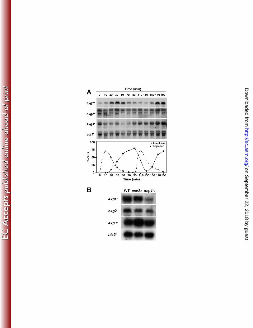
exg1+ expression peaks during the septation process 2
Northern blot analysis in asynchronous cultures indicated that the three ORFs were expressed 3
during the vegetative cycle (data not shown). When RNAs obtained from a synchronized cdc25-4
22 mutant strain were analyzed, a periodic cell cycle variation was found for exg1+ (Fig. 1A), the 5
maximum accumulation of mRNA occurring during the septation process, suggesting that the 6
product of this gene might exert its function during the last stages of the cell cycle; namely, 7
septum assembly or cell separation. No significant variations that correlated with cell cycle 8
progression were observed for the other two genes, exg2+ and exg3
+. These results are in good 9
agreement with the results obtained in a large-scale analysis (50). To study whether the periodic 10
expression of exg1+ was dependent on the transcription factors Ace2 or Sep1, Northern analyses 11
were performed to compare the expression in wild-type, ace2∆, and sep1∆ mutants (2, 50, 58). 12
The results revealed that the expression of exg1+ was clearly reduced in the sep1∆ mutant (Fig. 13
1B), suggesting that this transcription factors is required for its expression. As expected for genes 14
that do not show fluctuations during the cell cycle, no significant differences were seen in the 15
expression of exg2+ and exg3
+. 16
17
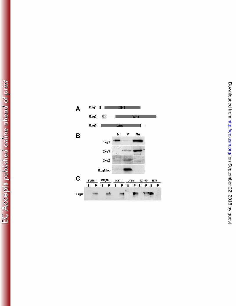
The three β-glucanases have different cellular fates 18
Analysis of the sequence of the three Exg proteins revealed the presence of different features 19
in addition to the GH5 domain. Thus, Exg1contained an N-terminal signal peptide for secretion, 20
with the likely cleavage site between positions 22 and 23 (AFS-YV). In contrast, Exg2 contained 21
a predicted transmembrane region (amino acids 39 to 60), while Exg3 did not contain any 22
sequence for extracellular localization (Fig. 2A). These observations suggested that the three 23
on Septem
ber 22, 2018 by guesthttp://ec.asm
.org/D
ownloaded from

12
proteins might have a different cellular localization: the cell wall, the plasma membrane and the 1
cytoplasm, respectively. To test these predictions, they were tagged with the HA epitope at their 2
C-terminus and the distribution of the proteins in culture supernatants, the membrane/cell wall 3
fraction and the cytoplasm was analyzed. The results confirmed that only Exg1 was present in 4
culture supernatants, indicating that the protein was secreted to the exterior of the cell and then 5
released to the surrounding medium (Fig. 2B). By contrast, Exg3 was mainly found in the 6
cytoplasm (supernatant) of the cell extracts, with a minor fraction in the pellet. Although Exg2 7
was difficult to detect, it was mainly present in the pellet fraction. To analyze the localization of 8
Exg2 with more detail, the gene was placed under the control of the nmt1 promoter and tagged 9
with the c-myc epitope. Overexpression of the gene confirmed that Exg2 was present in the 10
membrane/cell wall fraction (Exg2 hc in Fig. 2B). Thus, these results confirm the predictions of 11
the sequence analysis and indicate that Exg1 is a secreted protein, Exg2 a possible membrane-12
associated protein, and Exg3 a cytoplasmic protein. 13
To ascertain whether Exg2 was indeed an integral membrane protein, protein extracts were 14
prepared using different conditions. Cells were extracted with buffer alone, buffer containing 0.6 15
M NaCl or 1.6M Urea (to solubilize peripheral membrane proteins), 0.1 M Na2CO3 (to solubilize 16
intracellular vesicles), 4% Triton X-100 (a non-ionic detergent that solubilizes most membrane 17
proteins), or 2% SDS (an ionic detergent that solubilizes all membrane proteins) and extracts 18
were separated into the pellet and supernatant fractions. With the exception of SDS treatment, 19
Exg2 was found to be insoluble under all conditions tested and was detected in the pellet fraction 20
(Fig. 2C). Thus, Exg2 is a novel integral membrane protein that is solubilized only by strong 21
ionic detergents, but not by non-ionic detergents, as has been previously described for the 22
Cps1/Bgs1 subunit of the β-glucan synthase (28). 23
24
on Septem
ber 22, 2018 by guesthttp://ec.asm
.org/D
ownloaded from

13
1
Cellular localization of Exg proteins 2
To determine the in vivo localization of the three Exg proteins, the GFP was inserted in-frame 3
before the stop codon of each gene. The localization of the three proteins was monitored in live 4
cells, but only the fluorescence from Exg1-GFP and Exg3-GFP was detectable. In good 5
agreement with the peak of transcription during cytokinesis observed for exg1+, the protein was 6
mainly found at the center of the cells by the time the septum was starting to be visible by 7
differential interference contrast (DIC). Exg1 first started to accumulate as a ring surrounding the 8
septa during the initial steps of their assembly (Fig. 3A, asterisks). At later stages, when the 9
septum was clearly visible by DIC, Exg1 was found spanning the whole septum and it had 10
disappeared by the time the cells were separating (Fig. 3A, arrowhead). Staining of the cells with 11
Calcofluor or Aniline Blue to visualize the dynamics of septum assembly more clearly resulted in 12
a loss of the GFP fluorescence. 13
We have previously shown that the S. pombe endo-β(1,3)-glucanase Eng1 has a similar 14
localization to that found for Exg1 (33). To determine whether the two hydrolases co-localized, 15
we studied the localization of both proteins in the same cell using a strain that simultaneously 16
expressed Exg1-GFP and Eng1-RFP. Exg1-GFP was seen in the septum before the Eng1-RFP 17
fluorescence could be detected (Fig. 3B, asterisks), consistent with the fact that exg1+ expression 18
is dependent on Sep1 while that of eng1+ requires Ace2. Only when the septum was clearly 19
apparent by DIC did Eng1-RFP start to accumulate in the region, indicating that Eng1 secretion 20
occurs after septum assembly has been completed. Even though Eng1 and Exg1 were present in 21
the septum region, the distribution was different and there was no clear co-localization of either 22
protein. The fluorescence of Exg1-GFP was generally wider and less uniform than that of Eng1-23
RFP, suggesting that Exg1 localization might not be restricted to the primary septum, but also 24
on Septem
ber 22, 2018 by guesthttp://ec.asm
.org/D
ownloaded from

14
spread to the secondary septum. This is in contrast with the perfect co-localization found for the 1
two endo-glucanases involved in cell separation, Eng1 and Agn1 (36). 2
In the case Exg3, we found that this was a cytoplasmic protein, consistent with the 3
fractionation experiments, since the fluorescence was detected as a diffuse cytoplasmic staining 4
excluded from the nucleus, although no specific pattern or accumulations could be seen (Fig. 3C). 5
Exg2 could not be visualized when the Exg2-GFP protein was expressed from its own promoter, 6
suggesting that the gene is transcribed at low levels. When Exg2-GFP was mildly overexpressed 7
using the weak P41X-nmt1+ promoter, the localization of the protein could be determined. Exg2 8
was localized at the sites of cell growth, that is, the poles of the cell and the septum (Fig. 3D, 9
arrowheads). Thus, these results are in good agreement with the fractionation experiments, and 10
indicate that the three GH5 proteins have different cellular localizations. 11
12
Exg1 and Exg3 are β(1,6)-glucanases 13
The observed amino acid sequence similarity between S. pombe Exg proteins was an 14
indication that they might also be β(1,3)-glucanases. However, since S. pombe lacks any 15
detectable exo-β(1,3)-glucanase activity (48), two possibilities can be envisioned: First, it is 16
possible that the proteins might only be related in sequence but lacking any enzymatic activity. 17
Alternatively, they might have a different substrate specificity, since it has been described that 18
the S. cerevisiae and C. albicans Exg1 proteins are active not only against β(1,3) glucan but also 19
against β(1,6) glucans (54, 55). To check whether the S. pombe Exg proteins indeed contained 20
glucanase activity, Exg1 was purified from Pichia pastoris cells expressing an Exg1-His6-myc 21
construct. The enzymatic activity of the purified protein was assayed using laminarin (a β(1,3) 22
polymer), pustulan (a β(1,6) polymer), scleroglucan (a β(1,3)-glucan with β(1,6) branches), 23
lichenan (a mixed β(1,3)-β(1,4)-glucan), nigeran (an insoluble α(1,3) polymer) or the synthetic 24
on Septem
ber 22, 2018 by guesthttp://ec.asm
.org/D
ownloaded from

15
compound p-nitrophenyl-β-D-glucopyranoside (pNPG) as substrates. As shown in Fig. 4A, 1
activity was only detected when pustulan was used as a substrate, indicating that Exg1 was a 2
glucanase specific for β(1,6) linkages, in contrast to the S. cerevisiae and C. albicans proteins, 3
which are able to cleave both β(1,3) and β(1,6) polymers. In addition, no activity was detected 4
against pNPG, suggesting that the mechanism of action was not exo-hydrolytic. 5
To further confirm these results and to investigate the kinetics and degradation pattern of 6
different substrates, HPLC was used to analyze the reaction products. Soluble pustulan was used 7
as substrate for the assay. After 2 h of incubation, Exg1 had released small oligosaccharides (dp 8
2-30). The progressive degradation resulted in the formation of numerous reducing β(1,6)-9
oligosaccharides of varying size (Fig. 4B). Gentobiose to gentohexose (G2 to G6) were the main 10
reaction products after 4 h of incubation. The production of low amounts of glucose and high 11
amounts of oligosaccharides of different sizes proved that Exg1 degraded linear β(1,6)-glucan 12
substrates with an endolytic mode of action. No degradation of laminari-oligosaccharides (β(1,3)-13
glucans) such as laminarin, laminari-hexaose or octaose was observed (data not shown), 14
confirming that β(1,3)-glucans are not a substrate for Exg1. 15
Having determined that Exg1 was a β(1,6)-glucanase, we tested whether Exg2 and Exg3 16
possessed similar activity. To this end, extracts from cells overexpressing each of the three genes 17
under the control of the thiamine-repressible nmt1+ promoter were prepared and assayed using 18
pustulan as substrate. The results indicated that overexpression of exg1+ and exg3
+ produced a 3-19
fold and 6-fold increase in activity, respectively, in comparison with the activity found in cells 20
grown in the presence of thiamine (promoter repressed) or in wild-type cells, confirming that the 21
two proteins are β(1,6)-glucanases (Fig. 4C). However, no activity could be detected in cells 22
overexpressing exg2+. 23
24
on Septem
ber 22, 2018 by guesthttp://ec.asm
.org/D
ownloaded from

16
Deletion of exg genes presents no apparent phenotype 1
To determine the biological role of the three Exg proteins during the life cycle of the fission 2
yeast, mutant strains lacking each of the three genes were constructed. The strains were viable 3
and no growth defects were observed in different media (YES or Minimal medium) or at different 4
temperatures (25 to 37ºC). Growth on plates supplemented with cell wall-disturbing agents, such 5
as Calcofluor or Congo Red was also tested, and no significant differences with the wild-type 6
strain were detected. This could be due to a redundant function of the three Exg proteins. 7
However, similar results were obtained when the triple mutant exg1∆ exg2∆ exg3∆ was tested, 8
indicating that these proteins do not perform an essential role in cell wall construction or that the 9
cells have additional mechanisms to compensate for the absence of Exg proteins. Since Exg1 is 10
expressed during the cell cycle slightly before the endo-β(1,3)-glucanase Eng1 and since it also 11
localizes to the septum region during cell division, the double exg1∆ eng1∆ was constructed to 12
analyze whether Exg1 plays a minor role during cell separation. The phenotype of exg1∆ eng1∆ 13
cells was almost indistinguishable from that of eng1∆ cells. 14
15
Overexpression of exg2+ causes alterations in the cell wall 16
The effect of overexpression of the three exg+ genes was also assessed. Strains containing the 17
genes under the control of the strong version of the nmt1+ promoter were constructed and grown 18
in the absence of thiamine. Overexpression of exg1+ and exg3
+ produced a moderate defect in the 19
growth of the strains, but the microscopic appearance of the cells was similar to that of wild-type 20
cells (data not shown). In contrast, cells overexpressing exg2+ had a severe growth defect, and 21
they stopped growing at 10-12 h after induction (Fig. 5A). To determine the nature of the defect, 22
the morphology of cells was analyzed. By 12 h, the cells had an abnormal morphology and had 23
become rounded and irregularly shaped. Also, large amounts of abnormal material had 24
on Septem
ber 22, 2018 by guesthttp://ec.asm
.org/D
ownloaded from

17
accumulated at the poles of the cell. In an effort to determine the identity of the accumulated 1
material, cells overexpressing exg2
+ were stained with Aniline Blue, a dye that preferentially 2
binds to β(1,3)-glucans and is used to stain the cell wall and septum in S. pombe (25). The 3
material stained well with Aniline Blue (Fig. 5B), indicating
that it was cell wall and that at least 4
a portion of it was comprised of β-glucans. The excess of cell wall accumulated specifically at 5
the poles and septum of the cells. 6
The previous observations were confirmed when the cells were observed by electron 7
microscopy (Fig. 6A-F). In comparison with the normal appearance of the wall of wild-type cells 8
(Fig. 6A and E), the wall of cells overexpressing exg2+ clearly contained extra material that 9
accumulated at the poles of the cell and the separation septum (Fig. 6B-D, F). Interestingly, the 10
excess material was incorporated into the cell wall and had a similar appearance to the rest of the 11
cell wall. 12
13
Cells overexpressing exg2+ accumulate α- and β-glucans 14
To identify the nature of the material that accumulated in cells overexpressing exg2+ more 15
precisely, cell wall constituents were isolated and characterized after growing the cells in the
16
presence of (U-14
C) glucose. Incorporation of radioactive glucose into the wall of cells grown in 17
the presence of thiamine was similar to that of wild-type cells, but it increased considerably in the 18
absence of thiamine (from 34 to 47% of total glucose incorporated; Fig. 6G).
A dramatic increase 19
in the amount of α(1,3) and β(1,3) glucan was detected under conditions of exg2
+ overexpression 20
(Fig. 6H), but the β/α-glucan ratio was similar in the three strains, indicating a simultaneous 21
increase in both
glucan polymers. Additionally, the
amount of galactomannan was not 22
significantly affected, leading to an alteration in the glucan to galactomannan ratio (around 2
23
times more glucan than galactomannan). These results suggest that strains overexpressing exg2+ 24
on Septem
ber 22, 2018 by guesthttp://ec.asm
.org/D
ownloaded from

18
have lost the ability to properly coordinate glucan polymer synthesis with cell growth, and that 1
glucan synthases produce an excess of cell wall material. 2
3
The N-terminal region and the catalytic domain of Exg2 are required for cell wall 4
accumulation 5
To investigate the reason for increased cell wall accumulation in cells overexpressing exg2+, 6
versions of the protein in which different domains were deleted were constructed. When the exg2 7
sequence was analyzed using topology prediction programs such as TopPred 8
(http://bioweb.pasteur.fr/seqanal/ interfaces/toppred.html) or TMHMM (http://www.cbs.dtu.dk/ 9
services/TMHMM-2.0/), the N-terminus was predicted to be cytoplasmic while the catalytic 10
domain would be extracellular. Thus, mutant proteins lacking the putative cytoplasmic tail of the 11
protein (Exg2-∆N), the transmembrane domain (Exg2-∆TM), the spacer domain that separates 12
the transmembrane and catalytic domains (Exg2-∆out) or the catalytic domain (Exg2-∆GH5) 13
were generated (Fig. 7A). One possibility for explaining these results is that an excess of 14
enzymatic activity could weaken the cell wall, and as a consequence the synthesis of glucans 15
would be induced. To test this possibility, we also created proteins containing single amino acid 16
changes in the two glutamic acid residues that form part of the catalytic centre of GH5 proteins, 17
which are also conserved in exg2+. These mutations involved a substitution of E338 for Ala 18
(E338A, Exg2-A1), the replacement of E439 for Gln (E349Q, Exg2-A2) and the double mutant 19
(E338A-E349Q, Exg2-A1A2). As described for other members of this family (16, 30), these 20
mutations should completely eliminate the catalytic activity of the enzyme, if present. All the 21
constructs were cloned under the control of the strong version of the nmt1+
promoter and 22
introduced into the wild-type strain to test their effects. 23
Overexpression of Exg2-∆N resulted in cells with an abnormal morphology (the cells were 24
on Septem
ber 22, 2018 by guesthttp://ec.asm
.org/D
ownloaded from

19
rounder than wild-type cells) but that did not exhibit a large accumulation of cell wall material 1
(Fig. 7B). Overexpression of Exg2-∆TM resulted in almost wild-type cells, but since the protein 2
levels were significantly lower than those found in these latter cells (Fig. 7C), the absence of 3
phenotype could be due to the low amount of protein. The same reason -reduced protein levels- 4
could account for the fact that the overexpression of Exg2-∆out resulted in a modest phenotype, 5
with a minor accumulation of glucans in the septum region. When the construct lacking the 6
catalytic domain (Exg2-∆GH5) was overexpressed, the cells showed wild-type morphology, 7
indicating that this region of the protein is required for the activation of glucan synthesis. 8
However, the putative catalytic activity of the enzyme was not required for this effect, since cells 9
carrying the double mutant Exg2-A1A2 (and also the two single mutants Exg2-A1 and Exg2-A2) 10
were almost identical to those overexpression the full-length protein (Fig. 7B). Thus, these results 11
indicate that the N-terminal region and the GH5 domains are required to induce abnormal glucan 12
synthesis, but that the catalytic activity, if present, is not essential. 13
14
DISCUSSION 15
Cell wall growth and extension represents a delicate balance between the hydrolysis of 16
existing cell wall and the synthesis of new wall. A considerable body of evidence suggests that 17
fungal β(1,3)-glucanases play key roles in morphogenetic processes during development and 18
differentiation. Since β-glucans are major components of fungal and yeast cell walls, it seems 19
likely that β-glucanases would play a crucial role in this process, partially hydrolyzing localized 20
areas and enabling the insertion of new cell wall material, without disturbing the overall integrity 21
of the cell (1). The complement of glucan-modifying enzymes present in S. cerevisiae is very 22
complex, and a large number of proteins have been characterized, but much less is known about 23
the role of hydrolases and glucan-remodeling enzymes during the life cycle of fission yeasts. 24
on Septem
ber 22, 2018 by guesthttp://ec.asm
.org/D
ownloaded from

20
Only a few glycoside hydrolases have been studied in fission yeast, such as the endo-α(1,3)-1
glucanases Agn1 and Agn2, the endo-β(1,3)-glucanases Eng1 and Eng2, and the glycanosyl-2
transferases from family GH72 (19-22, 33-36). In this study, we have characterized the three 3
genes belonging to family GH5 that are present in the S. pombe genome. They were identified by 4
comparison of the S. cerevisiae Exg1 protein with the fission yeast genomic sequences, and were 5
named exg1+
(SPBC1105.05), exg2+ (SPAC12B10.11) and exg3
+ (SPBC2D10.05). 6
Family GH5 is a large and diverse group of hydrolases with different substrate specificities, 7
such as exo-β(1,3)-glucanases, endo-β(1,4)-glucanases (cellulases), endo-β(1,6)-glucanases, 8
endo-β(1,4)-xylanases, or β(1,3)-mannanases (see Carbohydrate Active Enzymes database, 9
http://www.cazy.org/). In many cases, hydrolysis of glycoside bonds takes place via a general 10
acid catalysis mechanism, which requires two acidic residues, one acting as an acid/base catalyst 11
(proton donor) and the other as a nucleophile (17). There is a low degree of conservation of the
12
primary sequence of these proteins, but all of them contain a conserved fold consisting of a (β/α)8 13
barrel. Despite its considerable sequence divergence, all share the signature [LIV]-14
[LIVMFYWGA](2)-[DNEQG]-[LIVMGST]-{SENR}-N-E-[PV]-[RHDNSTLIVFY] as well as 15
eight invariant residues that are involved in the catalysis and the recognition of the glycosyl 16
group attacked during cleavage. Comparison of the three S. pombe proteins to GH5 proteins 17
revealed that the signature and the eight invariant residues were conserved, suggesting that they 18
are new members of this family. 19
The S. pombe proteins share sequence homology to S. cerevisiae Exg1 and C. albicans Xog1, 20
which are exo-β(1,3)-glucanases that also act on β(1,6) linkages (15, 43, 57). However, since it 21
has been reported that S. pombe lacks any detectable exo-β(1,3)-glucanase activity (48), their 22
biochemical activity and substrate specificity were analyzed in strains overexpressing each of the 23
three proteins. In contrast to ScExg1 and CaXog1, the S. pombe Exg1 and Exg3 were highly 24
on Septem
ber 22, 2018 by guesthttp://ec.asm
.org/D
ownloaded from

21
specific for β(1,6)-glucans (pustulan), being unable to degrade linear or branched β(1,3)-glucans. 1
However, no enzymatic activity was detected for Exg2 in different assays or using different 2
substrates. Therefore, it is possible that Exg2 may have diverged from other GH5, losing its 3
catalytic activity along evolution. Alternatively, Exg2 might act on a substrate different from 4
those used in the assay or it could catalyze a transglycosidase reaction that cannot be detected 5
with the assay used. Indeed, transglycosidase activity has been reported for ScExg1 and CaXog1 6
(54, 55). Analysis of the reaction products released by Exg1 on β(1,6)-glucans by HPLC revealed 7
that the enzyme had endolytic activity, since the main products were gentooligosaccharides (dp 2 8
– 6). Therefore, the mode of action of S. pombe Exg1 is similar to that previously suggested for 9
the BGN16.2 glucanase from the filamentous fungus Trichoderma harzianum (18), and it is 10
different from that found in ScExg1 or CaXog1, which have non-specific β(1,3)-glucanase, 11
β(1,6)-glucanase, and β-glucosidase activities, with an exolytic mode of action (15, 43, 52). 12
Multiple sequence alignment of yeast and fungal GH5 proteins indicated that the S. pombe 13
proteins cluster in three different branches, but none of them associated with the branch that 14
contains β(1,6)-glucanases such as T. harzianum BGN16.2 (B9VQ16_TRIHA in Supplementary 15
Fig. 1). 16
Although no phenotypes have been detected for exg1∆ mutants, based on the fact that the 17
protein was secreted and that it localized to the cell wall it should be involved in the metabolism 18
of cell wall β(1,6)-glucans in vivo. Similarly, the physiological role of ScExg1 has not been 19
clearly established, but it has been proposed that in vivo it would be involved in the metabolism 20
of β(1,6)-glucans, since its deletion results in an increase in killer toxin sensitivity, while its 21
overexpression produces resistance (24). Furthermore, overproduction or deletion of the gene 22
leads to detectable in vivo alterations in the cell wall β(1,6)-glucan content, suggesting that its 23
function in the cell wall could be related to the metabolism of β(1,6)-glucan. This protein 24
on Septem
ber 22, 2018 by guesthttp://ec.asm
.org/D
ownloaded from
22
localized to the septum region in a pattern that was different from that found for Eng1, the endo-1
β(1,3)-glucanase responsible for primary septum degradation (33). Interestingly, the β(1,6)-2
branched β(1,3)-glucan spans the whole thickness of the septum, with a tendency to become more 3
concentrated in the primary septum, while β(1,6)-glucan is close to the cell membrane, labeling 4
only the secondary septum (23). Thus, it is possible that Exg1 could play a minor role during cell 5
separation, acting as an endo-β(1,6)-glucanase required for the hydrolysis the β(1,6)-glucans of 6
the secondary septum. 7
Exg2 protein is different from the other S. pombe GH5 proteins in that it contains a putative 8
transmembrane region before the catalytic domain. Sequence alignment indicates that Exg2 9
clusters in a branch containing a group of proteins of fungal origin (Aspergillus fumigatus, A. 10
nidulans, A. terreus, A. clavatus, Neurospora crassa or Magnaporthe grisea), all of which 11
contain a putative transmembrane domain before the catalytic domain (Supplementary Fig. 1). 12
However, the extent of the putative cytoplasmic region is longer in the fungal proteins than in S. 13
pombe Exg2, although no function has been described for any of them. We have demonstrated 14
that Exg2 is indeed an integral membrane protein that fractionates in the detergent-resistant 15
membrane fraction, a biochemical test for lipid-raft association (56), and that its overexpression 16
produces abnormal cell wall deposition. Interestingly, it has recently been reported that 17
overexpression of a catalytically inactive form of Gas3 is toxic for gas1∆ mutants in S. cerevisiae 18
(49), and it was proposed that hyperaccumulation of Gas3 might produce a physical disturbance 19
of the cell wall structure. Thus, it is possible that the defect in exg2+ overexpression in S. pombe 20
might be due to a similar cause. Alternatively, it is possible that Exg2 might activate cell wall 21
synthesis through an unknown mechanism. Unfortunately, attempts to identify proteins that 22
might interact with the Exg2 cytoplasmic tail by two-hybrid screenings were negative (data not 23
shown). Further analysis will be necessary to analyze this effect in more detail. 24
on Septem
ber 22, 2018 by guesthttp://ec.asm
.org/D
ownloaded from

23
Finally, Exg3 is a β(1,6)-glucanase that localizes to the cytoplasm. The function of 1
cytoplasmic glucanases is not currently known, although they are present in different yeast. 2
Indeed, Exg3 clusters in a tree branch in which all the members lack a putative signal secretion 3
sequence (Supplementary Fig. 1). Also, S. pombe contains cytoplasmic glucanases from other 4
families, such as the endo-α(1,3)-glucanase Agn2 (family GH71) and the endo-β(1,3)-glucanase 5
Eng2 (family GH81). It has been shown that these two proteins are required to hydrolyze the cell 6
wall of asci, allowing the dehiscence of spores and their dispersal (21, 22). Thus, cytoplasmic 7
glucanases might perform their function at specific moments of the life cycle. 8
9
ACKNOWLEDGEMENTS 10
We thank members of the lab for helpful comments and discussions on the manuscript 11
and Nick Skinner for revision of the manuscript This research was supported by grants from the 12
Comisión Interministerial de Ciencia y Tecnología (BFU2007-60390/BMC) and Junta de Castilla 13
y Leon (GR231). A.B. Martín-Cuadrado was recipient of a fellowship from Ministerio de 14
Educación y Ciencia (Spain). 15
16
REFERENCES 17
1. Adams, D. J. 2004. Fungal cell wall chitinases and glucanases. Microbiology 150:2029-2035. 18
2. Alonso-Núñez, M., H. An, A. B. Martín-Cuadrado, S. Mehta, C. Petit, M. Sipiczki, F. del 19
Rey, K. Gould, and C. R. Vázquez de Aldana. 2005. Ace2p controls the expression of genes 20
required for cell separation in Schizosaccharomyces pombe. Mol. Biol. Cell 16:2003-2017. 21
3. Arellano, M., A. Duran, and P. Perez. 1997. Localisation of the Schizosaccharomyces pombe 22
rho1p GTPase and its involvement in the organisation of the actin cytoskeleton. J. Cell Sci. 23
110:2547-2555. 24
on Septem
ber 22, 2018 by guesthttp://ec.asm
.org/D
ownloaded from

24
4. Bähler, J., J. Q. Wu, M. S. Longtine, N. G. Shah, A. McKenzie, A. B. Steever, A. Wach, P. 1
Philippsen, and J. R. Pringle. 1998. Heterologous modules for efficient and versatile PCR-2
based gene targeting in Schizosaccharomyces pombe. Yeast 14:943-951. 3
5. Baladrón, V., S. Ufano, E. Dueñas, A.B. Martín-Cuadrado, F. del Rey, and C. R. Vázquez 4
de Aldana. 2002. Eng1p, an endo-1,3-β-glucanase localized at the daughter side of the 5
septum, is involved in cell separation in Saccharomyces cerevisiae. Eukaryot. Cell 1:774-786. 6
6. Beauvais, A., R. Drake, K. Ng, M. Diaquin, and J. P. Latgé. 1993. Characterization of the 7
1,3-β-glucan synthase of Aspergillus fumigatus. J. Gen. Microbiol. 139:3071-3078. 8
7. Bush, D. A., M. Horisberger, I. Horman, and P. Wursch. 1974. The wall structure of 9
Schizosaccharomyces pombe. J. Gen. Microbiol. 81:199-206. 10
8. Cabib, E., N. Blanco, C. Grau, J. M. Rodríguez-Peña, and J. Arroyo. 2007. Crh1p and 11
Crh2p are required for the cross-linking of chitin to β(1-6)glucan in the Saccharomyces 12
cerevisiae cell wall. Mol. Microbiol. 63:921-935. 13
9. Cabib, E., V. Farkas, O. Kosik, N. Blanco, J. Arroyo, and P. McPhie. 2008. Assembly of 14
the yeast cell wall. Crh1p and Crh2p act as transglycosylases in vivo and in vitro. J. Biol. 15
Chem. 283:29859-29872. 16
10. Cid, V. J., A. Durán, F. del Rey, M. Snyder, C. Nombela, and M. Sánchez. 1995. 17
Molecular basis of cell integrity and morphogenesis in Saccharomyces cerevisiae. Microbiol. 18
Rev. 59:345-386. 19
11. Correa, J., C. R. Vázquez de Aldana, P. San Segundo, and F. del Rey. 1992. Genetic 20
mapping of 1,3-β-glucanase-encoding genes in Saccharomyces cerevisiae. Curr. Genet. 21
22:283-288. 22
12. Cortés, J. C., M. Konomi, I. M. Martins, J. Muñoz, M. B. Moreno, M. Osumi, A. Durán, 23
on Septem
ber 22, 2018 by guesthttp://ec.asm
.org/D
ownloaded from

25
and J. C. Ribas. 2007. The (1,3)β-D-glucan synthase subunit Bgs1p is responsible for the 1
fission yeast primary septum formation. Mol. Microbiol. 65:201-217. 2
13. Cortés, J. C. G., E. Carnero, J. Ishiguro, Y. Sánchez, A. Durán, and J. C. Ribas. 2005. 3
The novel fission yeast (1,3)β-D-glucan synthase catalytic subunit Bgs4p is essential during 4
both cytokinesis and polarized growth. J. Cell Sci. 118:157-174. 5
14. Craven, R. A., D. J. Griffiths, K. S. Sheldrick, R. E. Randall, I. M. Hagan, and A. M. 6
Carr. 1998. Vectors for the expression of tagged proteins in Schizosaccharomyces pombe. 7
Gene 221:59-68. 8
15. Chambers, R. S., M. J. Broughton, R. D. Cannon, A. Carne, G. W. Emerson, and P. A. 9
Sullivan. 1993. An exo-β-(1,3)-glucanase of Candida albicans: purification of the enzyme 10
and molecular cloning of the gene. J. Gen. Microbiol. 139:325-334. 11
16. Chambers, R. S., A. R. Walden, G. S. Brooke, J. F. Cutfield, and P. A. Sullivan. 1993. 12
Identification of a putative active site residue in the exo-β-(1,3)-glucanase of Candida 13
albicans. FEBS Lett. 327:366-369. 14
17. Davies, G., and B. Henrissat. 1995. Structures and mechanisms of glycosyl hydrolases. 15
Structure 3:853-859. 16
18. de la Cruz, J., J. A. Pintor-Toro, T. Benitez, and A. Llobell. 1995. Purification and 17
characterization of an endo-β-1,6-glucanase from Trichoderma harzianum that is related to its 18
mycoparasitism. J. Bacteriol. 177:1864-1871. 19
19. de Medina-Redondo, M., Y. Arnáiz-Pita, T. Fontaine, F. del Rey, J. P. Latgé, and C. R. 20
Vázquez de Aldana. 2008. The β-1,3-glucanosyltransferase gas4p is essential for ascospore 21
wall maturation and spore viability in Schizosaccharomyces pombe. Mol. Microbiol. 68:1283-22
1299. 23
on Septem
ber 22, 2018 by guesthttp://ec.asm
.org/D
ownloaded from

26
20. Dekker, N., D. Speijer, C. H. Grün, M. van den Berg, A. de Haan, and F. Hochstenbach. 1
2004. Role of the α-glucanase Agn1p in fission-yeast cell separation. Mol. Biol. Cell 15:3903-2
3914. 3
21. Dekker, N., J. van Rijssel, B. Distel, and F. Hochstenbach. 2007. Role of the α-glucanase 4
Agn2p in ascus-wall endolysis following sporulation in fission yeast. Yeast 24:279-288. 5
22. Encinar del Dedo, J., E. Dueñas, Y. Arnáiz, F. del Rey, and C. R. Vázquez de Aldana. 6
2009. β-glucanase Eng2 is required for ascus wall endolysis after sporulation in the fission 7
yeast Schizosaccharomyces pombe. Eukaryot. Cell 8:1278-1286. 8
23. Humbel, B. M., M. Konomi, T. Takagi, N. Kamasawa, S. A. Ishijima, and M. Osumi. 9
2001. In situ localization of β-glucans in the cell wall of Schizosaccharomyces pombe. Yeast 10
18:433-444. 11
24. Jiang, B., A. F. Ram, J. Sheraton, F. M. Klis, and H. Bussey. 1995. Regulation of cell wall 12
β-glucan assembly: PTC1 negatively affects PBS2 action in a pathway that includes 13
modulation of EXG1 transcription. Mol. Gen. Genet. 248:260-269. 14
25. Kippert, F., and D. Lloyd. 1995. The aniline blue fluorochrome specifically stains the 15
septum of both live and fixed Schizosaccharomyces pombe cells. FEMS Microbiol. Lett. 16
132:215-219. 17
26. Kopecka, M., G. H. Fleet, and H. J. Phaff. 1995. Ultrastructure of the cell wall of 18
Schizosaccharomyces pombe following treatment with various glucanases. J. Struct .Biol. 19
114:140-152. 20
27. Liu, J., X. Tang, H. Wang, and M. Balasubramanian. 2000. Bgs2p, a 1,3-β-glucan 21
synthase subunit, is essential for maturation of ascospore wall in Schizosaccharomyces pombe. 22
FEBS Lett. 478:105-108. 23
on Septem
ber 22, 2018 by guesthttp://ec.asm
.org/D
ownloaded from

27
28. Liu, J., X. Tang, H. Wang, S. Oliferenko, and M. K. Balasubramanian. 2002. The 1
localization of the integral membrane protein Cps1p to the cell division site is dependent on 2
the actomyosin ring and the Septation-Inducing Network in Schizosaccharomyces pombe. 3
Mol. Biol. Cell 13:989-1000. 4
29. Liu, J., H. Wang, D. McCollum, and M. K. Balasubramanian. 1999. Drc1p/Cps1p, a 1,3-5
β-glucan synthase subunit, is essential for division septum assembly in Schizosaccharomyces 6
pombe. Genetics 153:1193-1203. 7
30. Mackenzie, L. F., G. S. Brooke, J. F. Cutfield, P. A. Sullivan, and S. G. Withers. 1997. 8
Identification of Glu-330 as the catalytic nucleophile of Candida albicans exo-β-(1,3)-9
glucanase. J. Biol. Chem. 272:3161-3167. 10
31. Magnelli, P. E., J. F. Cipollo, and P. W. Robbins. 2005. A glucanase-driven fractionation 11
allows redefinition of Schizosaccharomyces pombe cell wall composition and structure: 12
assignment of diglucan. Anal. Biochem. 336:202-212. 13
32. Manners, D. J., and M. T. Meyer. 1977. The molecular structures of some glucans from the 14
cell wall of Schizosaccharomyces pombe. Carbohydr. Res. 57:189-203. 15
33. Martín-Cuadrado, A. B., E. Dueñas, M. Sipiczki, C. R. Vázquez de Aldana, and F. del 16
Rey. 2003. The endo-β-1,3-glucanase Eng1p is required for dissolution of the primary septum 17
during cell separation in Schizosaccharomyces pombe. J. Cell Sci. 116:1689-1698. 18
34. Martín-Cuadrado, A. B., J. Encinar del Dedo, M. de Medina-Redondo, T. Fontaine, F. 19
del Rey, J. P. Latgé, and C. R. Vázquez de Aldana. 2008. The Schizosaccharomyces pombe 20
endo-1,3-β-glucanase Eng1 contains a novel carbohydrate binding module required for septum 21
localization. Mol. Microbiol. 69:188-200. 22
35. Martín-Cuadrado, A. B., T. Fontaine, P. F. Esteban, J. Encinar del Dedo, M. de 23
on Septem
ber 22, 2018 by guesthttp://ec.asm
.org/D
ownloaded from

28
Medina-Redondo, F. del Rey, J. P. Latgé, and C. R. Vázquez de Aldana. 2008. 1
Characterization of the endo-β-1,3-glucanase activity of S. cerevisiae Eng2 and other members 2
of the GH81 family. Fungal Genet. Biol. 45:542-553. 3
36. Martín-Cuadrado, A. B., J. L. Morrell, M. Konomi, H. An, C. Petit, M. Osumi, M. 4
Balasubramanian, K. L. Gould, F. del Rey, and C. R. Vázquez de Aldana. 2005. Role of 5
septins and the exocyst complex in the function of hydrolytic enzymes responsible for fission 6
yeast cell separation. Mol. Biol. Cell 16:4867-4881. 7
37. Martín, V., B. García, E. Carnero, A. Durán, and Y. Sánchez. 2003. Bgs3p, a putative 8
1,3-β-glucan synthase subunit, is required for cell wall assembly in Schizosaccharomyces 9
pombe. Eukaryot. Cell 2:159-169. 10
38. Martín, V., J. C. Ribas, E. Carnero, A. Durán, and Y. Sánchez. 2000. bgs2+, a 11
sporulation-specific glucan synthase homologue is required for proper ascospore wall 12
maturation in fission yeast. Mol. Microbiol. 38:308-321. 13
39. Mitchison, J. M., and P. Nurse. 1985. Growth in cell length in the fission yeast 14
Schizosaccharomyces pombe. J. Cell Sci. 75:357-376. 15
40. Moreno, M. B., A. Durán, and J. C. Ribas. 2000. A family of multifunctional thiamine-16
repressible expression vectors for fission yeast. Yeast 16:861-872. 17
41. Moreno, S., A. Klar, and P. Nurse. 1991. Molecular genetics analysis of fission yeast 18
Schizosaccharomyces pombe. Methods Enzymol. 194:795-823. 19
42. Mouyna, I., T. Fontaine, M. Vai, M. Monod, W. A. Fonzi, M. Diaquin, L. Popolo, H. 20
R.P., and J. P. Latgé. 2000. Glycosylphosphatidylinositol-anchored glucanosyltransferases 21
play an active role in the biosynthesis of the fungal cell wall. J. Biol. Chem. 275:14882-14889. 22
43. Nebreda, A. R., C. R. Vázquez, T. G. Villa, J. R. Villanueva, and F. del Rey. 1987. 23
on Septem
ber 22, 2018 by guesthttp://ec.asm
.org/D
ownloaded from

29
Heterogeneous glycosylation of the EXG1 gene product accounts for the two extracellular exo-1
β-glucanases of Saccharomyces cerevisiae. FEBS Lett. 220:27-30. 2
44. Nelson, M. J. 1957. Colorimetric analysis of sugars. Methods Enzymol. 3:85-86. 3
45. Osumi, M., M. Sato, S. A. Ishijima, M. Konomi, T. Takagi, and H. Yaguchi. 1998. 4
Dynamics of cell wall formation in fission yeast, Schizosaccharomyces pombe. Fungal Genet. 5
Biol. 24:178-206. 6
46. Percival-Smith, A., and J. Segall. 1984. Isolation of DNA sequences preferentially 7
expressed during sporulation in Saccharomyces cerevisiae. Mol. Cell. Biol. 4:142-150. 8
47. Ragni, E., T. Fontaine, C. Gissi, J. P. Latgé, and L. Popolo. 2007. The Gas family of 9
proteins of Saccharomyces cerevisiae: characterization and evolutionary analysis. Yeast 10
24:297-308. 11
48. Reichelt, B. Y., and G. H. Fleet. 1981. Isolation, properties, function, and regulation of 12
endo-(1-3)-β-glucanases in Schizosaccharomyces pombe. J. Bacteriol. 147:1085-1094. 13
49. Rolli, E., E. Ragni, J. M. Rodriguez-Peña, J. Arroyo, and L. Popolo. 2010. GAS3, a 14
developmentally regulated gene, encodes a highly mannosylated and inactive protein of the 15
Gas family of Saccharomyces cerevisiae. Yeast 27:597-610. 16
50. Rustici, G., J. Mata, K. Kivinen, P. Lió, C. J. Penkett, G. Burns, J. Hayles, A. Brazma, 17
P. Nurse, and J. Bähler. 2004. Periodic gene expression program of the fission yeast cell 18
cycle. Nat. Genet. 36:809-817. 19
51. San Segundo, P., J. Correa, C. R. Vázquez de Aldana, and F. del Rey. 1993. SSG1, a gene 20
encoding a sporulation-specific 1,3-β-glucanase in Saccharomyces cerevisiae. J. Bacteriol. 21
175:3823-3837. 22
52. Sánchez, M., J. R. Villanueva, and T. G. Villa. 1982. Saccharomyces cerevisiae secretes 23
on Septem
ber 22, 2018 by guesthttp://ec.asm
.org/D
ownloaded from

30
two exoglucanases. FEBS Lett. 138:209-212. 1
53. Somogyi, M. 1952. Notes on sugar determination. J. Biol. Chem. 195:19-23. 2
54. Stubbs, H. J., D. J. Brasch, G. W. Emerson, and P. A. Sullivan. 1999. Hydrolase and 3
transferase activities of the β-1,3-exoglucanase of Candida albicans. Eur. J. Biochem. 4
263:889-895. 5
55. Suzuki, K., T. Yabe, Y. Maruyama, K. Abe, and T. Nakajima. 2001. Characterization of 6
recombinant yeast exo-β-1,3-glucanase (Exg1p) expressed in Escherichia coli cells. Biosci. 7
Biotechnol. Biochem. 65:1310-1314. 8
56. Takeda, T., T. Kawate, and F. Chang. 2004. Organization of a sterol-rich membrane 9
domain by cdc15p during cytokinesis in fission yeast. Nat. Cell. Biol. 6:1142-1144. 10
57. Vázquez de Aldana, C. R., J. Correa, P. San Segundo, A. Bueno, A. R. Nebreda, E. 11
Méndez, and F. del Rey. 1991. Nucleotide sequence of the exo-1,3-β-glucanase gene, EXG1, 12
of the yeast Saccharomyces cerevisiae. Gene 97:173-182. 13
58. Zilahi, E., E. Salimova, V. Simanis, and M. Sipiczki. 2000. The S. pombe sep1 gene 14
encodes a nuclear protein that is required for periodic expression of the cdc15 gene. FEBS 15
Lett. 481:105-108. 16
on Septem
ber 22, 2018 by guesthttp://ec.asm
.org/D
ownloaded from

31
FIGURE LEGENDS 1
Fig. 1: Transcription pattern of exg genes. (A) Expression during the cell cycle. Synchrony 2
was induced by arrest-release of a cdc25-22 mutant and samples were taken at the indicated time 3
points (minutes) after the release for RNA extraction. RNA was hybridized with specific probes 4
for exg1+, exg2
+, exg3
+ or act1
+. The graph represents the anaphase index (open circles) or 5
septation index (black circles) at each time-point. In this experiment, the peak of septum 6
formation occurred at 70-90 minutes. (B) Dependence on the Ace2 and Sep1 transcription 7
factors. RNA from wild-type, ace2∆ and sep1∆ mutants was extracted, transferred to 8
nitrocellulose membranes, and probed with specific probes for exg1+, exg2
+, exg3
+ or his3
+ as a 9
control. 10
11
Fig. 2: Cellular fractionation of the Exg proteins. (A) Schematic representation of the 12
characteristics of the Exg1, Exg2 and Exg3 proteins. The grey box indicates the GH5 domain, 13
common to the three proteins. The black box represents a hydrophobic region with the 14
characteristics of the signal secretion peptides and the hatched box a putative transmembrane 15
domain. (B) Cells expressing Exg1-HA, Exg2-HA or Exg3-HA were grown in minimal medium 16
to late-log phase (OD595=1.5). Those carrying Exg2-myc under the control of the nmt1 promoter 17
(Exg2 hc) were grown in the absence of thiamine for 22 h. Cells were collected by centrifugation 18
and broken with glass beads. Extracts were centrifuged at 10000 rpm for 10 min to separate the 19
cell wall and membranes (pellet, P) from the cytoplasmic content (supernatant, Sn). Concentrated 20
culture medium (M) was also loaded in the gels. Proteins were fractionated using SDS-PAGE 21
gels and immunoblotted using antibodies against the HA or c-myc epitopes. (C) Cells carrying 22
Exg2-myc under the control of the P3Xnmt1 promoter were extracted after lysis in buffer 23
containing 0.6M NaCl, 0.1M Na2CO3, 1.6 M Urea, 4% TritonX-100, and 2% SDS. Soluble and 24
on Septem
ber 22, 2018 by guesthttp://ec.asm
.org/D
ownloaded from

32
insoluble proteins were separated by centrifugation at 13000 g for 30 min, as indicated by 1
supernatant (S) and pellet (P), respectively. 2
3
Fig. 3: Localization of Exg proteins. (A) Wild-type cells expressing exg1-GFP were grown to 4
early-log phase. Photographs of differential interference contrast microscopy (DIC) or Exg1-GFP 5
are shown. (B) Exponentially growing wild-type cells expressing Exg1-GFP and Eng1-RFP were 6
imaged for differential interference contrast (DIC), RFP, and GFP fluorescence. Overlay of the 7
red and green channels is also shown in the merge images. (C) Wild-type cells expressing exg3-8
GFP were grown to early-log phase and live cells were used for microscopic observation. (D) 9
Wild-type cells expressing exg2-GFP under the control of the P41nmt1 promoter were grown in 10
medium without thiamine for 18 h and live cells were used for microscopic observation. 11
Representative cells at different stages of the cell cycle are shown. 12
13
Fig. 4: Exg proteins have activity against β(1,6) glucans. (A) β-glucanase activity of purified 14
Exg1 against laminarin (β(1,3) glucan), pustulan (β(1,6) glucan), schleroglucan (β(1,3) glucan 15
with β(1,6) ramifications), lichenan (β(1,3)-(1,4) glucan), nigeran (α(1,3) glucan), PNPG or S. 16
pombe cell wall (CW). 5 µg of protein was incubated with the substrates for 24 h before the 17
concentration of reducing sugars released was assayed. Activity is shown as a percentage of the 18
maximum activity detected for pustulan. (B) HPAE-PED chromatographic analysis of 19
oligosaccharides released by Exg1. The reaction was conducted in acetate buffer at 37ºC using 20
soluble pustulan as substrate for the indicated times (h) before the products were analyzed by 21
HPAE-PED chromatography. G2, gentobiose, G4, gentotetraose. (C) Enzymatic activity against 22
pustulan (β(1,6)-glucan) of cells overexpressing exg1+, exg2
+, or exg3
+. Wild-type cells 23
transformed with plasmids pED138 (carrying P3Xnmt1-exg1), pML2 (P3Xnmt1-exg2), pED139 24
on Septem
ber 22, 2018 by guesthttp://ec.asm
.org/D
ownloaded from

33
(P3Xnmt1-exg3) or vector alone (pJCR-3XL) were grown for 22 h in the presence (white bars) or 1
absence (black bars) of thiamine (T) to induce the expression of the genes. Cell extracts were 2
prepared and incubated in the presence of pustulan for different amounts of time. Activity is 3
presented as mU/mg. The result is the mean of two independent assays. Error bars indicate the 4
standard deviation. 5
6
Fig. 5: Overexpression of exg2+
produces severe defects in cell growth. (A) Growth of wild-7
type strains carrying P3Xnmt1-exg2 or vector alone grown in the presence or absence of 8
thiamine. Cells overexpressing exg2+ cease growth after 10-12 of induction. (B) Microscopic 9
appearance of wild-type (WT) and cells overexpressing exg2+ (OE exg2
+). Cells that had been 10
growing in the absence of thiamine for 22 h were stained with Aniline Blue, a fluorochrome that 11
preferentially binds β(1,3)-glucans. Photographs of differential interference contrast microscopy 12
(DIC) or Aniline Blue-stained cells (Aniline) are shown. Bar, 5µm. 13
14
Fig. 6: Effects of exg2+ overexpression. Wild-type cells containing vector (A and E, WT) or 15
plasmid pML2 (B-D, F, OE exg2+) were grown for 22 h in the absence of thiamine and prepared 16
for transmission electron microscopy. Images of panels A-D show a general view of the cells, 17
while those of panels E and F show details of the septa. Scale bars represent 0.6 µm (C-E) or 18
1.1µm (A-B). (G-H) Composition of the cell wall in strains overexpressing exg2+. The relative 19
levels of (14
C)glucose radioactivity incorporated into each cell wall polysaccharide in a 4-hr 20
labeling are shown for the wild-type strain (h20) containing vector (pJCR-3XL) or plasmid 21
pML2 grown in the presence (+T) or absence (-T) of thiamine. Values are the means of three 22
independent experiments with duplicate samples. Standard deviations are shown. 23
24
on Septem
ber 22, 2018 by guesthttp://ec.asm
.org/D
ownloaded from

34
Fig. 7: Overexpression of mutant forms of exg2+. (A) Schematic representation of Exg2 and 1
different mutant versions generated. The grey box represents the GH5 domain and the hatched 2
box the putative transmembrane domain. The different constructs were cloned under the control 3
of the nmt1 promoter and contained the c-myc epitope at the C-terminus. (B) Representative 4
images of cells carrying Exg2 (pED172), Exg2-∆N (pED178), Exg2-∆TM (pED179), Exg2-∆out 5
(pED177), Exg2-A1 (pED188), Exg2-A2 (pED193), Exg2-A1A2 (pED195) and Exg2-∆GH5 6
(pED215) grown for 22h in the absence of thiamine. Cells were stained with Aniline Blue. (C) 7
Exg2 protein levels. Protein extracts from the same strains were prepared, separated by SDS-8
PAGE electrophoresis (12% for Exg2-∆GH5 and 8% for the other constructs), transferred to 9
nitrocellulose membranes, and probed with anti-myc antibodies. 10
11
on Septem
ber 22, 2018 by guesthttp://ec.asm
.org/D
ownloaded from

35
Table 1. Yeast strains used in this study 1
Strain Genotype Source
h20 h- leu1-32 Lab stock
h123 h- ura4-∆18 Lab stock
PPG148 h- ura4-∆18 cdc25-22 Lab stock
LE1 h- leu1-32 exg1::KanMX4 This work
LE2 h- ura4-∆18 exg1::KanMX4 This work
LE3 h- leu1-32 exg2::KanMX4 This work
LE4 h- ura4-∆18 exg2::KanMX4 This work
LE5 h- leu1-32 exg3::KanMX4 This work
LE 6 h- ura4-∆18 exg3::KanMX4 This work
LE 14 h- ura4-∆18 exg1-HA::KanMX4 This work
LE 15 h- ura4-∆18 exg2-HA::KanMX4 This work
LE 16 h- ura4-∆18 exg3-HA::KanMX4 This work
LE19 h- leu1-32 exg1-GFP::KanMX4 This work
LE20 h- ura4-∆18 exg1-GFP::KanMX4 This work
LE21 h- leu1-32 exg1-GFP::KanMX4 This work
LE22 h- ura4-∆18 exg1-GFP::KanMX4 This work
LE23 h- leu1-32 exg3-GFP::KanMX4 This work
LE24 h- ura4-∆18 exg3-GFP::KanMX4 This work
LE48 h? ura4-∆18 exg1::KanMX, exg2::KanMX4 This work
LE50 h- exg1::KanMX4 exg3::ura4
+ This work
LE52 h- exg2::KanMX4 exg3::ura4
+ This work
LE54 h? exg1::KanMX4 exg2::KanMX4 exg3::ura4+ This work
YAB79 h- eng1::KanMX4 exg1::ura4
+ This work
YAB156 h? exg1-GFP::KanMX4 eng1-RFP::KanMX4 ura4-∆18 ade6-M210 leu1-32 This work
LE25 h- ura4-∆18 ace2::kanMX4 ura4
+ (33)
A131 h+
sep1::ura4 ura4-∆18 leu 1-32 M. Sipizcki
2
on Septem
ber 22, 2018 by guesthttp://ec.asm
.org/D
ownloaded from

36
Table 2. Oligonucleotides used in this study 1
Name Sequence
305 AAAGGATCCGCTGCACCATCCTCCTTCC
306 TAACTCGAGATCGATTTATATGTCCCTTCAA
424 TAACTCGAGATTAAAATGCTCTCTTTTACATCGG
425B TAAGGATCCACCTGTGGCTGAGTGAAAACCTA
426 TAACTCGAGTAAAATATGAGCAATCTTTTAGAA
427B TAAGGATCCATCGATTTATATGTCCCTTCAA (este es 427)
428 TAAGTCGACATTACGATGGGATTGAATAAACAA
429B TAAGGATCCTAACGTCAATAAATACTACTCCTT
640 ACGATTATAGGACAGTGGAGCCTTGCGGAT
641 CGCAAGGCTCCACTGTCCTATAATCGTCGG
643 AATGCCACTCAATGGAGTTAC
644 TAATTACCATCACATATGAAATTCAGATTG
645 CCATCTGAATTTCATATGTGATGGTAATTA
646 CCTTCGGATTCGTCAGCGTGT
647 CTAACGTCACAATGATGGATCGG
648 AAGAGCCTTCTTGTCTAAAAGATTGCTCAT
649 ATGAGCAATCTTTTAGACAAGAAGGCTCTT
650 GGTTCCATTGAAAGCCATCCACC
651 AGCGTGAGGAATAATGATAGTAATAAGAAG
652 CTTCTTATTACTATCATTATTCCTCACGCT
653 ATGTCTCGGGTATTGGGTGG
654 GAAGAAATTTGGTGCGTTAAGTGCACCGTA
655 GGTGCACTTAACGCACCAAATTTCTTCGTT
657 CTCGTTCAAAGGAGGAGCGTGAGGAATAAT
658 ATTATTCCTCACGCTCCTCCTTTGAACGAG
1010 GTACGGTCGACTTCCCCATATGGGTAGCAGCCACC
1011 TCTACGAGCTCAATGATGGAATCATTTTACAAAG
2
on Septem
ber 22, 2018 by guesthttp://ec.asm
.org/D
ownloaded from