characterization of bacterial species in … · 348 public health sanitation of commercially...
TRANSCRIPT

347Suranaree J. Sci. Technol. Vol. 21 No. 4; October - December 2014
CHARACTERIZATION OF BACTERIAL SPECIES IN COMMERCIALLY AVAILABLE DRINKING WATEROF BANGLADESH MD. Salahuddin1*, Ashit Kumar Paul2*, M. Salahuddin3, Napolean Bonaparte4 Abdus Samad5, M. Bahanur Rahman1, and M. Shahidur Rahman Khan1
Received: March 04, 2014; Revised: April 19, 2014; Accepted: April 24, 2014
Abstract
An investigation was carried out focusing on the characterization of bacterial spp. in the commercially available drinking water of Bangladesh. This study was conducted to characterize the bacteria species in cultural, biochemical, pathogenic, and antibiotic sensitivity analyses. A total of 50 commercial bottled water samples from 10 manufacturing companies were collected from the market. Out of these 50 samples, 4 (8%) were found to be positive for Staphylococcus aureus. All isolates fermented glucose, maltose, mannitol, lactose, and sucrose with the production of acid but did not ferment salicin, raffinose, or inulin. On the other hand, these isolates showed that the indole test was negative but the Voges-Proskauer test and the methyl-red test were positive. All these isolates were coagulase positive and pathogenic to mice. All isolates were highly sensitive to enrofloxacin and norfloxacin, moderately sensitive to ciprofloxacin and ampicillin, less sensitive to pefloxacin, and resistant to furazolidon. Based on the present study it might be concluded that the investigated bacteria species may be Staphylococcus aureus. This is a preliminary report focusing on the public health importance and further characterization using serological and molecular techniques is, therefore, required.
Keywords: Bacterial spp., commercial bottled water, public health
1 Department of Microbiology and Hygiene, Faculty of Veterinary Science, Bangladesh Agricultural University, Mymensingh-2202, Bangladesh. 2 Department of Medicine and Surgery, Faculty of Animal Science and Veterinary Medicine, Patuakhali Science and Technology University, Barisal-8210, Bangladesh. E-mail: [email protected]; Fax: +88-04427-56112 3 Department of Biological Sciences, Sungkyunkwan University, Gyeonggi-do, 440-746, South Korea. 4 School of Biotechnology, Suranaree University of Technology, Korat 30000, Thailand. 5 Upazila Livestock Office, Department of Livestock services, Ministry of Fisheries and Livestock, Gaibandha, Bangladesh. * Corresponding author
Suranaree J. Sci. Technol. 21(4):347-357

Public Health Sanitation of Commercially Available Drinking Water 348
Introduction Water is a molecular compound of hydrogen and oxygen, with the molecular formula H2O. Water has a number of roles in living organisms such as a solvent, temperature buffer, metabolite, living environment, etc. Water is one of the most important things on earth. Every living thing needs water for its survival. However, infectious diseases caused by pathogenic bacteria, viruses, and protozoa are the most common and widespread health risk associated with drinking water. Controlling the risks related to these pathogens is a permanent challenge for the water industry (Guillot and Loret, 2009) with the importance of the public health. The control of microorganisms is critical for the prevention and treatment of diseases; however, the increasing number and variety of drug- resistant pathogens is a serious public health problem (Prescott et al., 2005). Extensive bacteriological studies on the occurrence and distribution of microbes which have public health significance have been made which have revealed that most portions of the world’s population are suffering major epidemics caused by waterborne diseases (Ali, 2003). Bottled water is prepared from underground or surface water. So, if proper sanitary measures are not employed, it may harbor potential microbes (Amer et al., 2008). While traveling, most people drink bottled water. The enteric bacilli may contaminate this water through leakage and improper quality control by manufacturing companies. Bacterial contamination cannot be detected by sight, smell, or taste. The hygienic problems of bottled drinking-water are emphasized, especially on microbial contamination (Zhao et al., 2004). The United States Environmental Protection Agency (EPA) requires that all public water suppliers regularly test for coliform bacteria and deliver water that meets the EPA standards. Bottled water, because it is defined as a “food” under federal regulations,
is under the authority of the United States Food and Drug Administration (FDA) while the EPA–under much stricter standards- regulates tap water. Thus, bottled water, depending upon the brand, may actually be less clean and safe than tap water. The EPA mandates that local water treatment plants provide city residents with a detailed account of the tap water’s source and the results of any testing, including contaminant level violations. Bottled water companies are under no such directives. Also, while municipal water systems must be tested for harmful microbiological content in the water several times a day, bottled water companies are required to test for these microbes only once a week. Bacterial species associated with tap water, river water, pond water, and drinking water were characterized and counted by Ali, (2003) and Amer et al. (2008). Bacterial quality was studied associated with commercial fruit juice by Chung et al. (2003). But the characterization of the bacterial species in commercially available drinking water has not been done yet in respect to Bangladesh. Therefore, the present study has been undertaken to isolate and identify the bacterial species in the commercially available bottled water of Bangladesh and their antibiotic resistance patterns.
Materials and Methods The study was conducted in the bacteriological laboratory of the Department of Microbiology and Hygiene, Faculty of Veterinary Science, Bangladesh Agricultural University, Mymensingh, Bangladesh.
Collection of Samples
Different water samples were used for the characterization of the bacterial species. The list of commercially available bottled drinking water collected from the local market is mentioned in Table 1. After collection, the water samples were transported to the laboratory for detailed microbiological investigation.

349Suranaree J. Sci. Technol. Vol. 21 No. 4; October - December 2014
Preparation of Various Bacteriological Culture Media
Nutrient Broth Nutrient broth was prepared by dissolving 13 gof dehydrated nutrient broth (HiMedia Laboratories Pvt., Ltd., Mumbai, India (HiMedia)) in1000 ml of distilled water and was sterilized by autoclaving at 121oC under 15 psi for 15 min. Then the broth was dispensed into tubes (10 ml/tube).
Selenite Broth In 1000 ml of cold distilled water, 23 g of dehydrated selenite broth (HiMedia) was added and heated up to boiling to dissolve it completely. The solution was then shaken well and 5 ml of the solution was poured into sterile test tubes, stopped with cotton plugs, and sterilized in a boiling water bath or free flowing steam for 10 min and used for cultural characterization.
Plate Count Agar Plate count agar was prepared by dissolving 23.5 g of plate count agar powder (HiMedia), suspending it in 1000 ml of cold distilled water in a flask, and heating it upto boiling to dissolve it completely. The medium was then sterilized by autoclaving at 15 psiat
121oC for 15 min. After autoclaving, the medium was poured into each sterile petridish and allowed to solidify.
Nutrient Agar Twenty eight grams of nutrient agar powder (HiMedia) was suspended in 1000 ml of cold distilled water in a flask and heated to boiling to dissolve the medium completely. The medium was then sterilized by autoclaving. After autoclaving, the medium was poured into each sterile petridish and allowed to solidify.
MacConkey’s Agar In 1000 ml of distilled water, 51.5 g of MacConkey agar base powder (HiMedia)was added in a flask and heated until boiling to dissolve the medium completely. The medium was then sterilized by autoclaving at 15 psiat 121oC for 15 min. After autoclaving the medium was put into a water bath at 45o-50oC to decrease the temperature. Then the medium was poured in 10 ml quantities into sterile glass petridishes (medium sized) and in 15 ml quantities into sterile glass petridishes (large sized) to form thick layers therein. To ensure thatthe surface was quite dry, the medium was allowed to solidify for about 2 h with the covers of the petridishes partially removed.
Table 1. Bottled drinking water samples
Trade name Name of the manufacturer
Mum Partex Beverage Ltd. Jangaliapara, Bangla Bazar, Gazipur, Bangladesh.
Fresh United Mineral Water and PET Industries Ltd. Sonargaon, Narayangong, Bangladesh.
Jibon City PET Industries Ltd. Konapara, Demra, Dhaka, Bangladesh.
Acme The Acme Agrovet and Beverage Ltd. Thamri, Dhaka. Bangladesh.
Pran Pran Beverage Ltd. Property Heights, Dhaka-1203, Bangladesh.
Everest Everest Drinks and Dairy Products Ltd. Tejgaon , Dhaka- 1208, Bangladesh.
Boss Gazipur Beverage. Rajendrapur, Sripur, Gazipur, Bangladesh.
Shanti Dhaka WASA. Mirpur, Dhaka-1217, Bangladesh.
Niagra Jessor Corporation Ltd. Matuail, Jatrabari, Dhaka, Bangladesh
Spa Akij Food and Beverage Ltd. Krisnapur, Dhaka, Bangladesh.

Public Health Sanitation of Commercially Available Drinking Water 350
Eosine Methylene Blue (EMB) Agar Thirty six grams of powder of EMB agar base (HiMedia) was suspended in 1000 ml of distilled water. The suspension was heated to boil for a few minutes to dissolve the powder completely in the water. The medium was autoclaved for 30 min to make it sterile. After autoclaving the medium was put into a water bath at 45oC to cool down its temperature to 40oC. From the water bath, 10-20 ml of medium was poured into small and medium sized sterile petridishes to make EMB agar plates.
Brilliant Green Agar According to the directions of the manufacturer (HiMedia) 58 g of dehydrated medium was suspended in 1000 ml distilled water and heated to boiling to dissolve the medium completely. The medium was sterilized by autoclaving. After autoclaving the medium was put into a water bath at 45oC to decrease its temperature.
Blood Agar Forty grams of blood agar (BA) base (HiMedia) was suspended in 1000 ml of distilled water and heated to boiling to dissolve the medium completely. The base was then autoclaved and cooled at 50oC using a water bath. Then sheep blood collected aseptically was added at the rate of 5-7% of base. The medium was then poured in 20 ml quantities into 15×100 mm petridishes and allowed to solidify.
Salmonella-Shigella Agar Sixty three grams of salmonella-shigella agar base (HiMedia) powder was added to 1000 ml of distilled water in a flask and heated until boiling to dissolve the medium completely. The medium was put into a water bath at 50oC to decrease the temperature. Then the medium was poured in 10 ml quantities into sterile glass petridishes (medium sized) and in 15 ml quantities into sterile glass petridishes (large sized) to form thick layers therein. To ensure that the surface was quite dry, the medium was allowed to solidify for about 2 h with the covers of the petridishes partially removed.
Triple Sugar IronAgar Slant Sixty five grams of dehydrated medium (Difco Laboratories Inc., Sparks, MD, USA (Difco)) was mixed with 1000 ml cold distilled water in a flask and heated to boiling to dissolve the medium completely. The solution was distributed into tubes which were plugged with cotton. The tubes were then sterilized by autoclaving and slanted in such a manner as to allow a generous butt.
Sugar Solutions The medium consists of 1% peptone water to which fermentable sugars were added. The peptone water was prepared by adding 1 g of Bacto peptone (Difco) and 0.5 g of sodium chloride into 100 ml distilled water it was boiled for 5 min, adjusted to pH 7.6 with a phenol red (0.02%) indicator, cooled, and then filtered through filter paper. The solutionwasthen dispensed in 5 ml amounts into cotton plugged test tubes into which Durham’s fermentation tubes were inverted. Then the sugars, dextrose (Merck Ltd., Mumbai, India (Merck)), maltose (S.D. Fine- Chem Ltd., Mumbai, India), lactose (BDH Merck Ltd., Poole, UK), sucrose (Merck), and mannitol (PETERSTOL TENBEG) used for fermentation were prepared separately as 10% solutions in distilled water (10 gof sugar was dissolved in 100 ml of distilled water). A little heat was necessary to dissolve the sugar. These were then sterilized by autoclaving for 15 min. The sugar solutions were sterilized in an Arnold’s steam sterilizer at 100oC for 30 min for 3 consecutive days. An amount of 0.5 ml of sterile sugar solution was added aseptically in each of the culture tubes containing 4.5ml of sterile peptone water. The sugar solutions were incubated at 37oC for 24 h to check the sterility. These solutions were used for the biochemical test.
Methyl-Red Solution The indicator methyl-red (MR) solution was prepared by dissolving 0.1 gof Bacto methyl-red (Difco) in 300 ml of 95% alcohol and diluted to 500 ml with the addition of distilled water.

351Suranaree J. Sci. Technol. Vol. 21 No. 4; October - December 2014
Voges-Proskauer Solution The alpha-naphthol solution was prepared by dissolving 5 g of 1-naphthol in 100 ml of 95% ethyl alcohol. The potassium hydroxide solution was prepared by adding 40 g of potassium hydroxide crystals in 100 ml of cold distilled water.
Kovac’s Reagent This solution was prepared by dissolving 25 ml of concentrated hydrochloric acid in 75 ml of amyl alcohol and to this mixture 5 gof paradimethyl-amino-benzyldehyde crystals were added. The solution was then kept in a flask equipped with a rubber cork for future use.
Methyl-red and Voges’Proskauer (MR- VP) Broth A quantity of 3.4 g of MR-VP medium (HiMedia) was dissolved in 250 ml of distilled water and distributed in 2 ml quantities into test tubes and then autoclaved.
Phosphate Buffered Saline For preparation of the phosphate buffered saline, 8 g of sodium chloride (NaCl), 2.89 g of disodium hydrogen phosphate (Na2HPO4.12H2O), 0.2 g of potassium chloride (KCl), and 0.2 g of potassium hydrogen phosphate (KH2PO4) were suspended in 1000 ml of distilled water. The solution was heated to dissolve the contents completely and the pHwas adjusted with the help of a pH meter.
Sterilization and Storage of Media The sterility of the medium was checked by incubating at 37oC overnight. The contamination-free medium was used for cultural characterization or stored at 4oC in a refrigerator for future use.
Processing of Samples Proper care was taken during the processing of the samples for inoculation. Water bottles were first wiped with alcohol. A sterile pipette was used to collect water from a plastic bottle. The cover of the plastic bottle was opened carefully inside the hood. All commercially available bottled water was
inoculated in the petridishes containing the agar media.
Bacterial Isolation and Identification
The isolation and identification of the bacteria from different water samples were based on the morphology and staining (Gram’s staining) characteristics, colony characteristics, hemolytic activities, motility test, and biochemical tests, i.e. the sugar fermentation tests, catalase test, indole test, MR-VP test, and coagulase test One hundred microliters of the processed sample were inoculated into the NA media and EMB agar media by the spread plate technique. The inoculated media were incubated at 37oC overnight in an incubator. Different types of bacterial colonies were isolated in pure cultures.
Detection of Morphology of Bacteria by Gram’s Staining Method Different types of bacterial colonies found from each water sample were selected for Gram’s staining. The Gram’s staining method was performed for each individual colony as per the method described by Merchant and Packer (1967). The staining properties of isolated bacteria were studied. The procedures are as follows: A small colony was picked up from the cultural media with a sterile bacteriological loop, smeared on a separate glass slide, and fixed by gently heating. Crystal violet was then applied on each smear to stain for 2 min and then washed with running water. A few drops of Gram’s iodine were then added to act as a mordant for 1 min and then again washed with running tap water. Acetone alcohol was then added (to act as a decolorizer) for a few seconds. After washing with water, safranin was added as a counter- stain and allowed to stain for 2 min. The slides were then washed with water, blotted, and dried in air and then examined under a microscope with a high power objective (100X) using immersion oil.
Securing Pure Culture of Isolated Bacteria
For identification, an individual colony

Public Health Sanitation of Commercially Available Drinking Water 352
of each different type was selected from the primary culture for securing the pure culture. A single colony was then picked up by a sterile platinum loop and a subculture was performed in fresh nutrient agar (NA) media. After that the plate was labeled and incubated at 37oC for 24 h. The colonies were examined for the specific size and shape. Again, Gram’s staining was performed.
Hemolytic Activity
To determine the hemolytic property of the isolated bacteria, the colonies of bacteria were inoculated on to blood agar (BA) media and incubated at 37oC for 24 h. Various types of hemolysis were observed after the development of the bacterial colony on the BA.The hemolytic pattern of the bacteria was categorized according to the types of hemolysis produced on the BAand this was made as per the recommendation of Buxton and Fraser (1977).
Motility Test Using Hanging Drop Slide
The motility test was performed according to the method described by Cowan (1985) to differentiate motile bacteria from the non- motile bacteria. Before performing the test, a pure culture of the organism was allowed to grow in the nutrient broth. One drop of broth culture was placed on the cover slip and it was placed inverted over the concave depression of the hanging drop slide to make the hanging drop preparation. Vaseline was used around the concave depression of the hanging drop slide for better attachment of the cover slip and to prevent evaporation of fluid by the air current. The hanging drop slide was then examined carefully under the 100X power objective of the compound microscope using immersion oil. The motile and non-motile organisms were identified by observing the motility in contrast with the Brownian movement of the bacteria.
Biochemical Tests
All biochemical tests were performed according to the technique of Cheesbrough (1984).
Isolation and Identification of Staphylococcus spp. Staphylococcus spp. was isolated on the basis of the morphology, cultural characteristics, and biochemical characteristics. The colonies of staphylococci were round, glistening, convex, smooth, opaque, and golden-yellowish on the NA and BA. They were Gram-positive cocci arranged in clusters and catalase positive. Beta (β) hemolysis was produced by most of the strains on the BA. The coagulase test was performed for the identification of the pathogenic Staphylococcus spp.from the non- pathogenic one (Buxton and Fraser (1977).
Catalase Test This test was used to differentiate the bacteria which produce the enzyme catalase, such as staphylococci, from that of the non- catalase one, such asstreptococci. To perform this test, a small colony of good growth pure culture of the test organism was smeared on a slide. Then, 1 drop of catalase reagent (3%H2O2) was added on the smear. The slide was observed for bubble formation. Formation of a bubble within a few seconds was the indication of a positive test while the absence of a bubble formation indicated a negative result (Cheesbrough, 1985).
Coagulase Test For the coagulase test, rabbit plasma was used. Undiluted 0.5 rabbit plasma was mixed separately in 2 different test tubes containing an equal volume of 10 staphylococcal cultured broths and incubated at 37oC. The tubes were examined after 3-6 hof mixing the cultured broth to detect the presence of clots of plasma and the results were recorded according to the standard method described by Cowan (1985). The negatives tubes were left at room temperature overnight and then re-examined. A simple slide coagulase test (Carter, 1986) was also performed for the bacterial isolates. In this case 1-2 drops of diluted plasma were mixed with an equal volume of freshly cultured broth of a particular organism on a slide and examined under the microscope for the occurrence of any coagulation. Pathogenic Staphylococcus spp. are characterized by their coagulase positive reaction.
353Suranaree J. Sci. Technol. Vol. 21 No. 4; October - December 2014
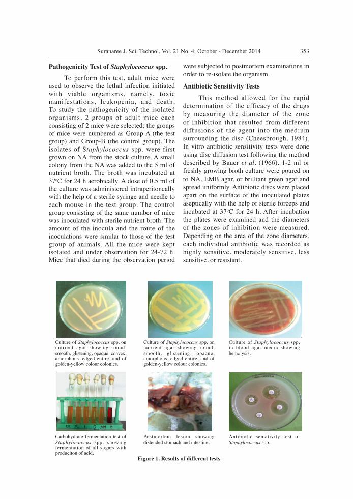
Pathogenicity Test of Staphylococcus spp. To perform this test, adult mice were used to observe the lethal infection initiated with viable organisms, namely, toxic manifestations, leukopenia, and death. To study the pathogenicity of the isolated organisms, 2 groups of adult mice each consisting of 2 mice were selected; the groups of mice were numbered as Group-A (the test group) and Group-B (the control group). The isolates of Staphylococcus spp. were first grown on NA from the stock culture. A small colony from the NA was added to the 5 ml of nutrient broth. The broth was incubated at 37oC for 24 h aerobically. A dose of 0.5 ml of the culture was administered intraperitoneally with the help of a sterile syringe and needle to each mouse in the test group. The control group consisting of the same number of mice was inoculated with sterile nutrient broth. The amount of the inocula and the route of the inoculations were similar to those of the test group of animals. All the mice were kept isolated and under observation for 24-72 h. Mice that died during the observation period
were subjected to postmortem examinations in order to re-isolate the organism.
Antibiotic Sensitivity Tests
This method allowed for the rapid determination of the efficacy of the drugs by measuring the diameter of the zone of inhibition that resulted from different diffusions of the agent into the medium surrounding the disc (Cheesbrough, 1984). In vitro antibiotic sensitivity tests were done using disc diffusion test following the method described by Bauer et al. (1966). 1-2 ml or freshly growing broth culture were poured on to NA, EMB agar, or brilliant green agar and spread uniformly. Antibiotic discs were placed apart on the surface of the inoculated plates aseptically with the help of sterile forceps and incubated at 37oC for 24 h. After incubation the plates were examined and the diameters of the zones of inhibition were measured. Depending on the area of the zone diameters, each individual antibiotic was recorded as highly sensitive, moderately sensitive, less sensitive, or resistant.
Figure 1. Results of different tests
Carbohydrate fermentation test of Staphylococcus spp. showing fermentation of all sugars with produciton of acid.
Culture of Staphylococcus spp. on nutrient agar showing round, smooth, glistening, opaque, convex, amorphous, edged entire, and of golden-yellow colour colonies.
Postmortem lesion showing distended stomach and intestine.
Culture of Staphylococcus spp. on nutrient agar showing round, smooth, glistening, opaque, amorphous, edged entire, and of golden-yellow colour colonies.
Antibiotic sensitivity test of Staphylococcus spp.
Culture of Staphylococcus spp. in blood agar media showing hemolysis.

Public Health Sanitation of Commercially Available Drinking Water 354
Results
Bacterial Species Isolated from Commercially Available Bottled Drinking Water
The bacterial species isolated from the commercially available bottled water were described in Table 2.
Isolation and Identification of Staphylococcus spp.
Cultural Examination Nutrient broth: Nutrient broth was inoculated separately with commercially available bottled water and incubated at 37oC for 24 h. The growth of bacteria was indicated by the presence of turbidity.
Nutrient Agar (NA):
The NA plates that were streaked separately with the organisms revealed the growth of bacteria after 24 h of incubation at 37oC aerobically and the growth was indicated by circular, small, smooth, convex, and gray-white or golden-yellowish colonies. Gram’s Staining Light microscopic examination after Gram’s staining revealed Gram-positive spherical cells arranged in pairs, tetrads, and clusters.
Biochemical Tests Sugars fermentation test: The isolates fermented 5 basic sugars (dextrose, maltose, lactose, sucrose, and mannitol) with production of acid which followed the description of Merchant and Packer (1967). The change of colour from reddish to yellow indicated acid production. Coagulase Test The isolates were coagulase positive and the isolated organism was Staphylococcus aureus which was similar to the description of Brooks et al. (2002) Catalase Test The isolates were catalase positive. Pathogenicity Test The pathogenicity test was conducted with the Staphylococcus spp. isolated from the bottled water. The organism was inoculated intraperitoneally in 2 adult mice and another 2 mice were kept as a control. The results are given in Table 3. The isolates of Staphylococcus spp. caused the death of the mice in the test group within 48 hand were categorized as pathogenic. Characteristic lesions in different organs were produced following the experimental inoculation of Staphylococcus spp. in the adult mice. The intestine was distended. No other signs were detected in the
Table 2 Bacterial species isolated from bottled water
Name of brand No. of samples examined
Positive samples (%)
Name of bacterial isolates
Acme 5 0 (0.0) nil
Fresh 5 0 (0.0) nil
Everest 5 4 (80.0) Staphylococcus spp.
Spa 5 0 (0.0) nil
Mum 5 0 (0.0) nil
Niagra 5 0 (0.0) nil
Shanti 5 0 (0.0) nil
Pran 5 0 (0.0) nil
Jibon 5 0 (0.0) nil
Boss 5 0 (0.0) nil

355Suranaree J. Sci. Technol. Vol. 21 No. 4; October - December 2014
visceral organs. The deaths of the mice might have been caused by neural toxicity.
Antibiotic Sensitivity and Resistance Pattern of Isolated Bacteria
Out of the total number of isolates, 4 Staphylococcus spp., irrespective of their sources, were tested for the antibiotic sensitivity and resistance against different antibiotics. The results of the sensitivity against antibiotic discs (the zones of inhibition) were recognized as resistant (-), less sensitive (+), moderately sensitive (++), and highly sensitive (+++). The results of the antibiotic sensitivity and resistance patterns are given in Table 4.
Antibiotic Sensitivity and Resistance Pattern of Staphylococcus spp.
Among the 4 isolates of Staphylococcu spp., 66.67% were highly sensitive to ampicillin, amoxicillin, gentamicin, furazolidon, norfloxacin, and pefloxacin and 33.33% to ciprofloxacin. One hundred percent of the isolates were
moderately sensitive to gentamicin, 66.67% to amoxicillin and furazolidon, and 33.33% to enrofloxacin and pefloxacin. Among the organisms 100.00% were less sensitive to furazolidon, 66.67% to amoxicillin, gentamicin, and pefloxacin, and 33.33% to ampicillin, ciprofloxacin, and norfloxacin. One hundred percent of the isolates were resistant to amoxicillin, gentamicin, and furazolidon, 66.67% to pefloxacin, and 33.33% to ciprofloxacin.
Discussion The present study was undertaken to isolate, identify, and characterize the bacterial spp. from commercially available bottleddrinking water in Bangladesh. The characterization procedure included cultural studies, morphological, staining properties, and biochemical tests and antibiotic sensitivity tests. The pathogenicity test of the isolated Staphylococcus spp.was done to determine their ability to produce diseases. The
Table 4. Antibiotic sensitivity and resistance patterns
Name of isolates AMP AML CIP CN ENR FR NOR PEF
Staphylococcus spp (n = 4).
+ + ++ + + + + + + +
+ ++ + + ++ - + + + + + + ++
++ + + + + + ++ - + + +
+++ - + + - + + + - + + + + ENR = Enrofloxacin, CIP = Ciprofloxacin, PEF = Pefloxacin, CN = Gentamicin, AMP = Ampicillin, NOR = Norfloxacin, AML = Amoxicillin, FR = Furazolidone
Table 3. Demonstration of the biochemical reactivity pattern of Staphylococcus spp. isolated from different sources
Fermentation reaction with five basic sugars Coagulase test
Coagulase test DX ML L S MN
A A A A A + + DX = Dextrose; ML = Maltose; L = Lactose; S = Sucrose; MN = Mannitol; A = Acid production + = Positive test; - =Negative test

Public Health Sanitation of Commercially Available Drinking Water 356
sensitivity and resistance patterns of the isolated organisms to different antibacterial agents available in the market were also a part of the study. Triplicate tests were done for all of the samples in all techniques. The bacteria isolated from the commercially available bottled water were Staphylococcus aureus. More than 8% of the water sampleswere found positive for Staphylococcus spp. which. was found to be pathogenic. The presence of these bacteria is alarming for human health. Ibekwe et al. (2004) isolated Staphyloccus from waste water in the accumulation pond and final discharge point of the Nigerian Bottling Company PLC in Owerri, Nigeria. Staphylococcus spp. were isolated from bottled mineral water by Tsai and Yu (1997), Okagbue et al. (2002), and by Abd El-Salam et al. (2008) The coagulase test was performed using a total of 4 Staphylococcus spp. to determine whether the organism was pathogenic or non-pathogenic. It was found that the isolated Staphylococcus spp. was coagulase positive i.e. it was the pathogenic Staphylococcus aureus. Brooks et al. (2002) described that Staphylococcus aureusas producing coagulase. For the bacteria isolated from the water samples, the pathogenicity test of Staphylococcus spp. was done on the mice because these organisms produce clinical diseases in man, livestock, and poultry. The organisms were found to be pathogenic for adult mice.
Among the 4 isolates of Staphylococcus spp., 66.67% were highly sensitive to ampicillin, amoxicillin, gentamicin, furazolidon, norfloxacin, and pefloxacin and 33.33% to ciprofloxacin. One hundred percent of the isolates were moderately sensitive to gentamicin, 66.67% to amoxicillin and furazolidon, and 33.33% to enrofloxacin and pefloxacin. Among the organisms, 100.00% were less sensitive to furazolidon, 66.67% to amoxicillin, gentamicin, and pefloxacin, and 33.33% to ampicillin, ciprofloxacin, and norfloxacin. One hundred percent of the isolates were resistant to amoxicillin, gentamicin, and furazolidon, 66.67% to pefloxacin, and 33.33% to ciprofloxacin. Gentilini et al. (2002) stated that resistance was detected in 34 (27.6%), 4 (3.2%), 7 (5.7%), and 6 (4.8%) of the isolates for penicillin, oxacillin, erythromycin,and pirlimycin, respectively. To our knowledge, this is the first study in Bangladesh regarding commercially bottled water. Furthermore,the research is promising for verifying all of the commercial drinking water with the molecular intervention.
Public Health Significance
It might be concluded that the presence of microorganisms in water has veterinary publichealth importance because it might play a direct or indirect role in the transmission of various enteric and waterborne diseases in man and other animals. Among the different brands of bottled water, all of them except
Table 5. Antibiotic sensitivity and resistance pattern in percent
Name of bacteria Sample Sensitivity/ resistance
% of isolated strains sensitive/ resistance to various antibiotics
AMP AML CIP CN ENR FR NOR PEF
Staphylococcus spp.
04
Resistance 0.00 100.00 33.33 100.00 0.00 100.00 0.00 66.67
Less sensitive 33.33 66.67 33.33 66.67 0.00 100.00 33.33 66.67
Moderately sensitive
0.00 66.67 0.00 100.00 33.33 66.67 0.00 33.33
Highly sensitive
66.67 66.67 33.33 66.67 0.00 66.67 66.67 66.67
ENR = Enrofloxacin, CIP = Ciprofloxacin, PEF = Pefloxacin, CN = Gentamicin, AMP = Ampicillin, NOR = Norfloxacin, AML = Amoxicillin, FR = Furazolidone

357Suranaree J. Sci. Technol. Vol. 21 No. 4; October - December 2014
“Everest mineral water” were found to be pathogen free.The isolated bacteria were Staphylococcus aureus which were found to be pathogenic. Among the 8 antibiotics, enrofloxacin, norfloxacin, and ciprofloxacin were found highly effective against most of the isolated bacteria.
Acknowledgements The authors express their cordial gratitude and thanks to the Bacteriological Laboratory, Department of Microbiology and Hygiene, Faculty of Veterinary Science, Bangladesh Agricultural University, Mymensingh, Bangladesh for financial support and providing scope for this research.
References Abd El-Salam, M.M., Al-Ghitany, E.M., and Kassem,
M.M. (2008). Quality of bottled water brands in Egypt. Part II: Biological water examination. J. Egypt. Public Health Assoc., 83:468-86.
Ali, M.A. (2003). Public private priority for water resources management in Bangladesh. Two billion people are dying for it. World Enviroment Day 5 June, 2003. Govt. Bangladesh, p. 43-46.
Amer, A.E., Soltan, E.M., and Gharbia, M.A. (2008). Molecular approach and bacterial quality of drinking water of urban and rural communities in Egypt. Acta Microbiol. Imm. H., 55:311-326.
Bauer, A.W., Kirby, W.M., Sherris, J.C., and Turck, M. (1966). Antibiotic susceptibility testing by a standardized single disk method. Am. J. Clin. Pathol., 45:493-496.
Brooks, B.F., Butel, J.S., and Morse, S.A. (2002). Jawetz, Melnick and Adelberg’s Medical Microbiology. 22nd ed. McGraw Hill, New Delhi, India, p.197-202.
Buxton, A. and Fraser, G. (1977). Animal Microbiology. Blackwell Scientific Publications, Oxford, UK, 1:85-110.
Carter, G.R. (1986). Studies on Pasteurella multocida. A haemagglutination test for the identification of serological types. Am. J. Vet. Res., 16:481-484.
Cheesbrough, M. (1984) Medical laboratory manual for tropical countries. Microbiology, 2:400-480.
Chung, Y.H., Kim, S.Y., and Chang, Y.H.(2003). Prevalence and antibiotic susceptibility of Salmonella isolated from foods in Korea from 1993 to 2001. J. Food Protect., 66:1154-1157.
Cowan, S.T. (1985). Cowan and Steel’s Manual for Identification of Bacteria. 2nd ed., Cambridge University Press, Cambridge, UK.
Gentilini, E., Denamiel, G., Betancor, A., Rebuelto, M., Fermepin, M.R., and Torres, R.A.D. (2002). Antimicrobial susceptibility of coagulase-negative staphylococci isolated from bovine mastitis in Argentina. J. Dairy Sci., 85:1913-1917.
Guillot, E. and Loret, J-F. (2009). Waterborne Pathogens: Review for the Drinking Water Industry. IWA Publishing, London, UK, 194p.
Ibekwe, V.I., Nwaiwu, O., and Offorbuike, J.O. (2004). Bacteriological and physico-chemical qualities of waste water from a bottling company in Owerri, Nigeria. J. Environ. Sci., 3:51-54.
Merchant, I.A. and Packer, R.A. (1967). Veterinary Bacteriology and Virology. 7th ed. The Iowa University Press, Ames, IA, USA, p.211-306.
Okagbue, R.N., Dlamini, N.R., Siwela, M., and Mpofu, F. (2002). Microbiological quality of water processed and bottled in Zimbabwe. Afr. J. Health Sci., 9:99-103.
Prescott, L.M., Harley, J.P., and Klein, D.A. (2005). Microbiology. 6th ed. McGraw-Hill Publishing, NY, USA, 992p.
Tsai, G.J. and Yu, S.C. (1997). Microbiological evaluation of bottled uncarbonated mineral water in Taiwan. Int. J. Food Microbiol., 37:137-143.
Zhao, Q., Shu, W., and Gao, J. (2004). Quality standards and hygienic problems of bottled drinking-water. Wei Sheng Yan Jiu, 33:386-388.
