characterization and sequence of the promoter region of the human
TRANSCRIPT

Proc. Natl. Acad. Sci. USAVol. 82, pp. 4920-4924, August 1985Biochemistry
Characterization and sequence of the promoter region of the humanepidermal growth factor receptor gene
(transcription/oncogene/DNase I hypersensitivity/simian virus 40/hydroxymethylglutaryl-CoA reductase)
SHUNSUKE ISHII*, YOUNG-HUA Xu*, RANDY H. STRATTONt, BRUCE A. ROEt, GLENN T. MERLINO*,AND IRA PASTAN*t*Laboratory of Molecular Biology, Division of Cancer, Biology and Diagnosis, National Cancer Institute, National Institutes of Health, Bethesda, MD 20205;and tDepartment of Chemistry, University of Oklahoma, Norman, OK 73019
Contributed by Ira Pastan, April 4, 1985
ABSTRACT The promoter region of the epidermal growthfactor (EGF) receptor has been identified by in vitro transcrip-tion using EGF receptor genomic DNA fragments as templateand by primer extension and nuclease S1 mapping using EGFreceptor mRNA. Six transcriptional start sites were identified.DNA sequence analysis shows that the promoter region con-tains neither a "TATA box" nor a "CAAT box," has anextremely high G+C content (88%), and contains fiveCCGCCC repeats and four (TCC)TCCTCCTCC repeats. Thispromoter region is situated close to or within a DNase I-hypersensitive site in A431 human epidermoid carcinoma cells,which overproduce the EGF receptor. The EGF receptor genepromoter has some resemblance to the promoter of thehydroxymethylglutaryl-CoA reductase gene and the early pro-moter of simian virus 40. This similarity may offer a clue to themechanism by which the receptor gene is regulated.
Epidermal growth factor (EGF) stimulates cell growth bybinding to specific membrane receptors (1, 2). The EGFreceptor is a membrane-spanning glycoprotein that has threefunctional domains: an EGF-binding domain located on theexternal cell surface, a transmembrane domain, and a cytoplas-mic tyrosine kinase domain (3-5). EGF binding induces inter-nalization of the receptor, which is ultimately delivered tolysosomes where it is destroyed (6), and induces accumulationsofEGF receptormRNA (unpublished observations). The bind-ing of EGF to the receptor activates a tyrosine kinase thatphosphorylates various cellular proteins, including the EGFreceptor itself (7, 8). The best characterized tyrosine-specificprotein kinase is pp6OsrC, the transforming protein of the Roussarcoma virus (9-11). The oncogene erbB, carried by the avianerythroblastosis retrovirus (AEV), is homologous to thetransmembrane and kinase domains of the EGF receptor gene(12-17). The EGF receptor is often overproduced in tumor cells(18); the human epidermoid carcinoma A431, a squamouscarcinoma cell line, and several glioblastomas contain a verylarge number of EGF receptors resulting from EGF receptorgene amplification (14, 15, 17, 19-21). Therefore, it seemsplausible that overexpression of the EGF receptor gene con-tributes to the phenotype of cellular transformation. To inves-tigate the regulation ofthe expression ofthe EGF receptor geneat the molecular level, we identified and characterized the 5'flanking region of this gene.
MATERIALS AND METHODSDNA analysis and sequencing were performed as described(22-25). Poly(A)+ RNA was isolated from A431 and epithelialcells (26). Extension of various primers was performed asdescribed (27, 28). Nuclease S1 mapping was done according
to a modification of procedures detailed elsewhere (28-30).For in vitro transcription studies, HeLa cell extracts wereprepared and incubations were performed as previouslyreported (27, 31). DNase I-hypersensitive sites were detectedas described (32); the nuclei were isolated according to Wu etal. (33), including centrifugation through a 1.5 M sucrosecushion at 100,000 x g for 30 min.
RESULTSIsolation of 5'-Specific Human EGF Receptor Genomic
Clones. Genomic clones containing the 5' end of the EGFreceptor gene were isolated, from a human genomic DNAfetal liver library (-34)by screening with a mixture of syntheticoligonucleotide probes [CTGGA(A/G)GA(A/G)AA(A/G)AA] that contains the possible codons of the amino acids atthe NH2 terminus of the EGF receptor (Leu-Glu-Glu-Lys-Lys-) (35). One positive clone was isolated (XER1). A950-base-pair (bp) EcoRI-Sst I (nucleotides -965 to -15)fragment of XER1 was found to hybridize with a 140-bp ClaI-Sst I (nucleotides -155 to -15) DNA fragment from thecDNA clone pE15 (36) that contains the 5' untranslatedsequences of the EGF receptor gene (Fig. 1). Additionalgenomic clones were selected in a second screening with this950-bp EcoRI-Sst I fragment as a probe. Two other clones,XER9 and XER14, were isolated and characterized (Fig. 1).Sequence analysis ofboth the 950-bp EcoRI-Sst I fragment
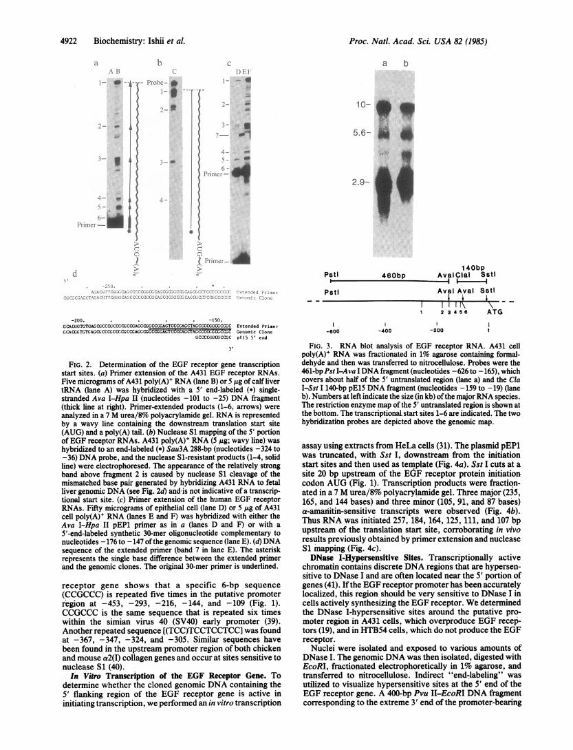
and the adjacent 3' region revealed that one exon is locatedin this area; this exon contains the 5' untranslated region, 72bp coding for the signal sequence, and 16 bp coding for theNH2-terminal portion of the mature EGF receptor (Fig. 1).The exon ends with the sequence AG and the adjacent intronbegins with the sequence GT.Primer Extension. To identify the 5' boundary of the EGF
receptor RNA, a 32P-labeled Ava I-Hpa II (Fig. 1, nucleo-tides -101 to -25) single-stranded DNA primer was preparedfrom plasmid pE15 and hybridized with A431 poly(A)+ RNA.The primer was extended with avian myeloblastosis virusreverse transcriptase, and the sizes of the resulting productswere determined by gel electrophoresis under denaturingconditions, followed by autoradiography. The most abundantextension products had lengths of 234, 160, 141, 93, 87, and83 bases (Fig. 2a, bands 1-6, respectively). These resultssuggest that the start site of EGF receptor gene transcriptionmay be heterogeneous, occurring 258, 184, 165, 117, 111, and107 bp upstream from the initiator AUG codon (Fig. 1).One possibility is that many of these sites are unique to
A431 cells, which possess amplified and rearranged EGF
Abbreviations: EGF, epidermal growth factor; HMG-CoA reduc-tase, hydroxymethylglutaryl-CoA reductase; SV40, simian virus 40;bp, base pair(s); kb, kilobase(s).tTo whom reprint requests should be addressed at: Building 37, Rm.4E-16, National Institutes of Health, Bethesda, MD 20205.
4920
The publication costs of this article were defrayed in part by page chargepayment. This article must therefore be hereby marked "advertisement"in accordance with 18 U.S.C. §1734 solely to indicate this fact.

Proc. Natl. Acad. Sci. USA 82 (1985) 4921
AER14
pEP6. pEP5. --4 p 1
EcoRI Exon 1 EcoRI
10Q..I- - ._ gOkb
, AER9AER 1
EcoRII _.-
5'end of pE15'
- _Exoni Sau3A --
Pvull Pstl-600
Sau3A-4b0
l l r, Aval Aval Sstl
-200 1Pvull
200
-400 .
CCT CCC'ATGCG COGCCCCACTrCGCCGGAGArTAGGCCCGCGGGGGCCACCGCrGTCCACOGCCTCCGGCC GCTGGCCrTGGGTCC6CCTG CrGGTTCrCCTrCCr ~CrCCrOGCATTCTCCTr CCTr ~CCCTGCTGCTCCCG.* -30 . . . . -400. , . . . .. -2 0
2. . -300. 1 . . . -200.
GCCGAGGGCGGCCGGAGTCCCGAGCrAGCCCCGGCGCCGCCGCCGCCCAGACCGGACACAGGCCACCr CGeG0GTGTCC CCCGAGTCCCCGCCTCGCCGCCAACGCCACAACCACCGCGCACGGccccccrCACICCGTCX O XRX WEM~
-1..+1 . . .XCAGTATTGATCGGCAGAGCCGGAGCGAGCrCrTCGGGGAGCAGCGATGCGACCCrCCGGGACGGCCGGGC CAGCGCrCGrGGCGCrGCrGCcGCGCrCrGCCCGGCAGTCG GGCrCTGGAGGAAAAGAAAGGTAAGGG
exon 1 | intron
FIG. 1. Structure of the human EGF receptor genomic clones. (Upper) A schematic representation of the three overlapping EGF receptorbacteriophage X recombinants is at the top. The subcloned genomic fragments are shown below the X recombinants (pEP1, pEP5, and pEP6).Below the EcoRI restriction map is an expanded map of the promoter and exon 1 region. Untranslated sequences are represented by a line,the exon by a box. The unshaded area of the box represents sequences that encode the 5' untranslated region; the stippled area of the box codesfor the signal peptide; the shaded area of the box codes for the mature EGF receptor. (Lower) Nucleotide sequence of the 5' portion ofthe humanEGF receptor gene, where + 1 corresponds to the A of the initiator methionine codon, and residues preceding it are represented by negativenumbers. The multiple initiation sites (numbered 1-6) demonstrated by primer extension, nuclease S1 mapping, and in vitro transcription areindicated by x, o, and I, respectively. The repeated CCGCCC sequences are underlined by a thick solid line, whereas the repeatedTCCTCCTCCsequences are underlined by a thin solid line.
receptor genes (14, 15, 17, 36). Normal human epithelial cell(37) poly(A)+ RNA was therefore isolated and primer-ex-tended. The same population of extended products is syn-thesized whether template RNA is from normal cells (Fig. 2c,lane D) or A431 cells (lane F). Therefore, the 5' end of theEGF receptor gene of A431 cells resembles that of a normalcell.
Nuclease S1 Mapping. When a 288-bp Sau3A DNA frag-ment from genomic subclone pEPI (Fig. 1, nucleotides -324to -36) was 5'-end-labeled, hybridized with A431 poly(A)+RNA, and digested with nuclease S1, multiple protectedfragments were detected (Fig. 2b). The major fragments hadlengths of 222, 148, 130, and 82 bases (Fig. 2b, bands 1-4),corresponding to initiation sites at nucleotides -257, -182,-164, and -116. Two fragments corresponding to the twodistal start sites were very weak but were seen after longerautoradiographic exposure.DNA Sequence Comparison. To determine whether the 5'
ends of the extended primers originate from within XER1genomic DNA, we compared the nucleotide sequence of anextended DNA primer to that of the cloned genomic DNA.By using a labeled synthetic oligonucleotide primer (30-mer)complementary to one part of the cDNA sequence (Fig. 2c,-176 to -147) a single major labeled extension product wassynthesized (Fig. 2c, lane E). The nucleotide sequence ofthisextended primer was found to be identical to the sequence ofthe genomic clone immediately adjacent to the primer, exceptfor position -214 (Fig. 2d, asterisk). The EGF receptorcDNA of A431 cells has a T; the genomic clone from fetalliver has a G. These results indicate that the genomic DNAcorresponding to the extended cDNA primer is not interrupt-ed by introns.The primer extension, nuclease S1 mapping, and sequenc-
ing data together strongly suggest that the synthesizedcDNAs extend to the extreme 5' end of the EGF receptorgene. Ullrich et al. (14) isolated an EGF receptor cDNA clonefrom an A431 library which contains 176 bp of 5' untranslatedcDNA. However, the 5'-most 10-bp sequence of their cDNAclone (5' GCCGCGCTGC 3') is not found in the sequence of
either the extended cDNA primer made from A431 RNA orthe XER1 genomic clone isolated from human fetal liver. Wedid find an inverted sequence (5' GCAGCGCGGC 3') of this10-bp region at nucleotide position -236 to -227.
Multiple EGF Receptor mRNAs Initiate from the Same5'-Most Site. A431 cells contain an aberrant 2.8- to 2.9-kilobase (kb) EGF receptor RNA which diverges from EGFreceptor RNAs found in other cell types (10 and 5.6 kb) at its3' end (14, 16, 17, 36). To determine whether different A431EGF receptorRNA species are transcribed from variant startsites, we performed RNA blot analysis by using a 641-bp PstI-Ava I probe (Fig. 1, -626 to -165) specific for the 5'portion of the 5' untranslated region; this DNA can onlyhybridize to transcripts starting from the two initation sites atnucleotide positions -258 and -184 (Fig. 1). The 461-bpprobe hybridized to all of the A431 EGF receptor RNAspecies (10, 5.6, and 2.9 kb; Fig. 3, lane a). A Cla I-Sst I140-bp DNA fragment (Fig. 1, -159 to -19) of the pE15cDNA clone, containing the 3' portion of the 5' untranslatedregion, also hybridized with all of the RNA species (Fig. 3,lane b). These results show that all three RNAs are at leasttranscribed from the start sites at nucleotides -258 and -184and subsequently processed into the three different-sizeRNA species.
Sequence Around the EGF Receptor Promoter Is G+C-Rich. The nucleotide sequence upstream of the EGF receptorRNA start sites contains neither a "TATA box" nor a"CAAT box" (Fig. 1). Most other characterized eukaryoticgenes have these two sequence elements "30 and 80 bp,respectively, upstream of the RNA initiation site (38). Sincein vitro transcriptional studies have shown that the TATAbox serves to fix the site at which transcription will start (39),the fact that the EGF receptor gene has numerous transcrip-tional initiation sites is not surprising.The sequence between nucleotides -540 and -1 (Fig. 1),
which contains the putative promoter and 5' untranslatedregion, has a G+C content of 88%; however, the regionfurther upstream of -540 has a G+C content of only about50%. Further analysis of the 5' flanking region of the EGF
55-6
5 end of pEP1 -
EcoRI-idoo
+T T T w
-800
Biochemistry: Ishii et al.
_ _
Proc. Natl. Acad. Sci. USA 82 (1985)
1) E.11- -.
i.ii04- _
i- 4W
t- Pime-
I m-lirllel,- APstl
C n: :(:Af:CCC(v(' 1',Cu :iAC,vc'{ ,.vCe: .'c vc c.xe:.,' i \ATw ,(AC
-200. . . . . -150.GCACGGTGTGAGCCGCCCGCCGrGCCGAGGCGGCCGGAGTCCCGAGCTAGCCCCGGCGCCGC Extended PrimerGCACGTGTCAGCGCCCGCOCGCCGACCCGCGCGGAGTCCcCGAGCTAGCCCCC.GCGCOGC Genomic Clone
GCCCCGGCGCCGC p15 5' end
3'
FIG. 2. Determination of the EGF receptor gene transcriptionstart sites. (a) Primer extension of the A431 EGF receptor RNAs.Five micrograms ofA431 poly(A)+ RNA (lane B) or 5 ,Ig of calf livertRNA (lane A) was hybridized with a 5' end-labeled (*) single-stranded Ava I-Hpa II (nucleotides -101 to -25) DNA fragment(thick line at right). Primer-extended products (1-6, arrows) wereanalyzed in a 7 M urea/8% polyacrylamide gel. RNA is representedby a wavy line containing the downstream translation start site(AUG) and a poly(A) tail. (b) Nuclease S1 mapping of the 5' portionof EGF receptor RNAs. A431 poly(A)+ RNA (5 ,ug; wavy line) washybridized to an end-labeled (*) Sau3A 288-bp (nucleotides -324 to-36) DNA probe, and the nuclease Si-resistant products (1-4, solidline) were electrophoresed. The appearance of the relatively strongband above fragment 2 is caused by nuclease S1 cleavage of themismatched base pair generated by hybridizing A431 RNA to fetalliver genomic DNA (see Fig. 2d) and is not indicative of a transcrip-tional start site. (c) Primer extension of the human EGF receptorRNAs. Fifty micrograms of epithelial cell (lane D) or 5 jug of A431cell poly(A)+ RNA (lanes E and F) was hybridized with either theAva I-Hpa II pEP1 primer as in a (lanes D and F) or with a5'-end-labeled synthetic 30-mer oligonucleotide complementary tonucleotides -176 to -147 ofthe genomic sequence (lane E). (d)DNAsequence of the extended primer (band 7 in lane E). The asteriskrepresents the single base difference between the extended primerand the genomic clones. The original 30-mer primer is underlined.
receptor gene shows that a specific 6-bp sequence(CCGCCC) is repeated five times in the putative promoterregion at -453, -293, -216, -144, and -109 (Fig. 1).CCGCCC is the same sequence that is repeated six timeswithin the simian virus 40 (SV40) early promoter (39).Another repeated sequence [(TCC)TCCTCCTCC] was foundat -367, -347, -324, and -305. Similar sequences havebeen found in the upstream promoter region of both chickenand mouse a2(I) collagen genes and occur at sites sensitive tonuclease S1 (40).In Vitro Transcription of the EGF Receptor Gene. To
determine whether the cloned genomic DNA containing the5' flanking region of the EGF receptor gene is active ininitiating transcription, we performed an in vitro transcription
10-
E.5.6- Jo
-..
2.9- _
460bp
Pstl
-600 -400
140bpAval Clal Sstl
Aval Aval Sstl
23 5IAIl\\1 2 3 4 56 ATG
-200
FIG. 3. RNA blot analysis of EGF receptor RNA. A431 cellpoly(A)+ RNA was fractionated in 1% agarose containing formal-dehyde and then was transferred to nitrocellulose. Probes were the461-bp Pst I-Ava I DNA fragment (nucleotides -626 to -165), whichcovers about half of the 5' untranslated region (lane a) and the ClaI-Sst I 140-bp pE15 DNA fragment (nucleotides -159 to -19) (laneb). Numbers at left indicate the size (in kb) of the major RNA species.The restriction enzyme map of the 5' untranslated region is shown atthe bottom. The transcriptional start sites 1-6 are indicated. The twohybridization probes are depicted above the genomic map.
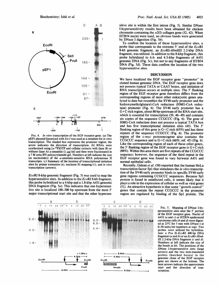
assay using extracts from HeLa cells (31). The plasmid pEP1was truncated, with Sst I, downstream from the initiationstart sites and then used as template (Fig. 4a). Sst I cuts at asite 20 bp upstream of the EGF receptor protein initiationcodon AUG (Fig. 1). Transcription products were fraction-ated in a 7 M urea/8% polyacrylamide gel. Three major (235,165, and 144 bases) and three minor (105, 91, and 87 bases)a-amanitin-sensitive transcripts were observed (Fig. 4b).Thus RNA was initiated 257, 184, 164, 125, 111, and 107 bpupstream of the translation start site, corroborating in vivoresults previously obtained by primer extension and nucleaseSi mapping (Fig. 4c).DNase I-Hypersensitive Sites. Transcriptionally active
chromatin contains discrete DNA regions that are hypersen-sitive to DNase I and are often located near the 5' portion ofgenes (41). If the EGF receptor promoter has been accuratelylocalized, this region should be very sensitive to DNase I incells actively synthesizing the EGF receptor. We determinedthe JDNase I-hypersensitive sites around the putative pro-moter region in A431 cells, which overproduce EGF recep-tors (19), and in HTB54 cells, which do not produce the EGFreceptor.
Nuclei were isolated and exposed to various amounts ofDNase I. The genomic DNA was then isolated, digested withEcoRI, fractionated electrophoretically in 1% agarose, andtransferred to nitrocellulose. Indirect "end-labeling" wasutilized to visualize hypersensitive sites at the 5' end of theEGF receptor gene. A 400-bp Pvu II-EcoRI DNA fragmentcorresponding to the extreme 3' end of the promoter-bearing
a bb
JIob-.)-c - *1- *
4-
ciI.\ B
1---_
I
4- _
Irlv-0-
Irt*
_
4922 Biochemistry: Ishii et al.
Proc. Natl. Acad. Sci. USA 82 (1985) 4923
a
EcoRI
Sstl, Pvull
EcoRI
b A B
235- 1
.w.
165- *X.
144- W-
105 -
91 -87-
- 1 I 1-125 1-107
1 114 56
ATG
Sstl
-200 -100 1
FIG. 4. In vitro transcription of the EGF receptor gene. (a) ThepEP1 plasmid linearized with Sst I was used as a template for in vitrotranscription. The shaded box represents the promoter region; thearrow indicates the direction of transcription. (b) RNAs weresynthesized using [a-32P]GTP and cellular extracts with (lane B) orwithout (lane A) a-amanitin (1 ug/ml) and then were fractionated ina 7 M urea/8% polyacrylamide gel. Numbers at left indicate the size(in nucleotides) of the a-amatinin-sensitive RNA polymerase IItranscripts. (c) Summary of the location of transcriptional initiationsites by primer extension (x), nuclease S1 mapping (o), and in vitrotranscription (arrows).
EcoRI 8-kbp genomic fragment (Fig. 5) was used to map thehypersensitive sites. In addition to the EcoRI 8-kb fragment,this probe hybridized to a 4-kbp and a 1.8-kbp A431 genomicDNA fragment (Fig. 5a). This indicates that one hypersensi-tive site is localized 100-300 bp upstream from the most 5'major transcriptional start site and that the other hypersen-
sitive site is within the first intron (Fig. 5). Similar DNaseI-hypersensitivity results have been obtained for chickenchromatin containing the a2(I) collagen gene (32, 42). WhenHTB54 nuclei were used, no obvious bands were generatedby DNase I digestion (Fig. 5b).To confirm the location of these hypersensitive sites, a
probe that corresponds to the extreme 5' end of the EcoRI8-kb genomic fragment, an EcoRI-HindIII 2.2-kbp DNAfragment, was utilized. In addition to the 8-kbp fragment, thisprobe hybridized to 6.6- and 4.3-kbp fragments of A431genomic DNA (Fig. 5c), but not to any fragments of HTB54DNA (Fig. Sd). These data confirm the location of the twohypersensitive sites.
DISCUSSIONWe have localized the EGF receptor gene "promoter" incloned human genomic DNA. The EGF receptor gene doesnot possess typical TATA or CAAT boxes, and initiation ofRNA transcription occurs at multiple sites. The 5' flankingregion of the EGF receptor gene therefore differs from thecorresponding regions of most other eukaryotic genes ana-lyzed to date but resembles the SV40 early promoter and thehydroxymethylglutaryl-CoA reductase (HMG-CoA reduc-tase) promoter (Fig. 6). The SV40 early promoter has aG+C-rich region, about 80 bp upstream of the RNA start site,which is essential for transcription (39, 46-49) and containssix copies of the sequence CCGCCC (Fig. 6). The gene ofHMG-CoA reductase does not possess a typical TATA boxand has five transcriptional initiation sites (43). The 5'flanking region of this gene is G+C-rich (65%) and has threerepeats of the sequence CCGCCC (Fig. 6). The promoterregion of the c-myc protooncogene also contains theCCGCCC sequence and is G+C-rich (66%; ref. 45) (Fig. 6).Like the corresponding region of each of these other genes,the 5' flanking region of the EGF receptor gene is G+C-rich(88%). Within this area there are five repeats of the CCGCCCsequence; however, the sequence of the third repeat in theEGF receptor gene was found to vary between A431 andnormal epithelial cells.
Recently, Gidoni et al. (50) reported that the human HeLatranscription factor Spl that stimulates the in vitro transcrip-tion of the SV40 early promoter binds to specific SV40 earlygene regions containing CCGCCC sequences. Because Splprotein is found in uninfected cells, it seems likely that itplays a role in the expression of cellular as well as viral genes(51). An attractive hypothesis is that some "growth control"genes that contain the repeat CCGCCC in the promoterregion are regulated by binding of the Spl protein. The
a b'
° 2 1 0 20 50 2 10 20 50
'b4H 4b,*
C dr- =10 2 10 20 50 0 2 1 0 20 50
8.0-6.6-
4.4-
.6. -40 04 *#4O. * ba
WIt- 1.8kb-*
v4.ukb I
+ EcoRI
Pvull
f - 6.6kb- 4.4kb ---
EcoRI f Ecc
Hindill8.0kb>
oRI
FIG. 5. Mapping of DNase I-hy-persensitive sites near the 5' portionof the EGF receptor gene. Nuclei ofA431 (a and c) or HTBS4 epidermoidcarcinoma cells (b and d) were digest-ed at 23°C for 5 min with DNase I at0-SO units/ml (numbers at top). Twoprobes were utilized for hybridiza-tion: a Pvu II-EcoRI 400-bp DNAfragment (a and b) or an EcoRI-Hind-III 2.2-kbp DNA fragment (c and d).Numbers at left indicate the size ofthe bands in kb. The positions of theDNase I-hypersensitive sites (largearrows) and the two nick-translatedprobes (hatched boxes) in thegenomic clone of the EGF receptorgene are shown at the bottom. Thebent arrows indicate the approximatestart and the direction of tran-scription.
C
-257
xo
-184 -1642 12 3
8.0-
4.0-
1.8-
EcoRI
8.0kb
Biochemistry: Ishii et al.

Proc. Natl. Acad. Sci. USA 82 (1985)
-292 -215 -144 -109recentor- I I ALo - LLL* Q'--107
-258 -1841 '_* 11-165 -117
89 68 47 0/5243101 80 58
TATA 523721
HMG-CoA -881 -812reductase m l a
-736
L.[j..O.LL ..- 640
-729 -655-660
-670-16
TATA 281
specific features ofthe 5' flanking region of the EGF receptorgene may prove to be important to our understanding of theregulation of protooncogene expression.
1. Savage, C. R. & Cohen, S. J. (1972) J. Biol. Chem. 247, 7609-7611.2. Carpenter, G. & Cohen, S. (1979) Annu. Rev. Biochem. 48,
193-216.3. Ushiro, H. & Cohen, S. (1980) J. Biol. Chem. 255, 8363-8365.4. Das, M., Miyakawa, T., Fox, C. F., Pruss, R. M., Aharonov, A. &
Herschman, H. R. (1977) Proc. Natl. Acad. Sci. USA 74,2790-2794.
5. Sahyoun, N., Hock, R. A. & Hollenberg, M. D. (1978) Proc. Natl.Acad. Sci. USA 75, 1675-1679.
6. Beguinot, L., Lyall, R. M., Willingham, M. C. & Pastan, I. (1984)Proc. Natl. Acad. Sci. USA 81, 2384-2388.
7. Hunter, T. & Cooper, J. A. (1981) Cell 24, 741-752.8. Cohen, S., Ushiro, H., Stoscheck, C. & Chinkers, M. (1982) J.
Biol. Chem. 257, 1523-1531.9. Collett, M. S., Purchio, A. F. & Erickson, R. L. (1980) Nature
(London) 285, 167-169.10. Levinson, A. D., Opperman, H., Varmus, H. E. & Bishop, J. M.
(1980) J. Biol. Chem. 255, 973-980.11. Richert, N. D., Blithe, D. L. & Pastan, I. (1982) J. Biol. Chem.
257, 7143-7150.12. Yamamoto, T., Nishida, T., Miyajima, N., Kawai, S., Ooi, T. &
Toyoshima, K. (1983) Cell 35, 71-78.13. Downward, J., Yarden, Y., Mayes, E., Scarce, G., Totty, N.,
Stockwell, P., Ullrich, A., Schlessinger, J. & Waterfield, M. D.(1984) Nature (London) 307, 521-527.
14. Ullrich, A., Coussens, L., Hayflick, J. S., Dull, T. J., Gray, A.,Tam, A. W., Lee, J., Yarden, Y., Libermann, T. A., Schlessinger,J., Downward, J., Mayes, E. L. V., Whittle, N., Waterfield, M. D.& Seeburg, P. H. (1984) Nature (London) 309, 418-425.
15. Merlino, G. T., Xu, Y.-h., Ishii, S., Clark, A. J. L., Semba, K.,Toyoshima, K., Yamamoto, T. & Pastan, I. (1984) Science 224,417-419.
16. Xu, Y.-h., Ishii, S., Clark, A. J. L., Sullivan, M., Wilson, R. K.,Ma, D. P., Roe, B. A., Merlino, G. T. & Pastan, I. (1984) Nature(London) 309, 806-810.
17. Lin, C. R., Chen, W. S., Kruiger, W., Stolarsky, L. S., Weber,W., Evans, R. M., Verma, I. M., Gill, G. N. & Rosenfeld, M. G.(1984) Science 224, 843-848.
18. Xu, Y.-h., Richert, N., Ito, S., Merlino, G. T. & Pastan, I. (1984)Proc. Natl. Acad. Sci. USA 81, 7308-7312.
19. Fabricant, R. N., DeLarco, J. E. & Todaro, G. J. (1977) Proc.Natl. Acad. Sci. USA 74, 565-569.
20. Merlino, G. T., Xu, Y.-h., Richert, N., Clark, A. J. L., Ishii, S.,Banks-Schlegel, S. & Pastan, I. (1985) J. Clin. Invest. 75,1077-1079.
21. Libermann, T. A., Nusbaum, H. R., Razon, N., Kris, R., Lax, I.,Soreq, H., Whittle, N., Waterfield, M. D., Ullrich, A. & Schles-singer, J. (1985) Nature (London) 313, 144-147.
22. Maniatis, T., Fritsch, E. F. & Sambrook, J. (1982) Molecular
FIG. 6. Diagram of promoter regions containing theCCGCCC sequence. The shaded and unshaded boxes representCCGCCC sequences and TATA boxes, respectively. The di-rection and location of the transcriptional initiation sites are
indicated by bent arrows. In both the EGF receptor andHMG-CoA reductase (43) genes, A of the initiator methionineis + 1. The nucleotide numbers of the SV40 genome are those ofthe specific SV40 numbering system (44). In the c-myc gene (45),the first T of the TATA box is numbered as +1.
Cloning: A Laboratory Manual (Cold Spring Harbor Laboratory,Cold Spring Harbor, NY).
23. Southern, E. M. (1975) J. Mol. Biol. 98, 503-517.24. Maxam, A. M. & Gilbert, W. (1980) Methods Enzymol. 65,
499-560.25. Sanger, R., Nicklen, S. & Coulson, A. R. (1977) Proc. NatI. Acad.
Sci. USA 74, 5463-5467.26. Chirgwin, J. M., Przybyla, A. E., MacDonald, R. J. & Rutter,
W. J. (1979) Biochemistry 18, 5294-5299.27. Merlino, G. T., Tyagi, J. S., deCrombrugghe, B. & Pastan, I.
(1982) J. Biol. Chem. 257, 7254-7261.28. Vogeli, G., Ohkubo, H., Sobel, M., Yamada, Y., Pastan, I. &
deCrombrugghe, B. (1981) Proc. Natl. Acad. Sci. USA 78,5334-5338.
29. Weaver, R. H. & Weissman, C. (1979) Nucleic Acids Res. 7,1175-1193.
30. Berk, A. & Sharp, P. (1977) Cell 12, 721-732.31. Manley, J. L., Fire, A., Cano, A., Sharp, P. A. & Gefter, M. L.
(1980) Proc. Natl. Acad. Sci. USA 77, 3855-3859.32. McKeon, C., Pastan, I. & deCrombrugghe, B. (1984) Nucleic Acids
Res. 12, 3491-3502.33. Wu, C., Bingham, P. M., Livak, K. J., Holmgren, R. & Elgin,
S. C. R. (1979) Cell 16, 797-806.34. Maniatis, T., Hardison, R. C., Lacy, E., Lauer, J., O'Connell, C.,
Quon, D., Sim, D. K. & Efstratiadis, A. (1978) Cell 15, 687-695.35. Weber, W., Gill, G. N. & Spiess, J. (1984) Science 224, 294-297.36. Merlino, G. T., Ishii, S., Whang-Peng, J., Knutsen, T., Xu, Y.-h.,
Clark, A. J. L., Stratton, R. H., Wilson, R. K., Ma, D. P., Roe,B. A., Hunts, J. H., Shimizu, N. & Pastan, I. (1985) Mol. Cell.Biol., in press.
37. Banks-Schlegel, S. P. & Harris, C. C. (1983) Exp. Cell. Res. 146,271-280.
38. Breathnach, R. & Chambon, P. (1981) Annu. Rev. Biochem. 50,349-383.
39. Fromm, M. & Berg, P. (1982) J. Mol. Appl. Genet. 1, 457-481.40. McKeon, C., Schmidt, A. & deCrombrugghe, B. (1984) J. Biol.
Chem. 259, 6636-6640.41. Elgin, S. C. R. (1981) Cell 27, 413-415.42. Merlino, G. T., McKeon, C., deCrombrugghe, B. & Pastan, I.
(1983) J. Biol. Chem. 258, 10041-10048.43. Reynolds, G. A., Basu, S. K., Osborne, T. H., Chin, D. J., Gil,
G., Brown, M. S., Goldstein, J. L. & Luskey, K. L. (1984) Cell 38,275-285.
44. Buchman, A., Burnett, L. & Berg, P. (1981) in DNA TumorViruses, Part II Revised, ed. Tooze, J. (Cold Spring HarborLaboratory, Cold Spring Harbor, NY), pp. 799-841.
45. Watt, R., Nishikura, K., Sorrentino, J., ar-Rushdi, A., Croce,C. M. & Rovela, G. (1983) Proc. Natl. Acad. Sci. USA 80,6307-6311.
46. Benoist, C. & Chambon, P. (1981) Nature (London) 290, 304-310.47. Hansen, U. & Sharp, P. A. (1983) EMBO J. 2, 2293-2303.48. Wasylyk, B. & Chambon, P. (1983) Cold Spring Harbor Symp.
Quant. Biol. 47, 921-934.49. Tjian, R. (1981) Cell 26, 1-2.50. Gidoni, D., Dynan, W. S. & Tjian, R. (1984) Nature (London) 312,
409-413.51. Dynan, W. S. & Tjian, R. (1983) Cell 32, 669-680.
EGF-452
SV40 early
4924 Biochemistry: Ishii et al.
s-ri-