characterising the anisotropic mechanical properties … · characterising the anisotropic...
TRANSCRIPT

Characterising the Anisotropic MechanicalProperties of Excised Human Skin
Aisling Nı Annaidha,b, Karine Bruyerec,Michel Destradea,d, Michael D. Gilchrista,e, Melanie Ottenio
aSchool of Mechanical & Materials Engineering,University College Dublin, Belfield, Dublin 4, Ireland
bInstitut Jean Le Rond D’Alembert,UMR 7190, CNRS-UPMC, 4 place Jussieu, 75005 Paris, France
c Ifsttar, LBMC, UMR T9406, F-69675, Bron,Universite Lyon 1, Villeurbanne, France
dSchool of Mathematics, Statistics and Applied Mathematics,National University of Ireland Galway, Galway, Ireland
eSchool of Human Kinetics,University of Ottawa, Ontario K1N 6N5, Canada
1
arX
iv:1
302.
3022
v1 [
q-bi
o.T
O]
13
Feb
2013
Abstract
The mechanical properties of skin are important for a number of appli-cations including surgery, dermatology, impact biomechanics and forensicscience. In this study we have investigated the influence of location and ori-entation on the deformation characteristics of 56 samples of excised humanskin. Uniaxial tensile tests were carried out at a strain rate of 0.012s−1 onskin from the back. Digital Image Correlation was used for 2D strain mea-surement and a histological examination of the dermis was also performed.The mean ultimate tensile strength (UTS) was 21.6±8.4MPa, the mean fail-ure strain 54±17%, the mean initial slope 1.18±0.88MPa, the mean elasticmodulus 83.3±34.9MPa and the mean strain energy was 3.6±1.6MJ/m3. Amultivariate analysis of variance has shown that these mechanical proper-ties of skin are dependent upon the orientation of Langer lines (P<0.0001-P=0.046). The location of specimens on the back was also found to have asignificant effect on the UTS (P =0.0002), the elastic modulus (P=0.001) andthe strain energy (P=0.0052). The histological investigation concluded thatthere is a definite correlation between the orientation of Langer Lines and thepreferred orientation of collagen fibres in the dermis (P<0.001). The dataobtained in this study will provide essential information for those wishingto model the skin using a structural constitutive model.
Keywords: Soft tissue, Langer lines, Tensile Properties, Histology.
1 IntroductionSkin is a complex multi-layered material which can broadly be divided into threemain layers, the epidermis, the dermis, and the hypodermis. The epidermis con-sists of cells and cellular debris, the dermis consists of mostly networks of thefibrous proteins collagen, reticulin and elastin (Wilkes et al. 1973), and the hypo-dermis is primarily made up of connective tissue and fat lobules. Collagen fibresaccount for 75% of the dry weight of dermal tissue (Wilkes et al. 1973) and it isthese fibres which are responsible for the strength of the skin.
Skin is a highly non-linear, anisotropic, viscoelastic and nearly incompressiblematerial. A typical stress-stretch graph for skin exhibits non-linear behaviourand its response is usually described as three-phase (Brown 1973). In the initialloading phase, skin is very compliant and large deformation occurs at a relativelylow applied load. At this stage the fibres are largely unaligned. In the secondphase the stiffness of the skin gradually increases as the fibres align themselves in
2

the direction of applied load. The third phase is an almost linear phase wherebythe stiffness increases rapidly as the collagen fibres are mostly aligned and theoverall mechanical response becomes dependent upon the mechanical propertiesof the collagen fibres, which are three grades of magnitude stiffer than elastin.
That skin is anisotropic was recognized as far back as the 19th century byKarl Langer (1861), who mapped the natural lines of tension which occur withinthe skin. These lines are identified by puncturing the skin with a circular device.The wounds then assume an elliptical shape and by joining the major axes of theellipses a system of lines can be drawn as shown in Fig. 1(a). These lines areknown as the Langer lines. While Langer lines are the best known skin tensionlines, many variations on the original lines proposed by Langer have been made.These include the Cox lines (Cox 1941), Kraissl lines (Kraissl 1951) and RelaxedSkin Tension Lines (Borges 1984).
Early tensile tests suggested that the deformation characteristics of skin aredependent upon specimen orientation with respect to the Langer lines (Ridge &Wright 1966). More recent work conducted using Optical Coherence Elastogra-phy in-vivo, indicates a large difference between the Young moduli of skin alongdirections parallel and orthogonal to the Langer lines (Liang & Boppart 2010). Inthis study, we provide new experimental data of in-vitro human skin focusing inparticular on the anisotropic properties of the skin. We also quantify the degree ofanisotropy of the mechanical properties with respect to their Langer line orienta-tion. Finally, we perform a quantitative histological analysis on the dermis of thetest samples to examine a possible correlation between the orientation of Langerlines and the preferred direction of collagen fibres.
While many recent publications on skin report in-vivo experiments (Delalleauet al. 2008, Flynn et al. 2011, Moerman et al. 2009), here we have limited ourexperiments to in-vitro uniaxial tensile tests. As discussed by Holzapfel, there area number of issues involved with tensile tests (Holzapfel 2006): The structuralintegrity of the samples may be disturbed at the lateral edges and due to the softnature of the test material it is difficult to insert samples without subjecting themto an axial load. Despite these shortcomings, it was decided to perform in-vitrotensile tests over in-vivo testing for a number of reasons. First, in-vitro tests pro-vide simple stress-strain relationships that can be modeled and quantified easily,since their boundary conditions are well defined. Second, for the application ofthis work in a further study into the dynamic rupture of skin, we are also interestedin failure and rupture of skin, and in-vitro testing makes it possible to test untilfailure.
Most in-vitro investigations use human skin substitutes such as pigskin, sili-
3

cone or polyurethane (McCarthy et al. 2007, Gilchrist et al. 2008, Jor et al. 2011,McCarthy et al. 2010) and in-vitro tests on human skin are particularly rare. Thoseclassical publications which do use human skin often rely on outdated equipmentand their results can be difficult to interpret (Cox 1941, Jansen & Rottier 1958b).However, the development of accurate constitutive models depends heavily upontheir material definitions. This study aims to provide new material data for hu-man skin which can be applied to constitutive models in a number of technicalareas such as razor blade manufacture, cosmetics, surgical simulation, forensicpathology and impact biomechanics. Uniaxial tensile tests alone are not suffi-cient to determine multi-dimensional material models for soft tissues and usinga simple non-linear regression analysis to determine constitutive parameters mayresult in non-unique solutions, ill-conditioned equations and slow convergencerates. Ideally, testing of skin would involve planar biaxial tests with an in-planeshear, and separate through-thickness shear tests. While advanced testing proto-cols like these are not yet available, simple tension tests remain important becausethey serve to evaluate the level of anisotropy and to provide data which can later beused as validation for models constructed using more complicated testing methods(Holzapfel & Ogden 2009). Moreover, model parameters can be provided directlyfrom a histological study of the collagen fibre alignment in the dermis which al-lows for reasonable determination of material responses (Holzapfel 2006).
2 Materials & Method
2.1 Specimen PreparationTensile tests and preparation were carried out in Ifsttar (Institut francais des sci-ences et technologies des transports, de l’amenagement et des reseaux), France.French law allows human corpses that have been donated to science to be used forresearch purposes. The ethics committee within Ifsttar approved the use of hu-man biological material. Skin was excised from seven subjects: three male, fourfemale. The average age of the subjects was 89±6 years and none had any relatedskin diseases. Table 1 provides details on the gender and age of each subject atthe time of death along with the number of days that elapsed prior to testing.
Only the skin from the back of the subject was available for use. This wasexcised from the body with a scalpel. Each sample of skin was cut into a dogboneshape specimen, according to the ASTM D412 Standard tensile test method forvulcanised rubber, using a custom made die (see Fig. 1). Each specimen then had
4

Table 1: Information on each test subject. ∗Number of days elapsed after deathprior to testing
Gender Age (years) TimeElapsed∗
Label
Male 97 9 AMale 91 3 B
Female 97 2 CFemale 81 3 DMale 89 3 E
Female 85 4 FFemale 86 4 G
the epidermis and any underlying adipose tissue carefully removed with a scalpel.The thickness of the skin after removal of adipose tissue was measured using aVernier Calipers and the mean thickness was 2.56±0.39mm. It was observed thatskin removed from the lumbar area of the back contained more adipose tissuethan skin form other areas of the back. The dimensions of the test specimens weremeasured before and after excision. Specimens were obtained in various orien-tations, shown in Fig. 1, to correlate the specimens with the direction of Langerlines. Each sample was grouped into one of three categories: parallel, perpendic-ular, or at 45◦ to the Langer lines. The skin was stored in moistened paper andrefrigerated at 4◦C until it was ready to be tested. A total of 56 samples weretested successfully. Although it is widely accepted that preconditioning is neces-sary at small and intermediate strains, at higher strains the stress-strain responseis not significantly affected by preconditioning (Eshel 2001). The samples weretherefore not pre-conditioned prior to testing.
2.2 Tensile TestsTensile tests were performed using a Universal Tensile Test machine. Sampleswere clamped using specially designed anti-slip clamps, to counteract the ten-dency of samples to slip in ordinary grips. The velocity of the cross-head was50mm/min. The tensile load was measured with a 1kN piezoelectric load cell.Each tensile test was videoed with two Dalsa Falcon 1.4M100 digital video cam-eras at 20 frames/second. This was to record any abnormal behaviour during theexperiments and also for the subsequent use of Digital Image Correlation. Thegauge length and width were both measured optically. For these tests, the strain
5

(a) (b)
Figure 1: (a) Orientation of samples from the back. Specimens are superimposedonto a map of the Langer lines to indicate their orientation with respect to the lines(Langer 1861). (b) Dimensions of custom made die (mm)
rate was 0.012s−1, nominally the same strain rate as in tests carried out by Jansen& Rottier (1958a).
The main focus of this study is an investigation into the hyperelastic proper-ties of skin and how these vary according to orientation and location. We thusconsidered the tensile tests to be taking place at quasi-static speed, and ignoredviscoelastic effects for the purpose of constitutive hyperelastic modelling (for ex-amples of studies where this route is chosen, see Jor et al. (2011), Pailler-Matteiet al. (2008), Evans & Holt (2009) or Cavicchi et al. (2009)). Further creep andstress relaxation tests, requiring additional skin samples, would need to be carriedout to characterize the viscoelasticty of skin.
2.3 Digital Image CorrelationThe stretch ratio was calculated via the displacement cell attached to the cross-head of the tensile machine. As validation, the stretch ratio was also calculated
6
via Digital Image Correlation (DIC). DIC is a full-field optical strain measurementtechnique which uses image registration to measure the 2D or 3D deformation ofa material. The technique works by tracking unique features on the surface of thematerial as it deforms. The correlations were based on images taken with the twoDalsa Falcon video cameras and processed using Vic2D Software (Version 2009-Correlated Solution, Inc.). Black spray paint was applied to the surface of the skinto create the desired random speckled pattern necessary for the correlation.
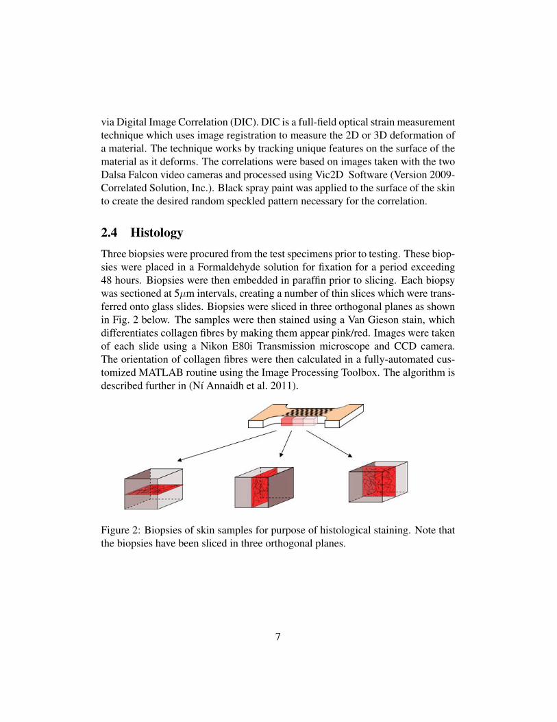
2.4 HistologyThree biopsies were procured from the test specimens prior to testing. These biop-sies were placed in a Formaldehyde solution for fixation for a period exceeding48 hours. Biopsies were then embedded in paraffin prior to slicing. Each biopsywas sectioned at 5µm intervals, creating a number of thin slices which were trans-ferred onto glass slides. Biopsies were sliced in three orthogonal planes as shownin Fig. 2 below. The samples were then stained using a Van Gieson stain, whichdifferentiates collagen fibres by making them appear pink/red. Images were takenof each slide using a Nikon E80i Transmission microscope and CCD camera.The orientation of collagen fibres were then calculated in a fully-automated cus-tomized MATLAB routine using the Image Processing Toolbox. The algorithm isdescribed further in (Nı Annaidh et al. 2011).
Figure 2: Biopsies of skin samples for purpose of histological staining. Note thatthe biopsies have been sliced in three orthogonal planes.
7

2.5 Statistical AnalysisA statistical analysis of the experimental results was carried out using the GeneralLinear Model procedure implemented in SAS 9.1 (SAS Institute Inc., USA). Amultivariate analysis of variance was utilised, followed by the Tukey-Kramer post-hoc test, to determine the influence of orientation and location of the various testsamples. Significance levels for these tests were set to P<0.05. When performingthis test a normal distribution was assumed. To ensure our data set followed anormal distribution, a Lilliefors test was performed using the lillietest function inMATLAB R2007b.
3 ResultsFor each tensile test performed a force-displacement curve was obtained. Thenominal stress was then calculated by dividing the force by the undeformed crosssectional area of the specimen. The stretch ratio was calculated by dividing thecurrent length of the specimen by the initial length. In this way nominal stress Vsstretch ratio graphs were plotted for each specimen. A number of characteristicsfrom these curves were identified as descriptive parameters. They are illustratedin Fig. 3.
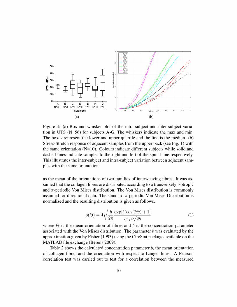
3.1 Intra-Subject and Inter-Subject VariationA difficulty often encountered with the testing of biological tissue is the large vari-ation in experimental results across samples. Fig. 4(a) indicates the intra-subjectand inter-subject variation in the UTS between samples. There is a large intra-subject variation for each subject which is primarily due to anisotropy. Fig. 4(b)shows the stress-stretch graphs of ten different samples taken from five subjects.Note that the left and right samples were adjacent to one another but also in thesame orientation with respect to the Langer lines. The similarity of these re-sponses between the left and right samples from the same subjects indicates goodrepeatability of our experiments.
3.2 Derformation of Specimens After ExcisionIt is known that both shrinkage and expansion of specimens can occur upon ex-cision from the body (Cox 1941, Jansen & Rottier 1958a, Ridge & Wright 1966,
8

Figure 3: Typical stress-stretch graph for the experiments. The ultimate tensilestrength is the maximum stress until failure of the specimen and is indicated byA. The elastic modulus is defined as the slope of the linear portion of the curveshown by B. The failure stretch is the maximum stretch obtained before failure andis shown by C. The initial slope is the slope of the curve at infinitesimal strainsand is shown by D. The strain energy is the energy per unit volume consumed bythe material during the experiment and is represented by the area under the curve.
Lanir & Fung 1974). This is due to the release of residual stresses within theskin. What remains unclear is the level of residual stress present within the skinand how this may vary with the orientation of specimens. It has been suggestedthat the shrinkage of excised specimens is greatest in the direction of Langer lines(Ridge & Wright 1966) however our results did not corroborate this. Our resultsshowed that 73% of all samples expanded upon excision, while only 27% of allsamples shrunk. Of the samples that expanded there was a mean expansion of5.1±3.4%. Amongst the samples that shrunk, a mean shrinkage of 3±2.9% wasrecorded. This is far below the reported maximum figure of 20% shrinkage forhuman skin (Jansen & Rottier 1958a) and 40% shrinkage provided for pig skin(Jor et al. 2011).
3.3 HistologyThe collagen fibres were assumed to form an interweaving lattice structure, as firstpostulated by Ridge & Wright (1966). Here, the preferred orientation is defined
9
(a)
1 1.1 1.2 1.3 1.4 1.5 1.6 1.7 1.8 1.90
5
10
15
20
25
Stretch ratioN
omin
al s
tres
s (M
Pa)
RightFLeftFRightELeftERightDLeftDRightALeftARightGLeftG
(b)
Figure 4: (a) Box and whisker plot of the intra-subject and inter-subject varia-tion in UTS (N=56) for subjects A-G. The whiskers indicate the max and min.The boxes represent the lower and upper quartile and the line is the median. (b)Stress-Stretch response of adjacent samples from the upper back (see Fig. 1) withthe same orientation (N=10). Colours indicate different subjects while solid anddashed lines indicate samples to the right and left of the spinal line respectively.This illustrates the inter-subject and intra-subject variation between adjacent sam-ples with the same orientation.
as the mean of the orientations of two families of interweaving fibres. It was as-sumed that the collagen fibres are distributed according to a transversely isotropicand π-periodic Von Mises distribution. The Von Mises distribution is commonlyassumed for directional data. The standard π-periodic Von Mises Distribution isnormalized and the resulting distribution is given as follows.
ρ(Θ) = 4
√b
2π
exp[b(cos(2Θ) + 1]
erfi√
2b(1)
where Θ is the mean orientation of fibres and b is the concentration parameterassociated with the Von Mises distribution. The parameter b was evaluated by theapproximation given by Fisher (1993) using the CircStat package available on theMATLAB file exchange (Berens 2009).
Table 2 shows the calculated concentration parameter b, the mean orientationof collagen fibres and the orientation with respect to Langer lines. A Pearsoncorrelation test was carried out to test for a correlation between the measured
10

preferred orientation obtained through histology and the perceived orientation ofLanger lines. The correlation was deemed to be significant (P<0.001) with aPearson R value of 0.9487 and an R2 value of 0.9000.
Table 2: Mean orientation of collagen fibres and their Langer Line orientations.(Note that the data given is axial data i.e. it represents undirected lines and doesnot distinguish between θ and π + θ (Jones 2006) e.g. the orientation of 0◦ and180◦ are equivalent).
Orientationwith respect toLanger Lines
PreferredOrientation, Θ
Level ofdispersion, b
0◦ 40◦ 0.52220◦ 163◦ 1.16050◦ 16◦ 1.14710◦ 168◦ 0.6004
45◦ 40◦ 0.838845◦ 61◦ 0.792645◦ 46◦ 0.745345◦ 53◦ 0.420945◦ 57◦ 0.832645◦ 83◦ 0.933090◦ 73◦ 0.900790◦ 120◦ 0.6197
3.4 Influence of OrientationSpecimens were analysed by considering their orientation with respect to theLanger lines (parallel, perpendicular or at 45◦ to the Langer lines, see Fig. 1).A multiway analysis of variance found the orientation of Langer lines to havea significant effect on the UTS (P<0.0001), the strain energy (P=0.0101), theelastic modulus (P=0.0002), the initial slope (P=0.0375), and the failure stretch(P=0.046). The interaction between orientation and location was also tested i.e.,whether the effect of orientation was dependent upon location. This interactionbetween orientation and location was found to be significant only for the failurestretch (P=0.0118). The presence of this interaction is probably due to small dif-ferences in the perceived orientation of Langer lines over the body. Fig. 5 and
11

Figure 5: Influence of orientation on the inital slope, elastic modulus, failurestretch, strain energy and UTS. Values given include mean and standard devia-tions
Table 3 display the variation in the mechanical properties between samples of dif-ferent orientations. The variations are substantial, except for the failure stretch,where the effect of the orientation is complicated by the existence of the interac-tion between orientation and location.
3.5 Influence of LocationWhen results were grouped into three locations (Upper Back, Middle Back andLower Back, see Fig. 1), a multiway analysis of variance also found the loca-tion of specimens to have a significant effect on the UTS (P=0.0002), the strainenergy (P=0.0052) and the Elastic Modulus (P=0.001), but neither on the failurestretch nor the initial slope. Fig. 6 displays variations in the mechanical propertiesbetween different locations on the back for the three properties which showed asignificant effect i.e., the elastic modulus, UTS and strain energy. Note that therewas no significant effect noted for either the initial slope or the failure stretch.
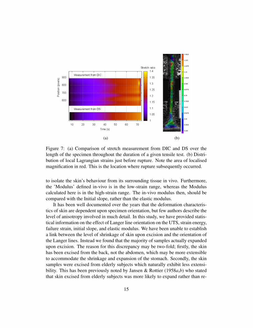
3.6 Digital Image CorrelationAn important feature of DIC is its ability to calculate local strains throughoutthe plane of the test specimens. Fig. 7(a) shows a comparison between the localstretch values, obtained through DIC, and the overall stretch values, obtained fromthe displacement sensor (DS), throughout the duration of a test. It can be seen that
12

Table 3: Mean ± standard deviation for each orientation/location group.LangerLine
Orientation
Location N UTS(MPa)
StrainEnergy
(MJ/m3)
FailureStretch
ElasticModulus
(MPa)
InitialSlope(MPa)
Parallel Middle 9 28.64±9.03
4.28±1.49
1.46±0.07
112.47±36.49
1.21±0.97
Parallel Bottom 7 17.60±4.77
2.54±0.76
1.74±0.32
73.81±19.41
1.95±1.34
45◦ Top 12 22.7±3.61
3.80±0.92
1.52±0.10
82.62±17.36
0.99±0.51
45◦ Middle 9 28.85±7.87
5.38±2.59
1.52±0.15
103.49±41.20
1.33±0.96
45◦ Bottom 5 20.23±4.18
3.31±0.67
1.43±0.04
82.81±18.43
1.30±0.60
Perpendic-ular
Middle 9 16.53±5.71
2.98±0.89
1.52±0.08
63.75±24.59
0.91±0.68
Perpendic-ular
Bottom 5 10.56±8.41
2.44±1.04
1.61±0.14
37.66±36.41
0.54±0.33
there is good agreement for most of the duration of the test. However, upon closerinspection it was found that a local magnification of the stretch ratio occurred justbefore rupture, close to the rupture site (for magnitudes, see Table 4). Fig. 7(b)shows the distribution of strains throughout the sample for a frame just beforerupture. For some samples this magnification was very localised while for othersit was distributed over a larger area. A similar result was reported by Jacquemoudet al. (2007) who found that at the rupture site, the local strain values can be up totwice as large as what is measured using overall specimen strains calculated overthe entire length of a specimen.
4 DiscussionWhen comparing our own data to that in the literature it can be seen that thereis much variation between authors. This is to be expected for biological tissues.First, because of the biological variability between subjects; second, because ofthe anisotropic nature of skin itself; and third, because of the sensitivity of bio-logical tissues to test conditions. In the absence of extensive published literatureon the properties of in vitro human skin from the back, Table 5 compares results
13

Figure 6: Influence of location on back on the elastic modulus, strain energy andUTS. Values given include mean and standard deviations.
Table 4: Local and overall maximum stretch ratios for five different samples.Local magnification of the stretch ratio occurs just before rupture.
Sample Local MaxStretch Ratio
Overall SpecimenStretch Ratio
1 1.47 1.382 1.45 1.403 1.65 1.454 1.70 1.485 1.70 1.50
obtained from the literature from a range of locations and test methods to our ownresults. The mean ultimate tensile strength was 21.6±8.4MPa, the mean failurestrain 54±17%, the mean initial slope 1.18±0.88MPa, the mean elastic modulus83.3±34.9MPa and the mean strain energy was 3.6±1.6MJ/m3. It can be seen thatour experimental results fall within the ranges found in the literature for in-vitrotests. However there is a large variation with the various types of in-vivo tests. Thereasons for this difference has been widely discussed and the primary causes aredue to the properties of skin changing once removed from the body; the differenttesting methods employed; and an inability to establish boundary conditions and
14
(a) (b)
Figure 7: (a) Comparison of stretch measurement from DIC and DS over thelength of the specimen throughout the duration of a given tensile test. (b) Distri-bution of local Lagrangian strains just before rupture. Note the area of localisedmagnification in red. This is the location where rupture subsequently occurred.
to isolate the skin’s behaviour from its surrounding tissue in vivo. Furthermore,the ’Modulus’ defined in-vivo is in the low-strain range, whereas the Moduluscalculated here is in the high-strain range. The in-vivo modulus then, should becompared with the Initial slope, rather than the elastic modulus.
It has been well documented over the years that the deformation characteris-tics of skin are dependent upon specimen orientation, but few authors describe thelevel of anisotropy involved in much detail. In this study, we have provided statis-tical information on the effect of Langer line orientation on the UTS, strain energy,failure strain, initial slope, and elastic modulus. We have been unable to establisha link between the level of shrinkage of skin upon excision and the orientation ofthe Langer lines. Instead we found that the majority of samples actually expandedupon excision. The reason for this discrepancy may be two-fold; firstly, the skinhas been excised from the back, not the abdomen, which may be more extensibleto accommodate the shrinkage and expansion of the stomach. Secondly, the skinsamples were excised from elderly subjects which naturally exhibit less extensi-bility. This has been previously noted by Jansen & Rottier (1958a,b) who statedthat skin excised from elderly subjects was more likely to expand rather than re-
15

Table 5: Comparison of results obtained to results in the literature. All experi-ments were carried out in-vitro on excised human skin. Results are displayed asmeans (±standard deviation) and ranges.
Author Test type UTS(MPa)
FailureStrain %
ElasticModulus
MPa
InitialSlopeMPa
Site∗ Age
(Jansen &Rottier1958b)
in vitrotension
1-24 17-207 2.9-54.0 0.69-3.7 Ab 0-99
(Dunn &Silver 1983)
in vitrotension
2-15 18.8 0.1AB&T
47-86
(Vogel 1987) in vitrotension
5-32 30-115 15-150 V 0-90
(Jacque-moud et al.
2007)
in vitrotension
5.7-12.6
27-59 19.5-87.1
F &A
62-98
(Agacheet al. 1980)
in vivotorsion
0.42-0.85
Back 3-89
(Diridollouet al. 1998)
in vivosuction
0.12-0.25
A&F 20-30
(Khatyret al. 2004)
in vivotension
0.13-0.66
Tibia 22-68
(Pailler-Mattei et al.
2008)
in vivo in-dentation
0.0045-0.008
A 30
(Zahouaniet al. 2009)
in vivo in-dentation& staticfriction
0.0062-0.0021
A 55-70
Our results in vitrotensile
21.6±8.4
54 ±17 83.3±34.9
1.18±0.88
Back 81-97
∗Ab = abdomen, T = thorax, V = various, F = forehead, A = arm
tract, when compared to younger subjects. Finally, the authors noted that whileshrinkage of the skin may have occurred directly after excision from the body,after subcutaneous fat was removed, the samples often expanded. This poses the
16

question what effect the subcutaneous fat has on the level of shrinkage/ expansionin the skin, usually thought to be solely due to the release of residual stresses inthe skin.
Previous studies show that the properties of skin are dependent upon speci-men location (Haut 1989, Sugihara et al. 1991). Our results also indicate that theUTS, strain energy and elastic modulus vary depending on location. However, asonly skin from the back was available for testing, the location of specimens couldonly be categorised into upper back, middle back and lower back regions. It wasnonetheless advantageous to gain information on skin from the back because it isseldom as readily available as that from the abdominal area.
All subjects were between the ages of 81 and 97. This meant that no compar-isons based on the age of subjects could be performed. Numerous other studieshave investigated the influence of age on the structure and mechanical behaviourof skin (Vogel 1987, Haut 1989, Sugihara et al. 1991). It has been established thatprogressive straightening of elastin fibres occurs over time (Lavker et al. 1987).This may cause the shortened phase I region of the stress-strain curve leading toan overall reduction in extensibility. The slope of the phase III portion of thecurve however remains relatively constant (Wilkes et al. 1973). Nonetheless, itmust be highlighted that the results of this study may not reflect the behaviourof younger skin and consideration must be taken to allow for age-related effectswhen interpreting these results.
A number of days had elapsed between the time of death and testing (seeTable 1) but it has been previously demonstrated that skin samples can be refrig-erated for up to 48 hours under the right conditions with no discernible effects onthe mechanical properties (Daly 1982) and for up to 21 days for thoracic aortasunder certain conditions (Adham et al. 1996).
DIC is a common strain measurement technique in biomechanics, howeveruntil now it has been used mostly for in-vivo experiments or at low strains (Moer-man et al. 2009). The results yielded from the present use of DIC may be useful inthe future as validation for a finite element approach. Also the reported local max-imum strain values are of interest to those wishing to model failure of biologicaltissues.
In this paper, our data has been examined with respect to the perceived orien-tation of Langer lines. However, the Langer lines are known to vary with location,age and gender and no universal pattern of maximum tensions exist (Brown 1973).The orientation of the Langer lines cannot be identified with certainty unless theskin of a whole cadaver is punctured, an option which was not feasible for thisstudy. Given the importance of the Langer lines on the mechanical properties of
17

skin there is a need for patient-specific maps to be established in-vivo and in realtime. These could be produced by relying on sophisticated imaging techniquessuch as those produced by ultrasonic surface wave propagation or optical coher-ence tomography and may eventually have applications in cosmetic surgery.
Cox (1941) and Stark (1977) both illustrated that the orientations of Langerlines remain even after the skin is removed from the body and the skin tension re-leased, and concluded that the lines have an anatomical basis. However it remainsunclear in the literature whether the Langer lines have a structural basis, and to theauthors’ knowledge there is no quantitative data published on this point. By iden-tifying the orientation of collagen fibres in the dermis we have shown that there isa correlation between their orientation and that of the Langer Lines. However, itis only when a quantitative study is performed that this becomes apparent.
Knowledge of the collagen fibre orientation along with the presented stress-strain data will also facilitate the determination of material parameters for struc-turally based constitutive models for skin. While the test method described hereis not sufficient to fully describe the full 3-D response of the material, Holzapfel(2006) has shown how a combination of tensile tests and histological data can beused to reasonably determine the material parameters of a 3D hyperelastic model.In a companion paper (Nı Annaidh et al. 2011), we use a similar approach in or-der to determine the material and structural parameters of a suitable anisotropicmodel, the so-called Gasser-Ogden-Holzapfel model (Gasser et al. 2006), readilyimplemented into the commercial Finite Element code ABAQUS.
AcknowledgmentsThis research was supported by a Marie Curie Intra European Fellowship withinthe 7th European Community Framework Programme, awarded to MD. It wasalso supported by IRCSET (Irish Research Council for Science, Engineering andTechnology) through the Ulysses Award, and IRCSET and the Office of the StatePathologist (Irish Department of Justice and Law Reform) through the EMBARKscholarship awarded to ANıA. This work is supported by grants from Ile-de-France.
18

ReferencesAdham, M., Gournier, J.-P., Favre, J.-P., De La Roche, E., Ducerf, C., Baulieux, J.,
Barral, X. & Pouyet, M. (1996), ‘Mechanical characteristics of fresh and frozenhuman descending thoracic aorta’, Journal of Surgical Research 64(1), 32–34.
Agache, P., Monneur, C., Leveque, J. & De Rigal, J. (1980), ‘Mechanical proper-ties and young’s modulus of human skin in vivo’, Archives of Dermatologicalresearch 269, 221–232.
Berens, P. (2009), ‘Circstat: A MATLAB toolbox for circular statistics’, Journalof Statistical Software 31(10).
Borges, A. (1984), ‘Relaxed skin tension lines (RSTL) versus other skin lines’,Plastic Reconstructive Surgery 73(1), 144–150.
Brown, I. A. (1973), ‘A scanning electron microscope study of the effects of uni-axial tension on human skin’, British Journal of Dermatology 89(383-393).
Cavicchi, A., Gambarotta, L. & Massab, R. (2009), ‘Computational modeling ofreconstructive surgery: The effects of the natural tension on skin wrinkling’,Finite Elements in Analysis and Design 45(8-9), 519 – 529.
Cox, H. (1941), ‘The cleavage lines of the skin’, British Journal of Surgery29(114), 234–240.
Daly, C. H. (1982), ‘Biomechanical properties of dermis’, Journal of InvestigativeDermatology 79, 17–20.
Delalleau, A., Josse, G., Lagarde, J. M., Zahouani, H. & Bergheau, J. M. (2008),‘Characterization of the mechanical properties of skin by inverse analysis com-bined with an extensometry test’, Wear 264(5-6), 405–410.
Diridollou, S., Berson, M., Vabre, V., Black, D., Karlsson, B., Auriol, F., Gregoire,J. M., Yvon, C., Vaillant, L., Gall, Y. & Patat, F. (1998), ‘An in vivo method formeasuring the mechanical properties of the skin using ultrasound’, Ultrasoundin Medicine & Biology 24(2), 215–224.
Dunn, M. G. & Silver, F. H. (1983), ‘Viscoelastic behavior of human connectivetissues: Relative contribution of viscous and elastic components’, ConnectiveTissue Research 12(1), 59–70.
19
Eshel, H.Lanir, Y. (2001), ‘Effects of strain level and proteoglycan depletionon preconditioning and viscoelastic responses of rat dorsal skin’, Annals ofBiomedical Engineering 29(2), 164–172.
Evans, S. L. & Holt, C. A. (2009), ‘Measuring the mechanical properties ofhuman skin in vivo using digital image correlation and finite element mod-elling’, The Journal of Strain Analysis for Engineering Design 44(5), 337–345.10.1243/03093247JSA488.
Fisher, N. (1993), Statistical Analysis of Circular Data, Cambridge UniversityPress, Cambridge.
Flynn, C., Taberner, A. & Nielsen, P. (2011), ‘Mechanical characterisation ofin vivo human skin using a 3d force-sensitive micro-robot and finite elementanalysis’, Biomechanics and Modeling in Mechanobiology 10, 27–38.
Gasser, T., Ogden, R. W. & Holzapfel, G. (2006), ‘Hyperelastic modelling ofarterial layers with distributed collagen fibre orientations’, Journal of the RoyalSociety Interface 3, 15–35.
Gilchrist, M. D., Keenan, S., Curtis, M., Cassidy, M., Byrne, G. & Destrade,M. (2008), ‘Measuring knife stab penetration into skin simulant using a novelbiaxial tension device’, Forensic Science International 177(1), 52–65.
Haut, R. C. (1989), ‘The effects of orientation and location on the strength ofdorsal rat skin in high and low speed tensile failure experiments’, Journal ofBiomechanical Engineering 111(2), 136–140.
Holzapfel, G. A. (2006), ‘Determination of material models for arterial walls fromuniaxial extension tests and histological structure’, Journal of Theoretical Biol-ogy 238(2), 290 – 302.
Holzapfel, G. A. & Ogden, R. W. (2009), ‘On planar biaxial tests for anisotropicnonlinearly elastic solids. a continuum mechanical framework’, Mathematicsand Mechanics of Solids 14(5), 474–489.
Jacquemoud, C., Bruyere-Garnier, K. & Coret, M. (2007), ‘Methodology to deter-mine failure characteristics of planar soft tissues using a dynamic tensile test’,Journal of Biomechanics 40(2), 468–475.
20

Jansen, L. & Rottier, P. (1958a), ‘Comparison of the mechanical properties ofstrips of human abdominal skin excised from below and from above the um-bilic’, Dermatologica 117, 252–258.
Jansen, L. & Rottier, P. (1958b), ‘Some mechanical properties of human abdomi-nal skin measured on excised strips’, Dermatologica 117, 65–83.
Jones, T. A. (2006), ‘Matlab functions to analyze directional (azimuthal) data–I:Single-sample inference’, Computers & Geosciences 32(2), 166–175.
Jor, J. W. Y., Nielsen, P. M. F., Nash, M. P. & Hunter, P. J. (2011), ‘Modellingcollagen fibre orientation in porcine skin based upon confocal laser scanningmicroscopy’, Skin Research and Technology .
Khatyr, F., Imberdis, C., Vescovo, P., Varchon, D. & Lagarde, J. M. (2004),‘Model of the viscoelastic behaviour of skin in vivo and study of anisotropy’,Skin research and technology 10, 96–103.
Kraissl, C. (1951), ‘The selection of appropriate lines for elective surgical inci-sions’, Plastic and Reconstructive Surgery 8(1), 1–28.
Langer, K. (1861), ‘On the anatomy and physiology of the skin’, The ImperialAcademy of Science, Vienna. Reprinted in (1978): British Journal of PlasticSurgery 17(31), 93–106.
Lanir, Y. & Fung, Y. C. (1974), ‘Two-dimensional mechanical properties of rabbitskin–ii. experimental results’, Journal of Biomechanics 7(2), 171–174.
Lavker, R., Zheng, P. & Dong, G. (1987), ‘Aged skin: a study by light, transmis-sion electron, and scanning electron microscopy’, J Investig Dermatol 88, 419–428.
Liang, X. & Boppart, S. A. (2010), ‘Biomechanical properties of in vivo hu-man skin from dynamic optical coherence elastography’, IEEE Transactionson Biomedical Engineering 57(4), 953–959.
McCarthy, C. T., Hussey, M. & Gilchrist, M. D. (2007), ‘On the sharpness ofstraight edge blades in cutting soft solids: Part I - Indentation experiments’,Engineering Fracture Mechanics 74(14), 2205–2224.
21

McCarthy, C. T., Nı Annaidh, A. & Gilchrist, M. D. (2010), ‘On the sharpness ofstraight edge blades in cutting soft solids: Part II - Analysis of blade geometry’,Engineering Fracture Mechanics 77(3), 437–451.
Moerman, K. M., Holt, C. A., Evans, S. L. & Simms, C. K. (2009), ‘Digital imagecorrelation and finite element modelling as a method to determine mechanicalproperties of human soft tissue in vivo’, Journal of Biomechanics 42(8), 1150–1153.
Nı Annaidh, A., Bruyere, K., Destrade, M., Gilchrist, M., Maurini, C., Ottenio,M. & Saccomandi, G. (2011), ‘Automated estimation of collagen fibre disper-sion in the dermis and its contribution to the anisotropic behaviour of skin’,Submitted .
Pailler-Mattei, C., Bec, S. & Zahouani, H. (2008), ‘In vivo measure-ments of the elastic mechanical properties of human skin by indenta-tion tests’, Medical Engineering & Physics 30(5), 599–606. doi: DOI:10.1016/j.medengphy.2007.06.011.
Ridge, M. & Wright, V. (1966), ‘The directional effects of skin. A bio-engineeringstudy of skin with particular reference to Langer’s lines’, Journal of Investiga-tive Dermatology 46(4), 341–346.
Silver, F. H., Freeman, J. W. & DeVore, D. (2001), ‘Viscoelastic properties ofhuman skin and processed dermis’, Skin Research and Technology 7(1), 18–23.
Stark, H. L. (1977), ‘Directional variations in the extensibility of human skin’,British Journal of Plastic Surgery 30(2), 105–114.
Sugihara, T., Ohura, T., Homma, K. & Igawa, H. (1991), ‘The extensibility inhuman skin: variation according to age and site’, British Journal of PlasticSurgery 44, 418–422.
Vogel, H. (1987), ‘Age dependence of mechanical and biochemical properties ofhuman skin’, Bioengineering and the skin 3, 67–91.
Wilkes, G., Brown, I. & Wildnauer, R. (1973), ‘The biomechanical properties ofskin’, CRC Critical Reviews in Bioengineering 1(4), 453–495.
Zahouani, H., Pailler-Mattei, C., Sohm, B., Vargiolu, R., Cenizo, V. & Debret, R.(2009), ‘Characterization of the mechanical properties of a dermal equivalent
22

compared with human skin in vivo by indentation and static friction tests’, Skinresearch and technology 15(1), 68–76.
23