chapters from root canal therapy, - … · chapters from root canal therapy, first edition chapters...
TRANSCRIPT

C H A P T E R S F R O M ROOT CANAL THERAPY, FIRST EDITION
C h a p t e r s 2 and 13 of the first edition of Root canal therapy ~ were selected for three reasons. First, they demonstrate the painstaking and meticulous preparation that went into Dr. Grossman's text. Second, they will give the reader the feeling of Dr. Grossman's scholarly pursuit of all aspects of the scientific background that ensured the validity of his recommendations. And finally, the reader is immediately struck by the presentation of basic endodontic practice that was so logically presented that, for the most part, there has been little changed in most of the basic techniques in the ensuing 40 years.
Chapter 2, an excellent summary of the status of the focal infection theory (circa 1940), is based on a paper authored hy Dr. Grossman that appeared in "Dental Cosmos" in 1925, titled "Focal infection and its oral significance." Although the focal infection theory has been discredited as propounded in those days, newer concepts in the principles of infectious diseases and immunology have revived interest in some aspects of this theory.
Permission to reprint these two chapters from Root Canal Therapy, 1940, has been given by Lea & Febiger, publishers.
The Editor
Chapter 2
Pulp less teeth and focal in fec t ion
One of the most controversial chapters in the history of dentistry is that of dealing with pulpless teeth and focal infection. At times, it has even sur- passed the "amalgam war" in intensity of feeling. It has undoubtedly contin- ued for a much longer time and is only now beginning to resolve itself.
Nearly 2000 papers dealing with focal infection have been published in the last twenty-five years--an average of about 100 papers a year. A goodly number of these are on oral focal infection. An excellent r+sum+ and interpretation of the more important contributions in this field has been given by MacNevin and Vaughn. 22
For nearly three decades, the pulp- less tooth has been intimately tied up with focal infection in the minds of
physicians and dentists alike. The sta- tus of the pulpless tooth has undergone a definite change in the last few years, demanding reeducation of the dentist, as well as the physician, on this point. It will be one of the objects of this chapter to define and interpret the pulpless tooth in the light of recent studies.
Many of the early reports dealing with dental focal infection were case histories in which spectacular im- provement in general health was ascribed to extraction of teeth. The chain of events given was somewhat as follows: (1) disease; (2) supportive treatment plus medical or surgical care, plus tooth extraction; (3) recov- ery. Ergo, tooth caused disease. It was not long before some began to question
what might have been the course of illness had the tooth not been removed and whether a later check-up would have found a sustained improvement or a remission. Despite the inherent weakness of such cases reports, it can- not be denied that periapical infection can, and sometimes does, cause infec- tion elsewhere in the body, or that it may contribute to ill health. The fre- quency with which such infection exists and causes systemic disease, is open to question, however, and has without doubt been greatly overem- phasized in the past.
Misconceptions Concerning the Pulpless Tooth . - - I t is desired first to clear up some misconceptions concern- ing the pulpless tooth. Since 1910 two popular misconceptions have persisted
S 18

JOURNAL OF ENDODONTICS ] SPECIAL ISSUE, JANUARY 1982, VOLUME 8
in the minds of dentists and physicians alike, namely: (1) that a pulpless tooth is a "dead" tooth, and (2) that Hunt- er ~9 aimed his shaft of criticism against the pulpless tooth.
A pulpless tooth is not a dead tooth. It still has a definite and vital relation- ship with the surrounding tissue. Because of the peculiar anatomic and structural make-up of the dental tis- sues, the erroneous concept of a "dead" tooth gained currency. Actual- ly, the life of the tooth depends on the attachment apparatus, i.e., periodon- tium and adnexa. In the words of Marshall, 23 "The life of the tooth is dependent upon the integrity of the peridental membrane and not upon the integrity of the pulp." As a matter of fact, many embryologists believe that the function of the pulp ceases when the tooth is completely calcified, shortly after eruption. If a pulpless tooth were a dead tooth, it should be exfoliated since .the body does not tolerate dead tissue. Tha t a pulpless tooth is not dead may be quickly demonstrated by an attempt to remove such a tooth without an anesthetic.
In his history-making address in Montreal in 1910, Hunter 19 did not specifically refer to the pulpless tooth as a source of oral sepsis when he condemned the kind of dentistry then prevalent. How the pulpless tooth came to be maligned instead of the accumulation of filth around ill-fitting crowns and bridgework, against which Hunter inveighed, will probably never be known. The entire matter is sum- med up in a much overlooked but very important editorial 1~ in the Journal of the American Dental Association. The editor referred to Hunter ' s criticism as follows: "The 'oral sepsis' of which he complains--and not without reason- - had nothing whatever to do with the thing that, during the decade that followed his address, caused the great-
est concern among the profession, i. e., focal infection from the apical ends of pulpless teeth. This is the thing that claimed our major consideration, and in connection therewith the name of Hunter has repeatedly been quoted. As a matter of fact, Hunte r never referred in the remotest way to the evils of pulpless teeth as such. He was concerned with the sepsis that came from accumulations around crowns, bridges, and artificial dentures, calling them 'gold traps of seps i s . ' . . . May- hap if this distinguished scientist had given his whole-hearted attention to this important question, the mental aberration and almost universal preju- dice against the pulpless tooth that has developed might have thereby to a certain degree been avoided." Tha t this misconception could have been perpetuated for so many years is indi- cative of the hysterical era which fol- lowed Hunter ' s criticism. Had his words been taken literally, the orgy of extraction that continued for more than two decades afterward might have been averted.
Still another misconception that gained some credence is that a pulpless tooth is an infected tooth. Granting that some pulpless teeth are infected, it does not follow that all are infected. The recovery of bacteria from pulpless teeth does not p e r se indicate that infection is present. Only when the presence of micro6rganisms produces a react ion--inflammation, at least- -can infection be said to exist. The surface of the body is covered with microgr- ganisms and the mouth is at all times a seething reservoir of bacteria, yet infection is absent under ordinary cir- cumstances. Infection not only depends upon the presence of organisms but upon the ability of such organisms to produce a local or systemic reaction. In the light of recent studies, which we shall discuss later, the finding of
microgrganisms upon the roots of extracted teeth is not necessarily sig- nificant of infection.
Unfortunately, today, we have no reliable laboratory or clinical test to determine whether a focus of infection exists, and if it does, where it is located. Various tests such as skin reactions, blood sedimentation rate, blood count, uric acid test, provocative short-wave tests, and others, have all been tried and found inadequate for diagnostic purposes. It would seem as if the inherent complexity of our bodi- ly mechanism is such as to make the development of a reliable diagnostic test highly improbable.
Investigations Prior to 1936.--It is a disconcerting but true statement to make, that practically every investiga- tion dealing with the pulpless tooth made prior to 1936 is invalid in the light of recent studies. It is neverthe- less desirable to review and evaluate some of the more important studies of the past, in order to understand better the events leading up to our present concept of the pulpless tooth.
One's attention might be called here to the fact that there are fashions in the healing, as well as in the sartorial art. A little more than fifty years ago hysterectomy was fashionable, and about thirty-five years ago nephropexy was in style. Many systemic ills, from simple fatigue to gastric disturbances and cardiac palpitation were attrib- uted to the movable kidney. Comment- ing on focal infection in the light of the past, Dr. O. H. Perry Pepper 26 said a decade ago, "I t is instructive and humilitating to go back into the litera- ture of twenty-five years ago and read the many articles describing the varie- ty of conditions and symptoms attrib- uted to the movable kidney and the remarkable therapeutic results ob- tained by nephropexy." He then asks the question, with regard to focal
S 19

JOURNAL OF ENDODONTICS [ SPECIAL ISSUE, JANUARY 1982, VOLUME a
infection, "Are we merely in the midst of a movement which will run its course and be looked back upon in ten years as we look back on the days of nephropexy?" As if in answer to this question, the following excerpt is tak- en from a recent paper by Cecil nd Angevine: 8 " M a n y of us who original- ly accepted the theory of focal infection with enthusiasm have watched with interest and some trepidation its rapid development in the various fields of medicine but are now wondering if the time has not arrived for a revaluation of the whole theory. Many thoughtful students today question seriously its validity, and some are quite willing to throw it completely overboard . . . Scientific men are becoming a little wearied of the universal acceptance of a theory as though it were an estab- lished fact."
In a most comprehensive review of the literature on focal infection, Mac- Nevin and Vaughn 22 analyzed the evi- dence for and against focal infection and concluded that the proof submitted by Price, Rosenow and others, has its limitations from the standpoint of both technic and interpretation of results. The interpretation of such experimen- tal work, they said, "must be pursued with extreme caution and open- mindedness. The deductions often made are unwarranted."
It is perhaps significant that the popularity of the focal infection theory was greatest in this country, where it was nurtured and publicized, while its importance has been minimized or practically disregarded in Europe. One thinks immediately of the work of Rosenow 27 in connection with focal infection, since he frequently suc- ceeded in showing an oral focal rela- tionship with systemic disease, by ani- mal experimentation. His work, how- ever, has not been generally accepted: (1) Because of the massive doses of
inoculant injected, as much as 10 cc being used at a time. An equivalent amount, if man were the laboratory animal, would be about one pint of inoculant. (2) Because the intravenous route was generally used for the injec- tions. (3) Because the organisms were at the height of virulence at the time of injection. Furthermore, it has been asserted that Rosenow's work has not been adequately controlled, and that the technic employed by him so devas- tates the laboratory animal that lesions are sometimes produced in almost ev- ery tissue and organ on the body. Rosenow's conclusions have not been confirmed, and are looked upon with much skepticism if not with actual disfavor by most bacteriologists. Com- menting on Rosenow's theory of elec- tive localization, Holman 18 stated, "The specificity of the bacteria involved has not been proved and the evidence favoring the theory of elective localization is so open to misinterpre- tation and so limited in its practical application that it cannot be consid- ered as a help in the solution of the problem. A certain general bacterial adaptation to environment is accepted by everyone, but the factors on the side of the host are more variable and far more important."
It is also significant, perhaps, that no definitely conclusive proof has yet been advanced that in any single case, a tooth or teeth were the direct cause of the systemic disturbance, excepting a priori proof. The extraction of a tooth followed by amelioration of symptoms does not necessarily prove that the tooth was the cause of the disease. This is only an assumption which may or may not be true.
Studies bearing on the pulpless tooth as a possible focus of infection may be divided into: I Clinical; I I Roentgenologic; I I l Histologic; IV Bacteriologic. Only the more impor-
tant findings in each group will be discussed here.
I. Clinical Studles . - - (1) A very large number of people having pulp. less teeth, show no apparent systemic involvement.
(2) During the World War draft, data were collected of more than two and one-half million men between the ages of eighteen and thirty years. An evaluation of the data, according to Appleton, 2 shows that "chronic infec- tion about the head is n o t . . , the sole determining factor in the contempora- neous occurrence of such conditions as gastric ulcer, appendicitis, endocardi- tis, arthritis, osteitis deformans, myosi. tis, and muscular rheumatism."
(3) Frankel ~2 studied the incidence of systemic disturbances in a group of nearly 17,000 people with and without so-called "heavy dentistry." By this term he meant the presence of crowns, bridges, pulpless teeth and any other dental evidence which might suggest the presence of a focus of infection. He concluded that the incidence of sys- temic disturbances in the "heavy den- tistry" group was not materially high- er than in the other group.
(4) Cecil and Angevine* recently analyzed their results after the elimi- nation of foci of infection in 200 cases of rheumatoid arthritis. They came to the conclusion that no benefit accrued to 47 out of 52 patients from whom dental foci of infection were removed. (See Fig. 1.)
(5) In two groups of clinical cases, in which root canal therapy was done in one and extraction in the other, Bryant and Polevitzky s found more improvement after root canal treat- ment than after extraction. The crite- rion used by them was Arneth blood counts taken at strategic periods before and after root treatment or extrac- tion.
S 20

JOURNAL OF ENDODONTICS I SPECIAL ISSUE, JANUARY 1982, VOLUME 8
II. Roentgenologic Studies.--(1) Complete roentgen-ray studies of more than 1500 subjects were made by Zis- kin 3' to determine whether a correla- tion existed between positive roentgen- ray findings of pulpless teeth and sys- temic disturbances. The subjects were patients in the wards of a city hospital, from the outpatient department of the same hospital, and ambulants who applied only for dental treatment and who were well otherwise. Ward and outpatient subjects were examined by the medical staff and complete medical histories were available. The following is a synopsis of the study: (a) 48 percent of the subjects studied were sick, 52 per cent were well; (b) 71 per cent of the "sick" group and 75 per cent of the "well" group had pulpless teeth. (c) Of those with pulpless teeth,
46 per cent were sick and 54 per cent were well, while of those without pulpless teeth 50 per cent were sick and 50 per cent were well. From these findings it is obvious that a relation- ship between pulpless teeth and sys- temic disease in the groups studied is certainly not apparent.
(2) Arnett and Ennis 3 made medi- cal and dental examinations, including full mouth roentgen-ray studies, of 883 college students. Although chronic rarefying osteitis was present in 20 per cent of cases, it was not associated with rheumatism, chorea, or heart disease. In 160 students studied, no relation- ship was found between the presence of chronic rarefying osteitis and elec- trocardiographic abnormalities.
(3) Both roentgenoiogic and clinical records of nearly 2000 patients have
Fig. 1.--Result) following treatment of tonsils and sinuses, and extraction of teeth in a group of 200 patients with rheumatoid arthritis. (Cecil and Angevine, courtesy of Ann. Int. Med).
CASES TREATED
] NO BENEFtT
] TEMPORARY BENEFIT
EXACERBATION
TONSILS T E E T H S I N U S E S
been carefully studied at the Universi- ty of Illinois. The status of these pulpless teeth has been followed up over a period from one to sixteen years after completion of treatment. At the last tabulation for which data are available (1936) 76 per cent either had remained or had become clinically and roentgenologically negative. In this group were included teeth which origi- nally had acute abscesses, chronic abscesses, or granulomas.
III. Histologic Studies.--(1) His- tologists who have studied sections of pulpless teeth, almost without excep- tion, are agreed upon three things: (a) that periapical bone restoration can and often does follow satisfactory root canal treatment of pulpless teeth; (b) that such pulpless teeth are commonly without histologic evidence of infec- tion; and (c) that they may be safely retained in the mouth.
(2) Of 250 pulpless roentgen-ray negative teeth, which were examined histologically, Skillen 28 found evidence of infection in only 6.
(3) Histologic evidence of periapi- cal repair following root canal therapy has been furnished by l~layney, 4 Cool- idge, 9 Kronfeld, 2~ Aisenberg, 1 Hatton, Skillen, Moen, 17 Orban, 2s Gottlieb, Schwarz and Stein, 13 and others.
Hatton ~6 has brought attention to the fact that the changes following successful root-canal therapy are simi- lar to repair following bone injury or fracture: (1) A destructive change; (2) a constructive change; (3) a resting stage. The first stage (1) is character- ized by removal, resorption and disso- lution of the damaged tissue in the pulp chamber or in the apical space. The next stage (2) is characterized by replacement of lost hard tissues of the tooth by osteoid cementum, and of bone by new bone. This calcifying process occurs in the accessory canals as well as in the main root canal and
S 21
JOURNAL OF ENDODONTICS I SPECIAL ISSUE, JANUARY 1982, VOLUME 8
extends out into the periapical tissues. (3) When the calcifying process ceases, the resting stage is reached. In this, the condition of the periapical tissue "per- sists for years without evidence of change, inflammatory or otherwise, except for a tendency of these tissues to undergo a fibrous atrophy . . . . Since the reactions at the apex of a pulpless tooth are essentially similar to those found in the healing of a broken bone, it is assumed that they are physiologic in character."
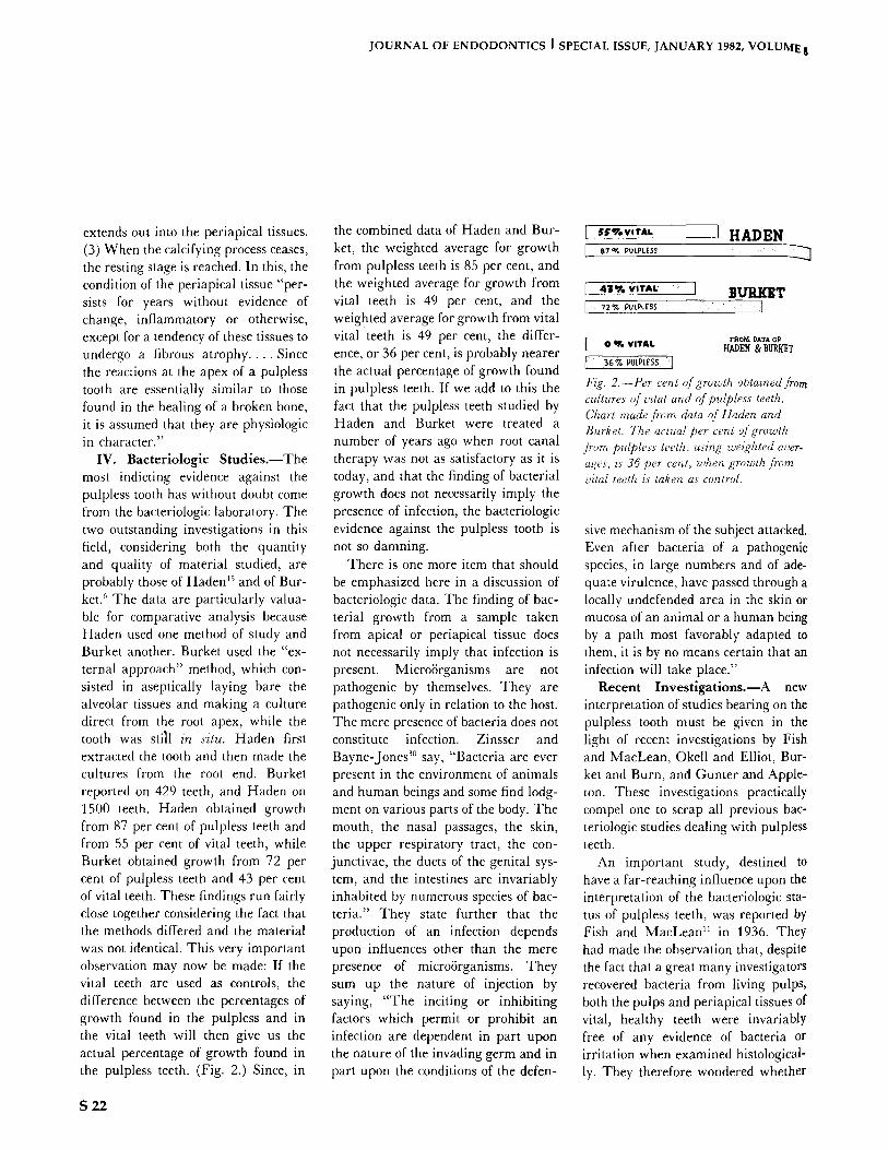
IV. Bacteriologic Studies . - -The most indicting evidence against the pulpless tooth has without doubt come from the bacteriologic laboratory. The two outstanding investigations in this field, considering both the quantity and quality of material studied, are probably those of Haden js and of Bur- ket. 6 The data are particularly valua- ble for comparative analysis because Haden used one method of study and Burket another. Burket used the "ex- ternal approach" method, which con- sisted in aseptically laying bare the alveolar tissues and making a culture direct from the root apex, while the tooth was still in situ. Haden first extracted the tooth and then made the cultures from the root end. Burket reported on 429 teeth, and Haden on 1500 teeth. Haden obtained growth from 87 per cent of pulpless teeth and from 55 per cent of vital teeth, while Burket obtained growth from 72 per cent of pulpless teeth and 43 per cent of vital teeth. These findings run fairly close together considering the fact that the methods differed and the material was not identical. This very important observation may now be made: If the vital teeth are used as controls, the difference between the percentages of growth found in the pulpless and in the vital teeth will then give us the actual percentage of growth found in the pulpless teeth. (Fig. 2.) Since, in
the combined data of Haden and Bur- ket, the weighted average for growth from pulpless teeth is 85 per cent, and the weighted average for growth from vital teeth is 49 per cent, and the weighted average for growth from vital vital teeth is 49 per cent, the differ- ence, or 36 per cent, is probably nearer the actual percentage of growth found in pulpless teeth. If we add to this the fact that the pulpless teeth studied by Haden and Burket were treated a number of years ago when root canal therapy was not as satisfactory as it is today, and that the finding of bacterial growth does not necessarily imply the presence of infection, the bacteriologic evidence against the pulpless tooth is not so damning.
There is one more item that should be emphasized here in a discussion of bacteriologic data. The finding of bac- terial growth from a sample taken from apical or periapical tissue does not necessarily imply that infection is present. MicroSrganisms are not pathogenic by themselves. They are pathogenic only in relation to the host. The mere presence of bacteria does not constitute infection. Zinsser and Bayne-Jones 3~ say, "Bacteria are ever present in the environment of animals and human beings and some find lodg- ment on various parts of the body. The mouth, the nasal passages, the skin, the upper respiratory tract, the con- junctivae, the ducts of the genital sys- tem, and the intestines are invariably inhabited by numerous species of bac- teria." They state further that the production of an infection depends upon influences other than the mere presence of microSrganisms. They sum up the nature of injection by saying, "The inciting or inhibiting factors which permit or prohibit an infection are dependent in part upon the nature of the invading germ and in part upon the conditions of the defen-
[ ~ ' V . v , ' r A L I HADEN B 7 ~ PULPLESS
[ 41% V'T**- . . . . I B U R I ~ T I ",2~. pULp~ESS " J
FROM DATA OP [ o ~. VITAL ladk.DEN & BUR~T t 36~PUtPtESs ] Fig. 2.--Per cent of growth obtained from cultures of vital and of pulpless teeth. Chart made from data of Haden and Burket. The actual per cent of growth from pulpless teeth, using wezghted aver- ages, is 36 per cent, when growth from vital teeth is taken as control.
sire mechanism of the subject attacked. Even after bacteria of a pathogenic species, in large numbers and of ade- quate virulence, have passed through a locally undefended area in the skin or mucosa of an animal or a human being by a path most favorably adapted to them, it is by no means certain that an infection will take place."
Recent Invest igat ions.--A new interpretation of studies bearing on the pulpless tooth must be given in the light of recent investigations by Fish and MacLean, Okell and Elliot, Bur- ket and Burn, and Gunter and Apple- ton. These investigations practically compel one to scrap all previous bac- teriologic studies dealing with pulpless teeth.
An important study, destined to have a far-reaching influence upon the interpretation of the bacteriologic sta- tus of pulpless teeth, was reported by Fish and MacLean 11 in 1936. They had made the observation that, despite the fact that a great many investigators recovered bacteria from living pulps, both the pulps and periapical tissues of vital, healthy teeth were invariably free of any evidence of bacteria or irritation when examined histological- ly. They therefore wondered whether
S 22

JOURNAL OF ENDODONTICS I SPECIAL ISSUE, JANUARY 1982, VOLUME 8
the micro/Srganisms gained access to the pulp during extraction and arrived too late to set up a reaction. This supposition was confirmed by experi- mental work in which they found that bacteria were "pumped" into the blood and lymph channels during manipula- tion of the tooth while it was being extracted. They found further that streptococci are present only in necrot- ic areas of bone and do not diffuse through the granulation tissue (if it be present) about the root apex. Such streptococci are only transient mi- grants in the environment of living tissue and require necrotic material in which to grow. In grossly infected pulpless teeth, the infection is limited to the root canals or can be found in the pus of the associated abscess. Even in such cases, the neighboring alveolar
bone and soft tissues are sterile, although they may be irritated by the diffusion of toxic products. The impor- tance of the work of Fish and MacLean lies in tlae fact that they have shown that it is practically impossible to remove a tooth aseptically (without actual cauterization of the crevicular gingiva) because bacteria are forced into the pulp and periapical tissues during the act of extraction.
Experimental evidence that bacteria may be "pumped" into a tooth during extraction is afforded by Kanner. 2~ That even the best bacteriologic tech- nic cannot obviate this risk of contam- ination is given by Tunnicliff and Hammond, z9 who found streptococci in the pulps of intact, vital teeth even after adequate surface sterilization.
Indirectly supporting the work of Fish and MacLean, are the studies of Okell and Elliot, 24 and of Burket and Burn] Okell and Elliot took blood cultures before, and ten minutes after, extraction of teeth, which were removed under general anesthesia. Positive blood cultures were obtained
in more than 60 per cent of cases after extraction, even though blood cultures were negative before extraction. When a local anesthetic, containing a vaso- constrictor, was substituted for general anesthesia, Burket and Burn obtained fewer positive cultures. Transient bac- teremias occurred, nevertheless, in 17 per cent of cases, even despite capillary constriction from the epinephrin con- tained in the local anesthetic solution. This supports the view that bacteria, which are not present in a pulp or pulpless tooth while the tooth is in situ, may be picked up during the act of extraction. Bacterial dissemination during extraction may also account for the relatively high percentage of posi- tive cultures recovered from both vital and pulpless teeth.
Summary. - - Inasmuch as the re- covery of bacteria from the blood stream following tooth extraction is an indication that dissemination of bac- teria from the dental tissues has oc- curred, and since contamination of the root surface or of the pulp during the act of extraction is by no means rare, the significance of finding growth within the pulp or upon the surfaces of extracted pulpless teeth must be ques- tioned. That the entire chapter on the bacteriology of vital and of pulpless teeth needs to be rewritten is further evidenced by the fact that recent studies on the bacteriology of the liv- ing, healthy pulp are at variance with those of the past.
Practically every investigator who at one time or another studied the bacte- riology of the vital pulp, found growth present in a greater or lesser percent- age of cases. Haden 15 believed that chronic infection occurs quite com- monly in the pulps of vital teeth and went so far as to say that "many believe there is a chronic infection in the pulp of every tooth in which the dentin is invaded by caries." If this
were taken literally and such teeth were condemned, we should have a toothless nation, since it is estimated that more than 90 per cent of the people have carious teeth or have had carious teeth at one time. In the light of the studies by Fish and MacLean, and of Okell and Elliot, it is easy to understand why bacteria were recov- ered from normal, healthy pulps fol- lowing tooth extraction. When vital pulps were exposed and cultured in situ according to the method of Gunter and Appleton TM whereby contamina- tion from the gingival crevice is elimi- nated no growth was obtained. This again points to the fact that in previous studies of vital teeth, bacteria were forced into the pulps during extrac- tion, which gave an erroneous indica- tion of the bacteriologic status of such teeth. By the same token, bacteria were forced within or upon the surfaces of extracted pulpless teeth, from which growth was afterward recovered. Cul- tures made from extracted teeth do not therefore reflect the true bacteriologic status, whether it be of vital or of pulpless teeth, unless, as shown by Fish and MacLean, the gingival tissue is first cauterized. Since pulpless teeth fell into some disrepute chiefly because of bacteriologic studies made in the last fifteen or twenty years, and since the findings were generally based upon bacteriologic examinations made after extraction, past interpretations can no longer be held valid in the light of recent knowledge. We must look to recent and future studies, rather than those of the past, to determine the real status of the pulpless tooth. From what is already known, it is expected that such studies will be in closer agree- ment with correlative studies made of pulpless teeth by clinical, roentgeno- logic and histologic examinations. To hold the pulpless tooth to the criticism of the past would be unjustified.
S 23

JOURNAL OF ENDODONTICS ] SPECIAL ISSUE, JANUARY 1982, VOLUME 8
References I. Aisenberg, M.S.: Jour. Am. Dent. Assn.,
18, 136, 1931. 2. Appleton, J.L.T.: Bacterial Infeetinn,
Philadelphia, Lea & Febiger, p. 491, 1933. 3. A,'nett, J.H., and Ennis, L.M.: Am..Jour.
Med. Sci, 185, 777, 1933. 4. Blayney, J.R.: Dent. Cosmos, 74, 635,
1932. 5. Bryant, (LK., and Polevitzky, K.: Dent.
Cosmos, 72, 363, 1930. 6. Burket, L.W.: Yale Jour. Biol. and Med.,
9, 271, 1937. 7. gurket, L.W., and Burn C.G.: Jour Dent.
Res., 16, 521, 1937. 8. Cecil, R.L., and Angevine, D. M.: Ann.
Int. Med., 12, 577, 1938. 9. Coolidge, E.D.: Jnuc. Am. Dent. Assn.,
18, 499, 1931. 10. Editorial: Journ. Am. Dent. Assn., 22,
659, 1935. 11. Fish, E.W., and MacLean, I.: Brit. Dent.
jnur., 61,336, 1936.
12. Frankel, L.K: Dent. Cosmos, 66, 35, 1924.
13. Gottlieb, B., Sehwarz, A.M., and Stein, G.: Ztsehr. f. Stomat., 26, 1151, 1928.
14. Gunter, J . t t . , and Appleton, J .LT . , el a/., Jnur. Dent. Res., 16, 310, 1937.
15. Haden, R.L.: L)emal Inflection and Sys- temic Disease, Philadelphia, Lea & Febiger, 1928.
16. 15/)2,
17. OH. :
18. 1928.
19. 20.
1938.
t fatton, E.H.: Jour. Ant. Dent. Assn., 18, 1931. Hatton, E.H., Skillen, W.G., and Moen, Jour. Am. Dent. Assn., 15, 56, 1928. Hohnan, W.L.: Arch. Path., 5, 68,
llunter, W.: Lancet, i, 79, I911. Karmer, O.: Jour. Dent. Res., 17, 47,
21. Kronlield, R.: Hismpathohlgy of the "I'emh, Philadelphia, Lea & Febiger, 1939.
22. MaeNevin, M.G., and Vaughn, HS.: "Month lnfeelions and Their Relation to Sys- temic Disease," New York, Joseph Purcell
Re.search Memorial, 1, 340, 1930. 23. Marshall, J.A.: Dent. Cosmos, 70, 253,
1928. 24. Okell, C.C., and Elliot, S.D.: Lancet, it,
869, 1935. 25. Orban, B.: Jour. Am. Dent. Assn., 19,
1348, 1932. 26. Pepper, O.tt.P.: Therap. Gaz., 42, 693,
1926. 27. Rosenow, E.C.: Dent. Cosmos, 59, 485,
1917. 28. Skil[en, W.G.: Jnur. Am. Dent. Assn.,
13, 291, 1926. 29. Tunnicliff, R., and Hammond, C.: Jour.
Am Dent. Assn., 24, 1663, 1937. 30. Zinsser, It., and Bayne-Jones, S.: Text-
book ~f' Bacteriology, 7th ed., New York, Apple- ton-Century Co., I934.
31 Ziskin, D.E.: Jour. Dent. Res., 11, 538, 1931.
Reprinted from R,ol Canal 77zerapy, Phila- delphia, Lea & Febiger, 1940, pp 15-26; 126- 142; 143-155.
I have known Lou Grossman for over fifty years, both as his student and disciple. I can portray him as an outstanding clinician, a pioneer in clinical endodontics, a renowned investigator, a superb motivational teacher, an excellent lecturer, and a concerned friend. Furthermore, the Divine Architect has seen fit to mould a personality with a sensitivity to social consciousness, fairness to students, regard for his colleagues, and recognition of the inadequacies of his specialty, endodontics.
Although Lou, by his own admission, is an irreligous man, he can be referred to as the prophet who stifled the influence of ignorance as was practiced by the folly of dogma. Every discipline has its giants of substantive literature. Clinical medicine has Osler, psychiatry has Freud, physiology has Starling, and clinical endodontics has Grossman. In none does he assume so lofty a place of honor and world influence as recorded by seven language translations of his text, Endodontic practice.
Mindful that he has been extolled with portraits, honorary degrees, and numerous awards, the highest accolade of dignity has been reserved by his disciples who have awarded him the title, The Grandfather of Endodontics, he who has sired the fathers of many new concepts. His students have benefitted most not only from his lucid presentations, but also from emulating his standards of excellence and exemplary guidelines of human behavior.
I. B. Bender, DDS Professor emeritus, University of Pennsylvania, Philadelphia
S 24