chapters 10, 11 the nervous system and clinical applications
TRANSCRIPT

Chapters 10, 11The Nervous Systemand Clinical Applications
COMD 6305 UTDALLAS
WILLIAM KATZ, PH.D.
1

Cells Of The Nervous SystemThe neuron
◦ Cell body
◦ Nucleus
◦ Nucleolus
◦ Dendrites
◦ Axon
◦ Terminal buttons
2

Myelin and GliaMyelin
•Fatty substance that forms insulation for axons
•Increases speed of transmission of neural impulses
•Damaged in Amyotrophic Lateral Sclerosis
Glia
•Supporting cells
•40 – 50x more prevalent than neurons
•Provide nutrients to neurons
3
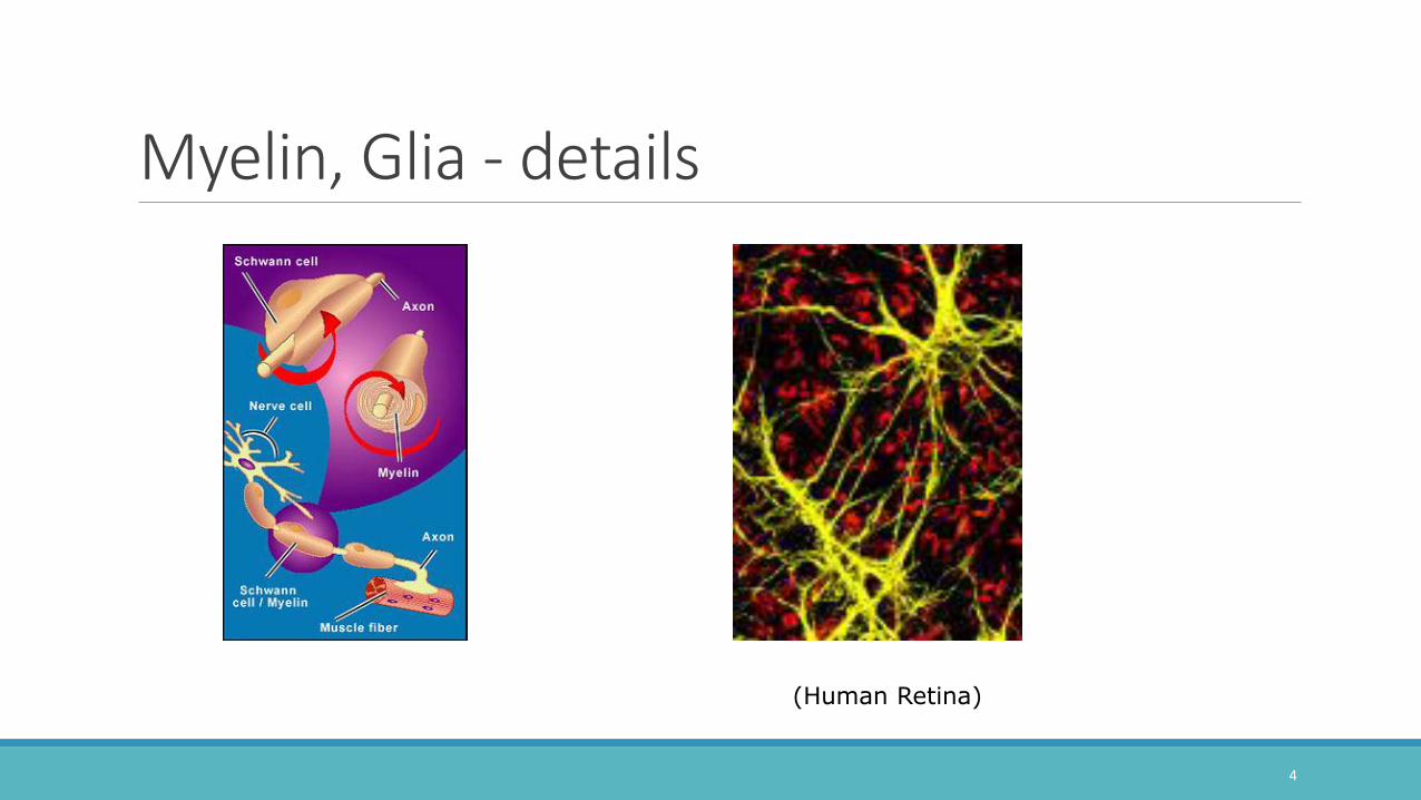
Myelin, Glia - details
4
(Human Retina)

Types of Neuron
(99% of all neurons in humans)
5

Neurons – polarity types
BIPOLAR
MULTIPOLAR
MONOPOLAR
6
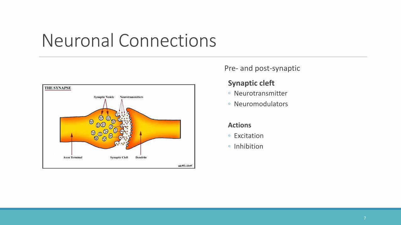
Neuronal ConnectionsPre- and post-synaptic
Synaptic cleft◦ Neurotransmitter
◦ Neuromodulators
Actions
◦ Excitation
◦ Inhibition
7

Synapse – details
8

Neural Impulse
Resting potential
Depolarization
Action potential
9

Neural Impulse•Extra and intracellular fluids Ions
•Resting membrane potential
•Sodium/ potassium pump
•Polarization
•Depolarization
•Action Potential
•Action spike
10
(fr. Kent, The Speech Sciences)

11

12

13

Schematic electrical signal
14

Anatomy Of The Nervous System•Central nervous system (CNS)
[brain + spinal cord]
•Peripheral nervous system (PNS)
[cranial nerves, spinal nerves]
15

Outline of NS
16

Divisions Of The CNSTELENCEPHALON
◦ (Cerebral hemispheres & basal nuclei)
DIENCEPHALON
◦ (Thalamus and hypothalamus)
MESENCEPHALON
◦ (Midbrain – tectum, tegmentum)
METENCEPHALON
◦ (Pons, cerebellum)
MYELENCEPHALON
◦ (Medula Oblongata)
17

Divisions – continued
18

Cerebral HemispheresGray matter
◦ LOBES
◦ GYRI
◦ SULCI
◦ FISSURES
19

Transverse viewWhite matter
◦ FASICULI
◦ COMMISSURE
20

Subcortical Structures
•Basal ganglia/basal nuclei
•Thalamus & hypothalamus
21
Animation showing Basal Ganglia - UBC


Limbic system - animations
23
Labelled limbic system animation - UBCAnimation of limbic system within cortex

Cerebellum•Part of the hindbrain
•Coordinates motor commands with sensory inputs to control movements
•Damage results in:
•Ataxia, gate deficits, problems with fine movements, control of rate and range of movement
•Dysarthria
24
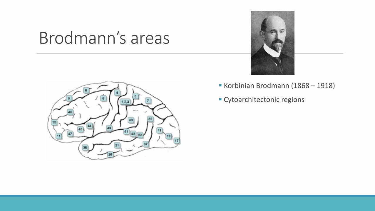
Brodmann’s areas
Korbinian Brodmann (1868 – 1918)
Cytoarchitectonic regions

Cerebellar ataxiaArticulation
imprecise consonant articulation
distorted vowels (slurred quality)
imprecise consonants, vowels
Prosody
equal and excess stress
prolonged phonemes
prolonged intervals between phonemes
monopitch, loudness
Ssow rate
Phonation: Harsh vocal quality, voice tremor
26
Video sample of speech

Brainstem
27

Brainstem -position
28

Sagittal view
29
(F. Netter)

Peripheral Nervous SystemSpinal nerves
Cranial nerves
30

12 Cranial Nerves - memory tools
31
The HIT SONG from UTSW Cranial Nerve Function!
Another way to memorize ‘em
“On Old Olympus Towering Tops A Finn And German Vended At Hops”
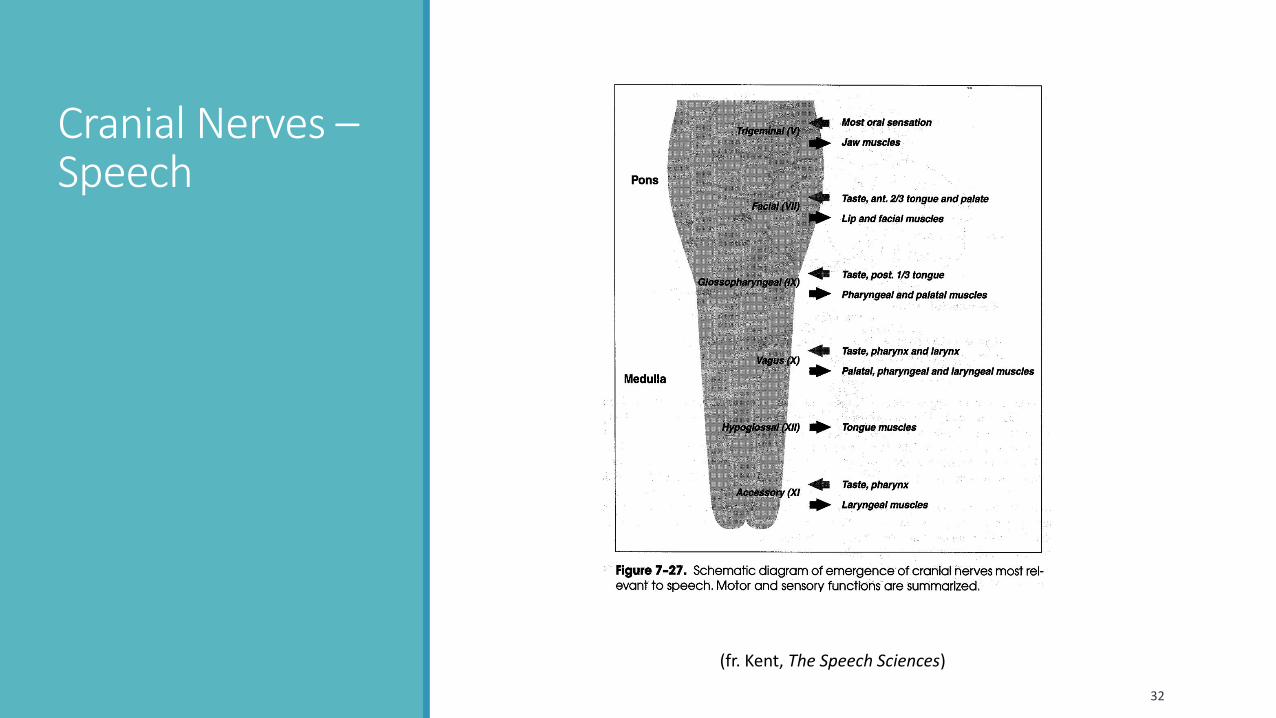
Cranial Nerves –Speech
32
(fr. Kent, The Speech Sciences)

Cranial Nerve Lesions (non-speech related)I. Olfactory- (Anosmia) loss of sense of smell
II. Optic- Visual deficits
III. Occulomotor- inability to turn eyes inward, dilation of pupil
IV. Trochlear- inability to pull eye down
VI. Abducens- Eye is rotated inwards as a results of paralysis. ◦ Diplopia-Inability to fuse images from both eyes
33

Cranial Nerve Lesion (speech-related)V. Trigeminal-Increased jaw jerk reflex
◦ Weakness of jaw, hypernasality, loss of sensation in anterior 2/3 tongue, altered sensation of E. tube, ear canal, tympanic membrane
VII. Facial- Bell’s palsy
◦ Inability to close eyelids, loss of tone of facial muscles, drooling, smiling affected.
34

Cranial Nerve Lesion (speech-related/ continued)VIII. Vestibulo-cochlear (auditory)-Hearing loss, disturbances in equilibrium, vertigo and nystagmus in case of head injury
IX. Glossopharyngeal -paralysis of stylopharyngeus muscles and loss of sensation of posterior 1/3 of tongue, absence of gag.
X. Vagus -Loss of gag, hypernasality, swallowing, damage to SLN and RLN branches of vagusaffect sensation and function of larynx.
35

Cranial Nerve Lesion (speech-related/ continued)XI. Accessory - Affects trapezius and sternocleidomastoid, thus unable to lift arm or turn head respectively. May affect movement of larynx and velum.
XII. Hypoglossus - Profound impact on articulation. Muscular weakness-affecting tongue movements, fasciculation or involuntary twitching, spasticity.
36

PNS - detailsSpinal nerves
37

Reflex arc
38

Brain CoveringsMeninges◦ Dura matter
◦ Arachnoid matter
◦ Pia matter
◦ Extradural space
◦ Subdural space
◦ Subarachnoid space
◦ Falx cerebelli
◦ Tentorium cerebelli
39

Ventricles
40

Nourishment Of The Brain20% of blood supply
Plasma
◦ Red corpuscles
◦ White corpuscles
◦ Platelets
Glycogen
41

Nourishment Of The BrainArteries
◦ Carotid
◦ Anterior cerebral
◦ Middle cerebral
◦ Subclavian
◦ Vertebral
◦ Basilar
◦ Posterior cerebral
◦ cerebellar
42
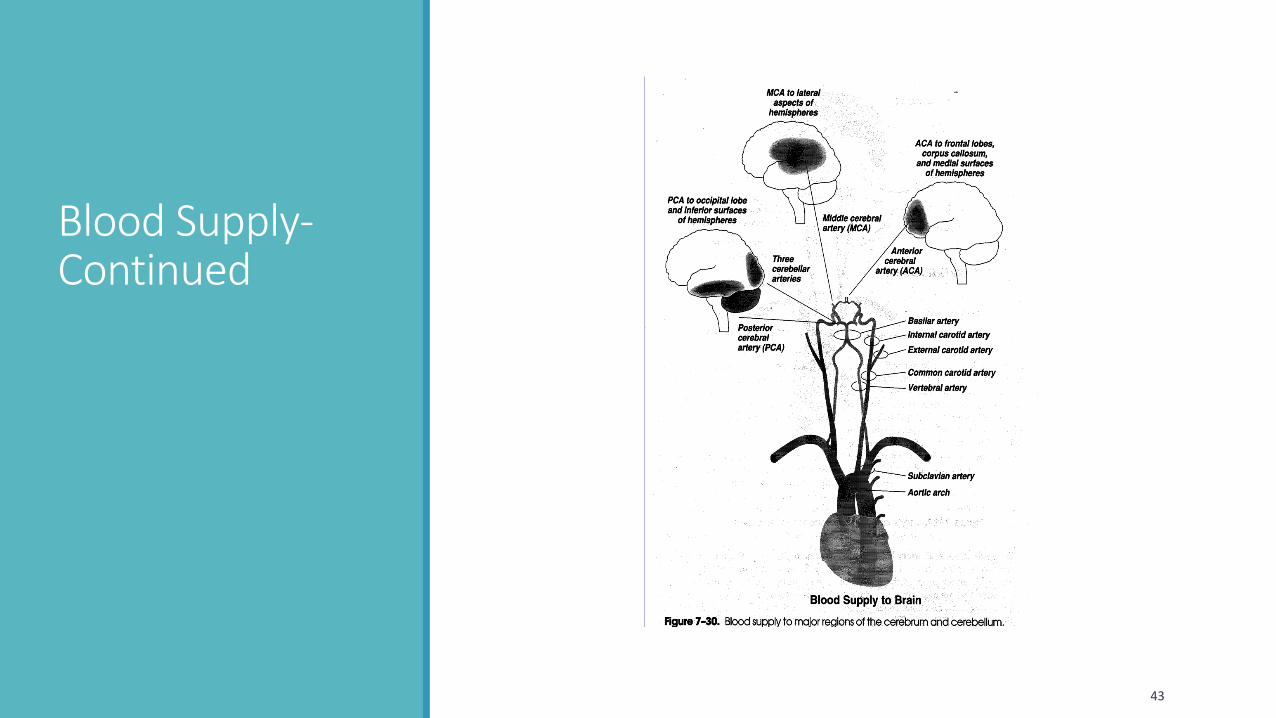
Blood Supply-Continued
43

Stroke - apoplexy
44


Neural pathways to motor control
46
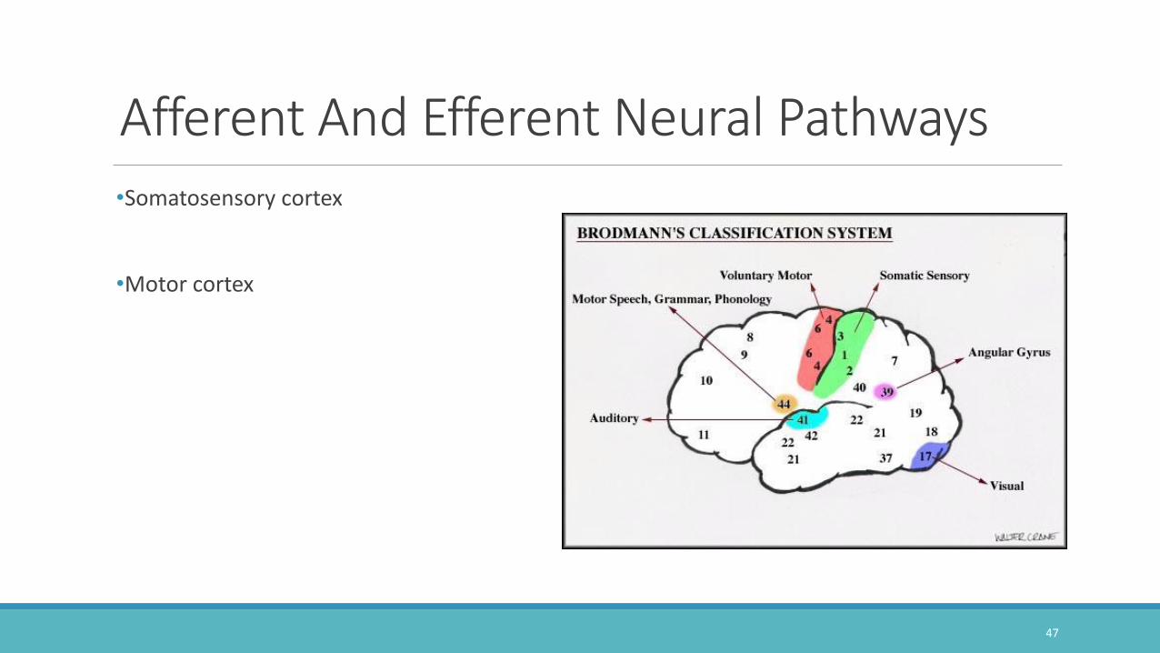
Afferent And Efferent Neural Pathways•Somatosensory cortex
•Motor cortex
47

Sensory Homunculus
48
A sensory“Mouseunculus”

Motor Homunculus
49

Somatosensory Pathways
50

Somatosensory Pathways First order sensory neuron
◦ -- Dorsal root ganglion
Second-order◦ -- Dorsal gray matter
Third-order
-- Thalamus
Primary sensory cortex
51
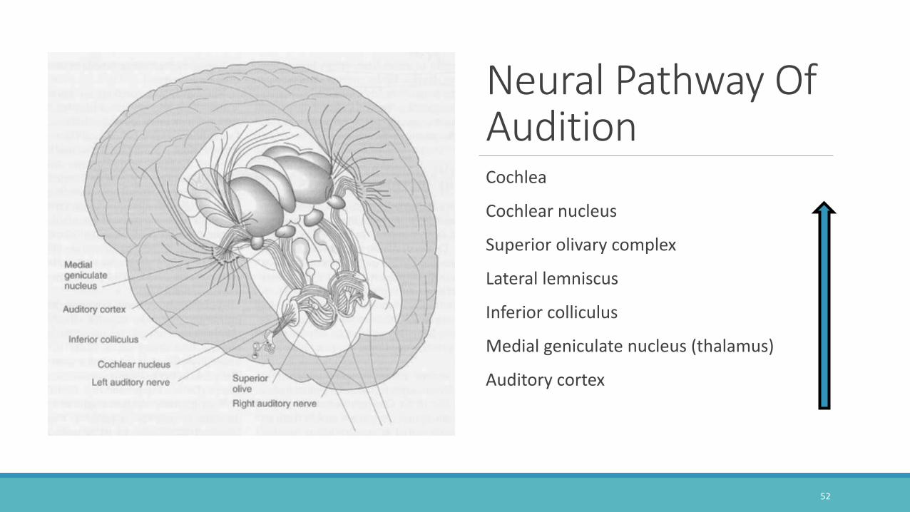
Neural Pathway Of AuditionCochlea
Cochlear nucleus
Superior olivary complex
Lateral lemniscus
Inferior colliculus
Medial geniculate nucleus (thalamus)
Auditory cortex
52

Auditory Neural Pathway -details•CN - actually a bundle of 3 nuclei
•SO - binaural interaction; sound localization
•LL- 6 parallel pathways projecting to IC
•IC- -biologically- significant sound processing
•MG -involved in reading disability(?)
53

Pyramidal SystemReceive input from cerebrum, thalamus
Carry efferent messages allowing voluntarily movement (muscles of face, trunk, arms, and legs)
Fibers converge in brainstem, then cross
Fibers visible as triangular, pyramid-like structures, hence the name.
54

Pathways Of Motor ControlPyramidal motor system
◦ Upper motor neuron
◦ * Lower motor neuron (common pathway)
◦ Motor unit
55
*

Pyramidal Motor System/ DamageHypertonic (spastic) behavior with damage to UMN system
Hypotonic (flaccid) behavior with damage to LMN [‘common pathway’]system
56

Extrapyramidal system
•In the brainstem
•All downward traveling fibers NOT inside the pyramids
•After receiving info from thalamus, regulates repetitive, rhythmical activity (e.g., walking, climbing, hopping, and turning).
•Voluntary or involuntary
•Disorders can result in involuntary, repetitive, rhythmical movements (e.g., tremor or twitching).
57

EPS –continued/ Flow chart◦ Complex!
◦ Influences motor signals sent to periphery
◦ Damage can cause characteristic dystonias
◦ (..next slide)
58

Extrapyramidal signs and symptoms
59
Reversible :
• Akinesia (lack of movement, Parkinson-like)
• Dystonic Reaction (muscle spasms of face, neck, back)
• Dyskinesia (Blinking or twitches)
• Akathesia (Inability to sit still)
Irreversible:
• Tardive Dyskinesia
• Hyperkinesia (lingual or facial)
• Blinking
• Lip smacking
• Sucking or chewing
• Rolls or protrudes Tongue
• Grimaces
• Choreathetoid extremity movement
• Clonic jerking fingers, ankles, toes
• Tonic contractions of neck or back

Sensorimotor RegulationJoint receptors
◦ Free nerve endings
◦ Golgi tendon organs
◦ Muscle spindles
◦ intrafusal fibers
◦ extrafusal fibers
◦ Alpha motor neurons
◦ Gamma motor neurons
60
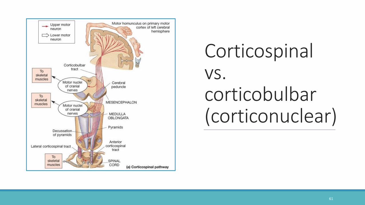
Corticospinal vs. corticobulbar (corticonuclear)
61

Neural Control Of Speech•Broca’s area
•Wernicke’s area
•Angular gyrus
•Supramarginal gyrus
•Supplementary motor cortex
•Orofacial motor area
62
“Traditional model”

Broca’s area
Wernicke’s Area
Arcuate Fascisculus
63
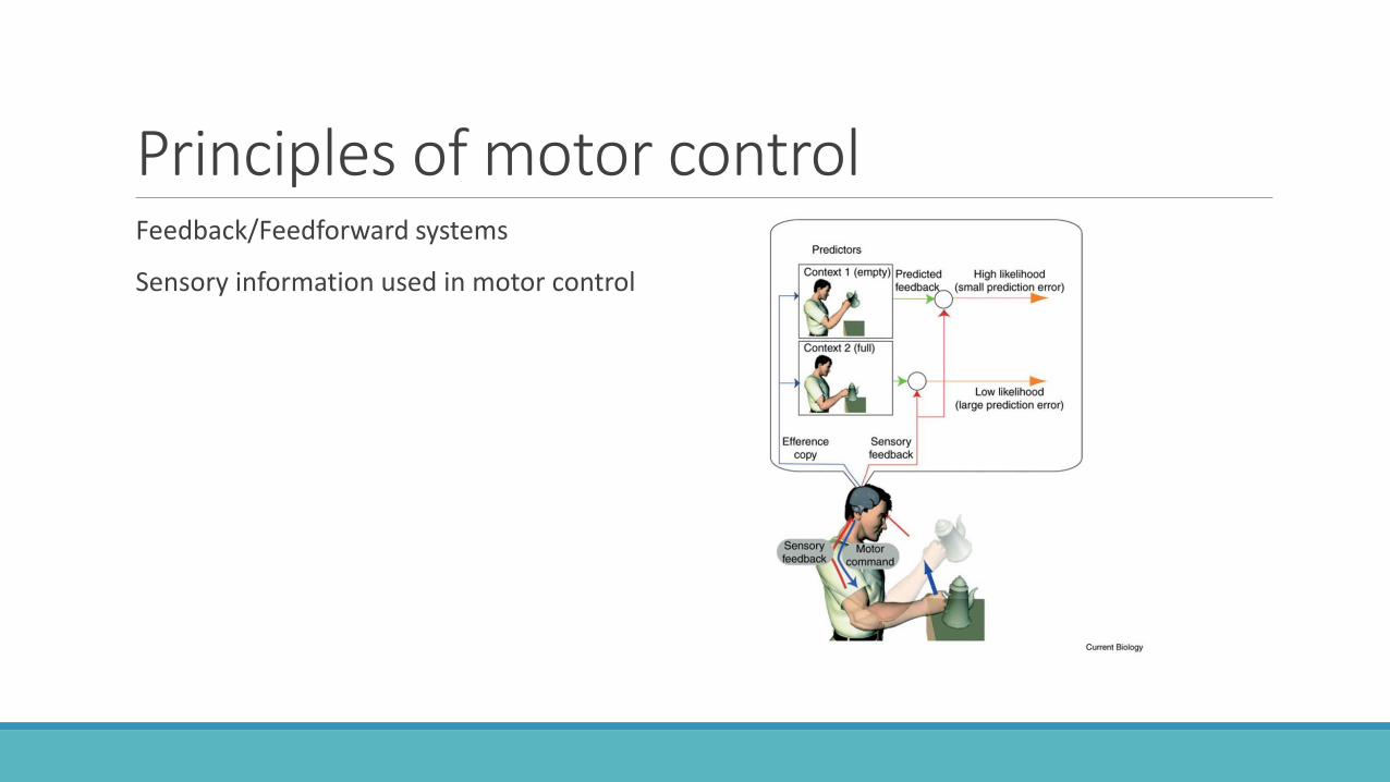
Principles of motor controlFeedback/Feedforward systems
Sensory information used in motor control

Efference copy

Chap 11Neuroimaging Methods
S T R U C T U R AL
Computed Tomography (CT)
Magnetic resonance imaging (MRI)
66
F U N C T I ONA L
Single photon emission computed tomography (SPECT)
Positron emission tomography (PET)
Functional magnetic resonance imaging (fMRI)
Electroencephalography (EEG)
Magnetoencephalography (MEG)

CT Scanner/ Principles
67

Computed Axial Tomoraphy (CAT or CT)
68

Oops..
69

Magnetic Resonance Imaging (MRI)
70
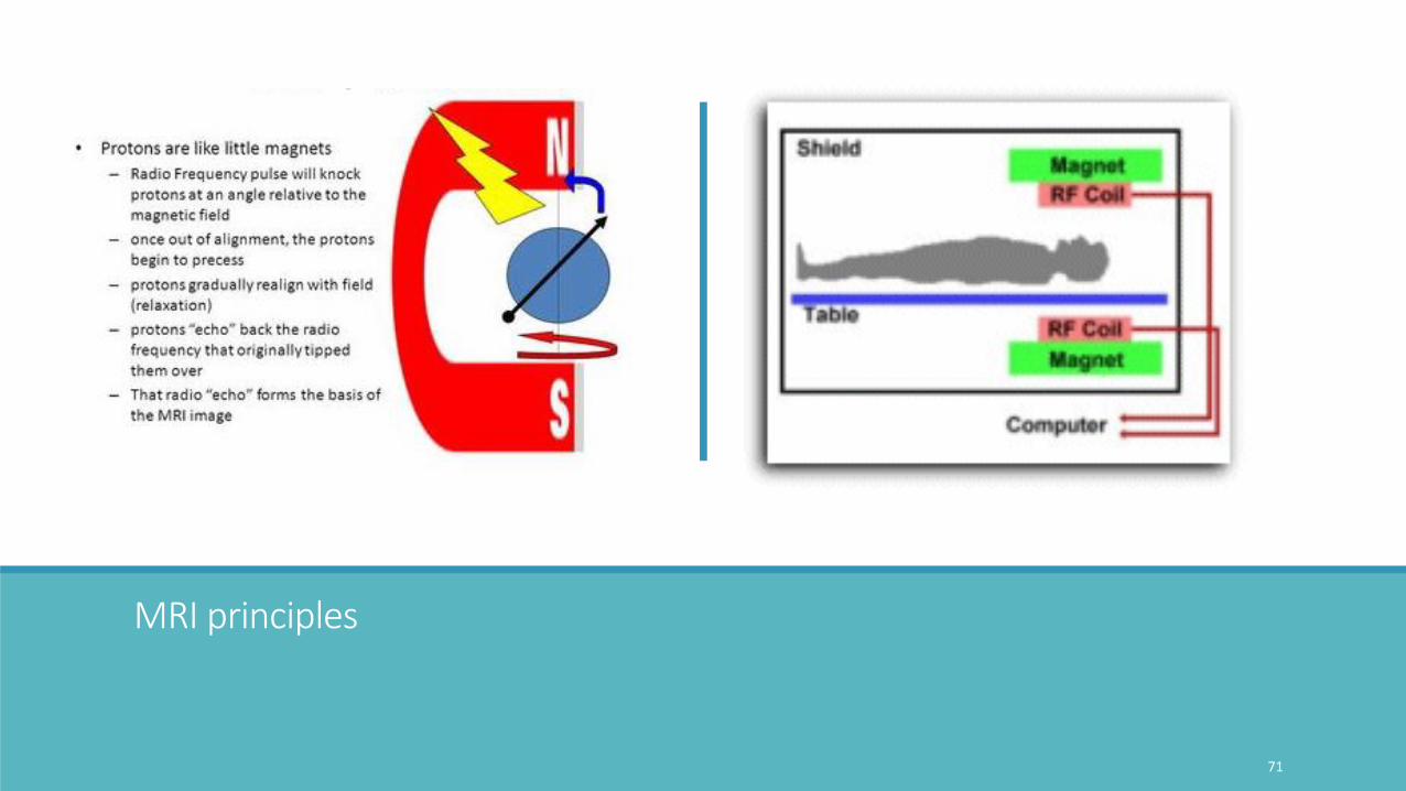
MRI principles
71

Positron Emission Tomography
72

PET imaging - principles
73

Visual Activation PET (overlaid on MR )
•Baseline condition: subjects viewed a simple white cross on a black background.
•
•Activation condition: view B&W drawings of animals.
•Red shows the increase of CBF in the associative visual cortex at the occipital part of the brain.
74

Functional imaging: Single Photon Emission Computed Tomography
75
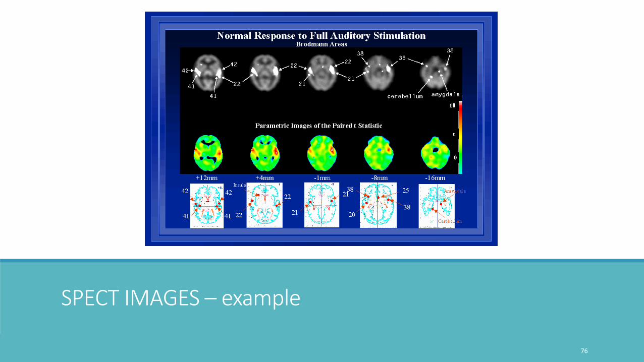
SPECT IMAGES – example
76

Functional Magnetic Resonance Imaging (fMRI)
77

Blood Oxygenation Level Dependent (BOLD) imaging
78
BOLD = (neural) “Cell poop” ??
79

fMRI example: sentence comprehensionSentence comprehension compared to pseudofont baseline in 15 young healthy adults
◦ A: subject-relative short linkage
◦ B: subject-relative long linkage
◦ C: object-relative short linkage
◦ D: object-relative long linkage
80

EEG - Electroencephalography
Electrodes, placed on or just under the scalp, are linked to an amplifier connected to a mechanism that converts electrical impulses into recorded images
81
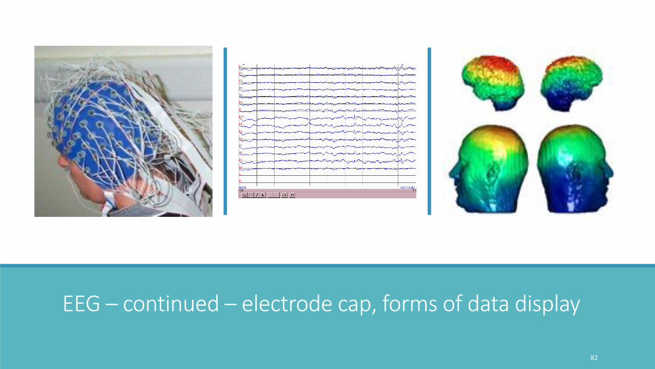
EEG – continued – electrode cap, forms of data display
82

83

84

P300, n400
85
unexpected/ oddball paradigm
Unexpected by sentence context

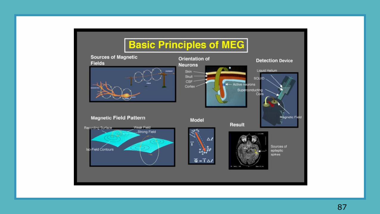
MEGMAGNETOENCEPHALOGRAPY
86
87

MEG –images A) Initial resting state MEG scan after mild TBI.B) Resting state MEG scan 26 months later showing improved connections with time.
(Dr. P. Mukherjee, UCSF, Dept. Radiology)
88

fNIRS –Functionalnear-infraredspectroscopy
Measuring speaker–listener neural coupling with functional near infrared spectroscopy
Yichuan Liu, Elise A. Piazza, Erez Simony, Patricia A. Shewokis, Banu Onaral, Uri Hasson & Hasan Ayaz
Scientific Reports volume7, Article number: 43293 (2017) Figure 1 : Listener-listener fNIRS inter-subject correlation.

Aphasia
•Literally “not speak” Gk. a phatos
•An acquired language disorder that results from damage to portions of the brain that are responsible for language (-NIH)
90

Aphasia: CausesStroke
Head injury
Tumors
Degenerative conditions (e.g. Alzheimer’s)
91

Aphasia: Traditional Distinction
Broca’s: Non-fluent speech; function words and morphemes omitted; comprehension ok.
Wernicke’s: Fluent speech, but filled with non-sense or filler words; comprehension impaired.
92

APHASIA SYNDROMESFLUENCY COMPREHENSION REPETITION NAMING
NON-FLUENT
Broca's poor good poor poor
Global poor poor poor poor
FLUENT
Wernicke's good poor poor poor
Conduction good good poor good
93
B W

94

Broca’s
95
Speech: Nonfluent, halting, agrammatic
Comprehension: Good, but difficulty with semantically difficult materials (e.g., reversible passives)
Basic Idea: Damage to areas in which speech motor programming takes place
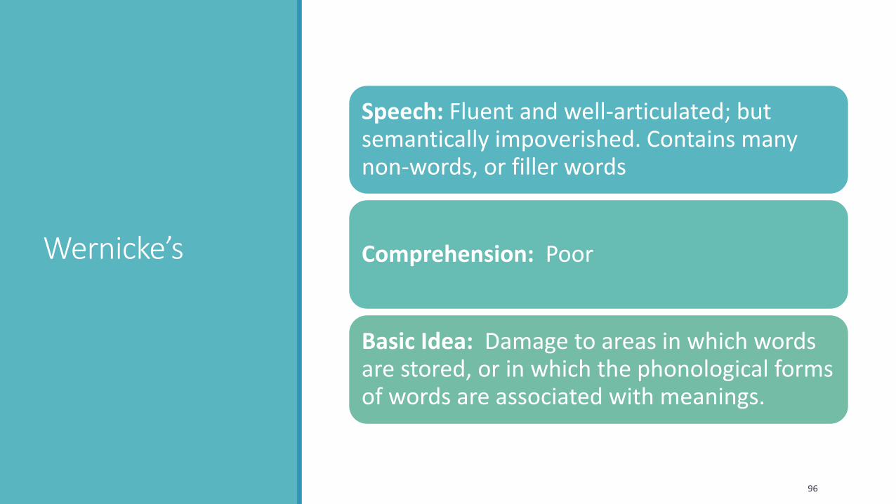
Wernicke’s
96
Speech: Fluent and well-articulated; but semantically impoverished. Contains many non-words, or filler words
Comprehension: Poor
Basic Idea: Damage to areas in which words are stored, or in which the phonological forms of words are associated with meanings.

Conduction Aphasia
97
Lesion: Affects areas connecting Wernicke’sand Broca’s areas (?)
Supramarginal gyrus
Also arcuate fasciculus, which is underneath

Conduction -continued
98
Speech: Relatively unimpaired; but many speech errors, or non-words are used. Also defective naming ability
Comprehension: Also good, but unlike Wernicke’s repetition is difficult
Idea: Network that builds meaningful units out of speech sounds is disabled

Global Aphasia
99
Lesion: Covers entire system of language areas in the dominant hemisphere (left perisylvian cortex)
Abilities: Almost total inability to produce or comprehend speech.
Idea: Combines features of Broca’s and Wernicke’s aphasias