chapter neuroimagingintheevaluation ...web.stanford.edu/~pbarnes/docs/lectures/fetalneonatal.pdf ·...
TRANSCRIPT

Chapter
18 Neuroimaging in the evaluationof pattern and timing of fetaland neonatal brain abnormalitiesPatrick D. Barnes
IntroductionIn this updated review, current and advanced neuroimagingtechnologies are discussed, along with the basic principles ofimaging diagnosis and guidelines for utilization in fetal, peri-natal, and neonatal brain abnormalities [1,2]. This includespattern of injury and timing issues, with special emphasis onneurovascular disease and the differential diagnosis. In thecausative differentiation of static encephalopathies (e.g., cerebralpalsy, CP) from progressive encephalopathies, specific categor-ies and timing are addressed. These include developmentalabnormalities, trauma, neurovascular disease, infections andinflammatory processes, and metabolic disorders. Although arare but important cause of progressive perinatal encephal-opathy, neoplastic processes are not considered in detailhere. Molecular and genetic technologies continue to advancetoward eventual clinical application.
Neuroimaging technologies and generalutilizationImaging modalities may be classified as structural or func-tional [1–12]. Structural imaging modalities provide spatialresolution based primarily on anatomic or morphologic data.Functional imaging modalities provide spatial resolutionbased upon physiologic, chemical, or metabolic data. Somemodalities may actually be considered to provide both struc-tural and functional information.
Ultrasonography (US) is primarily a structural imagingmodality with some functional capabilities (e.g., Doppler:Fig. 18.1a,b) [1–8,13–26]. It is readily accessible, portable, fast,real-time and multiplanar. It is less expensive than other cross-sectional modalities and relatively non-invasive (non-ionizingradiation). It requires no contrast agent and infrequentlyneeds patient sedation. The resolving power of US is basedon variations in acoustic reflectance of tissues. Its diagnosticeffectiveness, however, continues to be dependent upon theskill and experience of the operator and interpreter, and thereare issues regarding inter-observer reliability and accuracy
[1–8,13–26]. Also, US requires a window or path unimpededby bone or air for cranial and spinal imaging. The mostcommon uses of US are (1) fetal and neonatal screening,(2) screening of the infant who cannot be examined in theradiology department (e.g., premature neonate with intracra-nial hemorrhage, ECMO, intraoperative), (3) when importantadjunctive information is quickly needed (e.g., cystic versussolid, vascularity, vascular flow [Doppler: Fig. 18.1a,b], orincreased intracranial pressure), and (4) for real-time guidanceand monitoring of invasive diagnostic or therapeutic surgicaland interventional procedures [1–8,13].
In recent years, advanced US techniques have been intro-duced into clinical practice [1–8,13]. The development ofhigh-resolution transducers, improvements in color Dopplersignal processing, and new scanning techniques have signifi-cantly improved our ability to visualize structural, vascular,and cerebrospinal fluid abnormalities in the neonatal brain.Examples are the mastoid view to better visualize the posteriorfossa, power Doppler and transcranial Doppler (TCD) toevaluate intracranial hemodynamics (e.g., resistive indices,RI: Fig. 18.1a,b), and the graded fontanel compression Dop-pler technique to evaluate hydrocephalus. Another advance inUS technology that has yet to be translated is the developmentof vascular US contrast agents to amplify reflected soundwaves. Potential applications include the detection of slowflow and the assessment of organ perfusion. Computerizedanalysis of textural features is another development that haspromised increased sensitivity and specificity, but has notbeen translated to clinical practice [13].
Computed tomography (CT) is also primarily a structuralimaging modality that has some functional capabilities (e.g.,CT angiography) [1–8]. Although using ionizing radiation,current-generation multidetector CT (MDCT) effectively col-limates and restricts the x-ray exposure to the immediatevolume of interest, particularly when using the ALARA stand-ard for pediatric patients, which adjusts radiation dose relativeto age, size, and anatomic region [27]. Direct imaging is usuallyrestricted to the axial plane (Fig. 18.1c–e). Reformatting fromaxial sections to other planes (e.g., coronal or sagittal) isnow the MDCT standard. Projection scout images mayprovide information similar to plain films but with less spatialresolution. CT of the pediatric CNS is usually done usingeither the conventional or the helical/spiral technique. CT
Fetal and Neonatal Brain Injury, 4th edition, ed. David K. Stevenson, William E.Benitz, Philip Sunshine, Susan R. Hintz, and Maurice L. Druzin. Publishedby Cambridge University Press. # Cambridge University Press 2009.

requires sedation in infants and young children more oftenthan does US but less often than MRI. The more rapid MDCTtechnology, however, has allowed a significant reduction in theneed for sedation or anesthesia. The neonate or very younginfant, for example, may be examined bundled while asleepafter a feeding or during a nap. CT occasionally needs intra-venous iodinated contrast enhancement, but cerebrospinal(CSF) contrast opacification is rarely needed. High-resolutionbone and soft-tissue algorithms are important for demonstrat-ing fine anatomy (e.g., skull base). Advances in computerdisplay technology include image fusion, two-dimensional
reformatting, three-dimensional volumetric and reconstructionmethods, segmentation, and surface rendering techniques.These high-resolution display techniques are used for CTangiography and venography, craniofacial and spinal imagingfor surgical planning, and stereotactic image guidance ofradiotherapy, interventional, and neurosurgical procedures.
The role of CT has been further redefined in the contextof accessible and reliable US and MRI [1–12,15,28,29]. US isthe procedure of choice for primary imaging or screening ofthe brain and spinal neuraxis in the neonate and young infant.When US does not satisfy the clinical inquiry, or an acousticwindow is not available, then CT becomes the primary modal-ity for brain imaging in children, especially in acute or emer-gent presentations. This is especially important for acuteneurologic presentations. In these situations, CT is primarilyused to screen for acute or subacute hemorrhage, focal ordiffuse edema, herniation, fractures, hydrocephalus, tumormass, or abnormal collection (blood, pus, air, CSF, etc.). Otherprimary indications for CT include the evaluation of bonyor air-space abnormalities of the skull base, cranial vault,orbit, paranasal sinuses, facial bones, and temporal bone.Additionally, CT is the definitive procedure for detectionand confirmation of calcification. It is also important in thebony evaluation of a localized spinal column abnormality (e.g.,trauma). Contraindications to CT in childhood are unusual,particularly with the proper application of radiation protec-tion (ALARA standard), the appropriate use of non-ioniccontrast agents, the proper administration of sedation oranesthesia, and the use of vital monitoring.
When CT is used, intravenous enhancement for bloodpool effect (e.g., CT angiography) or blood–brain barrierdisruption is additionally recommended for the evaluation ofsuspected or known vascular malformation, neoplasm,abscess, or empyema [1–4,28,29]. Enhanced CT may helpevaluate a mass or hemorrhage of unknown etiology andidentify the membrane of a chronic subdural collection. Byidentifying the cortical veins, enhanced CT may distinguishprominent low-density subarachnoid collections (benignextracerebral collections or benign external hydrocephalus ofinfancy) from low-density subdural collections (e.g., chronicsubdural hematomas or hygromas). It also may help differen-tiate infarction from neoplasm or abscess, serve as an indicatorof disease activity, for example in degenerative or inflammatorydisease and vasculitis, or provide a high-yield guide for stereo-tactic or open biopsy. Ventricular or subarachnoid CSF-contrast opacification may further assist in evaluating orconfirming CSF compartment lesions or communication(e.g., arachnoid cyst or ventricular encystment). As a rule,MRI is the preferred alternative to contrast-enhanced CT inthe circumstances just enumerated.
Nuclear Medicine (NM) is primarily a functional imagingtechnology [1–8]. NM involves imaging of the biologicaldistributions of administered radioactive pharmaceuticals.Whereas positron emission tomography (PET) has the uniqueability to provide specific metabolic tracers (e.g., oxygen util-ization and glucose metabolism) the wider availability, relative
(a)
(c) (d) (e)
(b)
(f) (g)
(h) (i) (j)
Fig. 18.1. Normal infant and child brain. Ultrasound (US) images of termneonate: (a) coronal US; (b) sagittal US þ Doppler with resistive indices (RI).Computed tomography (CT) images of (c) term neonate, (d) 2-month-oldinfant, and (e) 2-year-old child show progress of maturation, includingmyelination. Sagittal T1 magnetic resonance images (MRIs) of (f) term neonateand (g) 1-year-old infant show progress in brain growth, myelination of thecorpus callosum (arrows), and pituitary maturation. T2 MRIs of (h) 20-weekfetus, (i) term neonate, and (j) 2-year-old child show progress in maturation,i.e., decreasing water content, increasing myelination and cortication.See color plate section.
Section 3: Diagnosis of the infant with brain injury
210
simplicity, and rapid technical advancement of single photonemission computed tomography (SPECT) allows more prac-tical functional assessment of the pediatric CNS. Clinical andinvestigative applications have included the assessment of braindevelopment and maturation, focus localization in refractorychildhood epilepsy (e.g., ictal perfusion SPECT, interictalPET), assessment of tumor progression versus treatmenteffects in childhood CNS neoplasia (perfusion and thalliumSPECT, 18FDG-PET), the evaluation of occlusive cerebrovas-cular disease for surgical revascularization (e.g., perfusionSPECT), the diagnosis of brain death (perfusion SPECT), theuse of brain activation techniques (e.g., perfusion SPECT,PET) in the elucidation of childhood cognitive disorders, theassessment of CSF kinetics (e.g., in hydrocephalus, CSF leaks),and spinal-column screening (skeletal SPECT) [1–8].
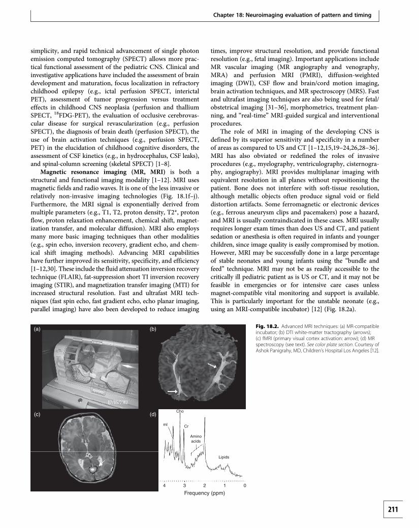
Magnetic resonance imaging (MR, MRI) is both astructural and functional imaging modality [1–12]. MRI usesmagnetic fields and radio waves. It is one of the less invasive orrelatively non-invasive imaging technologies (Fig. 18.1f–j).Furthermore, the MRI signal is exponentially derived frommultiple parameters (e.g., T1, T2, proton density, T2*, protonflow, proton relaxation enhancement, chemical shift, magnet-ization transfer, and molecular diffusion). MRI also employsmany more basic imaging techniques than other modalities(e.g., spin echo, inversion recovery, gradient echo, and chem-ical shift imaging methods). Advancing MRI capabilitieshave further improved its sensitivity, specificity, and efficiency[1–12,30]. These include the fluid attenuation inversion recoverytechnique (FLAIR), fat-suppression short TI inversion recoveryimaging (STIR), and magnetization transfer imaging (MTI) forincreased structural resolution. Fast and ultrafast MRI tech-niques (fast spin echo, fast gradient echo, echo planar imaging,parallel imaging) have also been developed to reduce imaging
times, improve structural resolution, and provide functionalresolution (e.g., fetal imaging). Important applications includeMR vascular imaging (MR angiography and venography,MRA) and perfusion MRI (PMRI), diffusion-weightedimaging (DWI), CSF flow and brain/cord motion imaging,brain activation techniques, and MR spectroscopy (MRS). Fastand ultrafast imaging techniques are also being used for fetal/obstetrical imaging [31–36], morphometrics, treatment plan-ning, and “real-time” MRI-guided surgical and interventionalprocedures.
The role of MRI in imaging of the developing CNS isdefined by its superior sensitivity and specificity in a numberof areas as compared to US and CT [1–12,15,19–24,26,28–36].MRI has also obviated or redefined the roles of invasiveprocedures (e.g., myelography, ventriculography, cisternogra-phy, angiography). MRI provides multiplanar imaging withequivalent resolution in all planes without repositioning thepatient. Bone does not interfere with soft-tissue resolution,although metallic objects often produce signal void or fielddistortion artifacts. Some ferromagnetic or electronic devices(e.g., ferrous aneurysm clips and pacemakers) pose a hazard,and MRI is usually contraindicated in these cases. MRI usuallyrequires longer exam times than does US and CT, and patientsedation or anesthesia is often required in infants and youngerchildren, since image quality is easily compromised by motion.However, MRI may be successfully done in a large percentageof stable neonates and young infants using the “bundle andfeed” technique. MRI may not be as readily accessible to thecritically ill pediatric patient as is US or CT, and it may not befeasible in emergencies or for intensive care cases unlessmagnet-compatible vital monitoring and support is available.This is particularly important for the unstable neonate (e.g.,using an MRI-compatible incubator) [12] (Fig. 18.2a).
ml
Cho
Cr
Aminoacids
Lipids
4
(a) (b)
(c) (d)
3 2
Frequency (ppm)1 0
Fig. 18.2. Advanced MRI techniques: (a) MR-compatibleincubator; (b) DTI white-matter tractography (arrows);(c) fMRI (primary visual cortex activation: arrow); (d) MRspectroscopy (see text). See color plate section. Courtesy ofAshok Panigrahy, MD, Children’s Hospital Los Angeles [12].
Chapter 18: Neuroimaging evaluation of pattern and timing
211

MRI demonstrates superior sensitivity and specificity ina number of circumstances, particularly with the additionof new structural and functional techniques such as FLAIR,STIR, MTI, DWI, PMRI, and MRS [1–12,15,19–24,26,28–36].The FLAIR sequence attenuates the signal from flowing water(i.e., CSF) and increases the conspicuity of non-fluid-water-containing lesions lying in close approximation to theCSF-filled subarachnoid and ventricular spaces. The STIRtechnique suppresses fat signal to provide improved con-spicuity of water-containing lesions in regions where fatdominates (e.g., orbit, head and neck, spine). The MTI methodsuppresses background tissues and increases conspicuity forvascular flow enhancement (e.g., MRA) and gadoliniumenhancement.
MRI is the imaging modality of choice in a number ofclinical situations [1–12,15,19–24,26,28–36]. These includedevelopmental delay (e.g., static encephalopathy vs. neuro-degenerative disease); unexplained seizures (especially focal),unexplained neuroendocrine disorder, or unexplained hydro-cephalus; the pretreatment evaluation of neoplastic processesand the follow-up of tumor response and treatment effects;suspected infectious, postinfectious, and other inflammatoryor non-inflammatory encephalitides (e.g., encephalitis, post-infectious demyelination, vasculitis); migrational and othersubmacroscopic dysgeneses (e.g., cortical dysplasia); neuro-cutaneous syndromes (e.g., neurofibromatosis 1, tuberoussclerosis); intractable or refractory epilepsy; vascular diseases,hemorrhage, and the sequelae of trauma.
MRI frequently offers greater diagnostic specificity thandoes CT or US for delineating vascular and hemorrhagicprocesses. This includes the clear depiction of vascular struc-tures and abnormalities based on proton flow parameters andsoftware enhancements not requiring the injection of contrastagents (e.g., MRA). MR angiography (MRA) techniques mayadditionally be used to differentiate arterial from venousocclusive disease [1–12,15,19–24,26,28–36]. Using gradientrecalled echo (GRE) magnetic susceptibility techniques, MRIalso provides more specific identification and staging ofhemorrhage and clot formation according to the evolution ofhemoglobin breakdown. MRI is often reserved for moredefinitive evaluation of hemorrhage and as an indicator orguide for angiography in a number of special situations. MRImay be used to evaluate an atypical or unexplained intracra-nial hemorrhage by distinguishing hemorrhagic infarctionfrom hematoma and by distinguishing among the typesof vascular malformations (e.g., cavernous vs. arteriovenousmalformations). MRA may obviate the need in some cases ofvascular malformation for conventional angiography in thefollow-up of surgery, interventional treatment, or radiosurgery.
In the evaluation of intracranial vascular anomalies(e.g., vascular malformation, aneurysm), MRI may identifyotherwise unsuspected prior hemorrhage (i.e., hemosiderin)[1–12,15,19–24,26,28–36]. When CT demonstrates a non-specific focal high density (calcification vs. hemorrhage),MRI may provide further specificity, for example, by distin-guishing an occult vascular malformation (e.g., cavernous
malformation) from a neoplasm (e.g., glioma). It may furtherassist US or CT in differentiating benign infantile collections(i.e., external hydrocephalus) from subdural hematomas[28,29]. MRI often also provides definitive evaluation of mus-cular and cutaneous vascular anomalies (i.e., hemangiomas,vascular malformations) that arise in parameningeal locations(e.g., head and neck, paraspinal) and extend to involve theCNS directly or are associated with other CNS vascular ornon-vascular abnormalities.
MR spectroscopy (MRS) offers a non-invasive in vivoapproach to biochemical analysis [1–12,37–40] (Fig. 18.2d).Furthermore, MRS provides additional quantitative informa-tion regarding cellular metabolites, since signal intensity islinearly related to steady-state metabolite concentration.MRS can detect cellular biochemical changes prior to thedetection of morphological changes by MRI or other imagingmodalities. MRS may therefore provide further insight intoboth follow-up assessment and prognosis. With recentadvances in instrumentation and methodology, and utilizingthe high inherent sensitivity of hydrogen 1, single-voxel andmultivoxel proton MRS is now carried out with relativelyshort acquisitions to detect low-concentration metabolites inhealthy and diseased tissues. Phosphorus-31 spectroscopy hasalso been developed for pediatric use. Currently, MRS hasbeen primarily used in the assessment of brain developmentand maturation (Fig. 18.3), perinatal brain injury, childhoodCNS neoplasia versus treatment effects, and metabolic andneurodegenerative disorders [39–40].
Perfusion MRI (PMRI) has been developed to evaluatecerebral perfusion dynamics through the application of adynamic contrast-enhanced T2*-weighted MR imagingtechnique [1–4,8,12,31,41]. This technique has been used toqualitate and quantitate normal and abnormal cerebrovasculardynamics of the developing brain by analyzing hemodynamicparameters including relative cerebral blood volume, relativecerebral blood flow, and mean transit time, all as comple-mentary to conventional MR imaging. Non-contrast-enhanced methods of PMRI have also been developed (e.g.,flow-activated inversion recovery, FAIR; arterial spin labeling,ASL; blood oxygen level determination, BOLD) [41]. Currentand advanced applications of these perfusion techniquesinclude the evaluation of ischemic cerebrovascular disease(e.g., hypoxia–ischemia, moyamoya, sickle cell disease), thedifferentiation of tumor progression from treatment effects,and brain activation imaging [1–4,8,12,31,41]. One of themost active areas of research is the localization of brain activ-ity, an area previously dominated by NM including SPECTand PET.
Functional MRI (fMRI) is the terminology often appliedto brain activation imaging in which local or regional changesin cerebral blood flow are displayed that accompany stimula-tion or activation of sensory (e.g., visual, auditory, somatosen-sory), motor, or cognitive centers [1–4,12]. fMRI is providingimportant information regarding the spatial distribution ofsensory, motor, and cognitive function and functional impair-ment (Fig. 18.2c). Also, it may serve as a guide for safer and
Section 3: Diagnosis of the infant with brain injury
212

more effective interventions including microneurosurgeryor conformal radiotherapy, for example in the ablation oftumors, vascular malformations, and seizure foci. Morerecently, fMRI is being used to evaluate brain developmentand maturation in the neonate and young infant, including theeffects of injury and post-injury repair and recovery [12].
Using echo planar or line-scan spin echo techniques,diffusion-weighted imaging (DWI) provides information basedupon differences in the rate of diffusion of water molecules, andit is especially sensitive to cellular changes [1–12,30,40,42–53].The rate of diffusion, or apparent diffusion coefficient (ADC),is higher for free or pure water than for macromolecularbound water. The ADC varies according to the microstruc-tural or physiologic state of a tissue. Fractional anisotropy(FA) is a vector measurement of the directionality of diffusionusing diffusion tensor imaging (DTI) methods and alsovaries with the microstructural environment, both develop-mentally and pathologically [53] (Fig. 18.2b). This methodis especially helpful in assessing axonal development andinjury, including myelination and synaptogenesis (connectiv-ity). Current clinical applications of DWI and DTI includethe assessment of brain maturation, the evaluation of acuteinjury, and the analysis of the sequelae of injury (Fig. 18.4).A particularly important application of DWI is in the earlydetection of diffuse and focal ischemic injury. The ADC ofwater is reduced within minutes of an ischemic insult andprogressively so within the first hour. High-intensity abnor-malities are demonstrated on DWI, along with low-intensityabnormalities on calculated ADC images, at a time whenconventional MRI is negative, and this likely reflects cellularinjury (e.g., necrosis) with primary or secondary energy fail-ure. Further investigations are under way regarding the roles
of DWI, PMRI, and MRS in the early diagnosis and treatmentof potentially reversible ischemic injury.
Motion-sensitive MRI techniques are not only used toevaluate vascular flow (e.g., MRA) and perfusion, but mayalso be used to demonstrate the effect of pulsatile cardiovascu-lar flow on other fluid tissues (e.g., CSF) and on non-fluidtissues such as the brain and spinal cord. Using cardiac orpulse gating, these MRI techniques may be used to evaluate,preoperatively and postoperatively, abnormalities of CSFdynamics (e.g., hydrocephalus, hydrosyringomyelia), as wellas abnormalities of brain motion (e.g., Chiari malformation),and spinal cord motion (e.g., tethered cord syndrome) [1–4].A number of non-gated MRI techniques (e.g., propellerimaging) are also being used to reduce motion artifact andimprove image quality.
Thalamus
(a) (b)
(c)
CrCr
Cho
ChoNAA
NAA
infant adult
Basal ganglia
Geniculocalcarine
Parietal
Paracentral
Frontal
0 100
48±10
NAA/Cho Maturation Time Constant
54±12
78±45
113±41
125±39
176±61
200Age (weeks)
Fig. 18.3. H-1 MR spectroscopy of thedeveloping brain with (a) infant spectra, (b) adultspectra, and (c) maturation time constants forbrain regions. Dermon J, Barnes PD, SpielmanD. Spatiotemporal mapping of cerebral maturationin childhood using 2D MR spectroscopic imaging.American Society of Neuroradiology, 2002.
Fig. 18.4. DTI with FAmap in preterm neonate as aquantitative display of white-matter development (arrows)of the internal capsule andcorpus callosum [12].
Chapter 18: Neuroimaging evaluation of pattern and timing
213

Guidelines and principles of imagingdiagnosisDevelopmental abnormalitiesCongenital abnormalities of the CNS may be developmentalor acquired in origin, and may result from defective forma-tion, postformational destruction, or disordered maturation[1,2,4,5,54]. These are probably best classified according togestational timing (Table 18.1), and include disorders of dorsaland ventral neural tube formation; disorders of neuronal, glial,and mesenchymal formation; neuroclastic processes (encepha-loclastic, myeloclastic); and disorders of maturation (myelin-ation and cortical maturation). These are classified in sixgroups (I–VI) in Table 18.1. Developmental anomalies oftendetected by US (prenatal or postnatal) or CT are the grossformational macrostructural defects of categories I–IV andthe gross neuroclastic macrostructural lesions of category V[1,2,4,5,54]. However, MRI always provides more completedelineation of these defects. This is especially true for thoseabnormalities involving the ventricular system or containingCSF. These include cephaloceles (Fig. 18.5), hydrocephalus,hydranencephaly, holoprosencephaly, absent septum pelluci-dum, hypogenesis of the corpus callosum (Figs. 18.5–18.7),porencephaly, open schizencephaly, the Dandy–Walker–Blakespectrum (Fig. 18.7), and arachnoid cysts. US may not clearlydistinguish hydranencephaly (absent cerebral mantle) fromsevere hydrocephalus (attenuated cerebral mantle). This isusually clarified by CT or MRI (Fig. 18.8). Other gross macro-structural anomalies often detected by US or CT includeChiari II malformation, lissencephaly, and vascular malforma-tions such as the Galenic malformation. Any “cystic” lesiondetected by US should be examined with Doppler to deter-mine if it is vascular in nature. Neuroclastic processesare destructive lesions of the already formed CNS andmay result from a variety of prenatal or perinatal insultsincluding hypoxia–ischemia and infection (Table 18.1). MRImay often demonstrate subtle macrostructural abnormalitiesnot revealed by US or CT (e.g., periventricular leukomalacia)[1–12,15,17,19–24,26,33].
MRI is important when the US or CT fails to satisfy theclinical investigation. MRI often provides a more completedelineation of complex macrostructural CNS anomalies fordiagnosis, treatment, prognosis, and genetic counseling[1,2,4,5,33,54]. In fact, ultrafast MRI techniques are beingincreasingly used prenatally to evaluate for fetal CNS abnor-malities in at-risk pregnancies or as detected by obstetrical US[31–36] (Figs. 18.5–18.7). Furthermore, MRI is often indicatedif more specific treatment is planned beyond simple shuntingof hydrocephalus. Intraoperative guidance may be providedby real-time and Doppler US. Patients with craniosynostosisare best evaluated with 3DCT. Those with multiple sutureinvolvement, especially when it is associated with craniofacialsyndromes, may require more extensive evaluation beyond3DCT, including CT venography, MRI, and MR venography(jugular venous steno-occlusive disease with collateralization).
MRI is often required to structurally delineate the moresubtle macrostructural anomalies arising as disorders of
Table 18.1. Classification of CNS malformations by gestational timing
I. Disorders of dorsal neural tube development (3–4 weeks)
Anencephaly
Cephaloceles
Dermal sinus
Chiari malformations
Spinal dysraphism
Hydrosyringomyelia
II. Disorders of ventral neural tube development (5–10 weeks)
Holoprosencephalies
Agenesis septum pellucidum
Optic and olfactory hypoplasia/aplasia
Pituitary – hypothalamic hypoplasia/aplasia
Cerebellar hypoplasia/aplasia
Dandy Walker spectrum
Craniosynostosis
III. Disorders of migration and cortical organization (2–5 months)
Schizencephaly
Neuronal heterotopia
Agyria/pachygyria
Lissencephaly
Polymicrogyria
Agenesis corpus callosum
IV. Disorders of neuronal, glial, and mesenchymal proliferation, differentiation,and histiogenesis (2–6 months)
Micrencephaly
Megalencephaly
Hemimegalencephaly
Aqueductal anomalies
Colpocephaly
Cortical dysplasias
Neurocutaneous syndromes
Vascular anomalies
Malformative tumors
Arachnoid cysts
V. Encephaloclastic processes (> 5–6 months)
Hydranencephaly
Porencephaly
Multicystic encephalopathy
Encephalomalacia
Leukomalacia
Hemiatrophy
Hydrocephalus
Hemorrhage
Infarction
VI. Disorders of maturation (7 months – 2 years)
Hypomyelination
Delayed myelination
Dysmyelination
Demyelination
Cortical dysmaturity
Note:See references [2,4,54].
Section 3: Diagnosis of the infant with brain injury
214

migration and cortical organization (category III: Fig. 18.9) oras disorders of proliferation, differentiation, and histiogenesis(category IV: Fig. 18.10) [1,2,4,5,33,54]. The perfusional andmetabolic characteristics of these anomalies (e.g., focal corticaldysgenesis, hemimegalencephaly) may be investigated withSPECT and PET, respectively, in children with medicallyrefractory partial epilepsy who are candidates for surgicalablation. For added precision, the SPECT or PET data maybe fused with the MRI data to provide a higher-resolutionspatial display of the functional information. MRS, DWI, DTI,and PMRI with fMRI are also contributing to the evaluationand treatment of these patients [4,12,30]. MRI is now thepreferred modality for the screening and definitive evaluation
of the dysgenetic, neoplastic, and vascular manifestationsof the neurocutaneous syndromes (Fig. 18.11). After initialscreening with US or CT, MRI is also considered the primarytechnology for treatment planning and follow-up of vascularmalformations and developmental tumors [1,2,4,30]. Althougharachnoid cysts are often readily delineated by US or CT, MRIis usually necessary for confirmation (i.e., to exclude solidtumor) and for surgical planning. FLAIR or DWI may readilydistinguish an arachnoid cyst from other lesions (e.g.,dermoid–epidermoid, fibrillary astrocytoma). Maturation(i.e., myelination and cortical maturation) and disorders ofmaturation (category VI) may be precisely assessed onlyby MRI [1,2,4,5,30,33,54] (Fig. 18.1f–j). The MRI findings,
(a) (b) (c)
C
C
C
Fig. 18.7. Dandy–Walker cyst (C): (a) fetal sagittalT2 MRI; (b) neonatal axial CT; (c) sagittal T1 MRI withhypogenesis of the corpus callosum (arrow).
(a) (b)
H
C
Fig. 18.5. (a) Fetal sagittal T2 MRI, showing cervico-occipital cephalocele(posterior arrow), Chiari III malformation (anterior arrow), agenesis of thecorpus callosum, and microcephaly. (b) Neonatal sagittal T1 MRI, showingoccipital meningoencephalocele (C) with kinked brainstem (arrow) andhydrocephalus (H).
(a) (b)
Fig. 18.6. Agenesis corpus callosum on (a) fetal axial T2 and (b) neonatalsagittal T1 MRI (see Fig. 18.1. f,g) [50].
(a) (b)
H
H
Fig. 18.8. Hydranencephaly on (a) sagittal and (b) axial T1 MRI. Only a smallportion of cortex is present frontally (arrows).
(a) (b)
Fig. 18.9. Microlissencephaly: neonatal (a) sagittal and (b) axial T1 MRI,showing microcephaly with agyric cortex.
Chapter 18: Neuroimaging evaluation of pattern and timing
215

however, are often non-specific regarding causation, particu-larly in the first year of life, because of the watery characterof the immature brain. DTI and MRS may add specificityto the diagnostic evaluation of these infants [1–12,37–40](Figs. 18.2–18.4).
Neurovascular diseaseNeurovascular disease characteristically presents as an acuteneurologic event (e.g., neonatal encephalopathy). However, arecently discovered but fixed deficit (e.g., hemiplegia, spasticdiplegia, hypotonia) may be the first indication of a remoteprenatal or perinatal neurovascular injury. Imaging assists inthe clinical evaluation and differentiation of hypoxia–ischemia,hemorrhage, and occlusive vascular disease [1–12,30].
Hypoxia–ischemiaIn general, the pattern of injury associated with hypoxic–ischemic encephalopathy (HIE), or other insults (e.g., reperfu-sion), varies with the severity and duration of the insult as wellas with the gestational (or corrected) age (GA) of the fetus,neonate, or infant at the time of the insult or insults [1–12](Table 18.2). Different brain structures are more vulnerablethan others to the different types of HIE insults (e.g., partialprolonged, profound, combined) at different stages of braindevelopment (e.g., formational vs. postformational GA, pre-term vs. term vs. full-term or post-term GA). Brain tissuesin the arterial border zones or watersheds (intervascularboundary zones), brain tissues with high metabolic demands,mature or actively maturing tissues, and tissues with higherconcentrations of neuroexcitatory amino acids are particularlyvulnerable to HIE and to other insults (e.g., hypoglycemia,
trauma, infection, seizures) [1–12,55–61]. Prenatal or perinatalpartial prolonged HIE (e.g., one or more insults of hypoxia/hypoperfusion) may be associated with periventricular border-zone/watershed injury to the preterm fetus or neonate (e.g., 27–35weeksGA) [1–12,55,57,62–75]. Subtypes of white-matter injuryof prematurity (i.e., “encephalopathy of prematurity”) includethe classic focal/multifocal “cystic” type of periventricularleukomalacia (PVL), the focal/multifocal “noncystic” (gliotic)form of PVL, and the diffuse white-matter gliosis injury pattern(Figs. 18.12–18.15). The pathogenesis may include not onlyhypoxia–ischemia but other factors such as infectious orinflammatory processes (e.g., maternal infection, chorioamnio-nitis, funisitis, fetal inflammatory response, cytokine-mediatedinjury) whether occurring in the preterm, or term, fetus orneonate [7,66,70]. Prenatal or perinatal partial prolonged HIEduring term gestation (e.g., 37–42 weeks GA) may produce acortical and subcortical border-zone/watershed cerebral injury(Figs. 18.16, 18.17). A transitional partial prolonged HIEpattern (cortical/subcortical/periventricular) may be seen inthe late preterm to early mid-term GA (e.g., 36–38 weeks) orwithmore severe injuries. Fetal or neonatal brain injurymay alsooccur with more profound HIE insults (e.g., anoxia or circulatoryarrest) and involve the thalami, basal ganglia (especiallyputamina), brainstem (especially midbrain), cerebellar vermis,
Fig. 18.10. Hemimegalencephaly:neonatal axial T2 MRI shows larger righthemisphere with unilateralventriculomegaly and abnormalcortication.
(a) (b)
Fig. 18.11. Tuberous sclerosis with periventricular and subcortical tubers(arrows) on (a) neonatal sagittal T1 and (b) axial T2 MRI.
Table 18.2. Imaging patterns of hypoxic–ischemic encephalopathy (HIE)
Hemorrhage
Germinal matrix – intraventricular hemorrhage
Choroid plexus – intraventricular hemorrhage
Subarachnoid hemorrhage
Hemorrhagic infarctions
Partial prolonged HIE
Preterm: White-matter injury of prematurity (e.g., periventricularleukomalacia)
Term/full-term/post-term: Cortical/subcortical injury (borderzone,watershed, parasagittal)
Intermediate: combined or transitional pattern
Ulegyria
Cystic encephalomalacia
Profound HIE
Thalamic and basal ganglia injury
Brainstem injury
Cerebellar vermian injury
Hippocampal injury
Cerebral white-matter injury
Paracentral injury
Global injury (prolonged profound)
Combined profound and partial prolonged (or prolonged profound) HIE
Total asphyxia pattern (including cystic encephalomalacia)
Notes:Depends on gestational age, chronological age, duration and severityof the insult.See references [2–8].
Section 3: Diagnosis of the infant with brain injury
216

hippocampi, paraventricular white matter, and perirolandiccortex [1–12,55,57,76–80] (Figs. 18.18, 18.19). This type ofinjury may also vary with GA (thalamic greater than putaminalinvolvement in the preterm GA; putaminal, hippocampal, andparacentral injury more common in the term GA). Combinedpartial prolonged plus profound HIE patterns (e.g., totalasphyxia) may also occur [1–12,76–80] (Fig. 18.20).
US, CT, or MRI (e.g., DWI) may demonstrate evolvingedema, necrosis, or hemorrhage in the hyperacute, acute,and subacute phases [1–12] (Figs. 18.12–18.20). The edemaof non-hemorrhagic HIE (e.g., partial prolonged type) usuallyevolves over 1–7 days and often peaks between 36 and 72 hours(2–4 days by US, CT) following the insult(s) and dependingupon reperfusion (and other “insults”). US may show hyper-echogenicity, and CT may show hypodensities with decreasedgray–white matter differentiation. Complete loss of gray–whitedifferentiation may correlate with peak edema [60,61]. In theearly phases of the injury, the neuroimaging findings maybe non-specific as to causation. The differential diagnosis
includes HIE, multifocal occlusive vascular infarction, infection,metabolic derangement (e.g., hypoglycemia, hyperbilirubin-emia, fluid-electrolyte imbalance),metabolic or connective tissuedisorder, and venous thrombosis (e.g., coagulopathy) [1–12].Associated hemorrhage may be subarachnoid, germinal matrixand intraventricular hemorrhage (e.g., preterm fetus or neo-nate), choroid plexus/intraventricular hemorrhage (e.g., termfetus or neonate), and cerebral or cerebellar hemorrhage (e.g.,hemorrhagic infarction). Their imaging characteristics aredescribed in the next section.
According to the evidence-based medical literature, thesensitivity and specificity of MRI depends on the techniquesused and the timing of the imaging [2,4,6,40,45,78]. Conven-tional MRI may show characteristic T1 hypointensities/T2 hyperintensities (12–48 hours), followed by T1 hyperinten-sities (as early as 2–4 days), and then T2 hypointensities(as early as 6–7 days). These T1 and T2 changes may lastfor a number of weeks to a month. DWI may be abnormalbefore conventional MRI and show restricted diffusion withdecreased ADC as increased intensity on DWI and decreasedintensity on ADC maps [2,8,40,45]. Diffusion abnormalitiesmay tend to evolve for up to 2–3 weeks. Knowledge of theseevolving intensity features is particularly important in order toavoid misinterpretation regarding pattern of injury and timing[40]. Doppler with resistive indices (e.g., RI< 60) or MRS (e.g.,elevated lactate, elevated glutamate, elevated lipids, decreasedN-acetyl-aspartate [NAA]) may provide additional early indi-cators of timing and outcome [2,6,8,14,37,40,81]. The moresubtle ischemic PVL lesions (e.g., cystic phase) may be betterdelineated by US (2–6 weeks after insult(s)) than by CT orMRI, in which the density and intensity character of immature
(a) (b) Fig. 18.12. Cystic PVL: (a) US, acute edema phase (confluentincreased echoes – arrows); (b) US, subacute cystic phase (hypoechoicfoci with surrounding increased echoes – arrows).
(a) (b)
Fig. 18.13. Cystic PVL: (a) axial T2 and (b) coronal FLAIR MRI, chroniccystic phase (arrows).
(a) (b) (c)Fig. 18.14. Non-cystic PVL with foci of gliosis(arrows) as high intensities on (a) sagittal and (b) axialT1 MRI, plus mineralization or hemorrhages (arrows)as low intensities on (c) axial GRE MRI.
Chapter 18: Neuroimaging evaluation of pattern and timing
217

white matter often obscures the injury. However, CT and MRIoften show gray-matter injury better than US, and MRI dem-onstrates non-cystic white-matter injury better than US or CT[4,8,19–24,26]. Further developments of PMRI, DWI, andMRS have further improved the diagnostic sensitivity andspecificity of MRI [4,8,12,30,40]. In fact, DWI has demon-strated restricted diffusion in the acute phase of PVL when US,CT, and conventional MRI are negative or non-specific. Suchadvances may facilitate the early institution of neuroprotectivemeasures to treat potentially reversible primary injury (necro-sis, apoptosis) and secondary injury (reperfusion, transneuraldegeneration) in HIE [82]. These advanced MRI techniquesmay also assist in distinguishing HIE from other causes of
encephalopathy, including common metabolic derangements(e.g., hypoglycemia, hyperbilirubinemia), rarer inborn errorsof metabolism, and non-metabolic conditions (e.g., infection).
The long-term result of HIE is a static encephalopathy (i.e.,CP) and imaging may demonstrate injury in the chronicphases (> 14–21 days after the insult(s)) including porence-phaly, hydranencephaly, atrophy, chronic periventricular leu-komalacia, cystic encephalomalacia, gliosis, or mineralizationin a characteristic distribution as described above [1–12](Figs. 18.12–18.20). The chronic changes are best demon-strated by MRI. In general, for pattern of injury and timingpurposes, two pieces of imaging evidence are optimally desired:(1) late imaging, and preferably MRI, beyond 2–3 years of age
(a) (b) (c)Fig. 18.15. Diffuse PVL (high intensities – arrows) on(a) near-term axial T2 and (b,c) older infant axial FLAIR MRI.
(a) (b) (c)
(d)
Fig. 18.16. Partial prolonged HIE: acute phase withwatershed injury (arrows) on (a) sagittal T1, (b) axial T2, and(c) axial DWI; (d) chronic phase with cortical atrophy andulegyria (arrow) on axial CT.
(a) (b) (c)Fig. 18.17. Very severe partial prolonged HIE: acute–subacute phase with (a) low-density cerebral peak edema(arrows) on axial CT and (b) high-intensity cerebral restricteddiffusion (arrows) on axial DWI; (c) chronic phase with cysticencephalomalacia (arrows) on axial T1.
Section 3: Diagnosis of the infant with brain injury
218

when the brain is greater than 90% mature (no more water ofimmaturity), in order to get a final, permanent injury patternfor causative etiology and GA timing; and (2) early postnatal(and/or prenatal) imaging, and preferably MRI, in order toevaluate evolution in the acute, subacute, and chronic phases,so that timing as to “day range” relative to the perinatal andperipartum periods may be assessed [1–12,40].
Intracranial hemorrhageIntracranial hemorrhage may result from parturitional trauma,HIE, a coagulopathy (e.g., thrombocytopenia, disseminatedintravascular coagulopathy [DIC], extracorporal membraneoxygenation [ECMO]), vasoocclusive disease (e.g., thrombo-philia with venous thrombosis), or it may be idiopathic[1–12,28,29,83]. Hemorrhage may occasionally be associatedwith infection (e.g., herpes simplex virus 2). Vascular malfor-mations producing intracranial hemorrhage are rare in theneonate and young infant and usually not encountered untillater childhood (i.e., arteriovenous malformations [AVM],
cavernous malformations, developmental venous anomalies,and telangiectasias) [1–12,28,29]. Aneurysms are exceedinglyrare in children but may be developmental, associated with asyndrome (e.g., Turner syndrome), or related to trauma (e.g.,dissection) or infection (i.e., mycotic aneurysm). The vein ofGalen malformations are subclassified as choroidal, mural, andAVM types. They rarely hemorrhage, and more commonlypresent in infancy with congestive heart failure, cerebral ische-mia, or hydrocephalus.
US or CT remains the primary imaging choice inacute situations [1–12,29]. As mentioned above, there maybe subarachnoid hemorrhage, germinal matrix and intraven-tricular hemorrhage (e.g., premature fetus or neonate), chor-oid plexus/intraventricular hemorrhage (e.g., term fetus orneonate), and cerebral or cerebellar hemorrhage (e.g., hemor-rhagic infarction). The hemorrhage usually appears hyper-echoic on US and high-density on CT in the acute to subacutephases (range 3 hours – 7 days) unless there is associatedcoagulopathy. With evolution and resolution, the hemorrhage
(b) (c) (d)
(e)Ch
CrNA
L
(a)Fig. 18.18. Profound HIE: acute phase with(a) bilateral basal ganglia and thalamichyperechogenicity (arrows) on US; (b) hypodensities(arrows) on CT; high intensities (arrows) on (c) axialDWI and (d) T1/FLAIR MRI; and (e) inverted lactatedoublet (L) on MRS.
(a) (b) (c)Fig. 18.19. Profound HIE: chronic phase with (a–c) bilateralhippocampal, putaminal, thalamic, and paracentral high intensities(arrows) on axial FLAIR MRI.
(a) (b) (c)
Fig. 18.20. Term combined HIE: subacute phase with (a) basalganglia and thalamic high densities (short arrows) plus cerebral lowdensities (long arrows) on CT; chronic phase with (b) basal gangliaand thalamic hypointense mineralization (arrows) on axial GRE plus(c) hypointense cystic encephalomalacia (arrows) on axial T1 MRI.
Chapter 18: Neuroimaging evaluation of pattern and timing
219
becomes isoechoic to hypoechoic and isodense to hypodense(> 7–10 days).
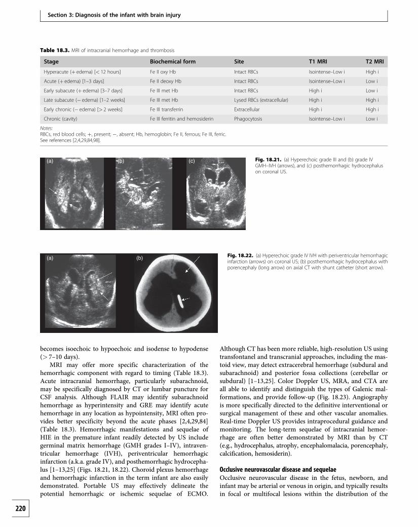
MRI may offer more specific characterization of thehemorrhagic component with regard to timing (Table 18.3).Acute intracranial hemorrhage, particularly subarachnoid,may be specifically diagnosed by CT or lumbar puncture forCSF analysis. Although FLAIR may identify subarachnoidhemorrhage as hyperintensity and GRE may identify acutehemorrhage in any location as hypointensity, MRI often pro-vides better specificity beyond the acute phases [2,4,29,84](Table 18.3). Hemorrhagic manifestations and sequelae ofHIE in the premature infant readily detected by US includegerminal matrix hemorrhage (GMH grades I–IV), intraven-tricular hemorrhage (IVH), periventricular hemorrhagicinfarction (a.k.a. grade IV), and posthemorrhagic hydrocepha-lus [1–13,25] (Figs. 18.21, 18.22). Choroid plexus hemorrhageand hemorrhagic infarction in the term infant are also easilydemonstrated. Portable US may effectively delineate thepotential hemorrhagic or ischemic sequelae of ECMO.
Although CT has been more reliable, high-resolution US usingtransfontanel and transcranial approaches, including the mas-toid view, may detect extracerebral hemorrhage (subdural andsubarachnoid) and posterior fossa collections (cerebellar orsubdural) [1–13,25]. Color Doppler US, MRA, and CTA areall able to identify and distinguish the types of Galenic mal-formations, and provide follow-up (Fig. 18.23). Angiographyis more specifically directed to the definitive interventional orsurgical management of these and other vascular anomalies.Real-time Doppler US provides intraprocedural guidance andmonitoring. The long-term sequelae of intracranial hemor-rhage are often better demonstrated by MRI than by CT(e.g., hydrocephalus, atrophy, encephalomalacia, porencephaly,calcification, hemosiderin).
Occlusive neurovascular disease and sequelaeOcclusive neurovascular disease in the fetus, newborn, andinfant may be arterial or venous in origin, and typically resultsin focal or multifocal lesions within the distribution of the
Table 18.3. MRI of intracranial hemorrhage and thrombosis
Stage Biochemical form Site T1 MRI T2 MRI
Hyperacute (þ edema) [< 12 hours] Fe II oxy Hb Intact RBCs Isointense–Low i High i
Acute (þ edema) [1–3 days] Fe II deoxy Hb Intact RBCs Isointense–Low i Low i
Early subacute (þ edema) [3–7 days] Fe III met Hb Intact RBCs High i Low i
Late subacute (� edema) [1–2 weeks] Fe III met Hb Lysed RBCs (extracellular) High i High i
Early chronic (� edema) [> 2 weeks] Fe III transferrin Extracellular High i High i
Chronic (cavity) Fe III ferritin and hemosiderin Phagocytosis Isointense–Low i Low i
Notes:RBCs, red blood cells; þ, present; �, absent; Hb, hemoglobin; Fe II, ferrous; Fe III, ferric.See references [2,4,29,84,98].
(a) (b) (c) Fig. 18.21. (a) Hyperechoic grade III and (b) grade IVGMH–IVH (arrows), and (c) posthemorrhagic hydrocephaluson coronal US.
(a) (b) Fig. 18.22. (a) Hyperechoic grade IV IVH with periventricular hemorrhagicinfarction (arrows) on coronal US; (b) posthemorrhagic hydrocephalus withporencephaly (long arrow) on axial CT with shunt catheter (short arrow).
Section 3: Diagnosis of the infant with brain injury
220

occluded vessel or vessels [1–12] (Table 18.4). Arterial occlu-sive disease may be partial or complete, and may be dueto embolization, thrombosis, or stenosis. The result may beischemic infarction or hemorrhagic infarction followedby atrophy. Arterial occlusive disease may occur as a prenatalor perinatal event (emboli of placental origin, fetal heart, orinvoluting fetal vessels), as a complication of infection (e.g.,meningitis), with congenital heart disease, or from a hyper-coagulopathy (thrombophilias, prothrombotic disorders)[85–93]. The thrombophilias may be genetic or acquired,
Table 18.4. Occlusive neurovascular disease in the fetus, neonate, and infant
Idiopathic
Cardiac disease
Congenital
Acquired
Vascular maldevelopment
Atresia
Hypoplasia
Traumatic
Dissection
Vascular distortion
Air or fat emboli
Vasculopathy
Moyomoya
Fibromuscular dysplasia
Marfan syndrome
Takayasu arteritis
Kawasaki disease
Vasculitis
Polyarteritis nodosa
Lupus
Vasospasm
Migraine
Ergot poisoning
Subarachnoid hemorrhage
Drugs
Cocaine
Amphetamines
l-asparaginase
Oral contraceptives
Hypercoagulopathy (thrombophilias)
Protein S deficiency
Protein C deficiency
Antithrombin III deficiency
Factor V (Leiden) & prothrombin mutations
Antiphospholipid antibody (lupus, anticardiolipin)
Heparin cofactor II deficiency
(a)
G G
(b) Fig. 18.23. Vein of Galen (G) vascular malformation(choroidal type) on (a) sagittal T2 and (b) lateral MRA.
Dehydration/hypernatremia
HIE/DIC
Sepsis/DIC
Polycythemia/hyperviscosity
Nephrotic syndrome
Oncologic disease
Hemolytic uremic syndrome
Hemoglobinopathies
Sickle cell disease
Infection
Meningoencephalitis
Sepsis
Metabolic disease
Homocystinuria
Dyslipoproteinemia
Fabry disease
Mitochondrial cytopathies
Familial lipid disorders
Other
Emboli from involuting fetal vasculature
Placental vascular anastomoses (twin gestation)
Co-twin fetal death
Fetofetal transfusion
ECMO
Catheterized vessel
Note:See references [2–5,90].
Chapter 18: Neuroimaging evaluation of pattern and timing
221

and include protein C and S deficiencies, activated protein Cresistance, antiphospholipid antibody (lupus, anticardiolipin),antithrombin III deficiency, factor V Leiden, prothrombin genemutation, methylene tetrahydrofolate reductase (MTHFR),homocysteine, factors VIII/IX/XI, anemia, polycythemia, andothers. These are risk factors that are often provoked by“triggers” that may include acute systemic disease (e.g., dehy-dration, infection, trauma, hypoxia–ischemia) and chronicsystemic disease (e.g., hematologic disorders, connective tissuedisorders, lupus) [90]. Other causes include trauma (e.g.,dissection), arteriopathies (e.g., moyamoya), and metabolicdisorders (e.g., mitochondrial cytopathies). Conditions com-monly associated with cortical or dural venous sinus occlusivedisease include infection, dehydration, perinatal encephalo-pathy, cyanotic congenital heart disease, polycythemia, otherhypercoagulable states, DIC, and trauma [1–12,85–94].Color Doppler US may be used as a non-invasive toolfor initial identification and monitoring of these infants.MRI is more sensitive and specific than US or CT for ischemicinfarction, hemorrhagic infarction, and venous thrombosis[2–5,7,12,30,88,90] (Figs. 18.24, 18.25). MRA or CTA mayalso contribute to the diagnosis of arterial or venous occlusionand clarify (or obviate) the need for cerebral angiography,particularly when anticoagulation or thrombolysis is beingconsidered. As mentioned earlier, PMRI, DWI, and MRS arecontributing to the early diagnosis and timely treatment ofischemic insults. The long-term sequelae of infarction includeatrophy, encephalomalacia, gliosis, mineralization, and poren-cephaly. These may be better shown by MRI than by CT.
Acute myelopathy due to HIE, vascular occlusion, hemor-rhage, or vascular malformation is extremely rare in the peri-natal period. Spinal MRI is the definitive procedure to evaluatespinal cord infarction or hemorrhage (see section on trauma,below). Spinal angiography is necessary to evaluate for vascu-lar malformation in anticipation of interventional or surgicaltherapy [2,4].
TraumaWith improvements in resolution and the use of additionalviews (e.g., mastoid view), US may be used as the primarymodality for evaluating the newborn with parturitionaltrauma. CT, however, is usually relied upon for delineatingskull and scalp injury (e.g., subgaleal hematoma: Fig. 18.26),extracerebral hemorrhage (e.g., subarachnoid or subdural),
posterior fossa hemorrhage, and direct (e.g., contusion, shear)versus indirect (e.g., HIE) brain injury [1–12,29,94–100]. CT issufficiently sensitive and specific for acute hemorrhage andthe complications or sequelae of fractures (e.g., depression,growing fracture, leptomeningeal cyst) (Fig. 18.27). Occasion-ally, skull films will demonstrate a skull fracture not shownby CT. It may occasionally be difficult to distinguish fracture
(a) (b) (c) Fig. 18.24. Middle cerebal arterial infarction (arrows):(a) acute phase with edema on axial DWI; (b) subacutephase on CT; (c) chronic phase on axial T2 withhemiatrophy.
(a) (b)
(c) (d)
Fig. 18.25. Venous thromboses (short arrows) with hemorrhages andinfarctions (long arrows) on (a,b) axial CT, (c) axial T1, (d) axial GRE in infantwith hypercoagulable state.
(a) (b)
Fig. 18.26. Large bilateral subgaleal hematomas (arrows) on CT with(a) bone and (b) soft tissue algorithms.
Section 3: Diagnosis of the infant with brain injury
222

from sutures, synchondroses, and their variants or anomalies(e.g., fissures, accessory sutures, intrasutural bones). MDCTwith 3D surface reconstructions may possibly be needed. MRIis probably necessary when neurologic deficits are present andthe CT is negative or non-specific. In this situation MRI mayreveal lesions such as brainstem infarction, traumatic axonal(shear) injury, and cortical contusion, or sequelae such asgliosis, microcystic encephalomalacia, and hemosiderindeposition. MRI is often more specific than CT for hemor-rhage beyond the hyperacute/acute stage (Table 18.3). ColorDoppler US, contrast-enhanced CT, or MRI may distinguishexternal hydrocephalus (dilated subarachnoid spaces) fromchronic subdural hematomas (e.g., child abuse and its mimics)when non-enhanced CT demonstrates non-specific extracer-ebral collections [29]. Furthermore, hemosiderin as demon-strated by MRI is confirmation of a previous hemorrhage. Inchildren with atypical intracranial hemorrhage on CT (e.g.,hemorrhage out of proportion to the history of trauma, hem-orrhage of varying age), MRI may show an existing vascularmalformation, a hemorrhagic neoplasm, or other findingsindicating the need for distinguishing child abuse from itsmimics [29].
Initial evaluation of spine trauma (e.g., fracture/disloca-tion) may include plain films or US, but MDCT (including 2Dreformatting and 3D surface reconstructions) is preferred.Abnormality on preliminary imaging, changing clinical signs,or unexplained brain injury may provide the indication forspinal MRI [2,4,29], including a STIR sequence. An existingspinal anomaly or mass should be ruled out. MRI is theprocedure of choice to fully evaluate acute spinal injury (e.g.,intraspinal hemorrhage, cord contusion, cord edema, transec-tion, brachial plexus injury, ligamentous injury), and thesequelae of spinal injury (e.g., hydrosyringomyelia, cystic mye-lopathy, myelomalacia).
Infections and inflammatory processesUS or CT is often initially used to delineate CNS infection andits sequelae or complications. However, MRI is clearly superiorfor early detection, including the use of diffusion imaging, andfor demonstrating the precise nature and extent of involvementusing T2, FLAIR, GRE, and gadolinium-enhanced sequences(e.g., CMV) [1–12,101–103]. This includes meningoencephalitis
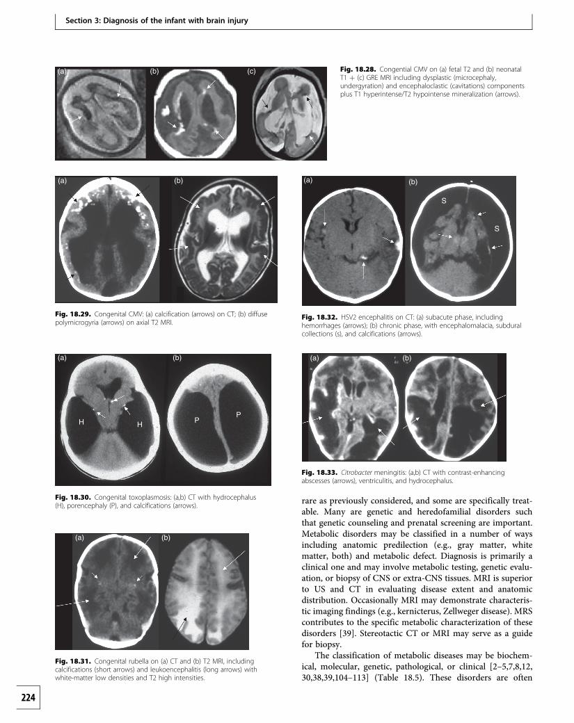
due to TORCH infections (i.e., toxoplasmosis, other [e.g.,syphilis], rubella, cytomegalovirus [CMV], herpes simplexvirus [HSV2], human immunodeficiency virus [HIV]), andneonatal meningitis (e.g., Group B Streptococcus, Listeria,Gram-negative) (Figs. 18.28–18.33). Less common but increas-ingly prevalent causes of subacute and chronic CNS inflam-mation are the granulomatous meningoencephalitic infections(e.g., tuberculosis, spirochete, fungal, parasitic), particularly inimmunocompromised hosts. The imaging pattern is oftenasymmetric and progressive. Such findings may include sub-arachnoid exudate, ventriculitis, edema, cerebritis, infarction,hydrocephalus, effusion, empyema, abscess, and in the chronicphase cystic encephalomalacia, atrophy, gliosis, and calcification.
Recurrent infectious or non-infectious CNS infection (e.g.,meningitis) may require investigation for a “parameningealfocus” (e.g., sinus or mastoid infection, dermal sinus, primitiveneurenteric connection, CSF leak after trauma, dermoid–epidermoid) [1–12]. Brain abscess or empyema may be asso-ciated with Gram-negative meningitis (e.g., Citrobacter) in theneonate. Suppurative collections related to sinus infection,trauma, surgery, sepsis, the immunocompromised state, oruncorrected cyanotic congenital heart disease primarily occurin older children. MRI is the imaging modality of choice fordefinitive evaluation and follow-up. Multiplanar T2, FLAIR,GRE, and DWI sequences are often necessary, along withgadolinium-enhanced T1 images, in order to delineate collec-tions requiring drainage. Contrast-enhanced stereotactic MRIor CT and intraoperative US may provide additional guidancefor surgery.
Plain films or SPECT have been used in the past forscreening of suspected spinal-column infection (discitis, osteo-myelitis) [2,5]. MRI, however, is now preferred for definitivediagnosis, treatment planning, and follow-up. CT may furtherassist in the delineation of bony involvement. MRI is also theprocedure of choice for evaluating spinal neuraxis infection.STIR sequences and fat-suppressed gadolinium-enhancedtechniques are particularly important for demonstratingsuppurative collections (e.g., epidural abscess).
Metabolic, toxic, and neurodegenerativedisordersIn the evaluation of neonatal encephalopathy and develop-mental delay (e.g., static encephalopathy vs. progressiveencephalopathy), MRI is the only modality that can providean accurate assessment of brain maturation based on myelin-ation and cortical development [2–5,7,8,12,30,38,39,104–107](Figs. 18.1–18.4). The clinical hallmark of a metabolic, toxic, orneurodegenerative disorder is “progressive” neurologic impair-ment in the absence of another readily identifiable process.These are to be distinguished from the “non-progressive”encephalopathies, for example, due to maldevelopment,hypoxia–ischemia, or infection. These disorders may beexogenous and internal (e.g., hypoglycemia, hyperbilirubine-mia) or external (e.g., fetal alcohol syndrome). The endogen-ous disorders (e.g., inborn errors of metabolism) are not as
(a) (b)
Fig. 18.27. Right frontal cranial depression (arrows) on CT with(a) soft tissue and (b) bone algorithms.
Chapter 18: Neuroimaging evaluation of pattern and timing
223
rare as previously considered, and some are specifically treat-able. Many are genetic and heredofamilial disorders suchthat genetic counseling and prenatal screening are important.Metabolic disorders may be classified in a number of waysincluding anatomic predilection (e.g., gray matter, whitematter, both) and metabolic defect. Diagnosis is primarily aclinical one and may involve metabolic testing, genetic evalu-ation, or biopsy of CNS or extra-CNS tissues. MRI is superiorto US and CT in evaluating disease extent and anatomicdistribution. Occasionally MRI may demonstrate characteris-tic imaging findings (e.g., kernicterus, Zellweger disease). MRScontributes to the specific metabolic characterization of thesedisorders [39]. Stereotactic CT or MRI may serve as a guidefor biopsy.
The classification of metabolic diseases may be biochem-ical, molecular, genetic, pathological, or clinical [2–5,7,8,12,30,38,39,104–113] (Table 18.5). These disorders are often
(a) (b) (c) Fig. 18.28. Congential CMV on (a) fetal T2 and (b) neonatalT1 þ (c) GRE MRI including dysplastic (microcephaly,undergyration) and encephaloclastic (cavitations) componentsplus T1 hyperintense/T2 hypointense mineralization (arrows).
(a) (b)
Fig. 18.29. Congenital CMV: (a) calcification (arrows) on CT; (b) diffusepolymicrogyria (arrows) on axial T2 MRI.
H H PP
(a) (b)
Fig. 18.30. Congenital toxoplasmosis: (a,b) CT with hydrocephalus(H), porencephaly (P), and calcifications (arrows).
(a) (b)
Fig. 18.31. Congenital rubella on (a) CT and (b) T2 MRI, includingcalcifications (short arrows) and leukoencephalitis (long arrows) withwhite-matter low densities and T2 high intensities.
(a) (b)
S
S
Fig. 18.32. HSV2 encephalitis on CT: (a) subacute phase, includinghemorrhages (arrows); (b) chronic phase, with encephalomalacia, subduralcollections (s), and calcifications (arrows).
(a) (b)
Fig. 18.33. Citrobacter meningitis: (a,b) CT with contrast-enhancingabscesses (arrows), ventriculitis, and hydrocephalus.
Section 3: Diagnosis of the infant with brain injury
224

Table 18.5. Metabolic, toxic, and neurodegenerative disorders
Lysosomal disorders
Lipidoses
Fabry, Gaucher's and Niemann–Pick disease
GM1 gangliosidosis
GM2 gangliosidosis (Tay–Sachs andSandhoff diseases)
Neuronal ceroid lipofuscinosis
Mucopolysaccharidoses (MPS)
Hurler, Scheie, Hurler–Scheie
Hunter
Sanfilippo A–D
Morquio A & B
Matoreaux-Lamy
Sly
Mucolipidoses
Mannosidosis, Fucosidosis, Sialidosis
Lysosomal leukodystrophies
Metachromatic leukodystrophy
Globoid cell leukodystrophy (Krabbe)
Peroxisomal disorders
Adrenoleukodystrophy complex
Neonatal leukodystrophy
Zellweger syndrome
Infantile Refsum's syndrome
Rhizomelic chondrodysplasia punctata
Hyperpipecolic acidemia
Cerebrotendinous xanthomatosis
Other leukodystrophies
Pelizaeus–Merzbacher disease
Canavan's disease
Alexander's disease
Cockayne's syndrome
Leukodystrophy with calcifications
Mitochondrial (respiratory oxidative) disorders
Leigh's disease
Kearns–Sayre syndrome
MELAS syndrome
MERRF syndrome
Alper's syndrome (poliodystrophy)
Menkes' disease (trichopoliodystrophy)
Marinesco-Sjogren syndrome
Infantile bilateral striatal necrosis
Lebers hereditary optic atrophy
L-Carnitine deficiency
Amino acid disorders
Phenylketonuria
Homocystinuria
Non-ketotic hyperglycinemia
Maple syrup urine disease
Glutaric aciduria, type I
Glutaric aciduria, type II
Methylmalonic and propionic acidurias
Urea cycle defects (e.g., OTC deficiency)
Oculocerebrorenal syndrome
Pyridoxine dependency
Carbohydrate and other storage disorders
Galactosemia
Glycogen storage diseases (i.e. Pompe)
Carbohydrate-deficient glycoproteinsyndrome
Niemann-Pick
Gaucher
Farber
Infantile sialidosis
Liver metabolic disorders
Wilson's disease (hepatolenticular degeneration)
PKAN
Hyperbilirubinemia (see toxic encephalopathies)
Hepatocerebral syndromes (see toxicencephalopathies)
Diseases of the cerebellum, brainstem, and spinal cord
Friedreich's ataxia
Olivopontocerebellar atrophies
Ataxia–telangiectasia
Carbohydrate-deficient glycoprotein syndrome
Infantile neuraxonal dystrophy
Other metabolic and neurodegenerative diseases
Juvenile multiple sclerosis
Molybdenum cofactor deficiency
3-Hydroxy-3-methylglutaryl-coenzyme A lyasedeficiency
Idiopathic leukoencephalopathy
Diseases of the basal ganglia
Sulfite oxidase deficiency
Parathyroid disease
Tuberous sclerosis
Down's syndrome
Progressive encephalopathy with basalganglia calcifications and CSF lymphocytosis
Inflammatory, toxic, and anoxic conditions
Radiation therapy
Renal tubular acidosis and osteoporosis
Huntington's disease
Fahr's disease
PKAN
Cockayne
Wilson
Toxic encephalopathies
Exogenous internal toxicities
Hyperbilirubinemia
Hepatocerebral syndromes
Hypoglycemia
Hypothermia and hyperthermia
Paraneoplastic toxins
Hemolytic uremic syndrome
Uremia
Ion imbalance disorders
Endocrinopathies
Porphyria
Exogenous external toxicities
Vitamin deficiencies/depletions
Vitamin B1
Folate
Vitamin B12
Biotin
Vitamin K
Vitamin C
Vitamin D
Toxins
Mercury poisoning
Methanol
Toluene
Carbon monoxide
Cyanides and sulfides
Lead
Alcohol
Cocaine and heroin
Anticonvulsants
Drug-induced
Methotrexate
Cyclosporine
Tacrolimus
Carmustine, cytosine arabiniside
Note:See references [2,4,5,106,107].
Chapter 18: Neuroimaging evaluation of pattern and timing
225

categorized according to the metabolic defect. Such a classifica-tion includes the lysosomal disorders (e.g., Krabbe), peroxisomaldefects (e.g., Zellweger), the mitochondrial disorders (e.g.,Leigh, Menkes), organic and aminoacidopathies (e.g., non-ketotic hyperglycinemia), disorders of carbohydrate metabol-ism (e.g., glycogen storage disease), liver metabolic disorders,and miscellaneous. Certain clinical features that assist indirecting the initial evaluation of these patients may alsoprovide a basis for classification (e.g., macrocephaly in maplesyrup urine disease). The ideal radiological classificationwould categorize the diseases by the anatomic distribution ofthe pathological process using CT and MRI. Unfortunately,most of these conditions affect multiple sites, and considerableoverlap in appearance is found. However, a practical imagingclassification may be based on the predominant areas ofinvolvement including the white matter (subcortical, periven-tricular), gray matter (cortical, deep), basal ganglia, brainstem,cerebellum, spinal cord, and peripheral nervous system[2,5,12,7,8,12,39, 104–107].
Disorders primarily affecting cortical gray matterEndogenous metabolic disorders which primarily, or predomi-nantly, affect the cortical gray matter include the storage dis-eases that result from lysosomal enzyme defects [2,5,39,107].However, these findings are often non-specific, and thedifferential diagnosis may include diffuse cortical atrophydue to any number of causes. Other considerations includethe end stage of a static encephalopathy (e.g., post-HIE orpost-infection), or “atrophy” related to chronic systemic dis-ease, malnutrition, or certain types of therapy (e.g., steroids).
Disorders primarily affecting deep gray matterMetabolic disorders may primarily involve the deep graymatter (including mineralization) [2,5,39,107,110–113]. Thosedisorders primarily involving the corpus striatum (i.e., caudateand putamen) include the mitochondrial disorders, organicand aminoacidopathies, juvenile Huntington's disease, Wilsondisease, and Cockayne's syndrome. Those disorders primarilyinvolving the globus pallidus include the aminoacidopathies,hyperbilirubinemia, pentothenate kinase associated neuro-degeneration (PKAN, formerly Hallervorden–Spatz disease),and toxic exposure (e.g., carbon dioxide) (Figs. 18.34, 18.35).It is unusual to see isolated involvement of the thalami in anyof the metabolic disorders. However, thalamic involvementmay be an early or dominant feature of Krabbe disease or
GM2 gangliosidosis. It may also be seen in the infantile formof Leigh disease along with extensive brainstem, basal ganglia,and cerebral white-matter involvement. In general, the differen-tial diagnosis, depending on the clinical picture and timing ofthe imaging, may also include profound HIE, hypoglycemia,toxic exposure (e.g., methane, cyanide), osmolar myelinolysis,striatal necrosis, and meningoencephalitis.
Disorders primarily affecting white matterThose disorders which primarily, or predominantly, affectthe white matter are known as the leukoencephalopathies[2,5,39,107–109]. Traditionally, leukoencephalopathies havebeen divided into dysmyelinating and myelinoclastic dis-orders. In dysmyelinating disorders an intrinsic (inherited)enzyme deficiency results in the disturbed formation, destruc-tion, or turnover of the essential components of myelin. Theyare also referred to as the leukodystrophies. The pattern ofdamage is symmetrical in both hemispheres, has diffusemargins, often spares the arcuate fibers, and consistentlyinvolves the cerebellar white matter. The leukodystrophiesare primarily associated with the lysosomal and peroxisomaldisorders (e.g., metachromatic leukodystrophy, Krabbe leuko-dystrophy, and the adrenoleukodystrophy complex [ALD]),and with diseases of white matter (e.g., Pelizaeus–Merzbacher,Canavan, Alexander, and Cockayne). Included in the differen-tial diagnosis is infantile-onset leukoencephalopathy with swell-ing (macrocephaly) and mild clinical course. Early centralwhite-matter involvement may suggest Krabbe (also, abnormalthalami), ALD, phenylketonuria, maple syrup urine disease(MSUD: Fig. 18.34), or Lowe syndrome. The lack of myelination
(a) (b)
Fig. 18.34. Maple syrup urine disease in neonate with globus pallidus(short arrows) and white matter (long arrows) edema on (a) axial CT and(b) axial T2 MRI.
(a) (b) (c) Fig. 18.35. Bilirubin encephalopathy and kernicterus on MRIwith (a,b) globus pallidus and subthalamic T1 hyperintensity(arrows) in subacute phase, and (c) T2 hyperintensity (arrows)plus atrophy in chronic phase.
Section 3: Diagnosis of the infant with brain injury
226

(hypomyelination) may suggest Pelizaeus–Merzbacher diseaseor Menkes' disease. In myelinoclastic disorders, the myelinsheath is intrinsically normal until it yields to exogenous orendogenous myelinotoxic factors. The pattern of damage isasymmetric, is sharply demarcated, irregularly involves thesubcortical arcuate fibers, and may spare the cerebellum.Examples are the infectious and postinfectious demyelinatingdiseases (e.g., TORCH, HIV, SSPE, ADEM) and the vasculi-tides (e.g., lupus). Non-specific white-matter abnormalitiesmay be seen with a variety of metabolic, neurodegenerative,infectious, postinfectious, toxic, and vascular processes. Inthis situation, the clinical findings must be relied upon. Animportant example is posterior reversible leukoencephalopathy(e.g., hypertension, transplant, cyclosporine, renal disease).Also, it is important to remember that the most common causesof cerebral white-matter abnormalities (particularly periventri-cular) and prominent Virchow–Robin spaces in children withdevelopmental delay are the static leukoencephalopathies
(e.g., maldevelopmental, undermyelination, postinflammatory,postischemic, idiopathic).
Disorders affecting both white matter and cortical gray matterA number of metabolic disorders involve both gray and whitematter [2,5,39,107–109]. Those disorders associated with cor-tical atrophy along with white-matter involvement includelysosomal disorders such as the lipidoses and mucopolysac-charidoses (also, associated skeletal dysplasia), and mitochon-drial disorders such as Alper disease and Menkes' disease.If there is a diffuse cortical dysgenesis (e.g., lissencephaly,polymicrogyria) associated with white-matter abnormalities,then peroxisomal disorders such as Zellweger syndromeshould be considered along with congenital infections (e.g.,cytomegaloviral), and the congenital muscular dystrophies(e.g., Fukuyama, Walker–Warburg, Santavuori) (Fig. 18.36).
In hypoglycemia, there is predominant involvement of theparieto-occipital gray and white matter, and especially theprimary visual cortex (Fig. 18.37). This pattern is to be distin-guished from a predominant posterior border-zone HIE andfrom dural venous sinus thrombosis primarily in the distribu-tion of the straight sinus, posterior superior sagittal sinus, and/or the inferior sagittal sinus.
Disorders affecting both white matter and deep gray matterDisorders associated with deep gray-matter involvement, inaddition to white-matter abnormalities, include those with pri-marily corpus striatum involvement (Leigh [Fig. 18.38], MELAS,Wilson's, Cockayne), those with predominant thalamic abnor-malities (Krabbe, GM2 gangliosidoses), and those with primarilyglobus pallidus involvement (Canavan, MSUD, methylmalonic/propionic acidopathy, Kearns–Sayre) [2,5,39,107–113]. Sulfiteoxidase deficiency also involves the basal ganglia and whitematter and can mimic HIE [114] (Fig. 18.39). Included in
(a) (b)
D H H
Fig. 18.36. Walker–Warburg with Dandy–Walker malformation (D) on(a) sagittal T1 plus (b) cobblestone lissencephaly (arrows) with extensivewhite-matter dysplasia and hydrocephalus (H) on axial T2 MRI.
(a) (b) (c)
(d) (e)
Fig. 18.37. Hypoglycemia with parieto-occipitalinvolvement (arrows) in the subacute phase withedema as (a) low density on CT, and as highintensity on (b) DWI and (c) T2 MRI; also (d,e) thechronic phase with atrophy and gliosis on T2 MRI.
Chapter 18: Neuroimaging evaluation of pattern and timing
227

the differential diagnoses, depending on the clinical context, areprofound HIE, osmolar myelinolysis, bilirubin encephalopathy(kernicterus), toxic exposure, and infectious or postinfectiousprocesses (e.g., TORCH, HIV, ADEM).
MR spectroscopy (MRS) in metabolic disordersProton (hydrogen-1) MRS using both short and long TEacquisitions (e.g., TEs 35, 144) is being increasingly usedclinically to evaluate brain development and maturation, aswell as patients with metabolic and other disorders [12,38,39].The normal MR brain spectra show an evolution from theimmature infantile pattern (e.g., decreased N-acetyl-aspartate[NAA] relative to choline [Ch]) to the mature, adult pattern(e.g., increased NAA to Ch) (Fig. 18.3). Although many ofthe metabolic and neurodegenerative disorders have specificbiochemical markers, most of the disorders have no differen-tiating features. Non-specific MRS abnormalities are thosethat reflect brain destruction and reactive changes includingdelayed maturation, neuronal loss, axonal degeneration,demyelination, and gliosis. Alterations in metabolites are oftendisplayed as a ratio relative to the reference metabolite, creat-ine (Cr), an energy marker. In disorders in which there ispredominant neuronal degeneration (i.e., loss of cell bodies,axons) and atrophy, or oligodendroglial loss, the major MRSfinding is a decrease in N-acetyl-aspartate (NAA), a neuronal(neurons including axons) and immature oligodendroglial
marker. In disorders in which there is predominant loss ofmyelin sheaths with secondary axonal degeneration and gliosis(e.g., demyelination), the characteristic spectral abnormalitiesare characterized by elevated lipids, a marker for myelindestruction; elevated choline (Ch), a marker of membraneturnover (e.g., myelin, glial); variable increases in lactate (L),a marker of anaerobic glycolysis; elevated glutamate/glutam-ine (Glx), neuroexcitatory amino acid markers; and elevatedmyoinositols (mI), also an osmolyte and glial marker. Associ-ated neuronal (e.g., axonal) or oligodendroglial damage isindicated by a decrease in NAA.
More specific MRS abnormalities may be seen in a numberof disorders [39]. Abnormal MRS spectra have been reportedwith some of the lysosomal defects such as Niemann–Pickdisease (abnormal lipid peak at 1.2 ppm), the mucopolysac-charidoses (decreased NAA late), and metachromatic leuko-dystrophy (decreased NAA, Ch, and Cr; increased mI and L).MRS abnormalities have been observed with a number ofthe peroxisomal disorders including adrenoleukodystrophy(decreased NAA with increased Ch, Glx, mI, lipids, and L)and Zellweger syndrome (decreased NAA with increasedlipids and Glx). Other leukodystrophies associated withobserved MRS findings are Canavan disease (increased NAAwith decreased Ch and Cr plus increased mI and L), Alexanderdisease (decreased NAA with increased L), and Pelizaeus–Merzbacher disease (normal early; decreased NAA andincreased Ch late). Primary and secondary disorders of energymetabolism have been associated with MRS findings ofdecreased NAA and increased L, including mitochondrialdisorders such as Leigh disease and MELAS (Fig. 18.38).Similar findings, however, are present with acute/subacutehypoxia–ischemia (plus elevated Glx, decreased Cr, andincreased lipids) (Fig. 18.18). Aminoacidopathies with report-edly abnormal spectra include phenylketonuria (increasedphenylalanine peak at 7.37 ppm), maple syrup urine disease(abnormal peak at 0.9 ppm), and non-ketotic hyperglycinemia(elevated glycine peak at 3.55 ppm). Other metabolic disordersassociated with abnormal MRS findings include the creatinedeficiencies (decreased, absent Cr), hepatic encephalopathy(increased Glx with decreased inositols and Ch), and hyper-osmolar states (increased inositols, Cr, and Ch). Neoplasticprocesses characteristically show elevated Ch/Cr, decreasedNAA/Cr, and decreased NAA/Cr ratios. Inflammatoryprocesses may be differentiated from neoplastic and otherprocesses by the suppressed mI peak [12].
SummaryUS and CT may provide important screening information,particularly with regard to hemorrhage, trauma, hydrocepha-lus, infection, and gross macrostructural anomalies. However,current and advanced MRI techniques provide more definitivemacrostructural, microstructural, and functional imaginginformation in both the early and late assessment of fetaland neonatal CNS injuries.
(a) (b)Ch
L
Cr
NA
Fig. 18.38. Leigh syndrome with basal-ganglia lesions (arrows) andwhite-matter involvement: (a) axial T2 MRI; (b) lactate doublet (L) on MRS.
(a) (b)
Fig. 18.39. Sulfite oxidase deficiency with thalamic (short arrows) anddiffuse white-matter involvement (long arrows) on (a) axial T1 and (b) T2 MRI.
Section 3: Diagnosis of the infant with brain injury
228

References1. Blankenburg FG, Barnes PD. Structural
and functional imaging of hypoxic–ischemic injury (HII) in the fetal andneonatal brain. In Stevenson D, Benitz W,Sunshine P, eds., Fetal and Neonatal BrainInjury, 3rd edn. Cambridge: CambridgeUniversity Press, 2003: 446–89.
2. Barnes P. State of the art: neuroimagingand the timing of fetal and neonatalbrain injury. J Perinatol 2001; 21: 44–60.
3. Winkler P, Zimmerman RA. PerinatalBrain Injury. Neuroimaging: Clinical andPhysical Principles. New York, NY:Springer, 2000: 531–83.
4. Barkovich A. Pediatric Neuroimaging,4th edn. Philadelphia, PA: Lippincott-Raven, 2005: 190–290.
5. Volpe JJ. Neurology of the Newborn, 4thedn. Philadelphia, PA: Saunders, 2001.
6. Ment L, Bada H, Barnes P, et al. Practiceparameter: neuroimaging of the neonate.Neurology 2002; 58: 1726–38.
7. Ferriero D. Neonatal brain injury.N Engl J Med 2004; 351: 1985–95.
8. Barkovich A. MR imaging of theneonatal brain. Neuroimaging ClinN Am 2006; 16: 117–36.
9. Rutherford M, Srinivasan L, Dyet L,et al. MRI in perinatal brain injury.Pediatr Radiol 2006; 36: 582–92.
10. Zimmerman R, Bilaniuk L.Neuroimaging evaluation of cerebralpalsy. Clin Perinatol 2006; 33: 517–44.
11. Triulzi F, Parazzini C, Righini A.Patterns of damage in the matureneonatal brain. Pediatr Radiol 2006;36: 608–20.
12. Panigrahy A, Blumel S. Advances in MRneuroimaging techniques in theevaluation of neonatal encephalopathy.Top Magn Reson Imaging 2007; 18: 3–30.
13. Barnes PD, Taylor GA. Imaging ofthe neonatal central nervous system.Neurosurgery Clin North Am 1998;1: 17–48.
14. Allison JW, Seibert JJ. TranscranialDoppler in the newborn with asphyxia.Neuroimaging Clin N Am 1999; 9: 11–16.
15. Blankenberg FG, Loh N, Bracci P, et al.Sonography, CT, and MR imaging: aprospective comparison of neonateswith suspected intracranial ischemia andhemorrhage. AJNR Am J Neuroradiol2000; 21: 213–18.
16. Debillon T, N'Guyen, Muet A, et al.Limitations of ultrasonography fordiagnosing white matter damage in
preterm infants. Arch Dis Child FetalNeonatal Ed 2003; 88: F275–9.
17. Laptook A, O'Shea M, Shankaran S, et al.Adverse neurodevelopmental outcomesamong extremely low birth weightinfants with normal head ultrasound.Pediatrics 2005; 115: 673–80.
18. Pinto-Martin JA, Riolo S, Cnaan A,et al. Cranial ultrasound prediction ofdisabling and nondisabling cerebralpalsy at age two in a low birthweightpopulation. Pediatrics 1995; 95: 249–54.
19. Maalouf EF, Duggan PJ, Counsell SJ,et al. Comparison of findings on cranialultrasound and magnetic resonanceimaging in preterm neonates. Pediatrics2001; 107: 719–27.
20. Sie LT, van der Knapp MD, van Wezel-Meijler G, et al. Early MR features ofhypoxic-ischemic brain injury inneonates with periventricular densitieson sonograms. AJNR Am J Neuroradiol2000; 21: 852–61.
21. Childs AM, Cornette L, RomenghiLA, et al. Magnetic resonance andcranial ultrasound characteristicsof periventricular white matterabnormalities in newborn infants.Clin Radiol 2001; 56: 647–55.
22. Roelants-van Rijn AM, Groenendaal F,Beek FJ, et al. Parenchymal brain injuryin the preterm infant: comparisonof cranial ultrasound, MRI andneurodevelopmental outcome.Neuropediatrics 2001; 32: 80–9.
23. Inder TE, Anderson NJ, Spencer C, et al.White matter injury in the prematureinfant: a comparison between serialcranial sonographic and MR findingsat term. AJNR Am J Neuroradiol 2003;24: 805–9.
24. Miller SP, Cozzio C, Goldstein RB, et al.Comparing the diagnosis of white matterinjury in premature newborns withserial MR imaging and transfontanelultrasonography findings. AJNRAm J Neuroradiol 2003; 24: 1661–9.
25. Hintz SR, Slovis T, Bulas D, et al.Interobserver reliability and accuracy ofcranial ultrasound scanninginterpretation in premature infants.J Pediatr 2007; 150: 592–6.
26. Mirmiran M, Barnes P, Keller K, et al.Neonatal brain MRI before discharge isbetter than serial cranial ultrasound inpredicting cerebral palsy in very lowbirth weight preterm infants. Pediatrics2004; 114: 992–8.
27. Slovis T. Children, computedtomography radiation dose, and the
As Low As Reasonably Achievable(ALARA) concept. Pediatrics 2003;112: 971–2.
28. Vertinzksy A, Barnes P. Macrocephaly,increased intracranial pressure, andhydrocephalus in the infant and youngchild. Top Magn Reson Imaging 2007;18: 31–52.
29. Barnes P, Krasnokutsky M. Imagingof the CNS in suspected or allegednonaccidental injury, including themimics. Top Magn Reson Imaging 2007;18: 53–74.
30. Mukherjee P. Advanced pediatricimaging. Neuroimaging Clin N Am 2006;16(1).
31. Levine D, Barnes PD, Robertson RR,et al. Fast MR imaging of fetal centralnervous system abnormalities. Radiology2003; 229: 51–61.
32. Garel C, Delezide A, Elmaleh-Berges M,et al. Contribution of fetal MRI in theevaluation of cerebral ischemic lesions.AJNR Am J Neuroradiol 2004; 25:1563–1568.
33. Levine D, Barnes P. MR imaging offetal CNS abnormalities. In Levine D,ed., Atlas of Fetal MRI. Boca Raton,FL: Taylor & Francis Group, 2005:25–72.
34. Griffiths P, Widjaja E, Paley M, et al.Imaging of the fetal spine in utero:diagnostic accuracy and impact onmanagement. Pediatr Radiol 2006;36: 927–33.
35. Garel C. New advances in fetal MRneuroimaging. Pediatr Radiol 2006;36: 621–5.
36. Glenn O, Barkovich J. Magneticresonance imaging of the fetal brain andspine. Part 1. AJNR Am J Neuroradiol2006; 27: 1604–11; Part 2. AJNR AmJ Neuroradiol 2006; 27: 1807–14.
37. Barkovich AJ, Baranski K, Vigneron D,et al. Proton MR spectroscopy in theevaluation of asphyxiated term neonates.AJNR Am J Neuroradiol 1999; 20:1399–405.
38. Vigneron DB. Magnetic resonancespectroscopic imaging of human braindevelopment. Neuroimag Clin N Am2006; 16; 75–86.
39. Cecil K. MR spectroscopy of metabolicdisorders. Neuroimaging Clin N Am2006; 16: 87–116.
40. Barkovich A, Miller S, Bartha A. MRimaging, MR spectroscopy, and diffusiontensor imaging of sequential studies inneonates with encephalopathy. AJNRAm J Neuroradiol 2006; 27: 533–47.
Chapter 18: Neuroimaging evaluation of pattern and timing
229

41. Wang J, Licht D. Pediatric perfusionMRI using arterial spin labeling.Neuroimaging Clin N Am 2006;16: 149–68.
42. Huppi PS, Maier SE, Peled S, et al.Microstructural development of humannewborn cerebral white matter assessedin vivo by diffusion tensor magneticresonance imaging. Pediatr Res 1998;44: 584–90.
43. Neil JJ, Shiran SI, McKinstry RC.Normal brain in human newborns:apparent diffusion coefficient anddiffusion anisotropy measured by usingdiffusion tensor MR imaging. Radiology1998; 209: 57–66.
44. Johnson AJ, Lee BCP, Lin W.Echoplanar diffusion-weighted imagingin neonates and infants with suspectedhypoxic–ischemic injury. AJNR AmJ Neuroradiol 1999; 172: 219–26.
45. Robertson RL, Ben-Sira L, Barnes PD,et al. MR line scan diffusion imagingof term neonates with perinatal brainischemia. AJNR Am J Neuroradiol 1999;20: 1658–70.
46. Phillips MD, Zimmerman RA. Diffusionimaging in pediatric hypoxic-ischemicinjury. Neuroimaging Clin N Am 1999;9: 41–52.
47. Inder T, Huppi PS, Zientara GP, et al.Early detection of periventricularleukomalacia by diffusion-weightedmagnetic resonance imaging techniques.J Pediatr 1999; 134: 631–4.
48. Inder TE, Huppi PS, Warfield S, et al.Periventricular white matter injury inthe premature infant followed byreduced cerebral cortical gray mattervolume at term. Ann Neurol 1999; 46:755–60.
49. Huppi PS, Murphy B, Maier SE, et al.Microstructural brain development afterperinatal cerebral white matter injuryassessed by diffusion tensor MR imaging.Pediatrics 2001; 107: 455–60.
50. Arzoumanian Y, Mirmiran M, Barnes P,et al. Diffusion tensor brain imagingfindings at term-equivalent age maypredict neurologic abnormalities in lowbirth weight preterm infants. AJNRAm J Neuroradiol 2003; 24: 1646–53.
51. Counsell S, Allsop J, Harrison M, et al.Diffusion-weighted imaging of the brainin preterm infants with focal and diffusewhite matter abnormality. Pediatrics2003; 112: 1–7.
52. Sagar P, Grant PE. Diffusion-weightedimaging: pediatric clinical applications.
Neuroimaging Clin N Am 2006;16: 45–74.
53. Mukherjee P, McKinstry R. Diffusiontensor imaging and tractographyof human brain development.Neuroimaging Clin N Am 2006;16: 19–44.
54. van der Knaap M, Valk J. Classificationof congenital abnormalities of theCNS. AJNR Am J Neuroradiol 1988;9: 315–26.
55. Barkovich AJ, Truwit CL. Brain damagefrom perinatal asphyxia: correlation ofMR findings with gestational age. AJNRAm J Neuroradiol 1990; 11: 1087–96.
56. Aida N, Nishimura G, Hachiya Y, et al.MR imaging of perinatal brain damage:comparison of clinical outcome withinitial follow-up MR findings. AJNRAm J Neuroradiol 1992; 19: 1909–22.
57. Barkovich A, Hallam D. Neuroimagingin perinatal hypoxic–ischemic injury.Ment Retard Dev Disabil Res Rev 1997;3: 28–41.
58. Cowan F, Rutherford M, Groenendaal F,et al. Origin and timing of brainlesions in term infants with neonatalencephalopathy.Lancet 2003; 361: 736–42.
59. Miller S, Ramaswamy V, Michelson D,et al. Patterns of brain injury in termneonatal encephalopathy. J Pediatr 2005;146: 453–60.
60. Lupton BA, Hill A, Roland EH, et al.Brain swelling in the asphyxiated termnewborn: pathogenesis and outcome.Pediatrics 1988; 82: 139–46.
61. Vannucci RC, Christensen MA, Jager JY.Nature, time-course, and extent ofcerebral edema in perinatal hypoxic–ischemic brain damage. Pediatr Neurol1993; 9: 29–34.
62. Pinto-Martin JA, Riolo S, Cnaan A,et al. Cranial ultrasound predictionof disabling and nondisabling cerebralpalsy at age two in a low birthweightpopulation. Pediatrics 1995; 95: 249–54.
63. Goetz MC, Gretebeck RJ, Oh KS, et al.Incidence, timing and follow-up ofperiventricular leukomalacia. AmJ Perinatol 1995; 12: 325–7.
64. Valkama AM, Pääkkö ELE, VainionpääLK, et al. Magnetic resonance imagingat term and neuromotor outcome inpreterm infants. Acta Paediatr 2000;89: 348–55.
65. Perlman JM, Rollins N. Surveillanceprotocol for the detection of intracranialabnormalities in premature neonates.ArchPediatr Adolesc Med 2000; 154: 822–6.
66. Volpe JJ. Neurobiology of periventricularleukomalacia in the premature infant.Pediatr Res 2001; 50: 553–62.
67. Panigrahy A, Barnes PD, Robertson RL,et al. Volumetric brain differences inchildren with periventricular T2-signalhyperintensities: a grouping bygestational age at birth. AJR AmJ Roentgenol 2001; 177: 695–702.
68. Miller SP, Hoque NN, Piecuch RE, et al.The spectrum of cerebellar hemorrhagein premature newborns. Pediatr Res2003; 53: 537A (abst #3040).
69. Austin NC, Woodward L, Spencer C,et al. Neurodevelopmental outcome atone year in a regional cohort of very lowbirth weight infants: correlation withMRI at term. Pediatr Res 2003; 53: 398A(abst #2253).
70. Volpe JJ. Cerebral white matter injuryof the premature infant: more commonthan you think. Pediatrics 2003; 112:176–80.
71. Inder TE, Wells SJ, Mogridge NB, et al.Defining the nature of the cerebralabnormalities in the premature infant: aqualitative magnetic resonance imagingstudy. J Pediatr 2003; 143: 171–9.
72. Panigrahy A, Barnes PD, Robertson RL,et al. Quantitative analysis of the corpuscallosum in children with cerebral palsyand developmental delay: correlationwith cerebral white matter volume.Pediatr Radiol 2005; 35: 1199–207.
73. Bodensteiner J, Johnsen S. MRI findingsin children surviving extremelypremature delivery and extremely lowbirthweight with cerebral palsy. J ChildNeurol 2006; 21: 743–7.
74. Dyet L, Kennea N, Counsell S, et al.Natural history of brain lesions inextremely preterm infants studied withserial MRI from birth andneurodevelopmental assessmentPediatrics 2006; 118: 536–48.
75. Khwaja O, Volpe J. Pathogenesis ofcerebral white matter injury ofprematurity. Arch Dis Child FetalNeonatal Ed 2008; 93: F153–61.
76. Barkovich AJ. MR and CT evaluationof profound neonatal and infantileasphyxia. AJNR Am J Neuroradiol 1992;13: 959–72.
77. Barkovich AJ, Sargent SK. Profoundasphyxia in the premature infant:imaging findings. AJNR AmJ Neuroradiol 1995; 16: 1837–46.
78. Barkovich AJ, Westmark K, Partridge C,et al. Perinatal asphyxia: MR findings in
Section 3: Diagnosis of the infant with brain injury
230

the first 10 days. AJNR Am J Neuroradiol1995; 16: 427–39.
79. Roland EH, Poskitt K, et al. Perinatalhypoxic–ischemic thalamic injury:clinical features and neuroimaging. AnnNeurol 1998; 44: 161–6.
80. Sargent M, Poskitt K, Roland E, et al.Cerebellar vermian atrophy afterneonatal hypoxic–ischemicencephalopathy. AJNR Am J Neuroradiol2004; 25: 1008–15.
81. Boichot C, Walker P, Durand C, et al.Term neonate prognosis after perinatalasphyxia: contributions of MR imaging,MR spectroscopy, relaxation times, andADCs. Radiology 2006; 239: 839–48.
82. Shankaran S, Laptook A, Ehrenkranz R,et al. Whole-body hypothermia forneonates with hypoxic–ischemicencephalopathy. N Engl J Med 2005; 353:1574–84.
83. Bulas D, Glass P. Neonatal ECMO:neuroimaging and neurodevelopmentaloutcome. Semin Perinatol 2005; 29: 58–65.
84. Zuerrer M, Martin E, Boltshauser E.MRI of intracranial hemorrhage inneonates and infants at 2.35 tesla.Neuroradiology 1991; 33: 223–9.
85. DeVeber G, Andrew M, Adams C,et al. Cerebral sinovenous thrombosisin children. N Engl J Med 2001; 345:417–23.
86. Carvalho K, Bodensteiner J, Connolly P,et al. Cerebral venous thrombosisin children. J Child Neurol 2001;16: 574–85.
87. Lynch J, Hirtz D, deVeber G, et al.Report of the National Institute ofNeurologic Disorders and StrokeWorkshop on Perinatal and ChildhoodStroke. Pediatrics 2002; 109: 116–23.
88. DeVeber G. Arterial ischemic strokes ininfants and children: an overview ofcurrent approaches. Semin ThrombosisHemostasis 2003; 29: 567–73.
89. Sebire G, Tabarki B, Saunders DE, et al.Cerebral venous thrombosis in children:risk factors, presentation, diagnosis, andoutcome. Brain 2005; 128: 477–89.
90. Kirton A, deVeber G. Cerebral palsysecondary to perinatal ischemic stroke.Clin Perinatol 2006; 33: 367–86.
91. Fitzgerald K,Williams LS, Garg BP, et al.Cerebral sinovenous thrombosis in theneonate. Arch Neurol 2006; 63: 405–9.
92. Barnes C, deVeber G. Prothromboticabnormalities in childhood ischaemicstroke. Thromb Res 2006; 118: 67–74.
93. Ehtisham A, Stern B. Cerebral venousthrombosis: a review. Neurologist 2006;12: 32–8.
94. Huang A, Robertson R. Spontaneoussuperficial parenchymal andleptomeningeal hemorrhage in termneonates. AJNR Am J Neuroradiol 2004;25: 469–75.
95. Govaert P, Vanhaesebrouck P, dePraeter C. Traumatic neonatalintracranial bleeding and stroke. ArchDis Child 1992; 67: 840–5.
96. Castillo M, Fordham LA. MR ofneurologically symptomatic newbornsafter vacuum extraction delivery. AJNRAm J Neuroradiol 1995; 16: 816–18.
97. Odita JC, Hebi S. CT and MRIcharacteristics of intracranialhemorrhage complicating breech andvacuum delivery. Pediatr Radiol 1996;26: 782–5.
98. Kleinman PK, Barnes PD. Headtrauma. In Kleinman PK, ed., Imagingof Child Abuse, 2nd edn. St Louis, MO:Mosby-Year Book, 1998.
99. Alexander J, Leveno K, Hauth J, et al.Fetal injury associated with cesareandelivery. Obstet Gynecol 2006; 108:885–90.
100. Doumouchtsis S, Arulkumaran S. Headtrauma after instrumental births. ClinPerinatol 2008; 35: 69–83.
101. Teixeira J, Zimmerman R, HaselgroveJ, et al. Diffusion imaging in pediatriccentral nervous system infections.Neuroradiology 2001; 43: 1031–9.
102. Jan W, Zimmerman R, Bilaniuk L, et al.Diffusion-weighted imaging in acutebacterial meningitis in infancy.Neuroradiology 2003; 45: 634–9.
103. DeVries L, Verboon-Maciolek M,Cowan F, et al. The role of cranial USand MRI in the diagnosis of infectionsof the central nervous system. EarlyHum Dev 2006; 82: 819–25.
104. Hoon AH. Neuroimaging in cerebralpalsy: patterns of brain dysgenesisand injury. J Child Neurol 2005;12: 936–9.
105. Rodriguez D, Young Poussaint T.Neuroimaging of the child withdevelopmental delay. Top Magn ResonImaging 2007; 18: 75–92.
106. Valk J, van der Knapp MS. Toxicencephalopathy. AJNR AmJ Neuroradiol 1992; 13: 747–60.
107. van der Knaap MS, Valk J. MagneticResonance of Myelination & MyelinDisorders, 3rd edn. New York, NY:Springer, 2005.
108. Barker P, Horska A. Neuroimaging inleukodystrophies. J Child Neurol 2004;19: 559–70.
109. Vanderver A. Tools for diagnosis ofleukodystrophies and other disorderspresenting with white matter disease.Curr Neurol Neurosci Rep 2005; 5:110–18.
110. Murakami Y, Yamashita Y, MatsuishiT, et al. Cranial MRI of neurologicallyimpaired children suffering fromneonatal hypoglycemia. Pediatr Radiol1999; 29: 23–7.
111. Alkalay A, Flores-Sarnat L, Sarnat H,et al. Brain imaging findings inneonatal hypoglycemia. Clin Pediatr2005; 44: 783–90.
112. Barkovich A, Ali F, Rowley H,et al. Imaging patterns of neonatalhypoglycemia. AJNR Am J Neuroradiol1998; 19: 523–8.
113. Govaert P, Lequin M, Swarte R, et al.Changes in globus pallidus with (pre)term kernicterus. Pediatrics 2003; 112:1256–63.
114. Dublin A, Hald J, Wootton-Gorges S.Isolated sulfite oxidase deficiency:MR imaging features. AJNR AmJ Neuroradiol 2002; 23: 484–5.
Chapter 18: Neuroimaging evaluation of pattern and timing
231