chapter chemical and physical characteristics of the principal mycotoxins · · 2016-10-26chapter...
TRANSCRIPT

Chapter 2. Chemical and physical characteristics of the principal mycotoxins 31
chapter 2.
Chemical and physical characteristics of the principal mycotoxins
Summary
This chapter provides information about the chemical and physical properties of the mycotoxins consid-ered in this book: aflatoxins; fumonisins; ochratoxin A; trichothecenes, espe-cially deoxynivalenol and nivalenol; zearalenone; and ergot alkaloids. This information about structures reveals the chemical diversity of mycotoxins, which is relevant to the wide range of toxicological effects in animals and humans discussed later in the book.
1. Aflatoxins
1.1 Formulae and structures
Aflatoxin B1. Chemical Abstracts (CA) name : (6aR,9aS)-2,3,6a,9a-tetrahydro-4-methoxycyclopenta[c]furo-(3′,2′:4,5]-furo[2,3-h][l]benzopyran-1,11-dione. Chemical Abstracts Service (CAS) regis-
try number : 1162-65-8. Molecular formu-la : C17H12O6. Molecular weight : 312.3.
Aflatoxin B2. CA name : (6aR,9aS)-2,3,6a,8,9,9a-hexahydro-4-methoxy-cyclopenta[c]furo(3′,2′:4,5]furo[2,3-h]-[l]benzopyran-1,11-dione. CAS registry number : 7220-81-7. Molecular formula : C17H14O6. Molecular weight : 314.3.
Aflatoxin G1. CA name : (7aR,10aS)-3,4,7a,10a-tetrahydro-5-methoxy-1H,12H-furo-(3′,2′:4,5]furo[2,3-h]pyrano-[3,4-c][l]benzopyran-1,12-dione. CAS registry number : 1165-39-5. Molecular formula : C17H12O7. Molecular weight : 328.3.
Aflatoxin G2. CA name : (7aR,10aS)-3,4,7a,9,10,10a-hexahydro-5-methoxy-1H,12H-furo-(3′,2′:4,5]furo[2,3-h]pyrano-[3,4-c][l]benzopyran-1,12-dione.CAS registry number : 7241-98-7. Molecular formula : C17H14O7. Molecular weight : 330.3.
Aflatoxin M1. CA name : (6aR,9aR)-2,3,6a,9a-tetrahydro-9a-hydroxy-4-methoxycyclopenta[c]furo-(3′,2′:4,5]-furo[2,3-h][l]benzopyran-1,11-dione. CAS registry number : 6795-23-9. Molecular formula : C17H12O7. Molec-ular weight : 328.3.
Structures of aflatoxins are shown in Fig. 2.1.
1.2 Physical data
Descriptions. Colourless to pale-yellow crystals. Fluorescence in ultra-violet (UV) light : aflatoxins B1 and B2, blue; aflatoxins G1 and G2, green; aflatoxin M1, blue–violet.
Melting-points. See Table 2.1.Spectral properties. For UV ab-
sorption, see Table 2.1. Fluorescence excitation and emission data are not listed in Table 2.1 because they depend on the type of instrument, the solvent, and the supporting media
CH
AP
TER
2

32
used. For those data, see Wogan (1966), Robertson and Pons (1968), Kiermeier and Kroczek (1974), and Uwaifo et al. (1977).
For mass and nuclear magnetic resonance (NMR) spectral data, see Bycroft et al. (1970), Stubblefield et al. (1970), and Cole and Schweikert (2003).
Specific rotation. [α]D in chloro-form, –558° (aflatoxin B1), –430° (aflatoxin B2), –556° (aflatoxin G1), –473° (aflatoxin G2); [α]D in dimeth-ylformamide, –280° (aflatoxin M1) (Cole and Schweikert, 2003).
1.3 Chemical data
Solubility. Insoluble in non-polar solvents. Slightly soluble in water (10–20 µg/mL). Freely soluble in moderately polar organic solvents (e.g. chloroform, methanol), espe-cially in dimethyl sulfoxide (Cole and Cox, 1981; O’Neil et al., 2001).
Stability. Unstable to UV light in the presence of oxygen. Unstable to extremes of pH (< 3 or > 10). Unstable in the presence of oxidizing agents (Castegnaro et al., 1980, 1991).
Reactivity. Under alkaline condi-tions, the lactone ring opens and the aflatoxins are apparently absent. However, the reaction is reversible upon acidification.
Ammoniation at high temperature and high pressure opens the lactone ring and results in decarboxylation. This reaction is not reversible.
2. Fumonisins
2.1 Formulae and structures
Fumonisin B1. CA name : 1,2,3-propane-tricarboxylic acid, 1, 1′-[1-(12-amino-4,9,11-trihydroxy-2-methyltridecyl)-2-(1-methylpentyl)-1,2-ethanediyl] ester.CAS registry number : 116355-83-0. Molecular formula : C34H59NO15. Molec-ular weight : 721.
Fumonisin B2. CA name : 1,2,3-propanetricarboxylic acid, 1,1′-[1-(12-amino -9,11-dihydroxy-2-meth-yltridecyl)-2-(1-methylpentyl)-1,2-ethanediyl] ester. CAS registry number: 116355-84-1. Molecular formula: C34H 59NO14. Molecular weight : 705.
Structures of fumonisins are shown in Fig. 2.2.
2.2 Physical data
Unless otherwise noted, data are from WHO (2000).
Description. White hygroscopic powder.
Melting-point. Not known (com-pounds have not been crystallized).
Spectral properties. For mass and NMR spectral data, see Bezuidenhout et al. (1988), Laurent et al. (1989), Plattner et al. (1990), Savard and Blackwell (1994), and Cole et al. (2003a).
2.3 Chemical data
Solubility. Soluble in methanol, in ace-tonitrile–water, and in water (at least 20 g/L) (NTP, 2001).
Stability. Stable in acetonitrile–water (1:1) at 25 °C. Unstable in meth-anol at 25 °C, forming monomethyl and dimethyl esters (Gelderblom et al., 1992; Visconti et al., 1994). Stable in methanol at –18 °C (Visconti et al., 1994). Stable in buffer solutions over the pH range 4.8–9 at 78 °C (Howard et al., 1998).
Octanol – water partition coeffi-cient for fumonisin B1. log P = 1.84 (Norred et al., 1997).
3. Ochratoxin A
3.1 Formula and structure
Ochratoxin A. CA name : N-[(5-chlo-ro-3,4-dihydro-8-hydroxy-3-methyl-1-oxo-1H-2-benzopyran-7-yl)carbonyl]-L-phenylalanine. CAS registry number:
Fig. 2.1. Structures of aflatoxins

Chapter 2. Chemical and physical characteristics of the principal mycotoxins 33
Aflatoxin Melting-point (°C)Ultraviolet absorption
λmax(nm) ε (L.mol–1.cm–1) × 10–3
B1 268–269 (decomposition)(crystals from CHCl3)
223 25.6
265 13.4
362 21.8
B2 286–289 (decomposition)(crystals from CHCl3–pentane)
265 11.7
363 23.4
G1 244–246 (decomposition)(crystals from CHCl3–methanol)
243 11.5
257 9.9
264 10.0
362 16.1
G2 237–240 (decomposition)(crystals from ethyl acetate)
265 9.7
363 21.0
M1 299 (decomposition)(crystals from methanol)
226 23.1
265 11.6
357 19.0
Table 2.1. Melting-points and ultraviolet absorption of aflatoxins
Data from O’Neil et al. (2001).
Fig. 2.2. Structures of fumonisins
CH
AP
TER
2

34
303-47-9. Molecular formula : C20H18ClNO6. Molecular weight : 403.8.
The structure of ochratoxin A (OTA) is shown in Fig. 2.3.
3.2 Physical data
Description. White odourless crys-talline solid (Pohland et al., 1982). Intensely fluorescent in UV light, emitting green and blue fluorescence in acid and alkaline solutions, respectively, due to two different forms, i.e. closed or open lactone ring, respectively.
Melting-point. 159 °C when re-crystallized from benzene–hexane (Natori et al., 1970); 169 °C when recrystallized from xylene (Van der Merwe et al., 1965a, 1965b); 168–173 °C after drying for 1 hour at 60 °C (Pohland et al., 1982).
Specific rotation. [α]20D –118° (c =
1.1 mmol/L in chloroform) (Van der Merwe et al., 1965a, 1965b); [α]21
D –46.8° (c = 2.65 mmol/L in chloroform) (Pohland et al., 1982).
UV spectrum. At λmax of 214, 282, and 332 nm, extinction coefficients of 37.2 × 10
–3, 0.89 × 10 –3, and 63.3 ×
10–3 L.mol–1.cm–1, respectively, have been reported (Cole and Cox, 1981).
Other spectral properties. For infrared (IR) spectra, see Van der Merwe et al. (1965a, 1965b), Steyn and Holzapfel (1967), and Pohland et al. (1982). For NMR spectra, see Pohland et al. (1982) and Cole et al. (2003b). For mass spectra, see Pohland et al. (1982) and Cole et al. (2003b).
3.3 Chemical data
Solubility. Moderately soluble in polar organic solvents (e.g. chloroform, ethanol, methanol).
Stability. OTA is partially de-graded under normal cooking conditions (Müller, 1983). The sta-bility of OTA to heating conditions depends on the water activity of the
medium (Subirade, 1996; Van der Stegen et al., 2001).
Reactivity. The lactone ring opens under alkaline conditions, but the reaction is reversible. Solutions of OTA are completely degraded by treatment with an excess of sodium hypochlorite.
4. Deoxynivalenol
4.1 Formula and structure
Deoxynivalenol. CA name: 12,13-ep-oxy-3,7,15-trihydroxy-(3α,7α)-trichothec-9-en-8-one. CAS registry number: 51481-10-8. Molecular formula: C15H20O6. Molecular weight: 296.32.
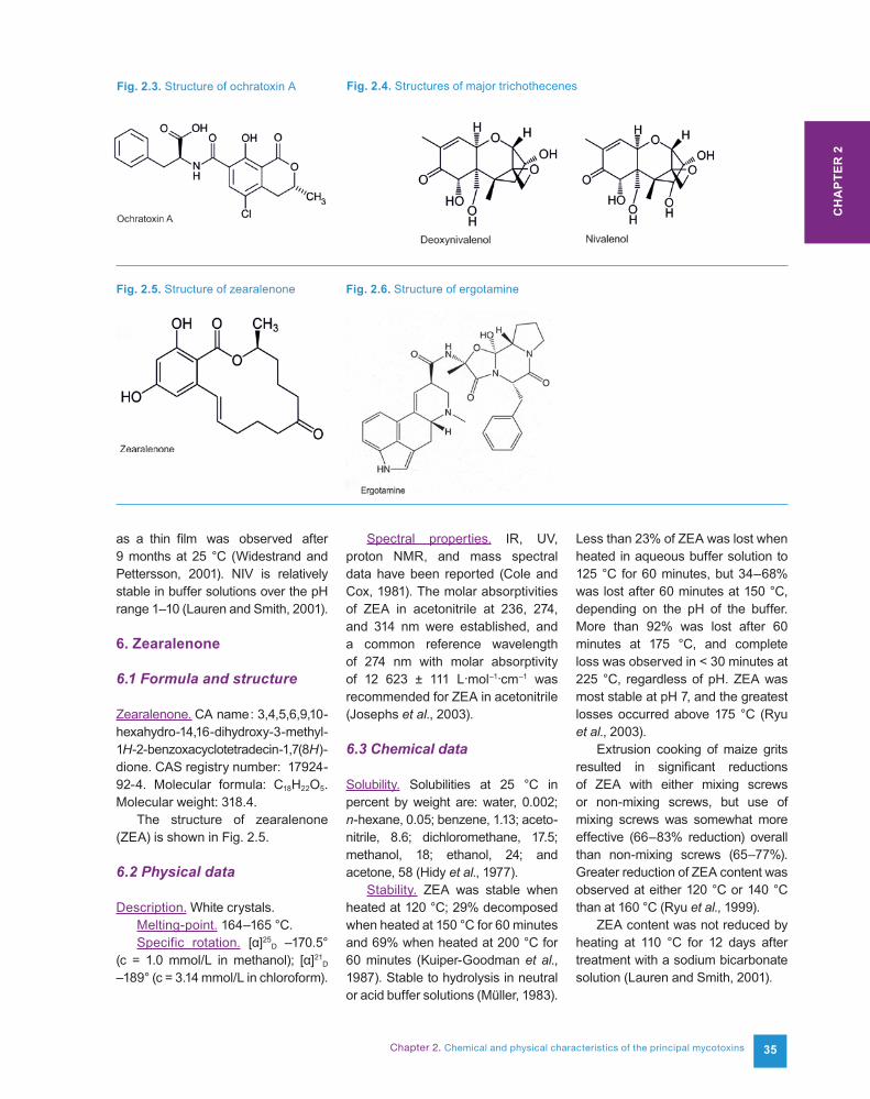
The structure of deoxynivalenol (DON) is shown in Fig. 2.4.
4.2 Physical data
Description. White needles.Melting-point. 151–153 °C. Specific rotation. [α]20
D +6.35° (c = 0.07 mmol/L in ethanol).
Spectral properties. IR, UV, NMR, and mass spectral data have been reported (Cole and Cox, 1981; Cole et al., 2003c).
4.3 Chemical data
Solubility. Soluble in chloroform, ethanol, methanol, and ethyl acetate.
Stability. Autoclaving creamed maize reduced DON content by only 12% (Wolf-Hall et al., 1999). At pH 4.0, DON appeared to be very stable, showing no destruction at 100 °C or 120 °C and only partial destruction at 170 °C after 60 minutes. At pH 7.0, DON was still stable but showed more destruction at 170 °C after 15 minutes. At pH 10.0, DON was partially destroyed at 100 °C after 60 minutes and was totally destroyed at 120 °C after 30 minutes and at 170 °C after 15 minutes (Wolf and Bullerman, 1998).
When DON was gamma-irradiated on maize, breakdown of DON began only after irradiation to 20 kGy, and 80–90% of the DON remained after irradiation to 50 kGy (O’Neill et al., 1993).
No significant decomposition of DON was observed when stored in ethyl acetate for 24 months at 25 °C or 3 months at 40 °C (Widestrand and Pettersson, 2001). DON was relatively stable in buffer solutions over the pH range 1–10 (Lauren and Smith, 2001).
5. Nivalenol
5.1 Formula and structure
Nivalenol. CA name : 12,13-epoxy-3,4,7,15-tetrahydroxy-(3α,4β,7α)-tricho-thec-9-en-8-one. CAS registry num-ber : 23282-20-4. Molecular formula : C15H20O7. Molecular weight : 312.32.
The structure of nivalenol (NIV) is shown in Fig. 2.4.
5.2 Physical data
Description. White crystals.Melting-point. 222 –223 °C (with
decomposition, after drying in the pres-ence of P2O5 at reduced pressure).
Specific rotation. [α]20D +21.54° (c =
1.3 mmol/L in ethanol).Spectral properties. IR, UV, NMR,
and mass spectral data have been reported (Cole and Cox, 1981; Brumley et al., 1982; Cole et al., 2003c).
5.3 Chemical data
Solubility. Soluble in chloroform, ethanol, methanol, and ethyl acetate; slightly soluble in water; soluble in polar organic solvents (Budavari, 1989).
Stability. No significant decompo-sition of NIV was observed when stored in ethyl acetate for 24 months at 25 °C or for 3 months at 40 °C. A significant decrease of NIV stored
Chapter 2. Chemical and physical characteristics of the principal mycotoxins 35
as a thin film was observed after 9 months at 25 °C (Widestrand and Pettersson, 2001). NIV is relatively stable in buffer solutions over the pH range 1–10 (Lauren and Smith, 2001).
6. Zearalenone
6.1 Formula and structure
Zearalenone. CA name : 3,4,5,6,9,10- hexahydro-14,16-dihydroxy-3-methyl-1H-2-benzoxacyclotetradecin-1,7(8H)-dione. CAS registry number: 17924-92-4. Molecular formula: C18H22O5. Molecular weight: 318.4.
The structure of zearalenone (ZEA) is shown in Fig. 2.5.
6.2 Physical data
Description. White crystals.Melting-point. 164–165 °C.Specific rotation. [α]25
D –170.5° (c = 1.0 mmol/L in methanol); [α]21
D –189° (c = 3.14 mmol/L in chloroform).
Spectral properties. IR, UV, proton NMR, and mass spectral data have been reported (Cole and Cox, 1981). The molar absorptivities of ZEA in acetonitrile at 236, 274, and 314 nm were established, and a common reference wavelength of 274 nm with molar absorptivity of 12 623 ± 111 L.mol–1.cm–1 was recommended for ZEA in acetonitrile (Josephs et al., 2003).
6.3 Chemical data
Solubility. Solubilities at 25 °C in percent by weight are: water, 0.002; n-hexane, 0.05; benzene, 1.13; aceto-nitrile, 8.6; dichloromethane, 17.5; methanol, 18; ethanol, 24; and acetone, 58 (Hidy et al., 1977).
Stability. ZEA was stable when heated at 120 °C; 29% decomposed when heated at 150 °C for 60 minutes and 69% when heated at 200 °C for 60 minutes (Kuiper-Goodman et al., 1987). Stable to hydrolysis in neutral or acid buffer solutions (Müller, 1983).
Less than 23% of ZEA was lost when heated in aqueous buffer solution to 125 °C for 60 minutes, but 34–68% was lost after 60 minutes at 150 °C, depending on the pH of the buffer. More than 92% was lost after 60 minutes at 175 °C, and complete loss was observed in < 30 minutes at 225 °C, regardless of pH. ZEA was most stable at pH 7, and the greatest losses occurred above 175 °C (Ryu et al., 2003).
Extrusion cooking of maize grits resulted in significant reductions of ZEA with either mixing screws or non-mixing screws, but use of mixing screws was somewhat more effective (66–83% reduction) overall than non-mixing screws (65–77%). Greater reduction of ZEA content was observed at either 120 °C or 140 °C than at 160 °C (Ryu et al., 1999).
ZEA content was not reduced by heating at 110 °C for 12 days after treatment with a sodium bicarbonate solution (Lauren and Smith, 2001).
Fig. 2.3. Structure of ochratoxin A Fig. 2.4. Structures of major trichothecenes
Fig. 2.5. Structure of zearalenone Fig. 2.6. Structure of ergotamine
CH
AP
TER
2
36
7. Ergot alkaloids
Ergots, the sclerotia produced by Claviceps purpurea and related species, contain a remarkable variety of compounds, which can be divided into three groups: derivatives of lysergic acid, derivatives of isolysergic acid, and clavines. The most impor-tant of these is ergotamine.
7.1 Formula and structure
Ergotamine. CA name: 12′-hydroxy-2′-methyl-5′-(phenylmethyl)-ergotaman-
3′,6′,18-trione. CAS registry number: 113-15-5. Molecular formula: C33H35N5O5. Molecular weight: 581.66.
The structure of ergotamine is shown in Fig. 2.6.
7.2 Physical data
Description. White powder.Melting-point. 180 °C.Spectral properties. UV, IR, and
fluorescence spectral data were reviewed by Hofmann (1964). The electron mass spectrum of ergot-amine was described by Vokoun and
Řehāček (1975), and the [1H]-NMR spectrum was reported by Pierri et al. (1982).
Specific rotation. [α]20D –160°.
7.3 Chemical data
Solubility. Some data on recrys-tallization, appearance, and solubility were reviewed by Hofmann (1964).

Chapter 2. Chemical and physical characteristics of the principal mycotoxins 37
References
Hidy PH, Baldwin RS, Greasham RL et al. (1977). Zearalenone and some derivatives: production and biological activities. Adv Appl Microbiol, 22:59–82. PMID:412398
Hofmann A (1964). Die Mutterkorn-alkaloide. Stuttgart: Ferdinand Enke Verlag.
Howard PC, Churchwell MI, Couch LH et al. (1998). Formation of N-(carboxymethyl)fumonisin B1, following the reaction of fumonisin B1 with reducing sugars. J Agric Food Chem, 46:3546–3557. doi:10.1021/jf980194q
Josephs RD, Krska R, MacDonald S et al. (2003). Preparation of a calibrant as certified reference material for determination of the Fusarium mycotoxin zearalenone. J AOAC Int, 86:50–60. PMID:12607740
Kiermeier F, Kroczek S (1974). Einfluss der Lösungsmittels auf die Fluorescenz von Aflatoxin B1. Z Lebensm Unters Forsch, 155:81–84. doi:10.1007/BF01460336
Kuiper-Goodman T, Scott PM, Watanabe H (1987). Risk assessment of the mycotoxin zearalenone. Regul Toxicol Pharmacol, 7:253–306. doi:10.1016/0273-2300(87)90037-7 PMID:2961013
Lauren DR, Smith WA (2001). Stability of the Fusarium mycotoxins nivalenol, deoxynivalenol and zearalenone in ground maize under typical cooking environments. Food Addit Contam, 18:1011–1016. doi:10.1080/02652030110052283 PMID:11665729
Laurent D, Platzer N, Kohler F et al. (1989). Macrofusine et micromoniline: deux nouvelles mycotoxines isolées de maïs infesté par Fusarium moniliforme Sheld [Macrofusin and micromonilin: two new mycotoxins isolated from corn infested with Fusarium moniliforme Sheld]. Microbiol Aliment Nutr, 7:9–l6.
Müller HM (1983). A survey of methods of decontaminating mycotoxins. Part II. Chemical methods and reactions with components of feedstuffs. Übersicht Tierernach, 11:7–37.
NTP (2001). NTP Technical Report on the Toxicology and Carcinogenesis Studies of Fumonisin B1 (CAS No. 116355-83-0) in F344/N Rats and B6C3F1 Mice (Feed Studies). Research Triangle Park, NC: National Toxicology Program, U.S. Department of Health and Human Services, National Institutes of Health (NTP Technical Report No. 496; NIH Publication No. 99-3955). Available at http://ntp.niehs.nih.gov/ntp/htdocs/lt_rpts/tr496.pdf.
Natori S, Sakaki S, Kurata H et al. (1970). Chemical and cytotoxicity survey on the production of ochratoxins and penicillic acid by Aspergillus ochraceus Wilhelm. Chem Pharm Bull (Tokyo), 18:2259–2268. doi:10.1248/cpb.18.2259 PMID:5494852
Norred WP, Plattner RD, Dombrink-Kurtzman MA et al. (1997). Mycotoxin-induced elevation of free sphingoid bases in precision-cut rat liver slices: specificity of the response and structure-activity relationships. Toxicol Appl Pharmacol, 147:63–70. doi:10.1006/taap.1997.8272 PMID:9356308
O’Neil MJ, Smith A, Heckelman PE, Budavari S, eds. (2001). The Merck Index, 13th ed. Whitehouse Station, NJ: Merck & Co.
O’Neill K, Damoglou AP, Patterson MF (1993). The stability of deoxynivalenol and 3-acetyl deoxynivalenol to gamma irradiation. Food Addit Contam, 10:209–215. doi:10.1080/02652039309374143 PMID:8314397
Pierri L, Pitman IH, Rae ID et al. (1982). Conformational analysis of the ergot alkaloids ergotamine and ergotaminine. J Med Chem, 25:937–942. doi:10.1021/jm00350a010 PMID:7120281
Plattner RD, Norred WP, Bacon CW et al. (1990). A method of detection of fumonisins in corn samples associated with field cases of equine leukoencephalomalacia. Mycologia, 82:698–702. doi:10.2307/3760156
Pohland AE, Schuller PL, Steyn PS, Van Egmond HP (1982). Physico-chemical data for selected mycotoxins. Pure Appl Chem, 54:2219–2284. doi:10.1351/pac198254112219
Robertson JA, Pons WA (1968). Solid state fluorescence emission of aflatoxin on silica gel. J Assoc Off Anal Chem, 51:1190–1192.
Ryu D, Hanna MA, Bullerman LB (1999). Stabil-ity of zearalenone during extrusion of corn grits. J Food Prot, 62:1482–1484. PMID:10606157
Ryu D, Hanna MA, Eskridge KM, Bullerman LB (2003). Heat stability of zearalenone in an aqueous buffered model system. J Agric Food Chem, 51:1746–1748. doi:10.1021/jf0210021 PMID:12617617
Savard ME, Blackwell BA (1994). Spectral characteristics of secondary metabolites from Fusarium fungi. In: Miller JD, Trenholm HL, eds. Mycotoxins in Grain: Compounds Other than Aflatoxin. St Paul, MN: Eagan Press, pp. 59–260.
Steyn PS, Holzapfel CW (1967). The synthesis of ochratoxins A and B metabolites of Aspergillus ochraceus Wilh. Tetrahedron, 23:4449–4461. doi:10.1016/S0040-4020(01) 88843-8 PMID:6077765
Stubblefield RD, Shotwell OL, Shannon GM et al. (1970). A new metabolite from Aspergillus parasiticus. J Agric Food Chem, 18:391–393. doi:10.1021/jf60169a025 PMID:5487091
Subirade I (1996). Fate of ochratoxin A during breadmaking. Food Addit Contam, 13 Suppl:25–26. PMID:8972345
Bezuidenhout SC, Gelderblom WCA, Gorst-Allman CP et al. (1988). Structure elucidation of the fumonisins, mycotoxins from Fusarium moniliforme. J Chem Soc Chem Commun, 11:743–745. doi:10.1039/c39880000743
Brumley WC, Andrzejewski D, Trucksess EW et al. (1982). Negative ion chemical ionization mass spectrometry of trichothecenes. Novel fragmentation under OH- conditions. Biomed Mass Spectrom, 9:451–457. doi:10.1002/bms.1200091008
Budavari S, ed. (1989). The Merck Index, 11th ed. Rahway, NJ: Merck & Co.
Bycroft BW, Hatton JR, Roberts JC (1970). Studies in mycological chemistry. XXV. Experiments directed towards a synthesis of aflatoxin-G2: synthesis of the coumarino-lactone system. J Chem Soc Perkin 1, 2:281–284. PMID:5460861
Castegnaro M, Barek J, Frémy JM et al., eds (1991). Laboratory Decontamination and Destruction of Carcinogens in Laboratory Wastes: Some Mycotoxins. Lyon: International Agency for Research on Cancer (IARC Scientific Publications Series, No. 113).
Castegnaro M, Hunt DC, Sansonne EB et al., eds (1980). Laboratory Decontamination and Destruction of Aflatoxins B1, B2, G1, G2
in Laboratory Wastes. Lyon : International Agency for Research on Cancer (IARC Scientific Publications Series, No. 37).
Cole RJ, Cox RH (1981). Handbook of Toxic Fungal Metabolites. New York : Academic Press.
Cole RJ, Jarvis BB, Schweikert MA (2003a). Fumonisins, AAL toxins, and related metabolites. In: Handbook of Secondary Fungal Metabolites, Vol. III. San Diego : Academic Press, pp. 561–612.
Cole RJ, Jarvis BB, Schweikert MA (2003b). Ochratoxins and related metabolites. In : Handbook of Secondary Fungal Metabolites, Vol. III. San Diego: Academic Press, pp. 613–624.
Cole RJ, Jarvis BB, Schweikert MA (2003c). Trichothecenes and related metabolites. In: Handbook of Secondary Fungal Metabolites, Vol. III. San Diego: Academic Press, pp. 199–324.
Cole RJ, Schweikert MA (2003). Aflatoxins. In: Handbook of Secondary Fungal Metabolites, Vol. I. San Diego: Academic Press, pp. 545–569.
Gelderblom WCA, Marasas WFO, Vleggaar R et al. (1992). Fumonisins: isolation, chemical characterization and biological effects. Mycopathologia, 117:11–16. doi:10.1007/BF00497273 PMID:1513367
CH
AP
TER
2

38
Uwaifo AO, Emerole GO, Bassir O (1977). Comparative study of the fluorescent characteristics of solutions of aflatoxins and palmotoxins in chloroform. J Agric Food Chem, 25. doi:10.1021/jf60213a021 PMID:893818
Van der Merwe KJ, Steyn PS, Fourie L (1965a). Mycotoxins. II. The constitution of ochratoxins A, B, and C, metabolites of Aspergillus ochraceus Wilh. J Chem Soc Perkin 1, 7083–7088. doi:10.1039/jr9650007083 PMID:5892024
Van der Merwe KJ, Steyn PS, Fourie L et al. (1965b). Ochratoxin A, a toxic metabolite produced by Aspergillus ochraceus Wilh. Nature, 205:1112–1113. doi:10.1038/2051112a0 PMID:5833211
Van der Stegen GH, Essens PJ, van der Lijn J (2001). Effect of roasting conditions on reduction of ochratoxin A in coffee. J Agric Food Chem, 49:4713–4715. doi:10.1021/jf0105586 PMID:11600012
Visconti A, Doko MB, Bottalico C et al. (1994). Stability of fumonisins (FB1 and FB2) in solution. Food Addit Contam, 11:427–431. doi:10.1080/02652039409374244 PMID:7958112
Vokoun J, Řehāček Z (1975). Mass spectra of ergot peptide alkaloids. Collect Czech Chem Commun, 40:1731–1737.
WHO (2000). Environmental Health Criteria 219: Fumonisin B1. Marasas WFO, Miller JD, Riley RT, Visconti A, eds. Geneva: United Nations Environment Programme, International Labour Organization, World Health Organization. Available at http://libdoc.who.int/ehc/WHO_EHC_219.pdf.
Widestrand J, Pettersson H (2001). Effect of time, temperature and solvent on the stability of T-2 toxin, HT-2 toxin, deoxynivalenol and nivalenol calibrants. Food Addit Contam, 18:987–992. doi:10.1080/02652030110050168 PMID:11665740
Wogan GN (1966). Chemical nature and biological effects of the aflatoxins. Bacteriol Rev, 30:460–470. PMID:5327461
Wolf CE, Bullerman LB (1998). Heat and pH alter the concentration of deoxynivalenol in an aqueous environment. J Food Prot, 61:365–367. PMID:9708313
Wolf-Hall CE, Hanna MA, Bullerman LB (1999). Stability of deoxynivalenol in heat-treated foods. J Food Prot, 62:962–964. PMID:10456755