chapter 9 melioidosis
TRANSCRIPT

223
Melioidosis
Chapter 9
MELIOIDOSIS
PAUL J. BRETT, PhD*; DAVID DeSHAZER, PhD†; and NICHOLAS J. VIETRI, MD‡
INTRODUCTION
THE INFECTIOUS AGENT
MILITARY RELEVANCE
THE DISEASEEpidemiologyClinical DiseaseDiagnosisTreatmentPrevention
SUMMARY
*Associate Professor, Department of Microbiology and Immunology, University of South Alabama, 610 Clinic Drive, Laboratory of Infectious Diseases Building, Mobile, Alabama 36688
†Microbiologist, Bacteriology Division, US Army Medical Research Institute of Infectious Diseases, 1425 Porter Street, Fort Detrick, Maryland 21702‡Colonel, Medical Corps, US Army; Infectious Diseases Officer, Bacteriology Division, US Army Medical Research Institute of Infectious Diseases, 1425 Porter Street, Fort Detrick, Maryland 21702; and Assistant Professor of Medicine, Uniformed Services University of the Health Sciences, 4301 Jones Bridge Road, Bethesda, Maryland 20814
244-949 DLA DS.indb 223 6/4/18 11:57 AM

224
Medical Aspects of Biological Warfare
INTRODUCTION
In 1911, Captain Alfred Whitmore and Dr CS Krishnaswami described a previously unrecognized disease that was prevalent among the ill-nourished and neglected inhabitants of Rangoon, Burma.1 The new disease resembled glanders, a zoonotic disease of equines,2 and the gram-negative bacillus they iso-lated post-mortem from tissue samples resembled the glanders bacillus, Bacillus mallei.3 However, the new bacillus could be differentiated from B mallei by its mo-tility, luxuriant growth on peptone agar, and wrinkled colony morphology, and was subsequently named Bacillus pseudomallei.3,4 Whitmore’s detailed account of the first 38 human cases of this disease demonstrated most were morphine injectors that died of septicemia with abscesses in multiple organs.4 As a result, the disease became known as “Whitmore’s disease” or “morphine injector’s septicemia.”5,6 In 1921, Stanton and Fletcher reported an outbreak of a septicemic dis-ease in a guinea pig colony at the Institute for Medical Research in Kuala Lumpur.7 The infectious agent they isolated from diseased animals was indistinguishable from Whitmore’s bacillus, and they coined the term “melioidosis” (a Greek term meaning glanders-like illness) to describe this new disease of the tropics.7 Stanton and Fletcher subsequently published a clas-
sic monograph in 1932 describing their observations of melioidosis in humans and animals occurring in Burma, Malaya, French Indochina, and Ceylon over a number of years.8
Today, melioidosis is regarded as an emerging in-fectious disease and a potential bioterrorism threat.9–11 The etiologic agent of melioidosis is present in water and soil in tropical and subtropical regions and is spread to humans through direct contact with the contaminated source. Clinical manifestations range from subclinical infection to overwhelming septicemia that resembles disseminated or localized, suppurative infection due to a variety of pathogens, resulting in the nickname “the remarkable imitator.”12 The majority of melioidosis cases have one or more identified risk factors, including diabetes, alcoholism, chronic renal disease, cystic fibrosis, and steroid abuse.13 Interest-ingly, acquired immunodeficiency syndrome does not seem to be a major risk factor for melioidosis. Healthy individuals can also get the disease, especially if they work in muddy soil without good hand and foot protection.14 Many animal species are susceptible to melioidosis, including sheep, goats, horses, swine, cattle, dogs, and cats.15 Numerous review articles on melioidosis have been published since 1990.11,13–30
THE INFECTIOUS AGENT
The bacterium that causes melioidosis, now des-ignated Burkholderia pseudomallei,31 has undergone numerous name changes since its original classification as Bacillus pseudomallei, including Bacterium whitmori, Bacillus whitmori, Pfeifferella whitmori, Pfeifferella pseu-domallei, Actinobacillus pseudomallei, Lofflerella whitmori, Flavobacterium pseudomallei, Malleomyces pseudomallei, and Pseudomonas pseudomallei. The non-sporulating, gram-negative bacillus is an environmental sapro-phyte found in surface waters and wet soils in endemic regions.32–39 Individual cells are approximately 0.8 × 1.5 µm with a polar tuft of two to four flagella and may exhibit bipolar staining with a “safety pin” appear-ance.40,41 B pseudomallei is metabolically versatile and can grow on numerous carbon sources.31,42 Anaerobic growth is possible, but only in the presence of nitrate or arginine.11 The microbe accumulates intracellular stores of poly-b-hydroxybutyric acid and can survive in distilled water for years.10,43,44 The optimal survival temperature for B pseudomallei is between 24°C and 32°C, but it can grow at temperatures up to 42°C.45,46 B pseudomallei demonstrate considerable interstrain and medium-dependent colony morphology.47–49 The oxidase-positive organism can grow on a variety of microbial media, but Ashdown’s selective medium is
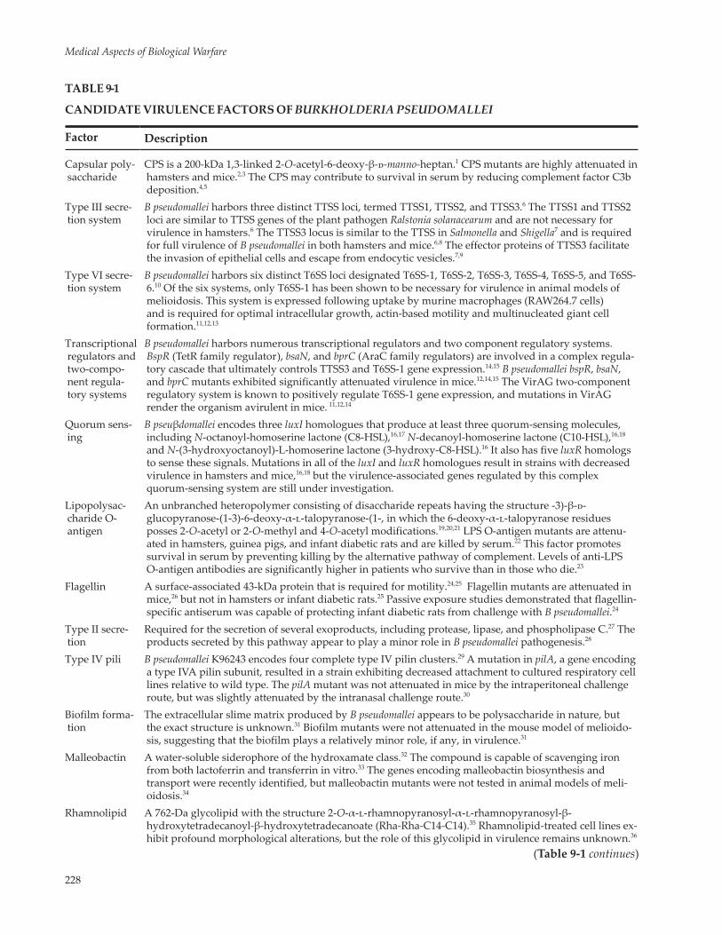
often used for isolating B pseudomallei from environ-mental and clinical specimens.50 Two distinct colony phenotypes are commonly observed on this medium (Figure 9-1a), presumably due to the differential up-take of crystal violet and neutral red or to the differen-tial production of ammonia and oxalic acid.50,51 Most strains appear lavender after 2 to 3 days of incubation at 37°C, but some isolates appear deep purple (see Figure 9-1a). After 3 days at 37°C, the colonies often become dull and wrinkled (Figure 9-1b) and emit a distinctive sweet, earthy smell. Other selective media have also been used to isolate B pseudomallei from contaminated specimens.52,53
The complete genome sequence of B pseudomallei K96243, a strain isolated in 1996 from a 34-year-old dia-betic patient in Khon Kaen, Thailand, was published in 2004.54 The 7.25-megabase pair (Mb) genome was comprised of two circular replicons, termed chromo-some 1 (4.07 Mb) and chromosome 2 (3.17 Mb). The G + C content of the genome is 68% and is predicted to encode 5,855 proteins. Chromosome 1 encoded a high proportion of core housekeeping functions (DNA rep-lication, transcription, translation, amino acid and nu-cleotide metabolism, basic carbohydrate metabolism, and cofactor synthesis), while chromosome 2 encoded
244-949 DLA DS.indb 224 6/4/18 11:57 AM

225
Melioidosis
Figure 9-1. Burkholderia pseudomallei colony morphologies as demonstrated on Ashdown’s selective medium50 supplemented with 100 µg/mL streptomycin. Plates were incubated for 3 days at 37°C (a) and 5 days at 37°C (b).
a
b
a high proportion of accessory functions (adaptation to atypical conditions, osmotic protection, and sec-ondary metabolism).54 Plasmid-like replication genes and accessory genes on chromosome 2 suggest it may have been derived from a plasmid (or megaplasmid) that became an indispensable replicon by acquiring essential functions such as transfer ribonucleic acid genes, amino acid biosynthesis genes, and energy me-tabolism genes. There are 16 “genomic islands” in the B pseudomallei K96243 genome that appear to have been acquired through horizontal gene transfer.54 Analysis of 11 additional B pseudomallei strains has led to the identification of at least 128 different genomic islands that are variably present among these strains.55,56 Mo-bile genetic elements, such as prophages, insertion sequences, and integrated plasmids, account for most of the laterally acquired genomic sequences. Recent
studies have shown that B pseudomallei strains exhibit significant genomic diversity and that much of the genetic heterogeneity is due to laterally acquired mo-bile genetic elements.54,57–61 These genomic islands may provide strains that give them a metabolic or virulence advantage over strains that do not contain such se-quences. Similarly, autonomously replicating plasmids are variably present in B pseudomallei isolates, but little is known about their biological significance.30,62–64 Com-plete genome sequences of 13 B pseudomallei isolates (K96243, 1026b, 1106a, 1710b, 668, BPC006, MSHR146, MSHR305, MSHR511, NAU20B-16, NCTC13178, NCTC13179, MSHR520) and draft genome sequences of an additional 54 B pseudomallei isolates are available in GenBank, dramatically enhancing the amount and diversity of genome sequence data available for study of B pseudomallei.65
MILITARY RELEVANCE
Throughout the 20th century, melioidosis had an impact on the health of soldiers serving in Asia during times of war and peace.66 Sporadic melioidosis infec-tions occurred in US and Japanese soldiers during World War II (WWII),41,67,68 and cases of recrudescent melioidosis in WWII veterans were also reported.69,70 During the French Indochina War (1946–1954), there were at least 100 cases of melioidosis in French forces during their fight against the resistance movement led by the Viet Minh.22,66 There were more than 300 cases of melioidosis in US soldiers during the Vietnam War22 and additional cases that did not surface until years after the war’s end, leading to the nickname “Vietnam Time Bomb.”71–73 Twenty-three melioidosis cases were reported in the Singapore Armed Forces from 1987 to 1994.74 The infection rate in these relatively healthy
Singapore Armed Forces was approximately fourfold the rate in the general population of Singapore, sug-gesting that close contact with the soil during military training may lead to an increased risk for melioidosis.
B pseudomallei is a Centers for Disease Control and Prevention Tier 1 select agent that must be handled in biosafety level 3 laboratories.9 Biosafety level 3 facilities incorporate specialized negative-air pressure ventila-tion systems and well-defined biosafety containment equipment and protocols to study agents that can be transmitted through the air and cause potentially lethal infection. Tier 1 agents present the greatest risk of deliberate misuse and pose a severe threat to public health and safety. B pseudomallei was studied by the United States, the former Soviet Union, and possibly Egypt as a potential biological warfare agent, but was
244-949 DLA DS.indb 225 6/4/18 11:57 AM

226
Medical Aspects of Biological Warfare
never used in this capacity.75–77 On the other hand, B mallei was used as a biological warfare agent several times in the past, including during the American Civil War, World War I, WWII, and in Afghanistan between 1982 and 1984.2,76,78,79 The usefulness of B pseudomallei as
a biological warfare agent is currently unknown, but the ease of acquiring strains from the environment, the ability to genetically manipulate the agent to be multiply antibiotic resistant, and the lack of a melioi-dosis vaccine make this possibility a serious concern.
THE DISEASE
Epidemiology
Melioidosis cases are most commonly reported from countries located between 20°N and 20°S in latitude, with the greatest concentration in Vietnam, Cambodia, Laos, Thailand, Malaysia, Singapore, and northern Australia.11,13,23 The disease has also been observed in the South Pacific, Africa, India, and the Middle East.80–83 In addition, sporadic cases of meli-oidosis have occurred in the Western Hemisphere in Aruba, Brazil, Mexico, Panama, Ecuador, Haiti, Peru, and Guyana.11,13,23,84,85 In endemic regions, the disease occurs in humans, sheep, goats, horses, swine, cattle, dogs, cats, deer, camels, monkeys, zebras, kangaroos, koalas, birds, and crocodiles.15,27,86–88 Melioidosis cases that occur in temperate regions are typically acquired in endemic areas. Human cases can often be attributed to recent travel to such areas.21,89–92 For example, the first case report of cerebral melioidosis in the United States was recently described in a individual that had immigrated from Cambodia and frequently visited his home country.93 A case of neurological melioidosis was reported in a pigtail macaque that was imported from Indonesia to the United States for research pur-poses.94 In addition, B pseudomallei infections have been described in pet green iguanas in California and, based on the multilocus sequence typing of the isolates in these cases, are believed to have originated in Central America.95 With the exception of a single case of melioidosis that occurred in Arizona in 2008, no B pseudomallei infections have been documented in US patients without a history of prior travel to an endemic region.96,97 While the source of the Arizona isolate could not be determined despite extensive investigation, molecular analysis indicated that it was consistent with Southeast Asian origin.97,98
Pathogenesis
Several animal models of melioidosis have been developed to study pathogenesis, virulence factors, and efficacy of antibiotics and vaccines.99–107 In gen-eral, hamsters and ferrets are highly susceptible to experimental melioidosis, while rats, pigs, and rhesus monkeys are relatively resistant. Infant rats can be made more susceptible to infection by intraperitoneal
injection of streptozotocin, a compound that induces diabetes.103,108 The LD50 (amount necessary to kill 50% of the subject population) of B pseudomallei for non-diabetic infant rats is greater than 108 bacteria, while the LD50 in streptozotocin-induced diabetic infant rats is approximately 104 bacteria. Mice and guinea pigs exhibit intermediate susceptibility to experimental infection with B pseudomallei, but the LD50 for mice var-ies widely depending on the route of infection, mouse strain, and bacterial strain.101,102,105,109 Recent reviews describe in detail the various mouse models of meli-oidosis that have been developed and the advantages and disadvantages of each.110,111
Due to fears that B pseudomallei might be used as a biological weapon, basic research on this pathogen has progressed rapidly over the past 10 years. The identification of virulence factors has been facilitated by the availability of genomic sequence data54 and the existence of a nonpathogenic B pseudomallei-like species designated B thailandensis.112–114 B pseudomallei and B thailandensis strains are genetically and immunologi-cally similar to one another, but B thailandensis is less virulent in animal models of infection and has only rarely been reported to cause disease in humans.115 Genetic determinants that confer enhanced virulence in B pseudomallei relative to B thailandensis have been identified by comparative analysis of genomic DNA from these species.58,116,117 Table 9-1 provides a brief de-scription of B pseudomallei virulence factors identified to date, their mechanisms of action, and their relative importance in animal models of melioidosis.
B pseudomallei is a facultative intracellular pathogen whose ability to survive and replicate in phagocytic and nonphagocytic cell lines has been well document-ed.118–123 The organism possesses a variety of mecha-nisms to adapt to the host environment and harbors an array of specialized secretory systems, which are required within this niche.124–128 Although relatively little is known about the initial interactions of B pseu-domallei with host cells, recent studies have identified a number of adhesins that mediate adherence of the organism to eukaryotic cells.129–133 Following internal-ization, B pseudomallei rapidly escapes from endocytic vacuoles and enters into the host cell cytosol, where it can replicate to high numbers, polymerize actin, and induce host cell fusion. These processes are believed
244-949 DLA DS.indb 226 6/4/18 11:57 AM

227
Melioidosis
to facilitate intracellular spread and multinucleated giant cell formation. Type III and type VI secretions systems have been shown to play key roles in endo-somal escape and multinucleated giant cell formation, respectively.124,134 The effector proteins delivered by these systems are predicted to modulate critical host proteins and pathways involved in cytoskeleton re-arrangement, cell signaling, and cell death, thereby enabling pathogen survival and proliferation within a host.17 It has been postulated that after the initial phase of infection, B pseudomallei can persist in a dormant stage in macrophages for months or years.123 Melioi-dosis has the potential for a long latency period and B pseudomallei’s intracellular persistence could provide a mechanism by which this occurs. Intracellular survival and cell-to-cell spread may also provide B pseudomallei protection from the humoral immune response.
Clinical Disease
Melioidosis is a tropical bacterial disease with primary endemic foci in southeast Asia, northern Australia, south Asia, and China. Hyperendemic areas for melioidosis include northern Australia and northeast Thailand, where the disease incidence peaks in the rainy season. The routes of infection include percutaneous inoculation, inhalation, or ingestion of contaminated food or water. Although percutaneous inoculation is the most common route of infection, heavy rainfall is associated with pneumonia and more severe disease and may represent a shift from percuta-neous inoculation to inhalation as the primary mode of infection.135 In hyperendemic areas, B pseudomallei causes a substantial burden of infectious disease. For example, at the Sappasit Prasong Hospital in Ubon Ratchatani, northeast Thailand, which serves a rural community of rice farmers and their families, nearly 20% of all community-acquired bacteremia is due to B pseudomallei.136 Likewise, melioidosis is the most common cause of fatal community-acquired bactere-mic pneumonia at the Royal Darwin Hospital in the Northern Territory of Australia.137
Cases of human-to-human transmission of B pseudo-mallei, although very rare, have been documented.138,139 The incubation period (time between exposure and appearance of clinical symptoms) varies, as infec-tious dose, route of infection, B pseudomallei strain characteristics, and host risk factors are all believed to play an important role. One study that looked at the incubation period after inoculation exposures in Darwin, Australia, revealed a mean incubation period of 9 days, with a range of 1 to 21 days.140 Although serologic studies suggest that most infections with B pseudomallei are asymptomatic,141 individuals with risk
factors such as diabetes mellitus, alcoholism, cirrhosis, thalassemia, or other immunosuppressed states are at an increased risk of developing symptomatic infec-tion. Other melioidosis-associated risk factors include chronic lung disease, kava consumption, and cystic fibrosis. Diabetes appears to be the most important of all the known risk factors, as up to 50% of patients with melioidosis have diabetes mellitus.27
Melioidosis, which presents as a febrile illness, has an unusually broad range of clinical presentations. The diversity of infectious presentations includes acute localized suppurative soft tissue infections, acute pulmonary infections, acute fulminant septicemia, and chronic localized infections. Clinical disease with B pseudomallei is generally caused by hematogenous spread of bacteria and seeding to various organs within the host.27 The Infectious Disease Association of Thai-land, the country with the largest number of reported cases (2,000–3,000 per year), divided 345 cases into the following categories: (a) disseminated septicemia, 45% of the cases with 87% mortality; (b) nondisseminated septicemia, 12% of the cases with 17% mortality; (c) localized septicemia, 42% of the cases with 9% mortal-ity; and (d) transient bacteremia, 0.3% of cases.142,143
Melioidosis is characterized by abscess formation, and the majority of patients with melioidosis are bacte-remic. The most commonly involved organ is the lung. The nidus of infection is either a primary pneumonia or lung abscess, or the infection results from hematog-enous seeding of the lung from bacteremia (Figures 9-2 and 9-3). For example, of the 540 cases of melioidosis analyzed in the 20-year Darwin Prospective Melioido-sis Study, pneumonia was the most common primary
Figure 9-2. Chest radiograph demonstrating a severe mul-tilobar pneumonia. Photograph courtesy of Bart Currie, MD, Royal Darwin Hospital, Australia.
244-949 DLA DS.indb 227 6/4/18 11:57 AM
228
Medical Aspects of Biological Warfare
TABLE 9-1
CANDIDATE VIRULENCE FACTORS OF BURKHOLDERIA PSEUDOMALLEI
Factor Description
Capsular poly-saccharide
CPS is a 200-kDa 1,3-linked 2-O-acetyl-6-deoxy-β-d-manno-heptan.1 CPS mutants are highly attenuated in hamsters and mice.2,3 The CPS may contribute to survival in serum by reducing complement factor C3b deposition.4,5
Type III secre-tion system
B pseudomallei harbors three distinct TTSS loci, termed TTSS1, TTSS2, and TTSS3.6 The TTSS1 and TTSS2 loci are similar to TTSS genes of the plant pathogen Ralstonia solanacearum and are not necessary for virulence in hamsters.6 The TTSS3 locus is similar to the TTSS in Salmonella and Shigella7 and is required for full virulence of B pseudomallei in both hamsters and mice.6,8 The effector proteins of TTSS3 facilitate the invasion of epithelial cells and escape from endocytic vesicles.7,9
Type VI secre-tion system
B pseudomallei harbors six distinct T6SS loci designated T6SS-1, T6SS-2, T6SS-3, T6SS-4, T6SS-5, and T6SS-6.10 Of the six systems, only T6SS-1 has been shown to be necessary for virulence in animal models of melioidosis. This system is expressed following uptake by murine macrophages (RAW264.7 cells) and is required for optimal intracellular growth, actin-based motility and multinucleated giant cell formation.11,12,13
Transcriptional regulators and two-compo-nent regula-tory systems
B pseudomallei harbors numerous transcriptional regulators and two component regulatory systems. BspR (TetR family regulator), bsaN, and bprC (AraC family regulators) are involved in a complex regula-tory cascade that ultimately controls TTSS3 and T6SS-1 gene expression.14,15 B pseudomallei bspR, bsaN, and bprC mutants exhibited significantly attenuated virulence in mice.12,14,15 The VirAG two-component regulatory system is known to positively regulate T6SS-1 gene expression, and mutations in VirAG render the organism avirulent in mice. 11,12,14
Quorum sens-ing
B pseuβdomallei encodes three luxI homologues that produce at least three quorum-sensing molecules, including N-octanoyl-homoserine lactone (C8-HSL),16,17 N-decanoyl-homoserine lactone (C10-HSL),16,18 and N-(3-hydroxyoctanoyl)-L-homoserine lactone (3-hydroxy-C8-HSL).16 It also has five luxR homologs to sense these signals. Mutations in all of the luxI and luxR homologues result in strains with decreased virulence in hamsters and mice,16,18 but the virulence-associated genes regulated by this complex quorum-sensing system are still under investigation.
Lipopolysac-charide O-antigen
An unbranched heteropolymer consisting of disaccharide repeats having the structure -3)-β-d-glucopyranose-(1-3)-6-deoxy-α-l-talopyranose-(1-, in which the 6-deoxy-α-l-talopyranose residues posses 2-O-acetyl or 2-O-methyl and 4-O-acetyl modifications.19,20,21 LPS O-antigen mutants are attenu-ated in hamsters, guinea pigs, and infant diabetic rats and are killed by serum.22 This factor promotes survival in serum by preventing killing by the alternative pathway of complement. Levels of anti-LPS O-antigen antibodies are significantly higher in patients who survive than in those who die.23
Flagellin A surface-associated 43-kDa protein that is required for motility.24,25 Flagellin mutants are attenuated in mice,26 but not in hamsters or infant diabetic rats.25 Passive exposure studies demonstrated that flagellin-specific antiserum was capable of protecting infant diabetic rats from challenge with B pseudomallei.24
Type II secre-tion
Required for the secretion of several exoproducts, including protease, lipase, and phospholipase C.27 The products secreted by this pathway appear to play a minor role in B pseudomallei pathogenesis.28
Type IV pili B pseudomallei K96243 encodes four complete type IV pilin clusters.29 A mutation in pilA, a gene encoding a type IVA pilin subunit, resulted in a strain exhibiting decreased attachment to cultured respiratory cell lines relative to wild type. The pilA mutant was not attenuated in mice by the intraperitoneal challenge route, but was slightly attenuated by the intranasal challenge route.30
Biofilm forma-tion
The extracellular slime matrix produced by B pseudomallei appears to be polysaccharide in nature, but the exact structure is unknown.31 Biofilm mutants were not attenuated in the mouse model of melioido-sis, suggesting that the biofilm plays a relatively minor role, if any, in virulence.31
Malleobactin A water-soluble siderophore of the hydroxamate class.32 The compound is capable of scavenging iron from both lactoferrin and transferrin in vitro.33 The genes encoding malleobactin biosynthesis and transport were recently identified, but malleobactin mutants were not tested in animal models of meli-oidosis.34
Rhamnolipid A 762-Da glycolipid with the structure 2-O-α-l-rhamnopyranosyl-α-l-rhamnopyranosyl-β-hydroxytetradecanoyl-β-hydroxytetradecanoate (Rha-Rha-C14-C14).35 Rhamnolipid-treated cell lines ex-hibit profound morphological alterations, but the role of this glycolipid in virulence remains unknown.36
(Table 9-1 continues)
244-949 DLA DS.indb 228 6/4/18 11:57 AM

229
Melioidosis
Exopolysaccha-ride
A linear, unbranched polymer of repeating tetrasaccharide units composed of d-galactose and 3-deoxy-d-manno-octulosonicacid (KDO), with the following structure: -3)-2-O-Ac-β-d-Galp-(1-4)-α-d-Galp-(1-3)-β-D-Galp-(1-5)-β-D-KDOp-(2-.37–39 EPS is not produced by the closely related nonpathogenic species B thailandensis, suggesting that it may be a virulence determinant of B pseudomallei. EPS is probably produced during infection because sera from melioidosis patients contain IgG and IgM antibodies to EPS.38,40
Endotoxin The lipid A portion of B pseudomallei LPS contains amide-linked 3-hydroxyhexadecanoic acids, which are longer than the fatty acid chains of enterobacterial LPS.41 The endotoxic activity of B pseudomallei LPS was 10- to 100-fold weaker than enterobacterial LPS in pyrogenic activity in rabbits, lethal toxic-ity in GalN-sensitized mice, and macrophage activation assays. However, the mitogenic activity of B pseudomallei LPS was much higher than enterobacterial LPS.41 The LD50 of purified B pseudomallei LPS in hamsters was 1,000 mg.42
Actin-based motility
Once B pseudomallei gains access to the host cell cytoplasm, it can replicate and exploit actin-based motil-ity for cell-to-cell spread and evasion of the humoral immune response.43–45 The autotransported protein bimA is located at the pole of the bacterial cell and is responsible for the formation of actin tails.44 It is currently unknown if actin-based motility is required for virulence in animal models of melioidosis.
Autotrans-porters and adhesins
B pseudomallei harbors 11 autotransporter analogs, including bimA, and two ATs (boaA and boaB) with roles in bacterial adhesion to epithelial cells, eight ATs (BpaA-F; BcaA-B) that contribute to adherence to and efficient invasion of A549 cells.46–48 BpaC and bcaA have been implicated in virulence in BALB/c mice, since mutants in these ATs displayed defects in dissemination to the liver or spleen, respectively.47 A B pseudomallei bbfA (also known as bpaF) mutant demonstrated a moderate attenuation in a murine model of melioidosis.48
Exotoxins There have been several reports in the literature about B pseudomallei exotoxins,49–53 but the genes encod-ing these exotoxins have not been identified and no defined exotoxin mutants have been constructed. The role of exotoxins as B pseudomallei virulence factors is highly controversial and there appears to be no correlation between in vitro cytotoxicity and in vivo virulence.42,54 In fact, the K96243 genome sequence does not encode any homologues of known major toxins produced by other pathogenic bacte-ria.29
Intracellular toxin
Burkholderia lethal factor 1 (BLF1; BPSL1549) is structurally related to cytotoxic necrotizing factor 1 and has been shown to specifically deamidate Gln339 of eukaryotic initiation factor 4A (a translation initia-tion factor) leading to inhibition of protein synthesis.55,56 Purified recombinant BLF1 was toxic to mice (via intraperitoneal injection), J774 macrophages, but not 3T3 cells.55,56 A B pseudomallei bfl1 mutant was significantly attenuated in mice and exhibited a 100-fold higher median lethal dose in comparison to the wild type strain.55,56
CPS: capsular polysaccharide; EPS: exopolysaccharide; Gln: glutamine; IgG: immunoglobulin G; IgM: immunoglobulin M; LPS: lipopolysac-charide; T6SS: type VI secretion system; TTSS: type III secretion system(1) Isshiki Y, Matsuura M, Dejsirilert S, Ezaki T, Kawahara K. Separation of 6-deoxy-heptan from a smooth-type lipopolysaccharide prepa-ration of Burkholderia pseudomallei. FEMS Microbiol Lett. 2001;199:21–25. (2) Reckseidler SL, DeShazer D, Sokol PA, Woods DE. Detection of bacterial virulence genes by subtractive hybridization: identification of capsular polysaccharide of Burkholderia pseudomallei as a major virulence determinant. Infect Immun. 2001;69:34–44. (3) Atkins T, Prior R, Mack K, et al. Characterization of an acapsular mutant of Burk-holderia pseudomallei identified by signature tagged mutagenesis. J Med Microbiol. 2002;51:539–547. (4) Reckseidler-Zenteno SL, DeVinney R, Woods DE. The capsular polysaccharide of Burkholderia pseudomallei contributes to survival in serum by reducing complement factor C3b deposition. Infect Immun. 2005;73:1106–1115. (5) Woodman ME, Worth RG, Wooten RM. Capsule influences the deposition of critical complement C3 levels required for the killing of Burkholderia pseudomallei via NADPH-oxidase induction by human neutrophils. PLoS One. 2012;7:e52276. (6) Warawa J, Woods DE. Type III secretion system cluster 3 is required for maximal virulence of Burkholderia pseudomallei in a hamster infection model. FEMS Microbiol Lett. 2005;242:101–108. (7) Stevens MP, Wood MW, Taylor LA, et al. An Inv/Mxi-Spa-like type III protein secretion system in Burkholderia pseudomallei modulates intracellular behaviour of the pathogen. Mol Microbiol. 2002;46:649–659. (8) Stevens MP, Haque A, Atkins T, et al. Attenuated virulence and protective efficacy of a Burkholderia pseudomallei bsa type III secretion mutant in murine models of melioidosis. Microbiology. 2004;150:2669–2676. (9) Stevens MP, Friebel A, Taylor LA, et al. A Burkholderia pseu-domallei type III secreted protein, BopE, facilitates bacterial invasion of epithelial cells and exhibits guanine nucleotide exchange factor activ-ity. J Bacteriol. 2003;185:4992–4996. (10) Schell MA, Ulrich RL, Ribot WJ, et al. Type VI secretion is a major virulence determinant in Burk-holderia mallei. Mol Microbiol. 2007;64:1466–1485. (11) Burtnick MN, Brett PJ, Harding SV, et al. The cluster 1 type VI secretion system is a major virulence determinant in Burkholderia pseudomallei. Infect Immun. 2011;79:1512–1525. (12) Chen Y, Wong J, Sun GW, Liu Y, Tan GY, Gan YH. Regulation of type VI secretion system during Burkholderia pseudomallei infection. Infect Immun. 2011;79:3064–3073. (13) Shalom G, Shaw JG, Thomas MS. In vivo expression technology identifies a type VI secretion system locus in Burkholderia pseudomallei that is induced upon invasion of macrophages. Microbiology. 2007;153:2689–2699. (14) Sun GW, Chen Y, Liu Y, et al. Identification of a regulatory cascade control-ling type III secretion system 3 gene expression in Burkholderia pseudomallei. Mol Microbiol. 2010;76:677–689. (15) Sun GW, Gan YH. Unravel-
(Table 9-1 continues)
Table 9-1 continued
244-949 DLA DS.indb 229 6/4/18 11:57 AM

230
Medical Aspects of Biological Warfare
ing type III secretion systems in the highly versatile Burkholderia pseudomallei. Trends Microbiol. 2010;18:561–568. (16) Ulrich RL, DeShazer D, Brueggemann EE, Hines HB, Oyston PC, Jeddeloh JA. Role of quorum sensing in the pathogenicity of Burkholderia pseudomallei. J Med Microbiol. 2004;53:1053–1064. (17) Song Y, Xie C, Ong YM, Gan YH, Chua KL. The BpsIR quorum-sensing system of Burkholderia pseudomal-lei. J Bacteriol. 2005;187:785–790. (18) Valade E, Thibault FM, Gauthier YP, Palencia M, Popoff MY, Vidal DR. The PmlI-PmlR quorum-sensing system in Burkholderia pseudomallei plays a key role in virulence and modulates production of the MprA protease. J Bacteriol. 2004;186:2288–2294. (19) Ulett GC, Currie BJ, Clair TW, et al. Burkholderia pseudomallei virulence: definition, stability and association with clonality. Microbes Infect. 2001;3:621–631. (20) Perry MB, MacLean LL, Schollaardt T, Bryan LE, Ho M. Structural characterization of the li-popolysaccharide O antigens of Burkholderia pseudomallei. Infect Immun. 1995;63:3348–3352. (21) Knirel YA, Paramonov NA, Shashkov AS, et al. Structure of the polysaccharide chains of Pseudomonas pseudomallei lipopolysaccharides. Carbohydrate Res. 1992;233:185–193. (22) De-Shazer D, Brett PJ, Woods DE. The type II O-antigenic polysaccharide moiety of Burkholderia pseudomallei lipopolysaccharide is required for serum resistance and virulence. Mol Microbiol. 1998;30:1081–1100. (23) Charuchaimontri C, Suputtamongkol Y, Nilakul C, et al. Antilipopoly-saccharide II: an antibody protective against fatal melioidosis. Clin Infect Dis. 1999;29:813–818. (24) Brett PJ, Mah DC, Woods DE. Isolation and characterization of Pseudomonas pseudomallei flagellin proteins. Infect Immun. 1994;62:1914–1919. (25) DeShazer D, Brett PJ, Carlyon R, Woods DE. Mutagenesis of Burkholderia pseudomallei with Tn5-OT182: Isolation of motility mutants and molecular characterization of the flagellin structural gene. J Bacteriol. 1997;179:2116–2125. (26) Chua KL, Chan YY, Gan YH. Flagella are virulence determinants of Burkhold-eria pseudomallei. Infect Immun. 2003;71:1622–1629. (27) Ashdown LR, Koehler JM. Production of hemolysin and other extracellular enzymes by clinical isolates of Pseudomonas pseudomallei. J Clin Microbiol. 1990;28:2331–2334. (28) DeShazer D, Brett PJ, Burtnick MN, Woods DE. Molecular characterization of genetic loci required for secretion of exoproducts in Burkholderia pseudomallei. J Bacteriol. 1999;181:4661–4664. (29) Holden MT, Titball RW, Peacock SJ, et al. Genomic plasticity of the causative agent of melioidosis, Burkholderia pseudomallei. Proc Natl Acad Sci U S A. 2004;101:14240–14245. (30) Essex-Lopresti AE, Boddey JA, Thomas R, et al. A type IV pilin, PilA, Contributes to adherence of Burkholderia pseudomallei and virulence in vivo. Infect Immun. 2005;73:1260–1264. (31) Taweechaisupapong S, Kaewpa C, Arunyanart C, et al. Virulence of Burkholderia pseudomallei does not correlate with biofilm formation. Microb Pathogen. 2005;39:77–75. (32) Yang HM, Chaowagul W, Sokol PA. Siderophore production by Pseudomonas pseudomallei. Infection & Immunity. 1991;59:776–780. (33) Yang H, Kooi CD, Sokol PA. Ability of Pseudomonas pseudomallei malleobactin to acquire transferrin-bound, lactoferrin-bound, and cell-derived iron. Infect Immun. 1993;61:656–662. (34) Alice AF, Lopez CS, Lowe CA, Ledesma MA, Crosa JH. Genetic and transcriptional analysis of the siderophore mal-leobactin biosynthesis and transport genes in the human pathogen Burkholderia pseudomallei K96243. J Bacteriol. 2006;188:1551–1566. (35) Haussler S, Nimtz M, Domke T, Wray V, Steinmetz I. Purification and characterization of a cytotoxic exolipid of Burkholderia pseudomallei. Infect. Immun. 1998;66:1588-1593. (36) Haussler S, Rohde M, von Neuhoff N, Nimtz M, Steinmetz I. Structural and functional cellular changes induced by Burkholderia pseudomallei rhamnolipid. Infect Immun. 2003;71:2970–2975. (37) Kawahara K, Dejsirilert S, Ezaki T. Char-acterization of three capsular polysaccharides produced by Burkholderia pseudomallei. FEMS Microbiol Lett. 1998;169:283–287. (38) Masoud H, Ho M, Schollaardt T, Perry MB. Characterization of the capsular polysaccharide of Burkholderia pseudomallei 304b. J Bacteriol. 1997;179:5663–5669. (39) Nimtz M, Wray V, Domke T, Brenneke B, Haussler S, Steinmetz I. Structure of an acidic exopolysaccharide of Burkholderia pseu-domallei. Eur J Biochem. 1997;250:608–616. (40) Steinmetz I, Nimtz M, Wray V, Haussler S, Reganzerowski A, Brenneke B. Exopolysaccharides of Burkholderia pseudomallei. Acta Trop. 2000;74:211–214. (41) Matsuura M, Kawahara K, Ezaki T, Nakano M. Biological activities of lipopoly-saccharide of Burkholderia (Pseudomonas) pseudomallei. FEMS Microbiol Lett. 1996;137:79–83. (42) Brett PJ, DeShazer D, Woods DE. Character-ization of Burkholderia pseudomallei and Burkholderia pseudomallei-like strains. Epidemiol Infect. 1997;118:137–148. (43) Kespichayawattana W, Rattanachetkul S, Wanun T, Utaisincharoen P, Sirisinha S. Burkholderia pseudomallei induces cell fusion and actin-associated membrane protrusion: a possible mechanism for cell-to-cell spreading. Infect Immun. 2000;68:5377–5384. (44) Stevens MP, Stevens JM, Jeng RL, et al. Identification of a bacterial factor required for actin-based motility of Burkholderia pseudomallei. Mol Microbiol. 2005;56:40–53. (45) Breitbach K, Rottner K, Klocke S, et al. Actin-based motility of Burkholderia pseudomallei involves the Arp 2/3 complex, but not N-WASP and Ena/VASP proteins. Cell Microbiol. 2003;5:385–393. (46) Balder R, Lipski S, Lazarus JJ, et al. Identification of Burkholderia mallei and Burkholderia pseudo-mallei adhesins for human respiratory epithelial cells. BMC Microbiol. 2010;10:250. (47) Campos CG, Borst L, Cotter PA. Characterization of BcaA, a putative classical autotransporter protein in Burkholderia pseudomallei. Infect Immun. 2013;81:1121–1128. (48) Lazar Adler NR, Dean RE, Saint RJ, et al. Identification of a predicted trimeric autotransporter adhesin required for biofilm formation of Burkholderia pseudomallei. PLoS One. 2013;8:e79461. (49) Colling M, Nigg C, Heckly RJ. Toxins of Pseudomonas pseudomallei I. Production in vitro. J Bacteriol. 1958;76:422–426. (50) Heckly RJ, Nigg C. Toxins of Pseudomonas pseudomallei II. Characterization. J Bacteriol. 1958;76:427–436. (51) Haase A, Janzen J, Barrett S, Currie B. Toxin production by Burkholderia pseudomallei strains and correlation with severity of melioidosis. J Med Microbiol. 1997;46:557–563. (52) Mohamed R, Nathan S, Embi N, Razak N, Ismail G. Inhibition of macromolecular synthesis in cultured macrophages by Pseudomonas pseudomallei exotoxin. Microbiol Immunol. 1989;33:811–820. (53) Nigg C, Heckly RJ, Colling M. Toxin produced by Malleomy-ces pseudomallei. Proc Soc Exp Biol Med. 1955;89:17–20. (54) Woods DE. The use of animal infection models to study the pathogenesis of melioidosis and glanders. Trends Microbiol. 2002;10:483–484. (55) Cruz-Migoni A, Hautbergue GM, Artymiuk PJ, et al. A Burkholderia pseu-domallei toxin inhibits helicase activity of translation factor eIF4A. Science. 2011;334:821–824. (56) Hautbergue GM, Wilson SA. BLF1, the first Burkholderia pseudomallei toxin, connects inhibition of host protein synthesis with melioidosis. Biochem Soc Trans. 2012;40:842–845.
Table 9-1 continued
clinical presentation, occurring in 278 (51%) of the patients.144 Patients with acute pulmonary melioidosis present with cough, fever, sputum production, and respiratory distress, and can present with or without shock. Chronic pulmonary melioidosis mimics tuber-culosis, with side effects including purulent sputum production, cough, hemoptysis, and night sweats.
Patients with the acute septic form of melioidosis present characteristically with a short history of
fever and no clinical evidence of focal infection. Most patients are profoundly ill with signs of sepsis. Septic shock may appear on presentation. In the Darwin Prospective Melioidosis Study, 298 (55%) of patients were bacteremic on presentation to the hospital. Septic shock, usually occurring on, or within 24 hours of, admission to the hospital, was associated with 50% mortality, while bactere-mia without septic shock was associated with a 7%
244-949 DLA DS.indb 230 6/4/18 11:57 AM

231
Melioidosis
Figure 9-3. Autopsy specimen demonstrating extensive pulmonary involvement with abscess formation due to B pseudomallei.Photograph courtesy of Bart Currie, MD, Royal Darwin Hospital, Australia.
mortality.144 Hematogenous seeding and abscess formation can occur in any organ (Figure 9-4); however, liver, spleen, skeletal muscle, prostate, and kidney are the most common abscess sites (Figures 9-5 and 9-6).27
Less common presentations of melioidosis include uncomplicated infections of the skin (Figure 9-7), subcutaneous tissues, or the eye. Corneal ulcerations resulting from trauma, which become secondarily infected with B pseudomallei, are rapidly destructive.145 Septic arthritis and osteomyelitis (Figure 9-8) have also been described, but cellulitis appears to be rare. In a prospective study of more than 2,000 patients with melioidosis in Thailand, primary meningitis
Figure 9-4. Pustules with an erythematous base due to sep-ticemic melioidosis. Photograph courtesy of Bart Currie, MD, Royal Darwin Hospital, Australia.
or endocarditis was not observed, but meningitis secondary to cerebral abscess rupture and mycotic aneurysms was seen.27 Other unusual melioidosis presentations include mediastinal masses, pericardial fluid collections, and adrenal abscesses. The clinical presentation of melioidosis also varies among differ-ent regions. In Thailand, 30% of the melioidosis cases in children present as acute suppurative parotitis.136 These children present with fever, pain, and swell-ing over the parotid (salivary) gland without other evidence of underlying predisposing conditions. In 10% of the cases, the swelling is bilateral.27 Although acute suppurative parotitis is unusual in Australia, approximately 4% of the melioidosis cases there pres-ent as brainstem encephalitis with peripheral motor
Figure 9-5. Computed tomography scan showing multilocu-lated liver abscess. Photograph courtesy of Bart Currie, MD, Royal Darwin Hospital, Australia.
Figure 9-6. Computer tomography scan showing prostatic abscess. Photograph courtesy of Bart Currie, MD, Royal Darwin Hospital, Australia.
244-949 DLA DS.indb 231 6/4/18 11:57 AM

232
Medical Aspects of Biological Warfare
weakness or flaccid paraparesis. Features associ-ated with this presentation include limb weakness, cerebellar signs, and cranial nerve palsies. Patients with this syndrome usually have an initial normal state of consciousness. Multiple focal B pseudomallei micro abscesses in the brainstem and spinal cord are probably responsible for this syndrome.27
Although acute infections in individuals with pre-disposing risk factors are the most common, latent infection with reactivation, resulting in an illness that can resemble tuberculosis, also occurs with melioido-sis. During the Vietnam War, large numbers of Western soldiers were exposed to B pseudomallei through inhala-
tion, contaminated wounds, or burns. A serologic sur-vey of US military personnel demonstrated that mild or unapparent infection was common, and estimated that 225,000 people with subclinical infection were potentially at risk for reactivation.146 Fortunately, the number of cases of reactivation melioidosis in these individuals has remained rare compared to the number of individuals exposed. Long latency periods between exposure and development of melioidosis in nonen-demic regions have been reported.70 Recently, a case of cutaneous melioidosis in a man taken prisoner by the Japanese during World War II was described. This man is presumed to have had reactivated melioidosis 62 years after exposure, as he had not returned to an area of melioidosis endemicity after being imprisoned in northwest Thailand.69
Diagnosis
Because of its protean clinical manifestations, the diagnosis of melioidosis depends on the isolation and identification of B pseudomallei from clinical specimens. Melioidosis should be suspected in any severely ill, febrile patient with an associated risk factor who has been in an endemic area. B pseudo-mallei can grow on most routine laboratory media and can be isolated from normally sterile sites, such as blood, by standard techniques.23 The organism is usually detected in blood culture within 48 hours. Isolator centrifugation blood culture systems result in quicker detection times, but are less sensitive com-pared to conventional broth-based blood culture.147 Ashdown’s medium, a crystal violet and gentamicin-containing medium that permits selective growth of B pseudomallei (see Figure 9-1), has been used to significantly increase the frequency of recovery of B pseudomallei from the rectum, wounds, and sputum compared to recovery on blood and MacConkey agars.50 Patients with suspected melioidosis should submit blood, sputum, urine, and abscess fluid, as well as throat wound and rectal swabs for culture.
B pseudomallei is intrinsically resistant to aminogly-cosides and polymyxins.148,149 This unusual antibiotic profile (gentamicin and colistin resistance, but amoxi-cillin-clavulanate susceptibility) in an oxidase-positive, gram-negative bacillus is helpful for identifying B pseudomallei in the microbiology laboratory. Commer-cially available kits for bacterial identification, such as the API 20NE (bioMérieux, Marcy l’Etoile, France), have been reported to reliably confirm the identity of B pseudomallei,49 although other investigators have reported mixed results.150 The Vitek 1 (bioMérieux) has also been found to be highly sensitive, having identi-fied 99% of the 103 B pseudomallei isolates tested.151
Figure 9-7. Skin lesions associated with melioidosis on the lower extremity. Photograph courtesy of Bart Currie, MD, Royal Darwin Hospital, Australia.
Figure 9-8. Chronic osteomyelitis of the lower extremity due to melioidosis. Photograph courtesy of Bart Currie, MD, Royal Darwin Hospital, Australia.
244-949 DLA DS.indb 232 6/4/18 11:57 AM

233
Melioidosis
However, in this same study, the Vitek 2 (bioMérieux) identified only 19% of these same isolates.151 Other studies continue to document difficulties in identify-ing B pseudomallei with the Vitek 2.152,153 It has also been recently reported that B pseudomallei specimens from infections that occurred in exotic locations, such as Malaysian Borneo, were misidentified as B cepacia by the Vitek 2.154
Serologic testing alone is not a reliable method of diagnosis. An indirect hemagglutination test and other serologic tests may produce false negatives in patients with sepsis, as well as false positives due to a high prevalence of antibodies to B pseudomallei in healthy individuals from endemic areas.143 A recently published paper from Australia proposed a highly sensitive B pseudomallei identification algorithm that makes use of screening tests (Gram stain, oxidase test, gentamicin, and polymyxin susceptibility testing) com-bined with monoclonal antibody agglutination testing and gas-liquid chromatography analysis of bacterial fatty acid methyl esters.155 Various polymerase-chain-reaction–based identification techniques have also been developed to aid in the identification of B pseudo-mallei.156,157 A recent comparison of the sensitivities and specificities of seven different real-time TaqMan (Life Technologies, Grand Island, New York) polymerase chain reaction assays for detecting B pseudomallei dem-onstrated that an assay targeting the type III secretion system (TTS1-ofr2) performed the best at detecting B pseudomallei directly from clinical samples.158
Treatment
Asymptomatic carriage of B pseudomallei appears to be a very rare event159; therefore, the isolation of B pseudomallei from a clinical specimen indicates that treat-ment is required. Melioidosis requires prolonged anti-biotic therapy to cure the infection and prevent relapse. Melioidosis cases should be treated with initial intensive therapy (at least 2 weeks of intravenous [IV] therapy) followed by oral eradication therapy for a minimum of 3 months. Cases presenting with localized mild disease can be treated with oral eradication therapy without initial parenteral treatment. The choice of therapy for treating melioidosis is complicated because B pseudomal-lei is resistant to many antibiotics, including penicillin, ampicillin, aminoglycosides, first- and second-gener-ation cephalosporins, and colistin.160,161 B pseudomallei is also relatively insensitive to quinolones and mac-rolides162; therefore, therapeutic options are limited.
The first study demonstrating the effectiveness of ceftazidime for severe melioidosis was published in 1989. In this study, ceftazidime treatment (120 mg/kg/day) was associated with a reduction of overall
mortality from 74% to 37% (P = 0.009) when compared to “conventional therapy” with chloramphenicol (100 mg/kg/day), doxycycline (4 mg/kg/day), trimethoprim (TMP; 10 mg/kg/day), and sulfamethoxazole (SMX; 50 mg/kg/day).163 In 1992, a second randomized clinical trial of treatment of severe melioidosis conducted in Thailand also demonstrated a substantial reduction in mortality when ceftazidime plus TMP-SMX was used, as compared to the four-drug conventional therapy.164
In 1999, a comparative treatment trial in Thailand found that imipenem/cilastatin was as effective as ceftazidime for the treatment of severe melioidosis. Although there was no difference in mortality, fewer treatment failures were observed in the patients given imipenem/cilastatin as compared to the ceftazidime group.165 Therefore, initial intensive therapy for meli-oidosis should consist of high doses of ceftazidime (50 mg/kg, up to 2 g IV every 6 hours), imipenem/cilastatin (25 mg/kg, up to 1 g IV every 6 hours), or meropenem (25 mg/kg, up to 1 g IV every 8 hours) combined with TMP-SMX (320 mg/1,600 mg IV or by mouth every 12 hours) for patients with severe infection involving the brain, prostate, or other privileged site, for at least 14 days.166 Critically ill patients with extensive pulmonary disease, organ abscesses, osteomyelitis, septic arthritis, or neurological melioidosis require longer intensive IV therapy (4 weeks or even longer).
The benefit of adding TMP-SMX to the initial antimicrobial regimen is supported by animal data and expert opinion.26 However, a 2005 paper from Thailand, which described two randomized controlled trials comparing ceftazidime alone versus ceftazidime combined with TMP-SMX for severe melioidosis, failed to demonstrate a mortality benefit associated with the addition of TMP-SMX, although the dose of TMP-SMX used in this study appears to be lower than that used in Australia.167 Nonetheless, patients in the Royal Darwin Hospital in the Northern Terri-tory of Australia with severe melioidosis involving the brain, bone, prostate or other sequestered site, or severe pulmonary disease are treated with meropenem and TMP-SMX (Bart Currie, MD, Royal Darwin Hos-pital, Australia, written communication, April 2013). Meropenem is used rather than imipenem/cilastatin because it has fewer neurological side effects.162
The median time to resolution of fever is 9 days, but patients with large abscesses or empyema often have fluctuating fevers lasting a month or more. In a 10-year prospective review of 252 melioidosis cases in Austra-lia, internal organ abscesses were common, with the largest majority found in the prostate. Although other internal collections frequently resolve with medical therapy, prostatic abscesses usually require drainage to prevent treatment failures.137
244-949 DLA DS.indb 233 6/4/18 11:57 AM

234
Medical Aspects of Biological Warfare
Adjunctive therapy with recombinant granulocyte colony-stimulating factor (G-CSF) is routinely used for melioidosis patients with septic shock in the Northern Territory of Australia. A retrospective review of mortal-ity rates before and after the addition of G-CSF therapy at the Royal Darwin Hospital was recently published. In this study, the introduction of G-CSF as adjunctive therapy for patients with septic shock was associated with a decrease in mortality from 95% to 10%.168 How-ever, a randomized controlled clinical trial of G-CSF for treating melioidosis sepsis in Thailand failed to demonstrate a significant mortality benefit to the group that received it in addition to ceftazidime. The authors pointed out that the resource-constrained environment where the trial took place (limited ventilator and ino-tropic support, no invasive monitoring, no dialysis) may have introduced confounding variables.169
After initial intensive therapy, oral maintenance therapy is given for another 12 to 20 weeks to prevent disease relapse.13 Oral maintenance therapy tradi-tionally consists of chloramphenicol (40 mg/kg/day), doxycycline (4 mg/kg/day), and TMP-SMX (10 mg/50 mg/kg/day).170 However, this combination frequently causes side effects that result in problems with compli-ance. Some experts recommend high-dose TMP-SMX (8 mg/40 mg/kg, up to 320/1,600 mg, by mouth twice daily) combined with doxycycline.142 The combina-tion of TMP-SMX with doxycycline was recently shown to be as effective and better tolerated than the conventional four-drug regimen (chloramphenicol, doxycycline, and TMP-SMX) for maintenance therapy in an open-labeled randomized trial conducted in Thailand.171 In this study, failure to complete at least 12 weeks of maintenance therapy was the most important determinate of relapse. In the Northern Territory of Australia, TMP-SMX (8 mg/40 mg/kg) given every 12 hours is used as monotherapy for maintenance for at least 3 to 6 months, with a low relapse rate (1 failure in fewer than 60 patients).13,137 It should be noted, however, that improvements in rates of relapse, from 6.4% (prior to September 2003) to 1.2% (after Sep-tember 2003) in the Darwin Prospective Melioidosis Study have been attributed to the improved used of efficacious antimicrobials as well as a lengthened IV treatment phase for complex cases.172 A recent, multi-center, double-blind, randomized placebo-controlled trial comparing the efficacy of 20 weeks of TMP-SMX plus placebo to TMP-SMX plus doxycycline for oral maintenance therapy was recently published. This trial, which enrolled 626 patients from five hospitals in northeast Thailand, demonstrated that TMP-SMX was noninferior to TMP-SMX plus doxycycline for the oral phase of melioidosis treatment. Adverse drug re-actions were less common in the TMP-STX group than
in the TMP-SMX plus doxycycline group, suggesting that TMP-STX monotherapy is preferred on the basis of safety and patient tolerance.173
Although evidence suggests that it is associated with a higher rate of relapse, amoxicillin-clavulanate can be used for oral maintenance therapy in indi-viduals with a sulfonamide allergy or in pregnant patients. The recommended dose of oral amoxicillin-clavulanate is 20 mg/5 mg/kg, three times a day—a dose of amoxicillin-clavulanate that is higher than usually prescribed.174
Quinolone antibiotics are not recommended as therapy for eradicating B pseudomallei. Ciprofloxacin and ofloxacin were found inferior, with a failure rate of 29%, when compared to a 20-week course of main-tenance therapy consisting of amoxicillin-clavulanate or the combination of chloramphenicol, doxycycline, and TMP-SMX.175 Another study also found that the combination of ciprofloxacin plus azithromycin was associated with an unacceptably high rate of relapse.176
Prevention
Several experimental melioidosis vaccines have been tested in rodent models of infection, including live attenuated vaccines, heterologous vaccines, acel-lular vaccines, and subunit vaccines.177–180 Variability in vaccination protocols, routes of challenge, and ani-mal models make it difficult to directly compare the experimental melioidosis vaccine studies published. In general, most vaccine candidates provided significant protection compared to unvaccinated controls, but none resulted in 100% protection and sterilizing immu-nity. Several recent comprehensive reviews thoroughly describe the melioidosis vaccine candidates that have been developed to date.178–180
Live attenuated vaccines have been shown to be im-munogenic and protective against a variety of faculta-tive intracellular pathogens, including Mycobacterium tuberculosis, Shigella, Salmonella, Yersinia, Listeria monocy-togenes, Francisella tularensis, and Brucella melitensis.181–185 B pseudomallei purine auxotrophic mutants generated by ultraviolet and chemical mutagenesis were highly attenuated in mice and provided significant protection against subsequent challenge with virulent strains.186,187 Unfortunately, the molecular nature of the purine-dependent mutations in these strains was unknown, and the possibility of reversion to wild-type could not be eliminated. A B pseudomallei temperature-sensitive mu-tant (chemically induced) and a branched-chain amino acid auxotroph (transposon mutant) were also tested as live attenuated vaccines and provided significant protec-tion in mice against challenge with virulent strains.186,188 Vaccination of mice with an attenuated strain harboring
244-949 DLA DS.indb 234 6/4/18 11:57 AM

235
Melioidosis
a suicide plasmid disruption of bipD, a gene encoding a type III secretion system translocation protein, resulted in partial protection against challenge with wild-type B pseudomallei.189 In contrast, vaccination with purified bipD protein did not significantly protect this animal model.189 More recently, a highly attenuated B pseu-domallei purM mutant (strain Bp82) was evaluated as a live attenuated vaccine and shown to provide mice significant protection against an intranasal challenge with wild-type B pseudomallei.190 These studies suggest that live attenuated vaccines are promising candidates for melioidosis vaccines; however, strains with defined deletion mutations would be preferred to prevent the possibility of reversion to wild-type.
Iliukhin et al vaccinated guinea pigs with live B thailandensis strains and protected less than 50% of the animals challenged with 200 times the LD
50 of wild-type B pseudomallei.191 B thailandensis and B pseudomallei produce identical lipopolysaccharide (LPS) O-antigens and contain immunologically related secreted and cell-associated antigens,112,113,192,193 which probably account for the protection that B thailandensis affords. The B pseudomallei exopolysaccharide and capsular polysac-charide (CPS; see Table 9-1) are not produced by most B thailandensis strains, and both polysaccharides may be necessary for full protection against challenge with B pseudomallei. Recently, B thailandensis strains (eg, E555) that express the CPS have been identified and tested as vaccine candidates.194,195 A study by Scott et al showed that immunization with E555 conferred significant protection against a lethal intraperitoneal challenge of B pseudomallei in mice.195 Live attenuated F tularensis strains were also tested as heterologous vaccine candidates against melioidosis in rodents.186,196 Attenuated F tularensis strains did afford some protec-tion against challenge with virulent B pseudomallei.
A crude acellular melioidosis vaccine was produced to protect captive cetaceans at Ocean Park in Hong Kong.197 The vaccine consisted of a protein-polysac-charide mixture (1:3), and it significantly protected hamsters against experimental challenge with virulent B pseudomallei. The acellular vaccine reduced melioido-sis mortality in cetaceans from 45% to less than 1%.197 Unfortunately, the exact chemical components of the vaccine were not well characterized, leaving a high probability of lot-to-lot variation. A naturally derived outer membrane vesicle vaccine has been developed and tested in BALB/c mice and shown to provide sig-nificant protection against a lethal B pseudomallei aero-sol challenge.198 In addition, studies have described the testing of purified protein antigens, including LolC (ABC transporter protein), PotF (periplasmic bind-ing protein), OppA (oligopeptide-binding protein), BimA (autotransporter protein), BopA (T3SS effector
protein), and Hcp1 (T6SS-1 component) as potential vaccine candidates.124,199,200 Significant protection was conferred by several of these protein antigens; steril-izing immunity was not.
In a study by Nelson et al, mice were actively im-munized with purified B pseudomallei CPS or LPS and challenged with virulent B pseudomallei by the intra-peritoneal or aerosol route.201 The LPS-vaccinated mice exhibited an increased mean time to death relative to controls, and 50% of the mice survived for 35 days after intraperitoneal challenge. By comparison, mice vaccinated with the purified CPS had an increased mean time to death, but 100% of the vaccinated mice were dead by day 28.201 Neither of the subunit vac-cines provided substantial protection against a lethal aerosol challenge, probably because B pseudomallei appears to be more virulent by this route of infec-tion.102,135 Improved subunit vaccines that generate both humoral and cell-mediated immune responses are probably necessary to protect against infection with B pseudomallei.202
Several studies have shown that CPS- and LPS-specific monoclonal antibodies (mAbs) can passively protect animals against challenge with B pseudomal-lei.203–205 The bactericidal and opsonophagocytic ac-tivities associated with various anti-LPS or anti-CPS mAbs correlated with their protective capacity in mice.203 CPS and LPS have also been shown to be ma-jor components of an outer membrane vesicle vaccine that provided significant protection in mice.198 Taken together, such findings indicate that these surface-exposed carbohydrates are protective antigens and support the rationale for developing LPS- and CPS-based vaccines for immunization against melioidosis. Both CPS- and OPS-based glycoconjugates have been produced and shown to be capable of eliciting high-titer, carbohydrate-specific antibody responses; how-ever, the protective capacity of these subunit vaccines remains to be reported.206,207
There is no licensed vaccine available to prevent human melioidosis and no definitive evidence that in-fection with B pseudomallei confers immunity, because reinfection with a different strain of B pseudomallei has occurred after successful melioidosis treatment.21 The only proven method of disease prevention for individ-uals with known risk factors is avoiding B pseudomallei in the environment. Recommendations for postexpo-sure prophylaxis (PEP) following a laboratory expo-sure or bioterrorism event involving B pseudomallei are complicated by the lack of efficacy data from either clinical studies or animal experiments. Recommenda-tions are therefore based on expert consensus opinion from physicians who frequently treat melioidosis cases and with data extrapolated from clinical studies on oral
244-949 DLA DS.indb 235 6/4/18 11:57 AM

236
Medical Aspects of Biological Warfare
REFERENCES
1. Whitmore A, Krishnaswami CS. An account of the discovery of a hitherto undescribed infective disease occurring among the population of Rangoon. Indian Med Gaz. 1912;47:262–267.
2. Waag DM, DeShazer D. Glanders: new insights into an old disease. In: Lindler LE, Lebeda FJ, Korch GW, eds. Biological Weapons Defense: Infectious Diseases and Counterbioterrorism. Totowa, New Jersey: Humana Press Inc; 2004: 209–237.
3. Whitmore A. On the bacteriology of an infective disease occurring in Rangoon. Br Med J. 1912;2:1306–1308.
4. Whitmore A. An account of a glanders-like disease occurring in Rangoon. J Hyg. 1913;13:1–35.
5. Krishnasawmy CS. Morphia injector’s septiaemia. Indian Med Gaz. 1917;52:296–299.
6. Knapp HHG. Morphine injector’s septicaemia (Whitmore’s disease). Indian Med Gaz. 1915;50:287–288.
maintenance therapy. The recommended antibiotic for PEP of B pseudomallei exposure is TMP-SMX. Dosing is as follows:
• adults greater than 60 kg: 2 × 160 mg/800 mg tablets every 12 hours;
• adults between 40 and 60 kg: 3 × 80 mg/400 mg tablets every 12 hours;
• adults less than 40 kg 2 × 80 mg/400 mg tablets every 12 hours; and
• children: 8 mg/40 mg/kg, maximum dose 320 mg/1,600 mg, every 12 hours.
If the organism is resistant to TMP-SMX or the patient is unable to take sulfa drugs, amoxicillin-clavulanate is the second choice. Dosing is as follows:
• adults greater than 60 kg: 3 × 500 mg/125 mg tablets every 8 hours;
• adults less than 60 kg: 2 × 500 mg/125 mg tablets every 8 hours; and
• children: 20 mg/5 mg/kg every 8 hours, with a maximum dose of 1,000 mg/250 mg every 8 hours.166,208
The recommended duration of PEP is 21 days.
SUMMARY
Melioidosis, a disease caused by the saprophytic gram-negative bacterium B pseudomallei, is regarded as an emerging infectious disease and a potential bioterrorism threat. B pseudomallei is present in water and soil samples in endemic tropical and subtropical regions, and is spread to humans through percutane-ous inoculation from a contaminated source or through inhalation or ingestion. The majority of individuals who develop melioidosis have an identifiable risk factor, such as diabetes mellitus, alcoholism, cirrhosis, or other immunosuppressed state, although healthy people may also develop disease. The incubation pe-riod is not clearly defined, but may range from 1 to 21 days. Exposed individuals with subclinical infection are potentially at risk for reactivation, which can occur many years later.
Melioidosis has an unusually broad range of clini-cal presentations. Clinical disease is generally caused by hematogenous seeding of bacteria to various or-gans within the host, resulting in abscess formation. The majority of patients with melioidosis present to the hospital with bacteremia. Because of its protean clinical manifestations, the diagnosis of melioidosis
depends on the isolation and identification of B pseu-domallei from clinical specimens. Ashdown’s selec-tive medium is often used to increase the recovery of B pseudomallei from nonsterile clinical specimens. Serologic testing alone is not a reliable method of diagnosis because there is a high prevalence of an-tibodies to B pseudomallei in healthy individuals in endemic areas, and false negative results in patients with sepsis.
All melioidosis cases should be treated with initial intensive therapy followed by oral eradication therapy. B pseudomallei is inherently resistant to many antibiot-ics, which complicates therapeutic decisions. Antibiot-ics recommended to treat melioidosis are ceftazidime, imipenem/cilastatin, meropenem, and TMP-SMX.
Various experimental melioidosis vaccines have been tested in animal models, but no licensed vaccine exists to prevent human infections. Avoidance of B pseudomallei by individuals with known risk factors is the only proven method of disease prevention. The efficacy of PEP in preventing human disease after exposure is unknown, although guidelines based on expert opinion have been published.
244-949 DLA DS.indb 236 6/4/18 11:57 AM
237
Melioidosis
7. Stanton AT, Fletcher W. Melioidosis, a new disease of the tropics. Trans 4th Congress Far East Assoc. Trop Med. 1921;2:196–198.
8. Stanton AT, Fletcher W. Melioidosis: Studies From the Institute of Medical Research, Federated Malay States. Vol 21. London, Eng: John Bale, Sons & Danielsson, Ltd; 1932: 1–97.
9. Rotz LD, Khan AS, Lillibridge SR, Ostroff SM, Hughes JM. Public health assessment of potential biological terrorism agents. Emerg Infect Dis. 2002;8:225–230.
10. Aldhous P. Tropical medicine: melioidosis? Never heard of it . . .. Nature. 2005;434(7034):692–693.
11. Yabuuchi E, Arakawa M. Burkholderia pseudomallei and melioidosis: be aware in temperate area. Microbiol Immunol. 1993;37:823–836.
12. Poe RH, Vassallo CL, Domm BM. Melioidosis: the remarkable imitator. Am Rev Respir Dis. 1971;104:427–431.
13. Cheng AC, Currie BJ. Melioidosis: epidemiology, pathophysiology, and management. Clin Microbiol Rev. 2005;18:383–416.
14. Dance DA. Pseudomonas pseudomallei: danger in the paddy fields. Trans R Soc Trop Med Hyg. 1991;85:1–3.
15. Sprague LD, Neubauer H. Melioidosis in animals: a review on epizootiology, diagnosis and clinical presentation. J Vet Med B Infect Dis Vet Public Health. 2004;51:305–320.
16. Asche V. Melioidosis—a disease for all organs. Today’s Life Sci. 1991;3:34–40.
17. Galyov EE, Brett PJ, DeShazer D. Molecular insights into Burkholderia pseudomallei and Burkholderia mallei patho-genesis. Annu Rev Microbiol. 2010;64:495–517.
18. Wiersinga WJ, Currie BJ, Peacock SJ. Melioidosis. N Engl J Med. 2012;367:1035–1044.
19. Jabbar Z, Currie BJ. Melioidosis and the kidney. Nephrology (Carlton). 2013;18:169–175.
20. Brett PJ, Woods DE. Pathogenesis of and immunity to melioidosis. Acta Trop. 2000;74:201–210.
21. Currie BJ. Melioidosis: an important cause of pneumonia in residents of and travellers returned from endemic regions. Eur Respir J. 2003;22:542–550.
22. Dance DA. Melioidosis. Rev Med Microbiol. 1990;1:143–150.
23. Dance DA. Melioidosis: the tip of the iceberg? Clin Microbiol Rev. 1991;4:52–60.
24. Dance DA. Melioidosis. Curr Opin Infect Dis. 2002;15:127–132.
25. Inglis TJJ, Mee BJ, Chang BJ. The environmental microbiology of melioidosis. Rev Med Microbiol. 2001;12:13–20.
26. Leelarasamee A. Recent development in melioidosis. Curr Opin Infect Dis. 2004;17:131–136.
27. White NJ. Melioidosis. Lancet. 2003;361:1715–1722.
28. Wiersinga WJ, van der Poll T, White NJ, Day NP, Peacock SJ. Melioidosis: insights into the pathogenicity of Burkholderia pseudomallei. Nat Rev Microbiol. 2006;4:272–282.
29. Woods DE, DeShazer D, Moore RA, et al. Current studies on the pathogenesis of melioidosis. Microbes and Infection. 1999;1:157–162.
30. Yang S. Melioidosis research in China. Acta Tropica. 2000;77:157–165.
244-949 DLA DS.indb 237 6/4/18 11:57 AM

238
Medical Aspects of Biological Warfare
31. Yabuuchi E, Kosako Y, Oyaizu H, et al. Proposal of Burkholderia gen. nov. and transfer of seven species of the genus Pseudomonas homology group II to the new genus, with the type species Burkholderia cepacia (Palleroni and Holmes 1981) comb. nov. Microbiol Immunol. 1992;36:1251–1275.
32. Finkelstein RA, Atthasampunna P, Chulasamaya M. Pseudomonas (Burkholderia) pseudomallei in Thailand, 1964–1967; geographic distribution of the organism, attempts to identify cases of active infection, and presence of antibody in representative sera. Am J Trop Med Hyg. 2000;62:232–239.
33. Ashdown LR, Clarke SG. Evaluation of culture techniques for isolation of Pseudomonas pseudomallei from soil. Appl Environ Microbiol. 1992;58:4011–4015.
34. Ellison DW, Baker HJ, Mariappan M. Melioidosis in Malaysia. I. A method for isolation of Pseudomonas pseudomallei from soil and surface water. Am J Trop Med Hyg. 1969;18:694–697.
35. Thomas AD. The isolation of Pseudomonas pseudomallei from soil in north Queensland. Aust Vet J. 1977;53:408.
36. Vuddhakul V, Tharavichitkul P, Na-Ngam N, et al. Epidemiology of Burkholderia pseudomallei in Thailand. Am J Trop Med Hyg. 1999;60:458–461.
37. Yang S, Tong S, Lu Z. Geographical distribution of Pseudomonas pseudomallei in China. Southeast Asian J Trop Med Public Health. 1995;26:636–638.
38. Wuthiekanun V, Mayxay M, Chierakul W, et al. Detection of Burkholderia pseudomallei in soil within the Lao People’s Democratic Republic. J Clin Microbiol. 2005;43:923–924.
39. Leelarasamee A, Trakulsomboon S, Kusum M, Dejsirilert S. Isolation rates of Burkholderia pseudomallei among the four regions in Thailand. Southeast Asian J Trop Med Public Health. 1997;28:107–113.
40. Brindle CS, Cowan ST. Flagellation and taxonomy of Whitmore’s bacillus. J Pathol Bacteriol. 1951;63:571–575.
41. Ziskind J, Pizzolato P, Buff EE. Characteristics of a strain of Malleomyces pseudomallei from chronic melioidosis. Amer J Clin Pathol. 1954;24:1241–1245.
42. Redfearn MS, Palleroni NJ, Stanier RY. A comparative study of Pseudomonas pseudomallei and Bacillus mallei. J Gen Microbiol. 1966;43:293–313.
43. Miller WR, Pannell L, Cravitz L, Tanner WA, Ingalls MS. Studies on certain biological characteristics of Malleomyces mallei and Malleomyces pseudomallei. I. Morphology, cultivation, viability, and isolation from contaminated specimens. J Bacteriol. 1948;55:115–126.
44. Wuthiekanun V, Smith MD, White NJ. Survival of Burkholderia pseudomallei in the absence of nutrients. Trans R Soc Trop Med Hyg. 1995;89:491.
45. Howe C, Sampath A, Spotnitz M. The pseudomallei group: a review. J Infectious Diseases. 1971;124:598–606.
46. Tong S, Yang S, Lu Z, He W. Laboratory investigation of ecological factors influencing the environmental presence of Burkholderia pseudomallei. Microbiol Immunol. 1996;40:451–453.
47. Nigg C, Ruch J, Scott E, Noble K. Enhancement of virulence of Malleomyces pseudomallei. J Bacteriol. 1956;71:530–541.
48. Nicholls L. Melioidosis, with special reference to the dissociation of Bacillus whitmori. Br J Exp Pathol. 1930;11:393–399.
49. Dance DA, Wuthiekanun V, Naigowit P, White NJ. Identification of Pseudomonas pseudomallei in clinical practice: use of simple screening tests and API 20NE. J Clin Pathol. 1989;42:645–648.
50. Ashdown LR. An improved screening technique for isolation of Pseudomonas pseudomallei from clinical specimens. Pathology. 1979;11:293–297.
244-949 DLA DS.indb 238 6/4/18 11:57 AM

239
Melioidosis
51. Rogul M, Carr SR. Variable ammonia production among smooth and rough strains of Pseudomonas pseudomallei: re-semblance to bacteriocin production. J Bacteriol. 1972;112:372–380.
52. Howard K, Inglis TJ. Novel selective medium for isolation of Burkholderia pseudomallei. J Clin Microbiol. 2003;41:3312–3316.
53. Walsh AL, Wuthiekanun V, Smith MD, Suputtamongkol Y, White NJ. Selective broths for the isolation of Pseudomonas pseudomallei from clinical samples. Trans R Soc Trop Med Hyg. 1995;89:124.
54. Holden MT, Titball RW, Peacock SJ, et al. Genomic plasticity of the causative agent of melioidosis, Burkholderia pseu-domallei. Proc Natl Acad Sci U S A. 2004;101:14240–14245.
55. Tuanyok A. Genomic islands in Burkholderia pseudomallei. In: Ketheesan N, ed. Melioidosis–A Century of Observation and Reserach. New York, NY: Elsevier; 2012: 82–86.
56. Tuanyok A, Leadem BR, Auerbach RK, et al. Genomic islands from five strains of Burkholderia pseudomallei. BMC Ge-nomics. 2008;9:566.
57. Fushan A, Monastyrskaya G, Abaev I, et al. Genome-wide identification and mapping of variable sequences in the genomes of Burkholderia mallei and Burkholderia pseudomallei. Res Microbiol. 2005;156:278–288.
58. Brown NF, Beacham IR. Cloning and analysis of genomic differences unique to Burkholderia pseudomallei by comparison with B. thailandensis. J Med Microbiol. 2000;49:993–1001.
59. DeShazer D. Genomic diversity of Burkholderia pseudomallei clinical isolates: subtractive hybridization reveals a Burk-holderia mallei-specific prophage in B. pseudomallei 1026b. J Bacteriol. 2004;186:3938–3950.
60. Ong C, Ooi CH, Wang D, et al. Patterns of large-scale genomic variation in virulent and avirulent Burkholderia species. Genome Res. 2004;14:2295–2307.
61. Ou K, Ong C, Koh SY, et al. Integrative genomic, transcriptional, and proteomic diversity in natural isolates of the human pathogen Burkholderia pseudomallei. J Bacteriol. 2005;187:4276–4285.
62. Zakharova IB, Viktorov DV, Zamaraev VS. Cloning and expression of a 7.55 kbp KpnI fragment of the pPM1 plasmid of Burkholderia pseudomallei in Escherichia coli. Mol Gen Mikrobiol Virusol. 2002;3:8–13.
63. Radua S, Ling OW, Srimontree S, et al. Characterization of Burkholderia pseudomallei isolated in Thailand and Malaysia. Diagn Microbiol Infect Dis. 2000;38:141–145.
64. Zamaraev VS, Antonov VA, Iliukhin VI, Zakharova IB. Isolation and primary characterization of Pseudomonas (Burk-holderia) pseudomallei plasmids (in Russian). Mol Gen Mikrobiol Virusol. 1998;4:28–33.
65. National Center for Biotechnology Information; US National Library of Medicine. GenBank. Burkholderia pseudomal-lei web site. http://www.ncbi.nlm.nih.gov/genome/genomes/476. Accessed July 16, 2014.
66. Rubin HL, Alexander AD, Yager RH. Melioidosis–a military medical problem? Mil Med. 1963;128:538–542.
67. Sakihara H. A case of melioidosis in a Japanese soldier in Malaya. Jpn J Med Sci Biol. 1952;5:425–432.
68. Cox CD, Arbogast JL. Melioidosis. Am J Clin Pathol. 1945;15:567–570.
69. Ngauy V, Lemeshev Y, Sadkowski L, Crawford G. Cutaneous melioidosis in a man who was taken as a prisoner of war by the Japanese during World War II. J Clin Microbiol. 2005;43:970–972.
70. Mays EE, Ricketts EA. Melioidosis: recrudescence associated with bronchogenic carcinoma twenty-six years following initial geographic exposure. Chest. 1975;68:261–263.
71. Gilbert DN, Moore WL, Hedberg CL, Sanford JP. Potential medical problems in personnel returning from Vietnam. Ann Intern Med. 1968;68:662–678.
244-949 DLA DS.indb 239 6/4/18 11:57 AM

240
Medical Aspects of Biological Warfare
72. Sanford JP, Moore WL. Recrudescent melioidosis: a Southeast Asian legacy. Am Rev Respir Dis. 1971;104:452–453.
73. Spotnitz M. Disease may be Vietnamese time bomb. Medical World News. 1966;7:55.
74. Lim MK, Tan EH, Soh CS, Chang TL. Burkholderia pseudomallei infection in the Singapore Armed Forces from 1987 to 1994–an epidemiological review. Ann Acad Med Singapore. 1997;26:13–17.
75. Kortepeter M, Christopher G, Cieslak T, et al, eds. USAMRIID’s Medical Management of Biological Casualties Handbook. 4th ed. Frederick, MD: US Army Medical Research Institute of Infectious Diseases; 2001.
76. Alibek K, Handelman S. Biohazard: The Chilling True Story of the Largest Covert Biological Weapons Program in the World. New York, NY: Random House; 1999.
77. Shoham D. Chemical and biological weapons in Egypt. Nonproliferation Rev. 1998;5:48–58.
78. Lehavi O, Aizenstien O, Katz LH, Hourvitz A. Glanders—a potential biological warfare against humans and animals. Harefuah. 2002;141:88–91.
79. Wheelis M. Biological sabotage in World War I. In: Geissler E, Moon JEvC, eds. Biological and toxin weapons: research, development, and use from the Middle Ages to 1945. Oxford, Eng: Oxford University Press; 1999:35–62.
80. Dance DA. Melioidosis as an emerging global problem. Acta Trop. 2000;74:115–119.
81. Katangwe T, Purcell J, Bar-Zeev N, et al. Human melioidosis, Malawi, 2011. Emerg Infect Dis. 2013;19:981–984.
82. Le Hello S, Currie BJ, Godoy D, et al. Melioidosis in New Caledonia. Emerg Infect Dis. 2005;11:1607–1609.
83. Vidyalakshmi K, Lipika S, Vishal S, Damodar S, Chakrapani M. Emerging clinico-epidemiological trends in melioidosis: analysis of 95 cases from western coastal India. Int J Infect Dis. 2012;16:e491–497.
84. Currie BJ, Dance DA, Cheng AC. The global distribution of Burkholderia pseudomallei and melioidosis: an update. Trans R Soc Trop Med Hyg. 2008;102 Suppl 1:S1–4.
85. Brilhante RS, Bandeira TJ, Cordeiro RA, et al. Clinical-epidemiological features of 13 cases of melioidosis in Brazil. J Clin Microbiol. 2012;50:3349–3352.
86. Millan JM, Mayo M, Gal D, Janmaat A, Currie BJ. Clinical variation in melioidosis in pigs with clonal infection fol-lowing possible environmental contamination from bore water. Vet J. Jul 2007;174:200–202.
87. Choy JL, Mayo M, Janmaat A, Currie BJ. Animal melioidosis in Australia. Acta Trop. 2000;74:153–158.
88. Limmathurotsakul D, Thammasart S, Warrasuth N, et al. Melioidosis in animals, Thailand, 2006-2010. Emerg Infect Dis. Feb 2012;18:325–327.
89. Shrestha N, Sharma S, Khanal B, Bhatta N, Dhakal S. Melioidosis imported into Nepal. Scand J Infect Dis. 2005;37:64–66.
90. Turner MO, Lee VT, FitzGerald JM. Melioidosis in a diabetic sailor. Chest. 1994;106:952–954.
91. Wilks D, Jacobson SK, Lever AM, Farrington M. Fatal melioidosis in a tourist returning from Thailand. J Infect. 1994;29:87–90.
92. Bouvy JJ, Degener JE, Stijnen C, Gallee MP, van der Berg B. Septic melioidosis after a visit to Southeast Asia. Eur J Clin Microbiol. 1986;5:655–656.
93. Vestal ML, Wong EB, Milner DA Jr, Gormley WB, Dunn IF. Cerebral melioidosis for the first time in the western hemisphere. J Neurosurg. 2013;119:1591–1595.
244-949 DLA DS.indb 240 6/4/18 11:57 AM

241
Melioidosis
94. Ritter JM, Sanchez S, Jones TL, Zaki SR, Drew CP. Neurologic melioidosis in an imported pigtail macaque (Macaca nemestrina). Vet Pathol. 2013;50:1139–1144.
95. Zehnder AM, Hawkins MG, Koski MA, Lifland B, Byrne BA, Swanson AA. Burkholderia pseudomallei isolates in 2 pet iguanas, California, USA. Emerg Infect Dis. 2014;20:304–306.
96. Inglis TJ, Rolim DB, Sousa Ade Q. Melioidosis in the Americas. Am J Trop Med Hyg. 2006;75:947–954.
97. Stewart T, Engelthaler DM, Blaney DD, et al. Epidemiology and investigation of melioidosis, Southern Arizona. Emerg Infect Dis. 2011;17:1286–1288.
98. Engelthaler DM, Bowers J, Schupp JA, et al. Molecular investigations of a locally acquired case of melioidosis in Southern AZ, USA. PLoS Negl Trop Dis. 2011;5:e1347.
99. Woods DE. The use of animal infection models to study the pathogenesis of melioidosis and glanders. Trends Microbiol. 2002;10:483–484.
100. Gauthier YP, Hagen RM, Brochier GS, et al. Study on the pathophysiology of experimental Burkholderia pseudomallei infection in mice. FEMS Immunol Med Microbiol. 2001;30:53–63.
101. Leakey AK, Ulett GC, Hirst RG. BALB/c and C57Bl/6 mice infected with virulent Burkholderia pseudomallei provide contrasting animal models for the acute and chronic forms of human melioidosis. Microb Pathog. 1998;24:269–275.
102. Jeddeloh JA, Fritz DL, Waag DM, Hartings JM, Andrews GP. Biodefense-driven murine model of pneumonic melioi-dosis. Infect Immun. 2003;71:584–587.
103. DeShazer D, Woods DE. Animal models of melioidosis. In: Zak O, Sande M, eds. Handbook of Animal Models of Infection. London, Eng: Academic Press Ltd; 1999: 199–203.
104. Miller WR, Pannell L, Cravitz L, Tanner WA, Rosebury T. Studies on certain biological characteristics of Malleomyces mallei and Malleomyces pseudomallei. II. Virulence and infectivity for animals. J Bacteriol. 1948;55:127–135.
105. Barnes JL, Ketheesan N. Route of infection in melioidosis. Emerg Infect Dis. 2005;11:638–639.
106. Najdenski H, Kussovski V, Vesselinova A. Experimental Burkholderia pseudomallei infection of pigs. J Vet Med B Infect Dis Vet Public Health. 2004;51:225–230.
107. Dannenberg AM Jr, Scott EM. Melioidosis: pathogenesis and immunity in mice and hamsters. I. Studies with virulent strains of Malleomyces pseudomallei. J Exp Med. 1957;107:153–187.
108. Woods DE, Jones AL, Hill PJ. Interaction of insulin with Pseudomonas pseudomallei. Infect Immun. 1993;61:4045–4050.
109. Ulett GC, Currie BJ, Clair TW, et al. Burkholderia pseudomallei virulence: definition, stability and association with clon-ality. Microbes Infect. 2001;3:621–631.
110. Sivalingam SP, Ulett GC, Nelson M. Models of infection. In: Keethesan N, ed. Melioidosis—A Century of Observation and Research. New York, NY: Elsevier; 2012:242–256.
111. Warawa JM. Evaluation of surrogate animal models of melioidosis. Front Microbiol. 2010;1:141.
112. Brett PJ, DeShazer D, Woods DE. Characterization of Burkholderia pseudomallei and Burkholderia pseudomallei-like strains. Epidemiol Infect. 1997;118:137–148.
113. Brett PJ, DeShazer D, Woods DE. Burkholderia thailandensis sp. nov., description of a Burkholderia pseudomallei-like spe-cies. Int J Syst Bacteriol. 1998;48:317–320.
114. Smith MD, Angus BJ, Wuthiekanun V, White NJ. Arabinose assimilation defines a nonvirulent biotype of Burkholderia pseudomallei. Infect Immun. 1997;65:4319–4321.
244-949 DLA DS.indb 241 6/4/18 11:57 AM
242
Medical Aspects of Biological Warfare
115. Glass MB, Gee JE, Steigerwalt AG, et al. Pneumonia and septicemia caused by Burkholderia thailandensis in the United States. J Clin Microbiol. 2006;44:4601–4604.
116. Reckseidler SL, DeShazer D, Sokol PA, Woods DE. Detection of bacterial virulence genes by subtractive hybridization: identification of capsular polysaccharide of Burkholderia pseudomallei as a major virulence determinant. Infect Immun. 2001;69:34–44.
117. Moore RA, Reckseidler-Zenteno S, Kim H, et al. Contribution of gene loss to the pathogenic evolution of Burkholderia pseudomallei and Burkholderia mallei. Infect Immun. 2004;72:4172–4187.
118. Stevens MP, Galyov EE. Exploitation of host cells by Burkholderia pseudomallei. Int J Med Microbiol. 2004;293:549–555.
119. Kespichayawattana W, Rattanachetkul S, Wanun T, Utaisincharoen P, Sirisinha S. Burkholderia pseudomallei induces cell fusion and actin-associated membrane protrusion: a possible mechanism for cell-to-cell spreading. Infect Immun. 2000;68:5377–5384.
120. Jones AL, Beveridge TJ, Woods DE. Intracellular survival of Burkholderia pseudomallei. Infect Immun. 1996;64:782–790.
121. Harley VS, Dance DA, Tovey G, Drasar BS. Interaction of Pseudomonas pseudomallei with macrophages. Biochem Soc Trans. 1994;22:88S.
122. Harley VS, Dance DA, Drasar BS, Tovey G. Effects of Burkholderia pseudomallei and other Burkholderia species on eu-karotic cells in tissue culture. Microbios. 1998;96:71–93.
123. Pruksachartvuthi S, Aswapokee N, Thankerngpol K. Survival of Pseudomonas pseudomallei in human phagocytes. J Med Microbiol. 1990;31:109–114.
124. Burtnick MN, Brett PJ, Harding SV, et al. The cluster 1 type VI secretion system is a major virulence determinant in Burkholderia pseudomallei. Infect Immun. 2011;79:1512–1525.
125. Chen Y, Wong J, Sun GW, Liu Y, Tan GY, Gan YH. Regulation of type VI secretion system during Burkholderia pseudo-mallei infection. Infect Immun. 2011;79:3064–3073.
126. French CT, Toesca IJ, Wu TH, et al. Dissection of the Burkholderia intracellular life cycle using a photothermal nano-blade. Proc Natl Acad Sci U S A. 2011;108:12095–12100.
127. Schell MA, Ulrich RL, Ribot WJ, et al. Type VI secretion is a major virulence determinant in Burkholderia mallei. Mol Microbiol. 2007;64:1466–1485.
128. Shalom G, Shaw JG, Thomas MS. In vivo expression technology identifies a type VI secretion system locus in Burk-holderia pseudomallei that is induced upon invasion of macrophages. Microbiology. 2007;153:2689–2699.
129. Balder R, Lipski S, Lazarus JJ, et al. Identification of Burkholderia mallei and Burkholderia pseudomallei adhesins for hu-man respiratory epithelial cells. BMC Microbiol. 2010;10:250.
130. Campos CG, Borst L, Cotter PA. Characterization of BcaA, a putative classical autotransporter protein in Burkholderia pseudomallei. Infect Immun. 2013;81:1121–1128.
131. Campos CG, Byrd MS, Cotter PA. Functional characterization of Burkholderia pseudomallei trimeric autotransporters. Infect Immun. 2013;81:2788–2799.
132. Lazar Adler NR, Dean RE, Saint RJ, et al. Identification of a predicted trimeric autotransporter adhesin required for biofilm formation of Burkholderia pseudomallei. PLoS One. 2013;8:e79461.
133. Lazar Adler NR, Stevens JM, Stevens MP, Galyov EE. Autotransporters and their role in the virulence of Burkholderia pseudomallei and Burkholderia mallei. Front Microbiol. 2011;2:151.
134. Stevens MP, Wood MW, Taylor LA, et al. An Inv/Mxi-Spa-like type III protein secretion system in Burkholderia pseu-domallei modulates intracellular behaviour of the pathogen. Mol Microbiol. 2002;46:649–659.
244-949 DLA DS.indb 242 6/4/18 11:57 AM

243
Melioidosis
135. Currie BJ, Jacups SP. Intensity of rainfall and severity of melioidosis, Australia. Emerg Infect Dis. 2003;9:1538–1542.
136. Dance DA, Davis TM, Wattanagoon Y, et al. Acute suppurative parotitis caused by Pseudomonas pseudomallei in chil-dren. J Infect Dis. 1989;159:654–660.
137. Currie BJ, Fisher DA, Howard DM, et al. Endemic melioidosis in tropical northern Australia: a 10-year prospective study and review of the literature. Clin Infect Dis. 2000;31:981–986.
138. Holland DJ, Wesley A, Drinkovic D, Currie BJ. Cystic fibrosis and Burkholderia pseudomallei: an emerging problem? Clin Infect Dis. 2002;35:e138–e140.
139. McCormick JB, Sexton DJ, McMurray JG, Carey E, Hayes P, Feldman RA. Human-to-human transmission of Pseudo-monas pseudomallei. Ann Intern Med. 1975;83:512–513.
140. Currie BJ, Fisher DA, Anstey NM, Jacups SP. Melioidosis: acute and chronic disease, relapse and re-activation. Trans R Soc Trop Med Hyg. 2000;94:301–304.
141. Wuthiekanun V, Chierakul W, Langa S, et al. Development of antibodies to Burkholderia pseudomallei during child-hood in melioidosis-endemic northeast Thailand. Am J Trop Med Hyg. 2006;74:1074–1075.
142. Leelarasamee A. Melioidosis in Southeast Asia. Acta Trop. 2000;74:129–132.
143. Currie BJ. Burkholderia pseudomallei and Burkholderia mallei: melioidosis and glanders. In: Mandell GL, Bennett JE, Dolin R, eds. Principles and Practice of Infectious Diseases. New York, NY: Churchill Livingstone; 2005: 2622–2632.
144. Currie BJ, Ward L, Cheng AC. The epidemiology and clinical spectrum of melioidosis: 540 cases from the 20 year Darwin prospective study. PLoS Negl Trop Dis. 2010;4:e900.
145. Siripanthong S, Teerapantuwat S, Prugsanusak W, et al. Corneal ulcer caused by Pseudomonas pseudomallei: report of three cases. Rev Infect Dis. 1991;13:335–337.
146. Clayton AJ, Lisella RS, Martin DG. Melioidosis: a serological survey in military personnel. Mil Med. 1973;138:24–26.
147. Simpson AJ, Howe PA, Wuthiekanun V, White NJ. A comparison of lysis centrifugation, pour plate, and conventional blood culture methods in the diagnosis of septicaemic melioidosis. J Clin Pathol. 1999;52:616–619.
148. Burtnick MN, Woods DE. Isolation of polymyxin B-susceptible mutants of Burkholderia pseudomallei and molecular characterization of genetic loci involved in polymyxin B resistance. Antimicrob Agents Chemother. 1999;43:2648–2656.
149. Moore RA, DeShazer D, Reckseidler SL, Weissman A, Woods DE. Efflux-mediated aminoglycoside and macrolide resistance in Burkholderia pseudomallei. Antimicrob Agents Chemother. 1999;43:465–470.
150. Inglis TJ, Chiang D, Lee GS, Chor-Kiang L. Potential misidentification of Burkholderia pseudomallei by API 20NE. Pathol-ogy. 1998;30:62–64.
151. Lowe P, Engler C, Norton R. Comparison of automated and nonautomated systems for identification of Burkholderia pseudomallei. J Clin Microbiol. 2002;40:4625–4627.
152. Lowe P, Haswell H, Lewis K. Use of various common isolation media to evaluate the new VITEK 2 colorimetric GN Card for identification of Burkholderia pseudomallei. J Clin Microbiol. 2006;44:854–856.
153. Kiratisin P, Santanirand P, Chantratita N, Kaewdaeng S. Accuracy of commercial systems for identification of Burk-holderia pseudomallei versus Burkholderia cepacia. Diagn Microbiol Infect Dis. 2007;59:277–281.
154. Podin Y, Kaestli M, McMahon N, et al. Reliability of automated biochemical identification of Burkholderia pseudomallei is regionally dependent. J Clin Microbiol. 2013;51:3076–3078.
155. Inglis TJ, Merritt A, Chidlow G, Aravena-Roman M, Harnett G. Comparison of diagnostic laboratory methods for identification of Burkholderia pseudomallei. J Clin Microbiol. 2005;43:2201–2206.
244-949 DLA DS.indb 243 6/4/18 11:57 AM

244
Medical Aspects of Biological Warfare
156. Gal D, Mayo M, Spencer E, Cheng AC, Currie BJ. Application of a polymerase chain reaction to detect Burkholderia pseudomallei in clinical specimens from patients with suspected melioidosis. Am J Trop Med Hyg. 2005;73:1162–1164.
157. Novak RT, Glass MB, Gee JE, et al. Development and evaluation of a real-time PCR assay targeting the type III secre-tion system of Burkholderia pseudomallei. J Clin Microbiol. 2006;44:85–90.
158. Kaestli M, Richardson LJ, Colman RE, et al. Comparison of TaqMan PCR assays for detection of the melioidosis agent Burkholderia pseudomallei in clinical specimens. J Clin Microbiol. 2012;50:2059–2062.
159. Price EP, Sarovich DS, Mayo M, et al. Within-host evolution of Burkholderia pseudomallei over a twelve-year chronic carriage infection. MBio. 2013;4.
160. Thibault FM, Hernandez E, Vidal DR, Girardet M, Cavallo JD. Antibiotic susceptibility of 65 isolates of Burkholderia pseudomallei and Burkholderia mallei to 35 antimicrobial agents. J Antimicrob Chemother. 2004;54:1134–1138.
161. Sookpranee T, Sookpranee M, Mellencamp MA, Preheim LC. Pseudomonas pseudomallei, a common pathogen in Thai-land that is resistant to the bactericidal effects of many antibiotics. Antimicrob Agents Chemother. 1991;35:484–489.
162. Cheng AC, Fisher DA, Anstey NM, Stephens DP, Jacups SP, Currie BJ. Outcomes of patients with melioidosis treated with meropenem. Antimicrob Agents Chemother. 2004;48:1763–1765.
163. White NJ, Dance DA, Chaowagul W, Wattanagoon Y, Wuthiekanun V, Pitakwatchara N. Halving of mortality of severe melioidosis by ceftazidime. Lancet. 1989;2:697–701.
164. Sookpranee M, Boonma P, Susaengrat W, Bhuripanyo K, Punyagupta S. Multicenter prospective randomized trial comparing ceftazidime plus co-trimoxazole with chloramphenicol plus doxycycline and co-trimoxazole for treatment of severe melioidosis. Antimicrob Agents Chemother. 1992;36:158–162.
165. Simpson AJ, Suputtamongkol Y, Smith MD, et al. Comparison of imipenem and ceftazidime as therapy for severe melioidosis. Clin Infect Dis. 1999;29:381–387.
166. Lipsitz R, Garges S, Aurigemma R, et al. Workshop on treatment of and postexposure prophylaxis for Burkholderia pseudomallei and B. mallei Infection, 2010. Emerg Infect Dis. 2012;18:e2.
167. Chierakul W, Anunnatsiri S, Short JM, et al. Two randomized controlled trials of ceftazidime alone versus ceftazi-dime in combination with trimethoprim-sulfamethoxazole for the treatment of severe melioidosis. Clin Infect Dis. 2005;41:1105–1113.
168. Cheng AC, Stephens DP, Anstey NM, Currie BJ. Adjunctive granulocyte colony-stimulating factor for treatment of septic shock due to melioidosis. Clin Infect Dis. 2004;38:32–37.
169. Cheng AC, Limmathurotsakul D, Chierakul W, et al. A randomized controlled trial of granulocyte colony-stimulating factor for the treatment of severe sepsis due to melioidosis in Thailand. Clin Infect Dis. 2007;45:308–314.
170. Chaowagul W. Recent advances in the treatment of severe melioidosis. Acta Trop. 2000;74:133–137.
171. Chaowagul W, Chierakul W, Simpson AJ, et al. Open-label randomized trial of oral trimethoprim-sulfamethoxazole, doxycycline, and chloramphenicol compared with trimethoprim-sulfamethoxazole and doxycycline for maintenance therapy of melioidosis. Antimicrob Agents. Chemother. 2005;49:4020–4025.
172. Sarovich DS, Ward L, Price EP, et al. Recurrent melioidosis in the darwin prospective melioidosis study: improving therapies mean that relapse cases are now rare. J Clin Microbiol. 2014;52:650–653.
173. Chetchotisakd P, Chierakul W, Chaowagul W, et al. Trimethoprim-sulfamethoxazole versus trimethoprim-sulfa-methoxazole plus doxycycline as oral eradicative treatment for melioidosis (MERTH): a multicentre, double-blind, non-inferiority, randomised controlled trial. Lancet. 2014;383:807–814.
174. Cheng AC, Chierakul W, Chaowagul W, et al. Consensus guidelines for dosing of amoxicillin-clavulanate in melioi-dosis. Am J Trop Med Hyg. 2008;78:208–209.
244-949 DLA DS.indb 244 6/4/18 11:57 AM

245
Melioidosis
175. Chaowagul W, Suputtamongkul Y, Smith MD, White NJ. Oral fluoroquinolones for maintenance treatment of meli-oidosis. Trans R Soc Trop Med Hyg. 1997;91:599–601.
176. Chetchotisakd P, Chaowagul W, Mootsikapun P, Budhsarawong D, Thinkamrop B. Maintenance therapy of meli-oidosis with ciprofloxacin plus azithromycin compared with cotrimoxazole plus doxycycline. Am J Trop Med Hyg. 2001;64:24–27.
177. Warawa J, Woods DE. Melioidosis vaccines. Expert Rev Vaccines. 2002;1:477–482.
178. Silva EB, Dow SW. Development of Burkholderia mallei and pseudomallei vaccines. Front Cell Infect Microbiol. 2013;3:10.
179. Sarkar-Tyson M, Titball RW. Progress toward development of vaccines against melioidosis: a review. Clin Ther. 2012;32:1437–1445.
180. Peacock SJ, Limmathurotsakul D, Lubell Y, et al. Melioidosis vaccines: a systematic review and appraisal of the po-tential to exploit biodefense vaccines for public health purposes. PLoS Negl Trop Dis. 2012;6:e1488.
181. Blasco JM. A review of the use of B. melitensis Rev 1 vaccine in adult sheep and goats. Prev Vet Med. 1997;31:275–283.
182. Drabner B, Guzman CA. Elicitation of predictable immune responses by using live bacterial vectors. Biomol Eng. 2001;17:75–82.
183. Garmory HS, Griffin KF, Brown KA, Titball RW. Oral immunisation with live aroA attenuated Salmonella enterica se-rovar Typhimurium expressing the Yersinia pestis V antigen protects mice against plague. Vaccine. 2003;21:3051–3057.
184. Mekalanos JJ. Live attenuated vaccine vectors. Int J Technol Assess Health Care. 1994;10:131–142.
185. Sandstrom G. Review: the tularemia vaccine. J Chem Tech Biotechnol. 1994;59:315–320.
186. Iliukhin VI, Kislichkin NN, Merinova LK, et al. The efficacy and outlook for the study of live vaccines for the preven-tion of melioidosis (in Russian). Zh Mikrobiol Epidemiol Immunobiol. 1999;2:49–51.
187. Levine HB, Maurer RL. Immunization with an induced avirulent auxotrophic mutant of Pseudomonas pseudomallei. J Immunol. 1958;81:433–438.
188. Atkins T, Prior RG, Mack K, et al. A mutant of Burkholderia pseudomallei, auxotrophic in the branched chain amino acid biosynthetic pathway, is attenuated and protective in a murine model of melioidosis. Infect Immun. 2002;70:5290–5294.
189. Stevens MP, Haque A, Atkins T, et al. Attenuated virulence and protective efficacy of a Burkholderia pseudomallei bsa type III secretion mutant in murine models of melioidosis. Microbiology. 2004;150:2669–2676.
190. Silva EB, Goodyear A, Sutherland MD, et al. Correlates of immune protection following cutaneous immunization with an attenuated Burkholderia pseudomallei vaccine. Infect Immun. 2013;81:4626–4634.
191. Iliukhin VI, Senina TV, Plekhanova NG, Antonov VA, Merinova LK, Seimova IK. Burkholderia thailandensis: biological properties, identification and taxonomy (in Russian). Mol Gen Mikrobiol Virusol. 2002;1:7–11.
192. Heiss C, Burtnick MN, Black I, Azadi P, Brett PJ. Detailed structural analysis of the O-polysaccharide expressed by Burkholderia thailandensis E264. Carbohydr Res. 2012;363:23–28.
193. Heiss C, Burtnick MN, Roberts RA, Black I, Azadi P, Brett PJ. Revised structures for the predominant O-polysaccharides expressed by Burkholderia pseudomallei and Burkholderia mallei. Carbohydr Res. 2013;381:6–11.
194. Sim BM, Chantratita N, Ooi WF, et al. Genomic acquisition of a capsular polysaccharide virulence cluster by non-pathogenic Burkholderia isolates. Genome Biol. 2010;11:R89.
195. Scott AE, Laws TR, D’Elia RV, et al. Protection against experimental melioidosis following immunization with live Burkholderia thailandensis expressing a manno-heptose capsule. Clin Vaccine Immunol. 2013;20:1041–1047.
244-949 DLA DS.indb 245 6/4/18 11:57 AM
246
Medical Aspects of Biological Warfare
196. Iliukhin VI, Plekhanova NG, Senina TV, Stanovaia OV, Kislichkin NN. Experimental study on the possibility of using live tularemia vaccine to increase resistance to heterologous infection disease (in Russian). Zh Mikrobiol Epidemio Im-munobiol. 2004;2:38–42.
197. Vedros NA, Chow D, Liong E. Experimental vaccine against Pseudomonas pseudomallei infections in captive cetaceans. Dis Aquat Org. 1988;5:157–161.
198. Nieves W, Asakrah S, Qazi O, et al. A naturally derived outer-membrane vesicle vaccine protects against lethal pul-monary Burkholderia pseudomallei infection. Vaccine. 2011;29:8381–8389.
199. Harland DN, Chu K, Haque A, et al. Identification of a LolC homologue in Burkholderia pseudomallei, a novel pro-tective antigen for melioidosis. Infect Immun. 2007;75:4173–4180.
200. Whitlock GC, Deeraksa A, Qazi O, et al. Protective response to subunit vaccination against intranasal Burkholderia mallei and B. pseudomallei challenge. Procedia Vaccinol. 2010;2:73–77.
201. Nelson M, Prior JL, Lever MS, Jones HE, Atkins TP, Titball RW. Evaluation of lipopolysaccharide and capsular poly-saccharide as subunit vaccines against experimental melioidosis. J Med Microbiol. 2004;53:1177–1182.
202. Healey GD, Elvin SJ, Morton M, Williamson ED. Humoral and cell-mediated adaptive immune responses are re-quired for protection against Burkholderia pseudomallei challenge and bacterial clearance postinfection. Infect Immun. 2005;73:5945–5951.
203. Zhang S, Feng SH, Li B, et al. In vitro and in vivo studies of monoclonal antibodies with prominent bactericidal activ-ity against Burkholderia pseudomallei and Burkholderia mallei. Clin Vaccine Immunol. 2011;18:825–834.
204. AuCoin DP, Reed DE, Marlenee NL, et al. Polysaccharide specific monoclonal antibodies provide passive protection against intranasal challenge with Burkholderia pseudomallei. PLoS One. 2012;7:e35386.
205. Bryan LE, Wong S, Woods DE, Dance DA, Chaowagul W. Passive protection of diabetic rats with antisera specific for the polysaccharide portion of the lipopolysaccharide isolated from Pseudomonas pseudomallei. Can J Infect Dis. Jul 1994;5:170–178.
206. Burtnick MN, Heiss C, Schuler AM, Azadi P, Brett PJ. Development of novel O-polysaccharide based glycoconjugates for immunization against glanders. Front Cell Infect Microbiol. 2012;2:148.
207. Burtnick MN, Heiss C, Roberts RA, Schweizer HP, Azadi P, Brett PJ. Development of capsular polysaccharide-based glycoconjugates for immunization against melioidosis and glanders. Front Cell Infect Microbiol. 2012;2:108.
208. Peacock SJ, Schweizer HP, Dance DA, et al. Management of accidental laboratory exposure to Burkholderia pseudomallei and B. mallei. Emerg Infect Dis. 2008;14:e2.
244-949 DLA DS.indb 246 6/4/18 11:57 AM