virulence of the melioidosis pathogen burkholderia ...virulence of the melioidosis pathogen...
TRANSCRIPT
Virulence of the Melioidosis Pathogen Burkholderia pseudomalleiRequires the Oxidoreductase Membrane Protein DsbB
Róisín M. McMahon,a,b Philip M. Ireland,c Derek S. Sarovich,d,e Guillaume Petit,a Christopher H. Jenkins,c Mitali Sarkar-Tyson,f
Bart J. Currie,d Jennifer L. Martina,b
aInstitute for Molecular Bioscience, The University of Queensland, St. Lucia, Queensland, AustraliabGriffith Institute for Drug Discovery, Griffith University, Nathan, Queensland, AustraliacDefence Science and Technology Laboratory, Chemical, Biological and Radiological Division, Porton Down,Salisbury, Wiltshire, United Kingdom
dGlobal and Tropical Health Division, Menzies School of Health Research, Darwin, Northern Territory, AustraliaeFaculty of Science, Health, Education and Engineering, University of the Sunshine Coast, Sippy Downs,Queensland, Australia
fMarshall Centre for Infectious Disease Research and Training, School of Pathology and Laboratory Medicine,University of Western Australia, Perth, Western Australia, Australia
ABSTRACT The naturally antibiotic-resistant bacterium Burkholderia pseudomallei isthe causative agent of melioidosis, a disease with stubbornly high mortality and acomplex, protracted treatment regimen. The worldwide incidence of melioidosis islikely grossly underreported, though it is known to be highly endemic in northernAustralia and Southeast Asia. Bacterial disulfide bond (DSB) proteins catalyze the oxi-dative folding and isomerization of disulfide bonds in substrate proteins. In the pres-ent study, we demonstrate that B. pseudomallei membrane protein disulfide bondprotein B (BpsDsbB) forms a functional redox relay with the previously characterized vir-ulence mediator B. pseudomallei disulfide bond protein A (BpsDsbA). Genomic analysisof diverse B. pseudomallei clinical isolates demonstrated that dsbB is a highly conservedcore gene. Critically, we show that DsbB is required for virulence in B. pseudomallei. Apanel of B. pseudomallei dsbB deletion strains (K96243, 576, MSHR2511, MSHR0305b, andMSHR5858) were phenotypically diverse according to the results of in vitro assays thatassess hallmarks of virulence. Irrespective of their in vitro virulence phenotypes, two de-letion strains were attenuated in a BALB/c mouse model of infection. A crystal structureof a DsbB-derived peptide complexed with BpsDsbA provides the first molecular charac-terization of their interaction. This work contributes to our broader understanding ofDSB redox biology and will support the design of antimicrobial drugs active against thisimportant family of bacterial virulence targets.
KEYWORDS Burkholderia pseudomallei, disulfide bond protein, melioidosis, X-raycrystallography, oxidoreductases, virulence determinants
Melioidosis is a tropical disease caused by the soil-dwelling bacterium and tier 1select agent Burkholderia pseudomallei. The clinical presentations of melioidosis
are myriad; however, the disease is classically characterized by pneumonia with orwithout sepsis. The disease can vary greatly in severity from a mild, self-resolving skinabscess to rapidly fatal, acute pneumonia with septic shock (1). Mortality rates are highand range from 10 to 40%, depending on the geographical region (2).
In tropical northern Australia, the bacterium is considered endemic (3) and is aleading cause of fatal pneumonia and sepsis (3, 4). Recent global modeling of diseasebased on environmental prevalence and confirmed cases in animals and humanssuggests that the global prevalence of B. pseudomallei and the incidence of melioidosisare likely much greater than previously thought, with an estimated 165,000 (95%
Received 21 December 2017 Returned formodification 18 January 2018 Accepted 4February 2018
Accepted manuscript posted online 12February 2018
Citation McMahon RM, Ireland PM, SarovichDS, Petit G, Jenkins CH, Sarkar-Tyson M, CurrieBJ, Martin JL. 2018. Virulence of the melioidosispathogen Burkholderia pseudomallei requiresthe oxidoreductase membrane protein DsbB.Infect Immun 86:e00938-17. https://doi.org/10.1128/IAI.00938-17.
Editor Vincent B. Young, University ofMichigan—Ann Arbor
© Crown copyright 2018. This is an open-access article distributed under the terms ofthe Creative Commons Attribution 4.0International license.
Address correspondence to Róisín M.McMahon, [email protected],Philip M. Ireland, [email protected], orJennifer L. Martin, [email protected].
R.M.M., P.M.I., and D.S.S. contributed equally tothis article.
BACTERIAL INFECTIONS
crossm
May 2018 Volume 86 Issue 5 e00938-17 iai.asm.org 1Infection and Immunity
on January 25, 2020 by guesthttp://iai.asm
.org/D
ownloaded from
credible interval, 68,000 to 412,000) human melioidosis cases per year worldwide, ofwhich 89,000 (95% credible interval, 36,000 to 227,000) are fatal (5). In Australia, therehas been a recent sharp increase in the infection rate. Between 2009 and 2012 therewere 252 cases, in comparison to 540 cases in the previous 2 decades (6). This wasattributed in part to the local emergence of a novel strain of apparent Asian origin (7)and increased heavy rainfall during the period and, thus, elevated exposure to thebacteria (6). Analysis of the association of melioidosis with climatic factors in Darwin,Australia, over a 23-year period identified a statistical association between the fre-quency of recorded cases and the nature and timing of rainfall-related events andsuggests that a future rise in the sea surface level and the ambient temperature maylead to an increased incidence of melioidosis (8). In addition, anthropogenic environ-mental change across northern Australia, such as the importation of pasture grassesthat appear to be hosts for the propagation of B. pseudomallei, may alter the soilecology in favor of B. pseudomallei (9).
Despite significant investment in research to identify a melioidosis vaccine, there arenone on the horizon. A number of vaccine candidates have been demonstrated toprovide partial protection in murine models of infection, but none has yet achievedsterilizing immunity or progressed to nonhuman primate or human trials (10). Treat-ment relies on prolonged intravenous/oral antibiotic regimes that can last up to 6months (11, 12).
The bacterial disulfide bond (DSB) machinery catalyzes the oxidation and isomer-ization of disulfides in secreted and membrane proteins and is a pivotal control pointfor bacterial virulence. The oxidative folding machinery in Gram-negative bacteriatypically comprises a soluble periplasmic oxidase (disulfide bond protein A [DsbA]) andits partner membrane protein (disulfide bond protein B [DsbB]) (13). Together with aquinone cofactor, DsbB generates de novo disulfide bonds which it donates to DsbA,which in turn oxidizes unfolded or partially folded substrate proteins (14). There is nowoverwhelming evidence for the role of DSB proteins in the pathogenicity of bacteria,including Vibrio cholerae (15), Escherichia coli (16), Francisella tularensis (17), and othersreviewed in reference 18.
In B. pseudomallei, deletion of dsbA has marked pleiotropic effects on virulence (19).B. pseudomallei strains deficient in dsbA show altered secretion of protease and areduced capacity to replicate in macrophages and are less motile than their wild-type(WT) counterparts. Strikingly, in a mouse model of infection, dsbA is required forvirulence; while all mice infected with WT bacteria die within 42 days, those infectedwith ΔdsbA bacteria survive. This differential outcome indicates the pivotal role of DSBproteins in disease.
In the present study, we demonstrate that B. pseudomallei membrane proteinDsbB (BpsDsbB) is the redox partner of B. pseudomallei DsbA (BpsDsbA) and ishighly conserved across a diverse range of isolates. B. pseudomallei with the dsbBdeletion is attenuated in a mouse model of infection, likely the consequence ofdisruption of BpsDsbA-mediated virulence. We also report a 2.5-Å resolution crystalstructure of BpsDsbA in complex with a peptide from the predicted interaction loopof BpsDsbB. This structure provides the first molecular snapshot of how BpsDsbBmay engage with BpsDsbA. Comparison with E. coli DsbA (EcDsbA) in complex witha peptide derived from its partner protein, E. coli DsbB, reveals commonalties ofbinding. This knowledge supports ongoing efforts to develop inhibitors of bothclasses of enzyme.
RESULTSDsbA and DsbB are highly conserved among a diverse collection of B. pseu-
domallei clinical isolates. Bioinformatic analyses predicted that B. pseudomallei en-codes a homolog of E. coli DsbB (EcDsbB) and a likely redox partner of BpsDsbA. Weanalyzed the genetic variation of DsbA and DsbB among 431 clinical isolates of B.pseudomallei sourced from the Darwin Prospective Melioidosis Study, which has doc-umented melioidosis cases occurring in northern Australia over the past 25 years (20).
McMahon et al. Infection and Immunity
May 2018 Volume 86 Issue 5 e00938-17 iai.asm.org 2
on January 25, 2020 by guesthttp://iai.asm
.org/D
ownloaded from
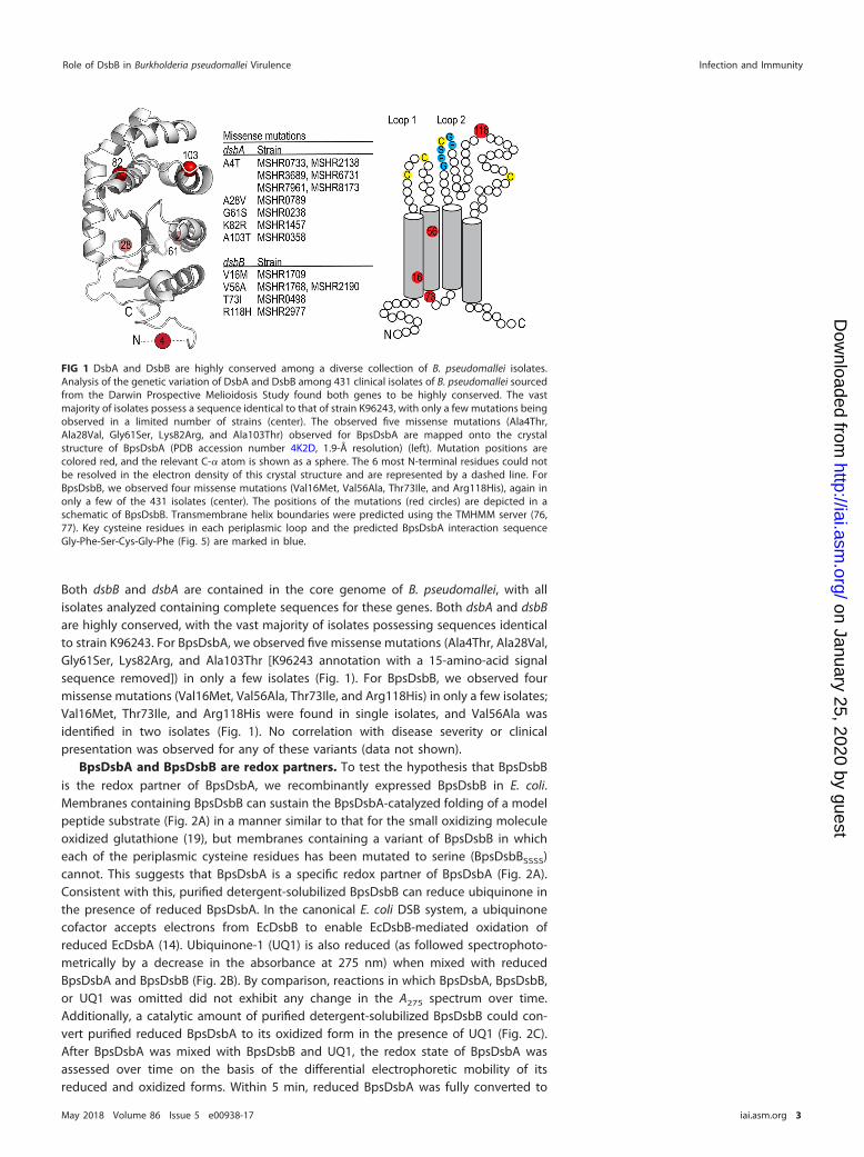
Both dsbB and dsbA are contained in the core genome of B. pseudomallei, with allisolates analyzed containing complete sequences for these genes. Both dsbA and dsbBare highly conserved, with the vast majority of isolates possessing sequences identicalto strain K96243. For BpsDsbA, we observed five missense mutations (Ala4Thr, Ala28Val,Gly61Ser, Lys82Arg, and Ala103Thr [K96243 annotation with a 15-amino-acid signalsequence removed]) in only a few isolates (Fig. 1). For BpsDsbB, we observed fourmissense mutations (Val16Met, Val56Ala, Thr73Ile, and Arg118His) in only a few isolates;Val16Met, Thr73Ile, and Arg118His were found in single isolates, and Val56Ala wasidentified in two isolates (Fig. 1). No correlation with disease severity or clinicalpresentation was observed for any of these variants (data not shown).
BpsDsbA and BpsDsbB are redox partners. To test the hypothesis that BpsDsbBis the redox partner of BpsDsbA, we recombinantly expressed BpsDsbB in E. coli.Membranes containing BpsDsbB can sustain the BpsDsbA-catalyzed folding of a modelpeptide substrate (Fig. 2A) in a manner similar to that for the small oxidizing moleculeoxidized glutathione (19), but membranes containing a variant of BpsDsbB in whicheach of the periplasmic cysteine residues has been mutated to serine (BpsDsbBSSSS)cannot. This suggests that BpsDsbA is a specific redox partner of BpsDsbA (Fig. 2A).Consistent with this, purified detergent-solubilized BpsDsbB can reduce ubiquinone inthe presence of reduced BpsDsbA. In the canonical E. coli DSB system, a ubiquinonecofactor accepts electrons from EcDsbB to enable EcDsbB-mediated oxidation ofreduced EcDsbA (14). Ubiquinone-1 (UQ1) is also reduced (as followed spectrophoto-metrically by a decrease in the absorbance at 275 nm) when mixed with reducedBpsDsbA and BpsDsbB (Fig. 2B). By comparison, reactions in which BpsDsbA, BpsDsbB,or UQ1 was omitted did not exhibit any change in the A275 spectrum over time.Additionally, a catalytic amount of purified detergent-solubilized BpsDsbB could con-vert purified reduced BpsDsbA to its oxidized form in the presence of UQ1 (Fig. 2C).After BpsDsbA was mixed with BpsDsbB and UQ1, the redox state of BpsDsbA wasassessed over time on the basis of the differential electrophoretic mobility of itsreduced and oxidized forms. Within 5 min, reduced BpsDsbA was fully converted to
FIG 1 DsbA and DsbB are highly conserved among a diverse collection of B. pseudomallei isolates.Analysis of the genetic variation of DsbA and DsbB among 431 clinical isolates of B. pseudomallei sourcedfrom the Darwin Prospective Melioidosis Study found both genes to be highly conserved. The vastmajority of isolates possess a sequence identical to that of strain K96243, with only a few mutations beingobserved in a limited number of strains (center). The observed five missense mutations (Ala4Thr,Ala28Val, Gly61Ser, Lys82Arg, and Ala103Thr) observed for BpsDsbA are mapped onto the crystalstructure of BpsDsbA (PDB accession number 4K2D, 1.9-Å resolution) (left). Mutation positions arecolored red, and the relevant C-� atom is shown as a sphere. The 6 most N-terminal residues could notbe resolved in the electron density of this crystal structure and are represented by a dashed line. ForBpsDsbB, we observed four missense mutations (Val16Met, Val56Ala, Thr73Ile, and Arg118His), again inonly a few of the 431 isolates (center). The positions of the mutations (red circles) are depicted in aschematic of BpsDsbB. Transmembrane helix boundaries were predicted using the TMHMM server (76,77). Key cysteine residues in each periplasmic loop and the predicted BpsDsbA interaction sequenceGly-Phe-Ser-Cys-Gly-Phe (Fig. 5) are marked in blue.
Role of DsbB in Burkholderia pseudomallei Virulence Infection and Immunity
May 2018 Volume 86 Issue 5 e00938-17 iai.asm.org 3
on January 25, 2020 by guesthttp://iai.asm
.org/D
ownloaded from
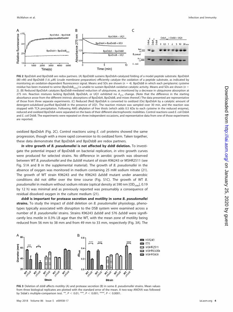
oxidized BpsDsbA (Fig. 2C). Control reactions using E. coli proteins showed the sameprogression, though with a more rapid conversion to its oxidized form. Taken together,these data demonstrate that BpsDsbA and BpsDsbB are redox partners.
In vitro growth of B. pseudomallei is not affected by dsbB deletion. To investi-gate the potential impact of BpsDsbB on bacterial replication, in vitro growth curveswere produced for selected strains. No difference in aerobic growth was observedbetween WT B. pseudomallei and the ΔdsbB mutant of strain K96243 or MSHR2511 (seeFig. S1A and B in the supplemental material). The growth of B. pseudomallei in theabsence of oxygen was monitored in medium containing 25 mM sodium nitrate (21).The growth of WT strain K96243 and the K96243 ΔdsbB mutant under anaerobicconditions did not differ over the time course (Fig. S1C). The growth of WT B.pseudomallei in medium without sodium nitrate (optical density at 590 nm [OD590], 0.19by 12 h) was minimal and as previously reported was presumably a consequence ofresidual dissolved oxygen in the culture medium (21).
dsbB is important for protease secretion and motility in some B. pseudomalleistrains. To study the impact of dsbB deletion on B. pseudomallei physiology, pheno-types typically associated with disruption to the DSB system were examined across anumber of B. pseudomallei strains. Strains K96243 ΔdsbB and 576 ΔdsbB were signifi-cantly less motile in 0.3% LB agar than the WT, with the mean zone of motility beingreduced from 56 mm to 38 mm and from 49 mm to 33 mm, respectively (Fig. 3A). The
FIG 2 BpsDsbA and BpsDsbB are redox partners. (A) BpsDsbB sustains BpsDsbA-catalyzed folding of a model peptide substrate. BpsDsbA(80 nM) and BpsDsbB (1.6 �M) (crude membrane preparation) efficiently catalyze the oxidation of a peptide substrate, as indicated bymonitoring an oxidation-dependent fluorescence signal. Means and SDs are shown (n � 4). BpsDsbB in which each periplasmic cysteineresidue has been mutated to serine (BpsDsbBSSSS) is unable to sustain BpsDsbA oxidative catalytic activity. Means and SDs are shown (n �2). (B) Reduced BpsDsbA catalyzes BpsDsbB-mediated reduction of ubiquinone, as monitored by a decrease in ubiquinone absorption at275 nm. Reaction mixtures lacking BpsDsbB, BpsDsbA, or UQ1 exhibited no A275 change. (Note that the difference in the startingabsorbance arose from the different intrinsic absorptions of BpsDsbA, BpsDsbB, and mixes thereof.) The data presented are representativeof those from three separate experiments. (C) Reduced (Red) BpsDsbA is converted to oxidized (Ox) BpsDsbA by a catalytic amount ofdetergent-solubilized purified BpsDsbB in the presence of UQ1. The reaction mixture was sampled over 30 min, and the reaction wasstopped with TCA precipitation. Following AMS alkylation of free thiols (which adds 0.5 kDa to each cysteine in the reduced enzyme),reduced and oxidized BpsDsbA were separated on the basis of their different electrophoretic mobilities. Control reactions used E. coli DsbAand E. coli DsbB. The experiments were repeated on three independent occasions, and representative data from one of those experimentsare reported.
FIG 3 Deletion of dsbB affects motility (A) and protease secretion (B) in some B. pseudomallei strains. Mean valuesfrom three biological replicates are plotted with the standard error of the mean. A two-way ANOVA was followedby Sidak’s multiple-comparison test. **, P � 0.01; ***, P � 0.001; ****, P � 0.0001.
McMahon et al. Infection and Immunity
May 2018 Volume 86 Issue 5 e00938-17 iai.asm.org 4
on January 25, 2020 by guesthttp://iai.asm
.org/D
ownloaded from
less severe reduction in motility observed for these dsbB deletion strains than for a dsbAdeletion strain (19) is consistent with motility data reported previously (22, 23). Incontrast, no difference in motility was observed between WT parental strains andtheir ΔdsbB variants in the Australian clinical isolates MSHR2511, MSHR0305b, andMSHR5858. Further motility studies using 0.3% Muller-Hinton agar and a 0.3%defined medium also showed no difference in motility between the WT and ΔdsbBmutant of the Australian clinical isolates.
Compounds such as cystine have previously been shown to chemically complementthe dsbB mutation in other bacteria (22, 23). Therefore, the minimal medium M9 wasselected for use for the preparation of a B. pseudomallei culture supernatant. Proteaseactivity was evident in culture supernatants prepared from all strains tested (Fig. 3B).Three of the dsbB deletion strains, including K96243, 576, and MSHR5858, exhibited asignificant reduction in protease activity, resulting in 1.76-, 4.65-, and 1.51-fold reduc-tions in activity, respectively, compared to that of the WT (Fig. 3B). Trypsin (28.6 units)was used as a positive control for azocasein hydrolysis, reaching a mean A440 value of0.33 � 0.009 (standard error of the mean [SEM], n � 3). Heat-treated culture superna-tant (80°C for 20 min) served as a negative control for each sample, with mean A440
values ranging from 0.01 � 0.003 to 0.02 � 0.009 (SEM). The effect of the dsbB deletionin B. pseudomallei strains grown in LB broth followed the same trend as the effect of thedeletion in B. pseudomallei strains grown in M9 medium.
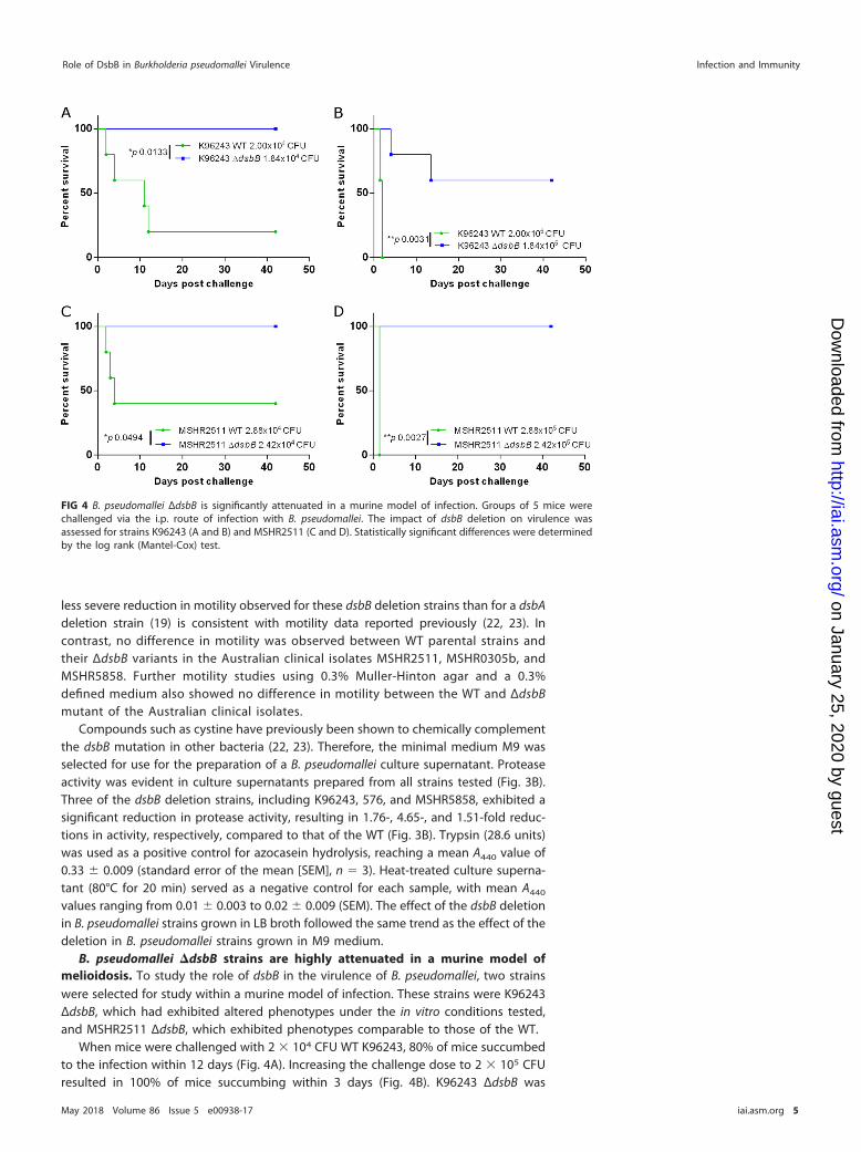
B. pseudomallei �dsbB strains are highly attenuated in a murine model ofmelioidosis. To study the role of dsbB in the virulence of B. pseudomallei, two strainswere selected for study within a murine model of infection. These strains were K96243ΔdsbB, which had exhibited altered phenotypes under the in vitro conditions tested,and MSHR2511 ΔdsbB, which exhibited phenotypes comparable to those of the WT.
When mice were challenged with 2 � 104 CFU WT K96243, 80% of mice succumbedto the infection within 12 days (Fig. 4A). Increasing the challenge dose to 2 � 105 CFUresulted in 100% of mice succumbing within 3 days (Fig. 4B). K96243 ΔdsbB was
FIG 4 B. pseudomallei ΔdsbB is significantly attenuated in a murine model of infection. Groups of 5 mice werechallenged via the i.p. route of infection with B. pseudomallei. The impact of dsbB deletion on virulence wasassessed for strains K96243 (A and B) and MSHR2511 (C and D). Statistically significant differences were determinedby the log rank (Mantel-Cox) test.
Role of DsbB in Burkholderia pseudomallei Virulence Infection and Immunity
May 2018 Volume 86 Issue 5 e00938-17 iai.asm.org 5
on January 25, 2020 by guesthttp://iai.asm
.org/D
ownloaded from
significantly attenuated within the mouse model at both challenge doses tested, with100% survival at the challenge dose of 1.84 � 104 CFU (Fig. 4A) and 60% survival at thehigher challenge dose of 1.84 � 105 CFU (Fig. 4B). MSHR2511 ΔdsbB was also signifi-cantly attenuated compared to the WT strain in the mouse model, with 100% survivalof mice at either challenge dose (Fig. 4C and D). For mice challenged with 2.88 � 104
CFU MSHR2511, 60% succumbed within 4 days, while at a challenge dose of 2.88 � 105
CFU, 100% of mice succumbed within 1.5 days.A peptide derived from the second periplasmic loop of BpsDsbB binds to the
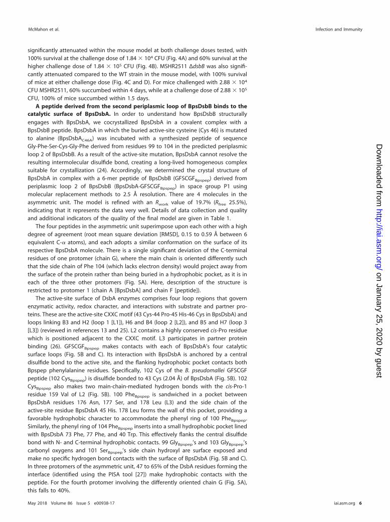
catalytic surface of BpsDsbA. In order to understand how BpsDsbB structurallyengages with BpsDsbA, we cocrystallized BpsDsbA in a covalent complex with aBpsDsbB peptide. BpsDsbA in which the buried active-site cysteine (Cys 46) is mutatedto alanine (BpsDsbAC46A) was incubated with a synthesized peptide of sequenceGly-Phe-Ser-Cys-Gly-Phe derived from residues 99 to 104 in the predicted periplasmicloop 2 of BpsDsbB. As a result of the active-site mutation, BpsDsbA cannot resolve theresulting intermolecular disulfide bond, creating a long-lived homogeneous complexsuitable for crystallization (24). Accordingly, we determined the crystal structure ofBpsDsbA in complex with a 6-mer peptide of BpsDsbB (GFSCGFBpspep) derived fromperiplasmic loop 2 of BpsDsbB (BpsDsbA-GFSCGFBpspep) in space group P1 usingmolecular replacement methods to 2.5 Å resolution. There are 4 molecules in theasymmetric unit. The model is refined with an Rwork value of 19.7% (Rfree 25.5%),indicating that it represents the data very well. Details of data collection and qualityand additional indicators of the quality of the final model are given in Table 1.
The four peptides in the asymmetric unit superimpose upon each other with a highdegree of agreement (root mean square deviation [RMSD], 0.15 to 0.59 Å between 6equivalent C-� atoms), and each adopts a similar conformation on the surface of itsrespective BpsDsbA molecule. There is a single significant deviation of the C-terminalresidues of one protomer (chain G), where the main chain is oriented differently suchthat the side chain of Phe 104 (which lacks electron density) would project away fromthe surface of the protein rather than being buried in a hydrophobic pocket, as it is ineach of the three other protomers (Fig. 5A). Here, description of the structure isrestricted to protomer 1 (chain A [BpsDsbA] and chain F [peptide]).
The active-site surface of DsbA enzymes comprises four loop regions that governenzymatic activity, redox character, and interactions with substrate and partner pro-teins. These are the active-site CXXC motif (43 Cys-44 Pro-45 His-46 Cys in BpsDsbA) andloops linking B3 and H2 (loop 1 [L1]), H6 and B4 (loop 2 [L2]), and B5 and H7 (loop 3[L3]) (reviewed in references 13 and 25). L2 contains a highly conserved cis-Pro residuewhich is positioned adjacent to the CXXC motif. L3 participates in partner proteinbinding (26). GFSCGFBpspep makes contacts with each of BpsDsbA’s four catalyticsurface loops (Fig. 5B and C). Its interaction with BpsDsbA is anchored by a centraldisulfide bond to the active site, and the flanking hydrophobic pocket contacts bothBpspep phenylalanine residues. Specifically, 102 Cys of the B. pseudomallei GFSCGFpeptide (102 CysBpspep) is disulfide bonded to 43 Cys (2.04 Å) of BpsDsbA (Fig. 5B). 102CysBpspep also makes two main-chain-mediated hydrogen bonds with the cis-Pro-1residue 159 Val of L2 (Fig. 5B). 100 PheBpspep is sandwiched in a pocket betweenBpsDsbA residues 176 Asn, 177 Ser, and 178 Leu (L3) and the side chain of theactive-site residue BpsDsbA 45 His. 178 Leu forms the wall of this pocket, providing afavorable hydrophobic character to accommodate the phenyl ring of 100 PheBpspep.Similarly, the phenyl ring of 104 PheBpspep inserts into a small hydrophobic pocket linedwith BpsDsbA 73 Phe, 77 Phe, and 40 Trp. This effectively flanks the central disulfidebond with N- and C-terminal hydrophobic contacts. 99 GlyBpspep’s and 103 GlyBpspep’scarbonyl oxygens and 101 SerBpspep’s side chain hydroxyl are surface exposed andmake no specific hydrogen bond contacts with the surface of BpsDsbA (Fig. 5B and C).In three protomers of the asymmetric unit, 47 to 65% of the DsbA residues forming theinterface (identified using the PISA tool [27]) make hydrophobic contacts with thepeptide. For the fourth protomer involving the differently oriented chain G (Fig. 5A),this falls to 40%.
McMahon et al. Infection and Immunity
May 2018 Volume 86 Issue 5 e00938-17 iai.asm.org 6
on January 25, 2020 by guesthttp://iai.asm
.org/D
ownloaded from
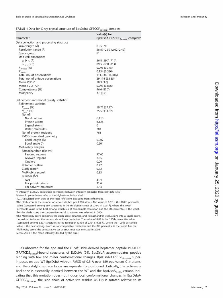
As observed for the apo and the E. coli DsbB-derived heptamer peptide PFATCDS(PFATCDSEcpep)-bound structures of EcDsbA (24), BpsDsbA accommodates peptidebinding with few and minor conformational changes. BpsDsbA-GFSCGFBpspep super-imposes on apo WT BpsDsbA with an RMSD of 0.3 Å over 189 equivalent C-� atoms,and the catalytic surface loops are equivalently positioned. Critically, the active-sitebackbone is essentially identical between the WT and the BpsDsbAC46A variant, indi-cating that this mutation does not induce local conformational changes. In BpsDsbA-GFSCGFBpspep, the side chain of active-site residue 45 His is rotated relative to its
TABLE 1 Data for X-ray crystal structure of BpsDsbA-GFSCGFBpspep complex
ParameterValue(s) forBpsDsbA-GFSCGFBpspep complexb
Data collection and processing statisticsWavelength (Å) 0.95370Resolution range (Å) 58.87–2.59 (2.62–2.49)Space group P1Unit cell dimensions
a, b, c (Å) 56.8, 59.7, 71.7�, �, � (°) 89.5, 67.8, 81.0
Rmerge (%) 0.095 (0.375)Rmeas 0.134 (0.530)Total no. of observations 111,338 (14,316)Total no. of unique observations 29,114 (3,835)Mean I/SD If 10.3 (3.0)Mean I CC(1/2)a 0.993 (0.856)Completeness (%) 96.6 (87.7)Multiplicity 3.8 (3.7)
Refinement and model quality statisticsRefinement statistics
Rfactor (%) 19.71 (27.17)Rfree
c (%) 25.50 (34.82)No. of:
Non-H atoms 6,410Protein atoms 6,126Ligand atoms 0Water molecules 284
No. of protein residues 781RMSD from ideal geometry
Bond length (Å) 0.002Bond angle (°) 0.50
MolProbity analysisRamachandran plot (%)
Favored regions 97.65Allowed regions 2.35Outliers 0.00
Rotamer outliers 0.77Clash scored 0.82MolProbity scoree 0.83B factor (Å2)
Avg 31.4For protein atoms 31.6For solvent molecules 27.4
aI, intensity; CC(1/2), correlation coefficient between intensity estimates from half data sets.bValues in parentheses refer to the highest-resolution shell.cRfree calculated over 5.0% of the total reflections excluded from refinement.dThe clash score is the number of serious clashes per 1,000 atoms. The value of 0.82 is the 100th percentilevalue (compared among 269 structures in the resolution range of 2.490 � 0.25 Å), where the 100thpercentile value is the best among structures of comparable resolution and the 0th percentile is the worst.For the clash score, the comparative set of structures was selected in 2004.
eThe MolProbity score combines the clash score, rotamer, and Ramachandran evaluations into a single score,normalized to be on the same scale as X-ray resolution. The value of 0.83 is the 100th percentile value(compared among 6,897 structures in the resolution range of 2.49 � 0.25 Å), where the 100th percentilevalue is the best among structures of comparable resolution and the 0th percentile is the worst. For theMolProbity score, the comparative set of structures was selected in 2006.
fMean I/SD I is the mean intensity divided by the error.
Role of DsbB in Burkholderia pseudomallei Virulence Infection and Immunity
May 2018 Volume 86 Issue 5 e00938-17 iai.asm.org 7
on January 25, 2020 by guesthttp://iai.asm
.org/D
ownloaded from
position in the apo WT BpsDsbA, thus accommodating 100 PheBpspep in the complexstructure. Equivalent active-site His residues have been observed to be mobile in otherDsbA structures (e.g., Pseudomonas aeruginosa DsbA [28]).
The surface area of the BpsDsbA-GFSCGFBpspep interface is 445 Å2 with a shapecomplementarity score of 1.0 (calculated using the PISA tool [27]). This compares to aninterface area of 784.3 Å2 and a shape complementarity score of 0.836 for EcDsbA andEcDsbB and is consistent with the hypothesis that DsbA-DsbB protein-protein interac-tions may be typified by relatively small surface areas but high shape complementarity(26).
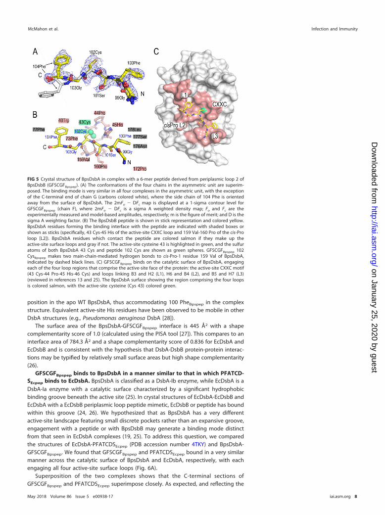
GFSCGFBpspep binds to BpsDsbA in a manner similar to that in which PFATCD-SEcpep binds to EcDsbA. BpsDsbA is classified as a DsbA-Ib enzyme, while EcDsbA is aDsbA-Ia enzyme with a catalytic surface characterized by a significant hydrophobicbinding groove beneath the active site (25). In crystal structures of EcDsbA-EcDsbB andEcDsbA with a EcDsbB periplasmic loop peptide mimetic, EcDsbB or peptide has boundwithin this groove (24, 26). We hypothesized that as BpsDsbA has a very differentactive-site landscape featuring small discrete pockets rather than an expansive groove,engagement with a peptide or with BpsDsbB may generate a binding mode distinctfrom that seen in EcDsbA complexes (19, 25). To address this question, we comparedthe structures of EcDsbA-PFATCDSEcpep (PDB accession number 4TKY) and BpsDsbA-GFSCGFBpspep. We found that GFSCGFBpspep and PFATCDSEcpep bound in a very similarmanner across the catalytic surface of BpsDsbA and EcDsbA, respectively, with eachengaging all four active-site surface loops (Fig. 6A).
Superposition of the two complexes shows that the C-terminal sections ofGFSCGFBpspep and PFATCDSEcpep superimpose closely. As expected, and reflecting the
FIG 5 Crystal structure of BpsDsbA in complex with a 6-mer peptide derived from periplasmic loop 2 ofBpsDsbB (GFSCGFBpspep). (A) The conformations of the four chains in the asymmetric unit are superim-posed. The binding mode is very similar in all four complexes in the asymmetric unit, with the exceptionof the C-terminal end of chain G (carbons colored white), where the side chain of 104 Phe is orientedaway from the surface of BpsDsbA. The 2mFo � DFc map is displayed at a 1-sigma contour level forGFSCGFBpspep (chain F), where 2mFo � DFc is a sigma A weighted density map; Fo and Fc are theexperimentally measured and model-based amplitudes, respectively; m is the figure of merit; and D is thesigma A weighting factor. (B) The BpsDsbB peptide is shown in stick representation and colored yellow.BpsDsbA residues forming the binding interface with the peptide are indicated with shaded boxes orshown as sticks (specifically, 43 Cys-45 His of the active-site CXXC loop and 159 Val-160 Pro of the cis-Proloop [L2]). BpsDsbA residues which contact the peptide are colored salmon if they make up theactive-site surface loops and gray if not. The active-site cysteine 43 is highlighted in green, and the sulfuratoms of both BpsDsbA 43 Cys and peptide 102 Cys are shown as green spheres. GFSCGFBpspep 102CysBpspep makes two main-chain-mediated hydrogen bonds to cis-Pro-1 residue 159 Val of BpsDsbA,indicated by dashed black lines. (C) GFSCGFBpspep binds on the catalytic surface of BpsDsbA, engagingeach of the four loop regions that comprise the active-site face of the protein: the active-site CXXC motif(43 Cys-44 Pro-45 His-46 Cys) and loops linking B3 and H2 (L1), H6 and B4 (L2), and B5 and H7 (L3)(reviewed in references 13 and 25). The BpsDsbA surface showing the region comprising the four loopsis colored salmon, with the active-site cysteine (Cys 43) colored green.
McMahon et al. Infection and Immunity
May 2018 Volume 86 Issue 5 e00938-17 iai.asm.org 8
on January 25, 2020 by guesthttp://iai.asm
.org/D
ownloaded from
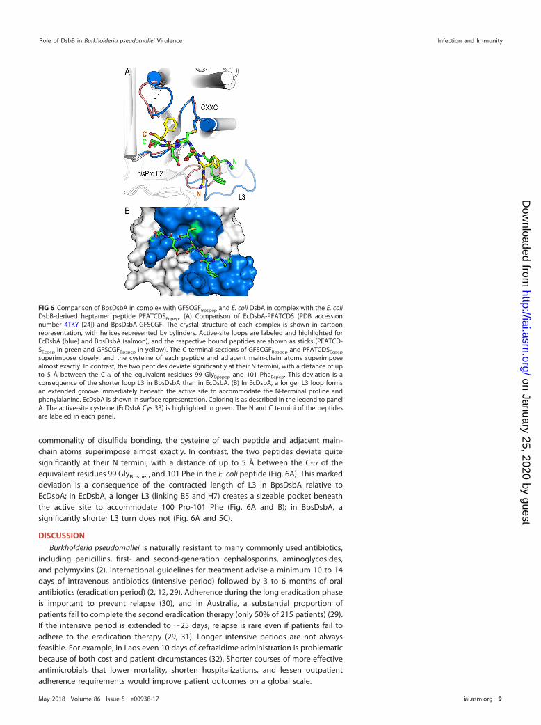
commonality of disulfide bonding, the cysteine of each peptide and adjacent main-chain atoms superimpose almost exactly. In contrast, the two peptides deviate quitesignificantly at their N termini, with a distance of up to 5 Å between the C-� of theequivalent residues 99 GlyBpspep and 101 Phe in the E. coli peptide (Fig. 6A). This markeddeviation is a consequence of the contracted length of L3 in BpsDsbA relative toEcDsbA; in EcDsbA, a longer L3 (linking B5 and H7) creates a sizeable pocket beneaththe active site to accommodate 100 Pro-101 Phe (Fig. 6A and B); in BpsDsbA, asignificantly shorter L3 turn does not (Fig. 6A and 5C).
DISCUSSION
Burkholderia pseudomallei is naturally resistant to many commonly used antibiotics,including penicillins, first- and second-generation cephalosporins, aminoglycosides,and polymyxins (2). International guidelines for treatment advise a minimum 10 to 14days of intravenous antibiotics (intensive period) followed by 3 to 6 months of oralantibiotics (eradication period) (2, 12, 29). Adherence during the long eradication phaseis important to prevent relapse (30), and in Australia, a substantial proportion ofpatients fail to complete the second eradication therapy (only 50% of 215 patients) (29).If the intensive period is extended to �25 days, relapse is rare even if patients fail toadhere to the eradication therapy (29, 31). Longer intensive periods are not alwaysfeasible. For example, in Laos even 10 days of ceftazidime administration is problematicbecause of both cost and patient circumstances (32). Shorter courses of more effectiveantimicrobials that lower mortality, shorten hospitalizations, and lessen outpatientadherence requirements would improve patient outcomes on a global scale.
FIG 6 Comparison of BpsDsbA in complex with GFSCGFBpspep and E. coli DsbA in complex with the E. coliDsbB-derived heptamer peptide PFATCDSEcpep. (A) Comparison of EcDsbA-PFATCDS (PDB accessionnumber 4TKY [24]) and BpsDsbA-GFSCGF. The crystal structure of each complex is shown in cartoonrepresentation, with helices represented by cylinders. Active-site loops are labeled and highlighted forEcDsbA (blue) and BpsDsbA (salmon), and the respective bound peptides are shown as sticks (PFATCD-SEcpep in green and GFSCGFBpspep in yellow). The C-terminal sections of GFSCGFBpspep and PFATCDSEcpep
superimpose closely, and the cysteine of each peptide and adjacent main-chain atoms superimposealmost exactly. In contrast, the two peptides deviate significantly at their N termini, with a distance of upto 5 Å between the C-� of the equivalent residues 99 GlyBpspep and 101 PheEcpep. This deviation is aconsequence of the shorter loop L3 in BpsDsbA than in EcDsbA. (B) In EcDsbA, a longer L3 loop formsan extended groove immediately beneath the active site to accommodate the N-terminal proline andphenylalanine. EcDsbA is shown in surface representation. Coloring is as described in the legend to panelA. The active-site cysteine (EcDsbA Cys 33) is highlighted in green. The N and C termini of the peptidesare labeled in each panel.
Role of DsbB in Burkholderia pseudomallei Virulence Infection and Immunity
May 2018 Volume 86 Issue 5 e00938-17 iai.asm.org 9
on January 25, 2020 by guesthttp://iai.asm
.org/D
ownloaded from
Bacterial DSB proteins are emerging as a promising new avenue for antivirulencetherapeutics (33). We have demonstrated here that B. pseudomallei DsbB is the partnerprotein of the known virulence regulator BpsDsbA (19) and contributes to virulence ina mouse model of melioidosis. Deletion of dsbB from B. pseudomallei K96243 andMSHR2511 protected mice from lethal peritoneal infection, demonstrating thatBpsDsbB is a potential drug target for antimicrobial development. Furthermore, theprotein sequence of BpsDsbB is highly conserved among 431 diverse clinical isolates ofB. pseudomallei, underpinning the importance of this enzyme in the bacterium andsuggesting that BpsDsbB inhibitors could be broadly effective in melioidosis. Theobserved mutations in BpsDsbA do not map to the active-site surface loops ofthe protein structure (Fig. 1) and thus are not predicted to alter protein function.The structure of BpsDsbB has not yet been determined. Three of the four observedmutations are predicted to be located on a transmembrane helix or to face thecytoplasm (Fig. 1). A fourth mutation (Arg118His) is predicted to be in the secondperiplasmic loop. This mutation has the potential to alter interactions with BpsDsbA,but we note that the modification is conservative and maintains a positively chargedside chain.
dsbB deletion likely impacts B. pseudomallei virulence because it interrupts thenormal redox activity of BpsDsbA. DsbA oxidatively folds disulfide bonds in substrateproteins and is itself reduced in the process. DsbB in turn accepts electrons from DsbAto restore it to its oxidized primed state, ready for the next substrate folding event.Consistent with this, the phenotype of K96243 ΔdsbB mimics the in vivo phenotype ofK96243 ΔdsbA in our infection model and broadly mimics the in vitro K96243 ΔdsbAphenotypes assayed, although we note that the magnitude of the effect on proteasesecretion and motility is reduced relative to that observed in K96243 ΔdsbA. Thisrecapitulation of the ΔdsbA mutant phenotypes is consistent with a BpsDsbB-mediatedloss of BpsDsbA activity in the current study.
We also showed that both the K96423 ΔdsbB and MSHR2511 ΔdsbB strains arehighly attenuated in an animal model of infection. Surprisingly, these dsbB deletionstrains exhibited very different effects in two standard in vitro virulence assays. Thissuggests that the in vitro experiments, which examine just two hallmarks of virulence,do not fully reflect the impact of BpsDsbB on virulence. We determined that deletionof dsbB does not affect the growth of B. pseudomallei in vitro under either aerobic oranaerobic conditions; wild-type and mutant growth curve profiles were equivalent, andthere was no intrinsic growth defect caused by the gene deletion (see Fig. S1 in thesupplemental material). Disruption of the DsbA/DsbB pathway is likely to impact manydisulfide bond-containing substrates, and the assessment of just two phenotypes doesnot characterize the broader impact of dsbB disruption. This highlights the challenge ofidentifying the mechanism(s) by which dsbB deletion leads to a reduction in virulence,as changes to virulence are likely to be the consequence of disruption of multiplepathways, potentially across time. A future study to unpick the precise impact of dsbBdeletion on B. pseudomallei infectivity, elicitation of an inflammatory response, andbacterial dissemination would be of great interest in light of reports that deletion ofindividual DSB proteins in a number of species results in pleiotropic phenotypes in vitro,including alterations to extracellular enzyme formation (34), adhesion (35), toxin pro-duction and secretion (36), competence (37), and antibiotic sensitivity (22). In vivo, theadditional environmental and immune system stresses encountered by the bacteriawithin the host add further complexity to understanding how dsbB loss disruptsvirulence. For example, quantification of bacterial numbers in infected organs ormacrophages can report changes in bacterial load but does not distinguish between agrowth defect (though our in vitro growth curve experiments do not suggest this wouldbe the case for ΔdsbB bacteria) and a reduced capacity to survive and disseminate inthe host environment. The loss of pathogenicity in the animal model reported here isconclusive evidence of the key role played by BpsDsbB in virulence in vivo, although theprecise pathway(s) by which the observed attenuation is achieved is yet to be elucidated.In future, it will also be important to analyze a panel of strains, as we have done, and to
McMahon et al. Infection and Immunity
May 2018 Volume 86 Issue 5 e00938-17 iai.asm.org 10
on January 25, 2020 by guesthttp://iai.asm
.org/D
ownloaded from
investigate the effect of deletions in animal models of infection to establish roles forvirulence and to test potential therapeutics. The phenotypic range observed in the in vitroassays likely also reflects the known diversity of the B. pseudomallei species (38, 39, 40).
There has recently been significant progress in the identification of inhibitors ofDsbB and DsbB-like bacterial proteins (33). The DsbB-like protein vitamin K epoxide(VKOR) in Mycobacterium tuberculosis can be inhibited by warfarin (41). Inhibitors of E.coli DsbB have been identified by fragment-based screening using purified protein(50% inhibitory concentration [IC50], 7 to 200 �M) (42). These were later advanced toan IC50 of �1 �M with a rational medicinal chemistry program (43). Additional EcDsbBinhibitors resulted from high-throughput screening in E. coli cells (44). Significantly, thelead compound in this study also inhibits DsbB proteins from other bacterial species.Together these data indicate that DsbB can be effectively inhibited and that successfulcompounds can have broad-spectrum activity.
The 2.5-Å-resolution structure of a BpsDsbB peptide in complex with BpsDsbA thatwe report provides an important framework for future rational efforts to identify anddevelop small-molecule inhibitors of BpsDsbA. This is the fourth reported structure ofa DsbA in a covalent (disulfide or thioether) complex with a peptide, joining EcDsbA incomplex with a DsbB-derived peptide (PDB accession number 4TKY [24]), EcDsbA incomplex with a substrate peptide from SigA (PDB accession number 3DKS [45]), andXylella fastidiosa DsbA in complex with a peptide of unknown sequence (PDB accessionnumber 2REM [46]). The peptide cysteine (or homoserine; PDB accession number 3DKS)and the flanking residues Cys �1 and Cys �1 are very similarly positioned in eachstructure. The peptide cysteine is engaged in a disulfide bond with the N-terminalactive-site Cys. It also makes two conserved main-chain hydrogen bonds to main-chainatoms of the neighboring cis-Pro-1 residue. Note that in the EcDsbA-SigA complex thedisulfide bond is replaced by a thioether bond and one of the main-chain bonds tocis-Pro-1 is water mediated, but the mode of interaction is highly similar to thatobserved in the disulfide-bonded DsbA-peptide complexes. There are a very limitednumber of DsbA-peptide structures from which to draw broad conclusions, but nev-ertheless, it seems that the conserved required DsbA-binding motif is minimal: adisulfide bond, and two hydrogen bonds between the backbone atoms of the incom-ing Cys and the DsbA cis-Pro-1 residue. Small molecules or peptidomimetics that blockthis site may inhibit BpsDsbA-BpsDsbB interaction and activity, though specificity withrespect to other thioredoxin fold proteins would also need to be considered. On thispoint, we note that small-molecule inhibitors of EcDsbA have been reported (47). Theseinhibitors recapitulate the hydrophobic interactions observed between an EcDsbB-derived peptide and the EcDsbA active-site surface groove, have inhibitory activity in amodel oxidative folding assay, and disrupt E. coli cell motility, presumably by disturbingproductive interactions between EcDsbA and EcDsbB and/or substrates.
DSB enzymes are not essential for aerobic growth in nutrient-rich media, and DsbA- andDsbB-deficient B. pseudomallei strains show no loss of viability or growth rate under theseconditions (19; the present study). Antimicrobials that inhibit DSB proteins under aerobicconditions would thus be antivirulence agents rather than antibiotics. The potential ad-vantages of antivirulence agents are manifold (48). Antivirulence agents do not kill bacteriaor prevent their growth, so it is likely that they will exert a reduced selection pressure fordevelopment of resistance (49), and as they target virulence effectors, they may sparecommensal bacteria. New antibiotic discovery is slow, and just five of the antibioticsapproved since 2000 have been first in class (50). New antimicrobials with a novel mode ofaction are urgently required to reinvigorate our antimicrobial resources. Antivirulencedrugs, used in combination with standard antimicrobials, may revitalize current therapeu-tics no longer deemed clinically useful due to widespread resistance.
MATERIALS AND METHODSBacterial strains and growth conditions. Five B. pseudomallei strains were selected for the current
work on the basis of their diverse genetic and clinical backgrounds: two clinical Thai isolates, K96243 (51)and 576 (52), and three Australian clinical isolates, MSHR0305b (53), MSHR2511, and MSHR5858 (54, 55).MSHR0305b is from patient P101, a nondiabetic male with fatal neurological melioidosis. Importantly,
Role of DsbB in Burkholderia pseudomallei Virulence Infection and Immunity
May 2018 Volume 86 Issue 5 e00938-17 iai.asm.org 11
on January 25, 2020 by guesthttp://iai.asm
.org/D
ownloaded from
P101 had a mixed infection with two genetically disparate strains, MSHR305 (56) and MSHR0305b.MSHR2511 was from a nondiabetic male with rapidly fatal pulmonary melioidosis. MSHR5858 is from amale diabetic patient (patient P730) who presented with acute pulmonary melioidosis. P730 ultimatelysurvived his infection. MSHR5858 was chosen for this study because it belongs to sequence type 562, agroup of strains isolated in Australia but hypothesized to be a recent introduction into Australia from Asia(57). Strains 576 and MSHR0305b contain lipopolysaccharide (LPS) type B, whereas strains MSHR2511,MSHR5858, and K96243 contain LPS type A. B. pseudomallei isolates were grown on Ashdown’s agar(Oxoid), chocolate agar (Oxoid), or LB agar at 37°C under aerobic conditions.
Ethics statement. Bacterial samples were obtained from the Darwin Prospective Melioidosis Studybacterial isolate collection, with ethical approval obtained through the Human Research Ethics Commit-tee of the Northern Territory Department of Health and the Menzies School of Health Research (approvalno. HREC 02/38, Clinical and epidemiological features of melioidosis). All patient data, with the exceptionof clinical presentation/severity, were deidentified prior to analysis.
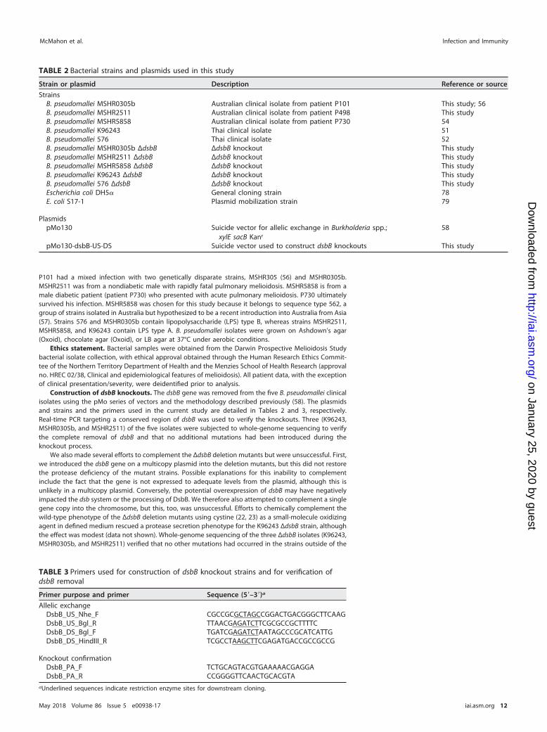
Construction of dsbB knockouts. The dsbB gene was removed from the five B. pseudomallei clinicalisolates using the pMo series of vectors and the methodology described previously (58). The plasmidsand strains and the primers used in the current study are detailed in Tables 2 and 3, respectively.Real-time PCR targeting a conserved region of dsbB was used to verify the knockouts. Three (K96243,MSHR0305b, and MSHR2511) of the five isolates were subjected to whole-genome sequencing to verifythe complete removal of dsbB and that no additional mutations had been introduced during theknockout process.
We also made several efforts to complement the ΔdsbB deletion mutants but were unsuccessful. First,we introduced the dsbB gene on a multicopy plasmid into the deletion mutants, but this did not restorethe protease deficiency of the mutant strains. Possible explanations for this inability to complementinclude the fact that the gene is not expressed to adequate levels from the plasmid, although this isunlikely in a multicopy plasmid. Conversely, the potential overexpression of dsbB may have negativelyimpacted the dsb system or the processing of DsbB. We therefore also attempted to complement a singlegene copy into the chromosome, but this, too, was unsuccessful. Efforts to chemically complement thewild-type phenotype of the ΔdsbB deletion mutants using cystine (22, 23) as a small-molecule oxidizingagent in defined medium rescued a protease secretion phenotype for the K96243 ΔdsbB strain, althoughthe effect was modest (data not shown). Whole-genome sequencing of the three ΔdsbB isolates (K96243,MSHR0305b, and MSHR2511) verified that no other mutations had occurred in the strains outside of the
TABLE 2 Bacterial strains and plasmids used in this study
Strain or plasmid Description Reference or source
StrainsB. pseudomallei MSHR0305b Australian clinical isolate from patient P101 This study; 56B. pseudomallei MSHR2511 Australian clinical isolate from patient P498 This studyB. pseudomallei MSHR5858 Australian clinical isolate from patient P730 54B. pseudomallei K96243 Thai clinical isolate 51B. pseudomallei 576 Thai clinical isolate 52B. pseudomallei MSHR0305b ΔdsbB ΔdsbB knockout This studyB. pseudomallei MSHR2511 ΔdsbB ΔdsbB knockout This studyB. pseudomallei MSHR5858 ΔdsbB ΔdsbB knockout This studyB. pseudomallei K96243 ΔdsbB ΔdsbB knockout This studyB. pseudomallei 576 ΔdsbB ΔdsbB knockout This studyEscherichia coli DH5� General cloning strain 78E. coli S17-1 Plasmid mobilization strain 79
PlasmidspMo130 Suicide vector for allelic exchange in Burkholderia spp.;
xylE sacB Kanr
58
pMo130-dsbB-US-DS Suicide vector used to construct dsbB knockouts This study
TABLE 3 Primers used for construction of dsbB knockout strains and for verification ofdsbB removal
Primer purpose and primer Sequence (5=–3=)a
Allelic exchangeDsbB_US_Nhe_F CGCCGCGCTAGCCGGACTGACGGGCTTCAAGDsbB_US_Bgl_R TTAACGAGATCTTCGCGCCGCTTTTCDsbB_DS_Bgl_F TGATCGAGATCTAATAGCCCGCATCATTGDsbB_DS_HindIII_R TCGCCTAAGCTTCGAGATGACCGCCGCCG
Knockout confirmationDsbB_PA_F TCTGCAGTACGTGAAAAACGAGGADsbB_PA_R CCGGGGTTCAACTGCACGTA
aUnderlined sequences indicate restriction enzyme sites for downstream cloning.
McMahon et al. Infection and Immunity
May 2018 Volume 86 Issue 5 e00938-17 iai.asm.org 12
on January 25, 2020 by guesthttp://iai.asm
.org/D
ownloaded from
dsbB region. Inspection of neighboring regions excluded the likelihood of polar effects of dsbB removal.We note that efforts to complement a B. pseudomallei ΔdsbA strain were also unsuccessful (19).
Whole-genome sequencing and comparative genomic analysis. Genomic data were generatedfrom paired-end Illumina reads using the HiSeq 2000 platform at the Australian Genome Research Facility(AGRF; Melbourne, Australia) or Macrogen, Inc. (Geumcheon-gu, Seoul, Republic of Korea). Analysis wasperformed with the SPANDx (version 3.0) pipeline (59), which wraps the BWA (60), SAMtools (61), GATK(62), and SnpEff (63) tools to identify genetic variants in comparison to a reference genome. Genomeswere assembled using the Microbial Genome Assembly pipeline (https://github.com/dsarov/MGAP-Microbial-Genome-Assembler-Pipeline).
BpsDsbB expression, membrane preparation, and purification. B. pseudomallei dsbB (UniProtaccession number Q63RY4) and a variant in which four cysteine residues predicted to be in theperiplasmic loops were mutated to serine (Cys41Ser, Cys44Ser, Cys102Ser, and Cys130Ser [BpsDsbBSSSS])were each inserted into a pET28a vector with a noncleavable C-terminal His8 tag. BpsDsbB andBpsDsbBSSSS were expressed in E. coli C41 cells in PASM autoinduction medium (64) with kanamycin(50 �g/ml) (16 h, 30°C, 220 rpm). Harvested cells were resuspended in phosphate-buffered saline (PBS;pH 7.4; 200 ml/liter culture) with DNase and protease inhibitors and lysed using a constant-pressure celldisrupter (two successive passages at 27,000 and 30,000 lb/in2, respectively). Large cellular debris wasremoved by centrifugation (15,000 � g, 4°C, 15 min). Membranes were isolated from the resultingsupernatant by ultracentrifugation (185,000 � g, 1 h 15 min, 4°C) and homogenized in 40 ml of PBS. Forpurification, membranes were solubilized in PBS and 1% (wt/vol) n-dodecyl-�-D-maltopyranoside (DDM;Glycon Biochemicals) with vigorous stirring (1 h, 4°C). Detergent-solubilized protein was isolated byultracentrifugation (170,000 � g, 1 h 15 min, 4°C), adjusted to a final concentration of 40 mM imidazole,and applied to a 5-ml HisTrap nickel affinity column (GE Healthcare) equilibrated in PBS, 40 mMimidazole, 10% (vol/vol) glycerol, and 0.3% n-decyl-�-D-maltopyranoside (DM; Glycon Biochemicals). Thecolumn was washed with 10% glycerol, 0.3% (wt/vol) DM in PBS (buffer 1) plus 40 mM imidazole (20 bedvolumes [bv] buffer 1 plus 60 mM imidazole [20 bv]). BpsDsbB was eluted in buffer 1 plus 0.5 M imidazole,followed by a final polishing size exclusion chromatography step (Superdex 200 16/60) in 25 mM MES(morpholineethanesulfonic acid; pH 6.5), 150 mM NaCl, 0.15% (wt/vol) DM. Protein purity was assessedby SDS-PAGE.
BpsDsbA expression and purification. Codon-optimized B. pseudomallei dsbA (UniProt accessionnumber Q63Y08) and a variant in which Cys 46 was mutated to alanine (C46A) were each inserted intoa modified pET21a vector with a tobacco etch virus (TEV) protease-cleavable N-terminal His6 tag asdescribed in reference 19. BpsDsbA and BpsDsbAC46A were expressed and purified as described previ-ously (19) with modifications. Harvested cells were resuspended in 25 mM HEPES, pH 7.5, 150 mM NaCl(buffer 2), DNase, and protease inhibitors and lysed using a constant-pressure cell disrupter (18,000ln/in2). Clarified lysate was adjusted to a final concentration of 5 mM imidazole and purified with Talonresin (Clontech, Australia) equilibrated in buffer 2 with 5 mM imidazole, washed with 25 mM HEPES, pH7.5, 500 mM NaCl, 10 mM imidazole (5 bv), buffer 2 plus 10 mM imidazole (5 bv), and eluted with buffer2 plus 500 mM imidazole (5 bv). Purified protein was exchanged into buffer 2 using a column packedin-house with Sephadex G-25 resin, to remove imidazole prior to cleavage and removal of the N-terminalHis6 tag (described in reference 18) prior to a final size exclusion step in buffer 2 (Superdex S75 16/60column).
Oxidation and reduction of DSB proteins. B. pseudomallei and E. coli DsbA proteins were reducedwith 10 mM dithiothreitol (DTT; reduced) or oxidized with 20 mM oxidized glutathione (GSSG). Sampleswere incubated for at least 30 min on ice. Redox agents were removed from the protein preparationsusing a column (a PD-10 desalting column or an S75 16/60 size exclusion chromatography column)equilibrated in degassed buffer without redox agents. An Ellman assay was performed to verify theprotein redox state (65).
BpsDsbA-mediated oxidative folding assay. B. pseudomallei DSB redox activity was assessed usingan oxidative folding assay as described previously (19, 43) in which DSB-catalyzed folding of a modelpeptide is monitored fluorometrically over time. BpsDsbA (80 nM) and 1.6 �M either WT BpsDsbB orBpDsbBSSSS (crude membrane extracts) were combined with peptide substrate at a final concentration of8 �M. Plotted data show the means and standard deviations (SDs) for four (WT BpsDsbB) or two(BpsDsbBSSSS) biological replicates.
BpsDsbB-catalyzed reduction of ubiquinone. We assessed the ability of purified detergent-solubilized DsbB to reduce ubiquinone-1 (UQ1; coenzyme Q1; Sigma) in the presence of DsbA using aCary 50 UV-visible spectrophotometer (Varian) (14, 66). Reduction of UQ1 is accompanied by a decreasein its absorbance at 275 nm. The assay was performed in a final volume of 100 �l at 30°C in a reactionmixture of 30 �M reduced BpsDsbA (BpsDsbAreduced), 1 �M BpsDsbB, and 45 �M UQ1 in 25 mM MES, 150mM NaCl, 0.3% (wt/vol) DM, pH 6.5. Control reactions omitted BpsDsbA, BpsDsbB, or UQ1. The reactionmixtures with E. coli proteins had 30 �M EcDsbAreduced, 80 nM EcDsbB, and 45 �M UQ1. Threeindependent experiments were conducted, and representative data from one of these independentexperiments are reported.
Redox reactions between BpsDsbA and BpsDsbB. The ability of purified detergent-solubilizedDsbB to oxidize reduced DsbA was assessed by alkylation with AMS (4=-acetamido-4=-maleimidylstilbene-2,2=-disulfinic acid) and protein separation by SDS-PAGE. A 200-�l reaction mixture of 30 �M BpsDs-bAreduced, 0.5 �M BpsDsbB, and 45 �M UQ1 in 25 mM MES, 150 mM NaCl, 0.3% DM, pH 6.5, was incubatedwith gentle mixing at 30°C. After 5 and 30 s and 1, 2, 5, 10, and 30 min, 20-�l samples were mixed with40 �l of 10% (wt/vol) trichloroacetic acid (TCA) to stop the reaction and precipitate the proteins. Sampleswere incubated on ice for 15 min and then centrifuged (18,000 � g, 10 min), and the supernatant was
Role of DsbB in Burkholderia pseudomallei Virulence Infection and Immunity
May 2018 Volume 86 Issue 5 e00938-17 iai.asm.org 13
on January 25, 2020 by guesthttp://iai.asm
.org/D
ownloaded from
decanted. The pellet was washed (200 �l of 100% ice-cold acetone) and centrifuged again (18,000 � g,10 min), the acetone was decanted, and the tubes were air dried for 30 min. The pellets were mixed with20 �l of 5 mM AMS in 1% SDS, 50 mM Tris, pH 7.0, and 20 �l of loading dye (without reducing agent).Five microliters of this mixture was analyzed on a 12% bis-Tris gel (NuPAGE; Invitrogen) (100 V in MOPS[morpholinepropanesulfonic acid] buffer [Novex, Life Technologies], 180 min). For E. coli protein refer-ence reactions, 2.5 �l of the final sample was analyzed by SDS-PAGE. Experiments were repeated onthree independent occasions, and representative data from one of those experiments are reported.
In vitro growth analysis of B. pseudomallei �dsbB strains. B. pseudomallei strains were initiallygrown overnight in 50 ml LB broth in 250-ml flasks at 180 rpm and 37°C. Aerobic growth curves wereinitiated by adjusting the OD590 of 50 ml of LB to 0.10 with the appropriate dilution of overnight culture.The OD590 was recorded hourly, and the experiment was repeated on three separate occasions.Anaerobic cultures were adjusted to an OD590 of 0.10 in 50 ml of LB containing 0.75% glucose and 25mM sodium nitrate (21). Each flask was incubated at 37°C and 180 rpm in O-ring-sealed biojarscontaining GENbag Anaer gas packs (bioMérieux, Basingstoke, United Kingdom). A separate flask wasprepared for each time point, and the experiment was repeated on two separate occasions. Statisticalanalysis was performed by two-way analysis of variance (ANOVA) following log transformation of thedata.
Protease assay. An azocasein hydrolysis assay was performed as previously described with modi-fication (19, 67). B. pseudomallei strains were grown from glycerol stocks in LB broth for 18 h at 37°C and200 rpm. Cells were harvested by centrifugation and resuspended in M9 minimal medium, the bacterialconcentration was adjusted to 5 � 107 CFU/ml by measurement of the optical density, and the culturewas incubated for 18 h at 37°C and 200 rpm. The supernatant from each culture was prepared bycentrifugation for 15 min at 13,000 rpm, and the pellet was discarded. The protease assay was performedby adding 125 �l of supernatant to 125 �l of azocasein (5 mg/ml) prepared in 0.05 M Tris-HCl, pH 8.0.The reaction mixture was incubated at 37°C for 2.5 h and stopped by the addition of 250 �l 10% TCA,followed by centrifugation for 15 min to remove insoluble azocasein. The absorbance at 440 nm wasmeasured following the addition of 500 �l of 0.5 M NaOH. Mean values from three biological replicateswith standard errors of the means were calculated. A two-way ANOVA was followed by Sidak’smultiple-comparison test to assess significance.
Motility assay. B. pseudomallei strains were grown from glycerol stocks and adjusted to 5 � 107
CFU/ml as described above for the protease assay. Each strain (5 �l) was stab inoculated into the centerof a semisolid motility agar plate (0.3% LB agar). Motility assay plates were incubated at 37°C for 20 h,and the zones of motility were measured. Mean values from three biological replicates with standarderrors of the means were calculated. A two-way ANOVA was followed by Sidak’s multiple-comparison testto assess significance.
Animals. Age-matched female BALB/c mice, approximately 6 weeks old, were obtained from CharlesRiver (Margate, United Kingdom). The mice had access to food and water and were grouped together incages of five mice each with 12-h light and 12-h dark cycles. The animals were handled under biosafetylevel III containment conditions within a half-suit isolator, compliant with British Standard BS5726. Allprocedures within this study were carried out according to the requirements of the Animal (ScientificProcedures) Act 1986. The bacterial challenge was prepared by suspending LB agar plate-grown B.pseudomallei strains (37°C, 20 h) into LB broth to a concentration of 1 � 108 CFU/ml. The cells werediluted 1/100 and incubated for 18 h at 37°C and 180 rpm. The challenge doses were prepared bydilution to the desired concentration, and 100 �l was used to inoculate animals via the intraperitoneal(i.p.) route of infection. Humane endpoints were strictly observed, and animals assessed to be incapableof survival were humanely killed by cervical dislocation. The animals were monitored twice daily for 42days. Data were evaluated for statistically significant differences by the log rank (Mantel-Cox) test.
BpsDsbA-peptide crystal structure determination and refinement. A peptide (GFSCGFBpspep) wasdesigned on the basis of the sequence flanking the first cysteine (Cys 102) of periplasmic loop 2 inBpsDsbB and synthesized (�95% purity) by GenicBio Ltd. Lyophilized GFSCGFBpspep was solubilized in a0.1 M ammonium bicarbonate, pH 8.0, solution at a final concentration of 15 mM and air oxidizedovernight at room temperature with stirring. GFSCGFBpspep was mixed in an �3.4-fold molar ratio withBpsDsbAC46A (final concentrations, 7.5 mM peptide and 2.2 mM protein), and the mixture was incubated(2 h, 20°C). This mixture was buffer exchanged to 25 mM HEPES, pH 7.5, 150 mM NaCl using an AmiconUltra 0.5-ml centrifugal filter (10-kDa-molecular-mass cutoff), the volume was reduced, and the volumewas topped up with the target buffer three times, before final concentration to 64 mg/ml for crystalli-zation. BpsDsbAC46A-GFSCGFBpspep was crystallized using The University of Queensland Remote OperatedCrystallization and X-Ray Diffraction Facility and the vapor diffusion method, and hanging drops weredispensed with a Mosquito crystallization robot (TTP Labtech). The temperature of all crystallizationexperiments was maintained at 293 K. Crystals of BpsDsbAC46A-GFSCGFBpspep (200 nl of 64-mg/mlBpsDsbAC46A-GFSCGFBpspep plus 200 nl of reservoir solution) grew in 0.1 M HEPES, pH 7.0, 0.5% (vol/vol)Jeffamine ED-2001 reagent, and 1.74 M sodium malonate, pH 7.0.
Perfluoropolyether (1 �l; Hampton Research Inc.) was added to the drops as a cryoprotectant, andcrystals were flash frozen in liquid nitrogen. Diffraction data were measured at 100 K at the AustralianSynchrotron MX2 beamline using an ADSC Quantum 315r detector. Data were collected over a 360°rotation at a wavelength of 0.95370 Å. Using the autoPROC software toolbox (68), the data were indexedand integrated with the XDS program package (69) prior to further processing with the Pointlessprogram and scaling with the Aimless program (70). Inspection of the diffraction images and theproportion of reflections included in the final processing indicated diffraction from more than one latticein the collected data but just one lattice in the selected data. Diffraction from ice rings was not detected.
McMahon et al. Infection and Immunity
May 2018 Volume 86 Issue 5 e00938-17 iai.asm.org 14
on January 25, 2020 by guesthttp://iai.asm
.org/D
ownloaded from
Following data assessment with the programs Pointless, Aimless, and Xtriage (including probabilisticMatthews coefficient analysis), the space group was determined to be P1 with four copies of BpsDsbA-GFSCGFBpspep in the asymmetric unit. A high-resolution limit of 2.49 Å was applied to the data followingevaluation of the half data set correlation coefficient, Rmeas, and completeness values in Aimless.Molecular replacement using BpsDsbA (PDB accession number 4K2D) as a template structure yielded asuccessful solution in PHASER (71) within the Phenix suite (72). The resulting model was subjected toiterative rounds of refinement (Phenix.Refine [73])—including translation/libration/screw (TLS) parame-ters and addition of hydrogen atoms using a riding model—and model building using the COOTprogram (74). The quality of the final model was assessed with MolProbity (75) throughout therefinement process.
Accession number(s). The structure of BpsDsbA-GFSCGFBpspep has been deposited in the ProteinData Bank under accession number 5VYO.
SUPPLEMENTAL MATERIAL
Supplemental material for this article may be found at https://doi.org/10.1128/IAI.00938-17.
SUPPLEMENTAL FILE 1, PDF file, 0.4 MB.
ACKNOWLEDGMENTSWe acknowledge use of the Australian Synchrotron and the support of MX2 beam-
line scientist Tom Caradoc-Davies. We also thank Karl Byriel and Gordon King for theirsupport and access to The University of Queensland Remote Operated Crystallizationand X-Ray Diffraction Facility and Mark Mayo for long-term curation of the MenziesSchool of Health Research B. pseudomallei isolate collection.
This work was supported by funding from the Australian Government National Healthand Medical Research Council (NHMRC): NHMRC project grants 1099151 (to J.L.M.), 1061241(to J.L.M., M.S.-T., and B.J.C.), 1098337 (to B.J.C. and D.S.S.), and 1131932 (the HOT NORTHinitiative) (to B.J.C.). D.S.S is supported by an Advance Queensland Fellowship (AQRF13016-17RD2). Additional funding was provided by the United Kingdom Ministry of Defense (PI;www.gov.uk/government/organisations/ministry-of-defence).
The funders had no role in study design, data collection and analysis, decision topublish, or preparation of the manuscript.
REFERENCES1. Sarovich DS, Price EP, Webb JR, Ward LM, Voutsinos MY, Tuanyok A,
Mayo M, Kaestli M, Currie BJ. 2014. Variable virulence factors in Burk-holderia pseudomallei (melioidosis) associated with human disease.PLoS One 9:e91682. https://doi.org/10.1371/journal.pone.0091682.
2. Wiersinga WJ, Currie BJ, Peacock SJ. 2012. Melioidosis. N Engl J Med367:1035–1044. https://doi.org/10.1056/NEJMra1204699.
3. Cheng AC, Currie BJ. 2005. Melioidosis: epidemiology, pathophysiology,and management. Clin Microbiol Rev 18:383– 416. https://doi.org/10.1128/CMR.18.2.383-416.2005.
4. Chaowagul W, White NJ, Dance DA, Wattanagoon Y, Naigowit P, DavisTM, Looareesuwan S, Pitakwatchara N. 1989. Melioidosis: a major causeof community-acquired septicemia in northeastern Thailand. J Infect Dis159:890 – 899. https://doi.org/10.1093/infdis/159.5.890.
5. Limmathurotsakul D, Golding N, Dance DA, Messina JP, Pigott DM, MoyesCL, Rolim DB, Bertherat E, Day NP, Peacock SJ, Hay SI. 2016. Predicted globaldistribution of Burkholderia pseudomallei and burden of melioidosis. NatMicrobiol 1:15008. https://doi.org/10.1038/nmicrobiol.2015.8.
6. Parameswaran U, Baird RW, Ward LM, Currie BJ. 2012. Melioidosis atRoyal Darwin Hospital in the big 2009-2010 wet season: comparisonwith the preceding 20 years. Med J Aust 196:345–348. https://doi.org/10.5694/mja11.11170.
7. Price EP, Sarovich DS, Smith EJ, MacHunter B, Harrington G, Theobald V,Hall CM, Hornstra HM, McRobb E, Podin Y, Mayo M, Sahl JW, Wagner DM,Keim P, Kaestli M, Currie BJ. 2016. Unprecedented melioidosis cases innorthern Australia caused by an Asian Burkholderia pseudomallei strainidentified by using large-scale comparative genomics. Appl EnvironMicrobiol 82:954 –963. https://doi.org/10.1128/AEM.03013-15.
8. Kaestli M, Grist EPM, Ward L, Hill A, Mayo M, Currie BJ. 2016. Theassociation of melioidosis with climatic factors in Darwin, Australia: a23-year time-series analysis. J Infect 72:687– 697. https://doi.org/10.1016/j.jinf.2016.02.015.
9. Kaestli M, Schmid M, Mayo M, Rothballer M, Harrington G, Richardson L,Hill A, Hill J, Tuanyok A, Keim P, Hartmann A, Currie BJ. 2012. Out of theground: aerial and exotic habitats of the melioidosis bacterium Burk-holderia pseudomallei in grasses in Australia. Environ Microbiol 14:2058 –2070. https://doi.org/10.1111/j.1462-2920.2011.02671.x.
10. Peacock SJ, Limmathurotsakul D, Lubell Y, Koh GCKW, White LJ, Day NPJ,Titball RW. 2012. Melioidosis vaccines: a systematic review and appraisalof the potential to exploit biodefense vaccines for public health pur-poses. PLoS Negl Trop Dis 6:e1488. https://doi.org/10.1371/journal.pntd.0001488.
11. Currie BJ. 2015. Melioidosis: evolving concepts in epidemiology, patho-genesis, and treatment. Semin Respir Crit Care Med 36:111–125. https://doi.org/10.1055/s-0034-1398389.
12. Lipsitz R, Garges S, Aurigemma R, Baccam P, Blaney DD, Cheng AC,Currie BJ, Dance D, Gee JE, Larsen J, Limmathurotsakul D, Morrow MG,Norton R, O’Mara E, Peacock SJ, Pesik N, Rogers LP, Schweizer HP,Steinmetz I, Tan G, Tan P, Wiersinga WJ, Wuthiekanun V, Smith TL. 2012.Workshop on treatment of and postexposure prophylaxis for Burkhold-eria pseudomallei and B. mallei infection, 2010. Emerg Infect Dis 18:e2.https://doi.org/10.3201/eid1812.120638.
13. Shouldice SR, Heras B, Walden PM, Totsika M, Schembri MA, Martin JL.2011. Structure and function of DsbA, a key bacterial oxidative foldingcatalyst. Antioxid Redox Signal 14:1729 –1760. https://doi.org/10.1089/ars.2010.3344.
14. Bader MW. 2000. Disulfide bonds are generated by quinone reduction.J Biol Chem 275:26082–26088. https://doi.org/10.1074/jbc.M003850200.
15. Peek JA, Taylor RK. 1992. Characterization of a periplasmic thiol:disulfideinterchange protein required for the functional maturation of secretedvirulence factors of Vibrio cholerae. Proc Natl Acad Sci U S A 89:6210 – 6214.
16. Totsika M, Heras B, Wurpel DJ, Schembri MA. 2009. Characterization of
Role of DsbB in Burkholderia pseudomallei Virulence Infection and Immunity
May 2018 Volume 86 Issue 5 e00938-17 iai.asm.org 15
on January 25, 2020 by guesthttp://iai.asm
.org/D
ownloaded from
two homologous disulfide bond systems involved in virulence factorbiogenesis in uropathogenic Escherichia coli CFT073. J Bacteriol 191:3901–3908. https://doi.org/10.1128/JB.00143-09.
17. Qin A, Scott DW, Thompson JA, Mann BJ. 2009. Identification of anessential Francisella tularensis subsp. tularensis virulence factor. InfectImmun 77:152–161. https://doi.org/10.1128/IAI.01113-08.
18. Heras B, Shouldice SR, Totsika M, Scanlon MJ, Schembri MA, Martin JL.2009. Dsb proteins and bacterial pathogenicity. Nat Rev Microbiol7:215–225. https://doi.org/10.1038/nrmicro2087.
19. Ireland PM, McMahon RM, Marshall LE, Halili M, Furlong E, Tay S, MartinJL, Sarkar-Tyson M. 2014. Disarming Burkholderia pseudomallei: struc-tural and functional characterization of a disulfide oxidoreductase(DsbA) required for virulence in vivo. Antioxid Redox Signal 20:606 – 617.https://doi.org/10.1089/ars.2013.5375.
20. Currie BJ, Ward L, Cheng AC. 2010. The epidemiology and clinicalspectrum of melioidosis: 540 cases from the 20 year Darwin ProspectiveStudy. PLoS Negl Trop Dis 4:e900. https://doi.org/10.1371/journal.pntd.0000900.
21. Hamad MA, Austin CR, Stewart AL, Higgins M, Vazquez-Torres A, VoskuilMI. 2011. Adaptation and antibiotic tolerance of anaerobic Burkholderiapseudomallei. Antimicrob Agents Chemother 55:3313–3323. https://doi.org/10.1128/AAC.00953-10.
22. Hayashi S, Abe M, Kimoto M, Furukawa S, Nakazawa T. 2000. TheDsba-DsbB disulfide bond formation system of Burkholderia cepacia isinvolved in the production of protease and alkaline phosphatase, mo-tility, metal resistance, and multi-drug resistance. Microbiol Immunol44:41–50. https://doi.org/10.1111/j.1348-0421.2000.tb01244.x.
23. Bardwell JC, Lee JO, Jander G, Martin N, Belin D, Beckwith J. 1993. Apathway for disulfide bond formation in vivo. Proc Natl Acad Sci U S A90:1038 –1042.
24. Duprez W, Premkumar L, Halili MA, Lindahl F, Reid RC, Fairlie DP, MartinJL. 2015. Peptide inhibitors of the Escherichia coli DsbA oxidative ma-chinery essential for bacterial virulence. J Med Chem 58:577–587.https://doi.org/10.1021/jm500955s.
25. McMahon RM, Premkumar L, Martin JL. 2014. Four structural subclassesof the antivirulence drug target disulfide oxidoreductase DsbA providea platform for design of subclass-specific inhibitors. Biochim BiophysActa 1844:1391–1401. https://doi.org/10.1016/j.bbapap.2014.01.013.
26. Inaba K, Murakami S, Suzuki M, Nakagawa A, Yamashita E, Okada K, ItoK. 2006. Crystal structure of the DsbB-DsbA complex reveals a mecha-nism of disulfide bond generation. Cell 127:789 – 801. https://doi.org/10.1016/j.cell.2006.10.034.
27. Krissinel E, Henrick K. 2007. Inference of macromolecular assembliesfrom crystalline state. J Mol Biol 372:774 –797. https://doi.org/10.1016/j.jmb.2007.05.022.
28. McMahon RM, Coincon M, Tay S, Heras B, Morton CJ, Scanlon MJ, MartinJL. 2015. Sent packing: protein engineering generates a new crystal formof Pseudomonas aeruginosa DsbA1 with increased catalytic surfaceaccessibility. Acta Crystallogr D Biol Crystallogr 71:2386 –2395. https://doi.org/10.1107/S1399004715018519.
29. Pitman MC, Luck T, Marshall CS, Anstey NM, Ward L, Currie BJ. 2015.Intravenous therapy duration and outcomes in melioidosis: a new treat-ment paradigm. PLoS Negl Trop Dis 9:e0003586. https://doi.org/10.1371/journal.pntd.0003586.
30. Limmathurotsakul D, Chaowagul W, Chierakul W, Stepniewska K, Maha-rjan B, Wuthiekanun V, White NJ, Day NPJ, Peacock SJ. 2006. Risk factorsfor recurrent melioidosis in northeast Thailand. Clin Infect Dis 43:979 –986. https://doi.org/10.1086/507632.
31. Sarovich DS, Ward L, Price EP, Mayo M, Pitman MC, Baird RW, Currie BJ.2014. Recurrent melioidosis in the Darwin Prospective Melioidosis Study:improving therapies mean that relapse cases are now rare. J Clin Micro-biol 52:650 – 653. https://doi.org/10.1128/JCM.02239-13.
32. Dance DAB. 2015. Editorial commentary: melioidosis in Puerto Rico: theiceberg slowly emerges. Clin Infect Dis 60:251–253. https://doi.org/10.1093/cid/ciu768.
33. Smith RP, Paxman JJ, Scanlon MJ, Heras B. 2016. Targeting bacterial Dsbproteins for the development of anti-virulence agents. Molecules 21:E811. https://doi.org/10.3390/molecules21070811.
34. Urban A, Leipelt M, Eggert T, Jaeger KE. 2001. DsbA and DsbC affectextracellular enzyme formation in Pseudomonas aeruginosa. J Bacteriol183:587–596. https://doi.org/10.1128/JB.183.2.587-596.2001.
35. Jacob-Dubuisson F, Pinkner J, Xu Z, Striker R, Padmanhaban A, HultgrenSJ. 1994. PapD chaperone function in pilus biogenesis depends on
oxidant and chaperone-like activities of DsbA. Proc Natl Acad Sci U S A91:11552–11556.
36. Foreman DT, Martinez Y, Coombs G, Torres A, Kupersztoch YM. 1995.ToiC and DsbA are needed for the secretion of Stb, a heat-stableenterotoxin of Escherichia coli. Mol Microbiol 18:237–245. https://doi.org/10.1111/j.1365-2958.1995.mmi_18020237.x.
37. Meima R, Eschevins C, Fillinger S, Bolhuis A, Hamoen LW, Dorenbos R,Quax WJ, van Dijl JM, Provvedi R, Chen I, Dubnau D, Bron S. 2002. TheBdbDC operon of Bacillus subtilis encodes thiol-disulfide oxidoreductasesrequired for competence development. J Biol Chem 277:6994 –7001.https://doi.org/10.1074/jbc.M111380200.
38. Duangsonk K, Gal D, Mayo M, Hart CA, Currie BJ, Winstanley C. 2006. Use ofa variable amplicon typing scheme reveals considerable variation in theaccessory genomes of isolates of Burkholderia pseudomallei. J Clin Micro-biol 44:1323–1334. https://doi.org/10.1128/JCM.44.4.1323-1334.2006.
39. Ulett G. 2001. Burkholderia pseudomallei virulence: definition, stabilityand association with clonality. Microbes Infect 3:621– 631. https://doi.org/10.1016/S1286-4579(01)01417-4.
40. DeShazer D. 2004. Genomic diversity of Burkholderia pseudomallei clin-ical isolates: subtractive hybridization reveals a Burkholderia mallei-specific prophage in B. pseudomallei 1026b. J Bacteriol 186:3938 –3950.https://doi.org/10.1128/JB.186.12.3938-3950.2004.
41. Dutton RJ, Wayman A, Wei JR, Rubin EJ, Beckwith J, Boyd D. 2010.Inhibition of bacterial disulfide bond formation by the anticoagulantwarfarin. Proc Natl Acad Sci U S A 107:297–301. https://doi.org/10.1073/pnas.0912952107.
42. Fruh V, Zhou Y, Chen D, Loch C, Ab E, Grinkova YN, Verheij H, Sligar SG,Bushweller JH, Siegal G. 2010. Application of fragment-based drugdiscovery to membrane proteins: identification of ligands of the integralmembrane enzyme DsbB. Chem Biol 17:881– 891. https://doi.org/10.1016/j.chembiol.2010.06.011.
43. Halili MA, Bachu P, Lindahl F, Bechara C, Mohanty B, Reid RC, Scanlon MJ,Robinson CV, Fairlie DP, Martin JL. 2015. Small molecule inhibitors ofdisulfide bond formation by the bacterial DsbA-DsbB dual enzymesystem. ACS Chem Biol 10:957–964. https://doi.org/10.1021/cb500988r.
44. Landeta C, Blazyk JL, Hatahet F, Meehan BM, Eser M, Myrick A, BronstainL, Minami S, Arnold H, Ke N, Rubin EJ, Furie BC, Furie B, Beckwith J,Dutton R, Boyd D. 2015. Compounds targeting disulfide bond formingenzyme DsbB of Gram-negative bacteria. Nat Chem Biol 11:292–298.https://doi.org/10.1038/nchembio.1752.
45. Paxman JJ, Borg NA, Horne J, Thompson PE, Chin Y, Sharma P, SimpsonJS, Wielens J, Piek S, Kahler CM, Sakellaris H, Pearce M, Bottomley SP,Rossjohn J, Scanlon MJ. 2009. The structure of the bacterial oxidoreduc-tase enzyme DsbA in complex with a peptide reveals a basis for sub-strate specificity in the catalytic cycle of DsbA enzymes. J Biol Chem284:17835–17845. https://doi.org/10.1074/jbc.M109.011502.
46. Rinaldi FC, Meza AN, Guimarães BG. 2009. Structural and biochemicalcharacterization of Xylella fastidiosa DsbA family members: new insightsinto the enzyme-substrate interaction. Biochemistry 48:3508 –3518.https://doi.org/10.1021/bi801899x.
47. Adams LA, Sharma P, Mohanty B, Ilyichova OV, Mulcair MD, Williams ML,Gleeson EC, Totsika M, Doak BC, Caria S, Rimmer K, Horne J, ShouldiceSR, Vazirani M, Headey SJ, Plumb BR, Martin JL, Heras B, Simpson JS,Scanlon MJ. 2015. Application of fragment-based screening to the de-sign of inhibitors of Escherichia coli DsbA. Angew Chem Int Ed Engl54:2179 –2184. https://doi.org/10.1002/anie.201410341.
48. Clatworthy AE, Pierson E, Hung DT. 2007. Targeting virulence: a newparadigm for antimicrobial therapy. Nat Chem Biol 3:541–548. https://doi.org/10.1038/nchembio.2007.24.
49. Allen RC, Popat R, Diggle SP, Brown SP. 2014. Targeting virulence: canwe make evolution-proof drugs? Nat Rev Microbiol 12:300 –308. https://doi.org/10.1038/nrmicro3232.
50. Butler MS, Blaskovich MA, Cooper MA. 2017. Antibiotics in the clinicalpipeline at the end of 2015. J Antibiot 70:3–24. https://doi.org/10.1038/ja.2016.72.
51. Holden MTG, Titball RW, Peacock SJ, Cerdeño-Tárraga AM, Atkins T,Crossman LC, Pitt T, Churcher C, Mungall K, Bentley SD, Sebaihia M,Thomson NR, Bason N, Beacham IR, Brooks K, Brown KA, Brown NF,Challis GL, Cherevach I, Chillingworth T, Cronin A, Crossett B, Davis P,DeShazer D, Feltwell T, Fraser A, Hance Z, Hauser H, Holroyd S, Jagels K,Keith KE, Maddison M, Moule S, Price C, Quail MA, Rabbinowitsch E,Rutherford K, Sanders M, Simmonds M, Songsivilai S, Stevens K, TumapaS, Vesaratchavest M, Whitehead S, Yeats C, Barrell BG, Oyston PCF,Parkhill J. 2004. Genomic plasticity of the causative agent of melioidosis,
McMahon et al. Infection and Immunity
May 2018 Volume 86 Issue 5 e00938-17 iai.asm.org 16
on January 25, 2020 by guesthttp://iai.asm
.org/D
ownloaded from
Burkholderia pseudomallei. Proc Natl Acad Sci U S A 101:14240 –14245.https://doi.org/10.1073/pnas.0403302101.
52. Atkins T, Prior R, Russell P, Nelson M, Prior J, Ellis J, Oyston PC, DouganG, Titball RW. 2002. Characterisation of an acapsular mutant of Burk-holderia pseudomallei identified by signature tagged mutagenesis. JMed Microbiol 51:539 –547. https://doi.org/10.1099/0022-1317-51-7-539.
53. Tuanyok A, Leadem BR, Auerbach RK, Beckstrom-Sternberg SM,Beckstrom-Sternberg JS, Mayo M, Wuthiekanun V, Brettin TS, NiermanWC, Peacock SJ, Currie BJ, Wagner DM, Keim P. 2008. Genomic islandsfrom five strains of Burkholderia pseudomallei. BMC Genomics 9:566.https://doi.org/10.1186/1471-2164-9-566.
54. Daligault HE, Davenport KW, Minogue TD, Bishop-Lilly KA, Broomall SM,Bruce DC, Chain PS, Coyne SR, Frey KG, Gibbons HS, Jaissle J, Koroleva GI,Ladner JT, Lo C-C, Munk C, Palacios GF, Redden CL, Rosenzweig CN,Scholz MB, Johnson SL. 2014. Whole-genome assemblies of 56 Burk-holderia species. Genome Announc 2:e01106-14. https://doi.org/10.1128/genomeA.01106-14.
55. Welkos SL, Klimko CP, Kern SJ, Bearss JJ, Bozue JA, Bernhards RC, TrevinoSR, Waag DM, Amemiya K, Worsham PL, Cote CK. 2015. Characterizationof Burkholderia pseudomallei strains using a murine intraperitonealinfection model and in vitro macrophage assays. PLoS One 10:e0124667.https://doi.org/10.1371/journal.pone.0124667.
56. Stone JK, Johnson SL, Bruce DC, Detter JC, Mayo M, Currie BJ, GelhausHC, Keim P, Tuanyok A. 2013. Complete genome sequence of theencephalomyelitic Burkholderia pseudomallei strain MSHR305. GenomeAnnounc 1:e00656-13. https://doi.org/10.1128/genomeA.00656-13.
57. Sarovich DS, Garin B, De Smet B, Kaestli M, Mayo M, Vandamme P, JacobsJ, Lompo P, Tahita MC, Tinto H, Djaomalaza I, Currie BJ, Price EP. 2016.Phylogenomic analysis reveals an Asian origin for African Burkholderiapseudomallei and further supports melioidosis endemicity in Africa.mSphere 1:e00089-15. https://doi.org/10.1128/mSphere.00089-15.
58. Hamad MA, Zajdowicz SL, Holmes RK, Voskuil MI. 2009. An allelic ex-change system for compliant genetic manipulation of the select agentsBurkholderia pseudomallei and Burkholderia mallei. Gene 430:123–131.https://doi.org/10.1016/j.gene.2008.10.011.
59. Sarovich DS, Price EP. 2014. Spandx: a genomics pipeline for compara-tive analysis of large haploid whole genome re-sequencing datasets.BMC Res Notes 7:618. https://doi.org/10.1186/1756-0500-7-618.
60. Li H, Durbin R. 2009. Fast and accurate short read alignment withBurrows-Wheeler transform. Bioinformatics 25:1754 –1760. https://doi.org/10.1093/bioinformatics/btp324.
61. Li H, Handsaker B, Wysoker A, Fennell T, Ruan J, Homer N, Marth G,Abecasis G, Durbin R, 1000 Genome Project Data Processing Subgroup.2009. The sequence alignment/map format and SAMtools. Bioinformat-ics 25:2078 –2079. https://doi.org/10.1093/bioinformatics/btp352.
62. McKenna A, Hanna M, Banks E, Sivachenko A, Cibulskis K, Kernytsky A,Garimella K, Altshuler D, Gabriel S, Daly M, DePristo MA. 2010. TheGenome Analysis toolkit: a MapReduce framework for analyzing next-generation DNA sequencing data. Genome Res 20:1297–1303. https://doi.org/10.1101/gr.107524.110.
63. Cingolani P, Platts A, Wang LL, Coon LM, Nguyen T, Wang L, Land SJ, LuX, Ruden DM. 2012. A program for annotating and predicting the effectsof single nucleotide polymorphisms, SnpEff: SNPs in the genome ofDrosophila melanogaster strain W1118; iso-2; iso-3. Fly (Austin) 6:80 –92.https://doi.org/10.4161/fly.19695.
64. Studier FW. 2005. Protein production by auto-induction in high-density
shaking cultures. Protein Expr Purif 41:207–234. https://doi.org/10.1016/j.pep.2005.01.016.
65. Ellman GL, Courtney KD, Andres V, Jr, Feather-Stone RM. 1961. A new andrapid colorimetric determination of acetylcholinesterase activity. BiochemPharmacol 7:88–95. https://doi.org/10.1016/0006-2952(61)90145-9.
66. Kadokura H, Bader M, Tian H, Bardwell JC, Beckwith J. 2000. Roles of aconserved arginine residue of DsbB in linking protein disulfide-bond-formation pathway to the respiratory chain of Escherichia coli. Proc NatlAcad Sci U S A 97:10884 –10889. https://doi.org/10.1073/pnas.97.20.10884.
67. Charney J, Tomarelli RM. 1947. A colorimetric method for the determi-nation of the proteolytic activity of duodenal juice. J Biol Chem 171:501–505.
68. Vonrhein C, Flensburg C, Keller P, Sharff A, Smart O, Paciorek W, WomackT, Bricogne G. 2011. Data processing and analysis with the autoPROCtoolbox. Acta Crystallogr D Biol Crystallogr 67:293–302. https://doi.org/10.1107/S0907444911007773.
69. Kabsch W. 2010. XDS. Acta Crystallogr D Biol Crystallogr 66:125–132.https://doi.org/10.1107/S0907444909047337.
70. Evans P. 2006. Scaling and assessment of data quality. Acta Crystallogr DBiol Crystallogr 62:72– 82. https://doi.org/10.1107/S0907444905036693.
71. McCoy AJ, Grosse-Kunstleve RW, Adams PD, Winn MD, Storoni LC, ReadRJ. 2007. Phaser crystallographic software. J Appl Crystallogr 40:658 – 674. https://doi.org/10.1107/S0021889807021206.
72. Adams PD, Afonine PV, Bunkóczi G, Chen VB, Echols N, Headd JJ, HungL-W, Jain S, Kapral GJ, Grosse-Kunstleve RW, McCoy AJ, Moriarty NW,Oeffner RD, Read RJ, Richardson DC, Richardson JS, Terwilliger TC, ZwartPH. 2011. The Phenix software for automated determination of macro-molecular structures. Methods 55:94 –106. https://doi.org/10.1016/j.ymeth.2011.07.005.
73. Afonine PV, Grosse-Kunstleve RW, Echols N, Headd JJ, Moriarty NW,Mustyakimov M, Terwilliger TC, Urzhumtsev A, Zwart PH, Adams PD.2012. Towards automated crystallographic structure refinement withPhenix.Refine. Acta Crystallogr D Biol Crystallogr 68:352–367. https://doi.org/10.1107/S0907444912001308.
74. Emsley P, Cowtan K. 2004. Coot: model-building tools for moleculargraphics. Acta Crystallogr D Biol Crystallogr 60:2126 –2132. https://doi.org/10.1107/S0907444904019158.
75. Chen VB, Arendall WB, III, Headd JJ, Keedy DA, Immormino RM, Kapral GJ,Murray LW, Richardson JS, Richardson DC. 2010. MolProbity: all-atom struc-ture validation for macromolecular crystallography. Acta Crystallogr D BiolCrystallogr 66:12–21. https://doi.org/10.1107/S0907444909042073.
76. Krogh A, Larsson B, von Heijne G, Sonnhammer EL. 2001. Predictingtransmembrane protein topology with a hidden Markov model: appli-cation to complete genomes. J Mol Biol 305:567–580. https://doi.org/10.1006/jmbi.2000.4315.
77. Sonnhammer EL, von Heijne G, Krogh A. 1998. A hidden Markov modelfor predicting transmembrane helices in protein sequences. Proc IntConf Intell Syst Mol Biol 6:175–182.
78. Hanahan D. 1983. Studies on transformation of Escherichia coli withplasmids. J Mol Biol 166:557–580. https://doi.org/10.1016/S0022-2836(83)80284-8.
79. Simon R, Priefer U, Pühler A. 1983. A broad host range mobilizationsystem for in vivo genetic engineering: transposon mutagenesis in gramnegative bacteria. Nat Biotechnol 1:784 –791. https://doi.org/10.1038/nbt1183-784.
Role of DsbB in Burkholderia pseudomallei Virulence Infection and Immunity
May 2018 Volume 86 Issue 5 e00938-17 iai.asm.org 17
on January 25, 2020 by guesthttp://iai.asm
.org/D
ownloaded from