chapter 9 cardiac physiologybiology/.../past-classes/295/chap9_cardiac_physiology.pdfchapter 9...
TRANSCRIPT

Chapter 9Chapter 9
Cardiac PhysiologyCardiac Physiologyby Dr. Jay M. Templin
© Brooks/Cole - Thomson Learning

Circulatory System
Heart
Blood Vessels
Blood
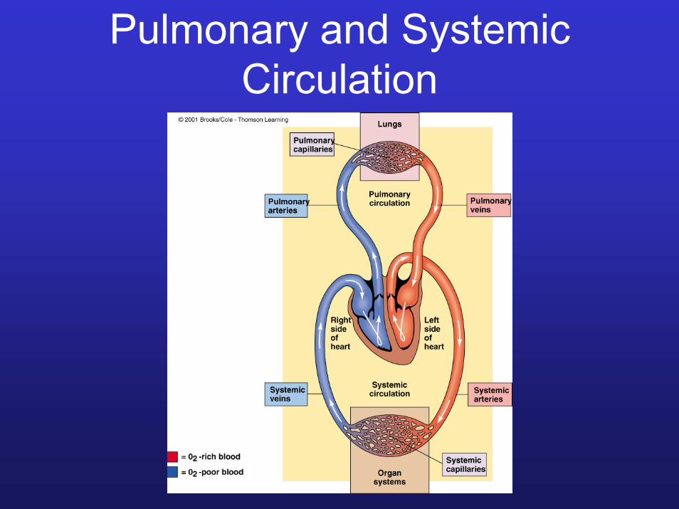
Pulmonary and Systemic Circulation

Location of the Heart

Anatomy of the Heart: Chambers

Anatomy of the Heart: Blood Vessels
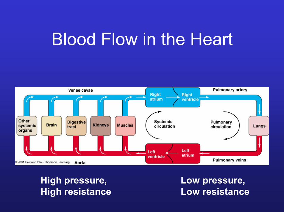
Blood Flow in the Heart
High pressure, High resistance
Low pressure, Low resistance

Heart Valve Action

Valve System in the Heart
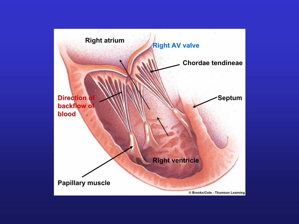
Right atriumRight AV valve
Direction ofbackflow ofblood
Right ventricle
Papillary muscle
Chordae tendineae
Septum

Transverse Heart Structure
© Brooks/Cole - Thomson Learning
Tricuspid valveMitral valve
Semilunar valve
Semilunar valve

Valve SystemNotice:
Valves are found between atria and ventricles or between ventricles and arteries (aorta and pulmonary vein)
No valves between atria and veins (pulmonary vein and vena cava) WHY?
http://www.smm.org/heart/heart/pumping-f.htmMOVIE

Tissue Structure in the Heart
Endocardium-Epithelial tissue
Myocardium-Cardiac muscle tissue
Epicardium-Connective tissue
Pericardial sac and Pericardial fluid

Cell Structure in the MyocardiumPlasma membranes of adjacentcardiac muscle fibers
Desmosome
Actionpotential
Gap junction

Cells in MyocardiumContractile cells
Pacemaker cells
1) Sinoatrial node
2) Atrioventricularnode
3) Atrioventricularbundle
4) Purkinje cells

Function of Pacemaker CellsSelf-inducedaction potential
Slowdepolarization(pacemakerpotential)
Thresholdpotential
http://www.interactivephysiology.com/demo/systems/buildframes.html?cardio/actnpot/01
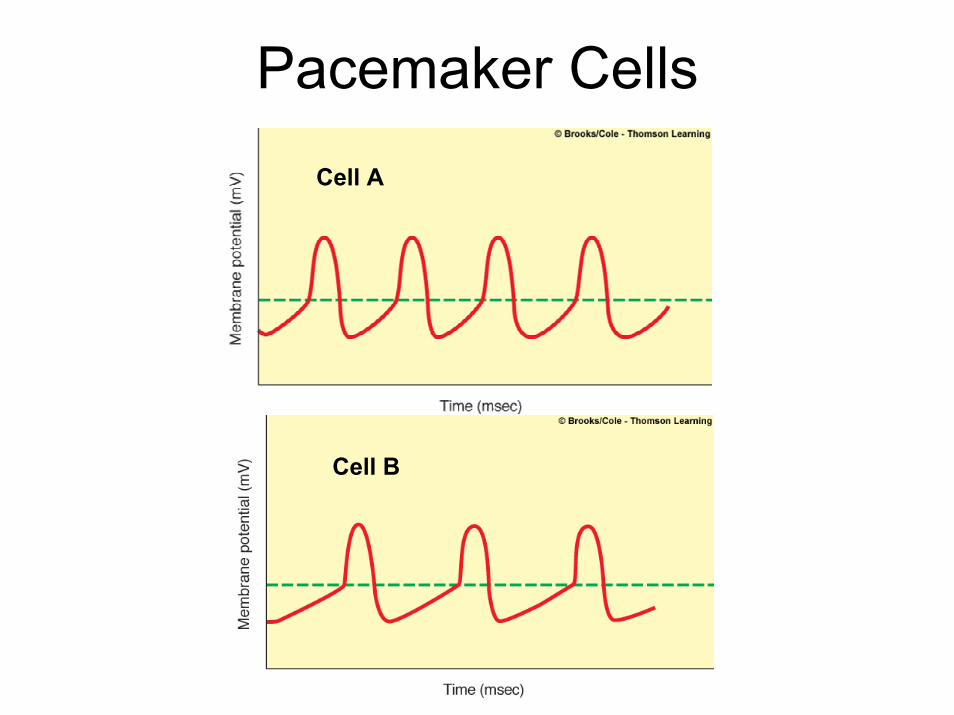
Pacemaker CellsCell A
Cell B


Complete Heart Block
Premature Beat (Extrasystole)

Spreading of Cardiac Excitation

Cardiac Contraction
1) Atrial excitation should be completed before ventricular excitation
AV nodal delay~100 msec

Cardiac Contraction
2) Each chamber should contract as a unit
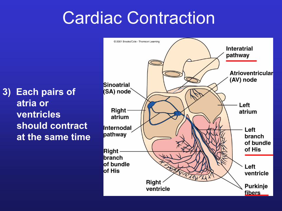
Cardiac Contraction
3) Each pairs of atria or ventricles should contract at the same time

Interatrial pathway
Right atrium Left atrium
Right ventricle Left ventricle
SA node
AV node Internodalpathway
Purkinjefibers Bundle
of His

Action Potential in Cardiac Muscle
Plateauphase ofaction potential
Thresholdpotential
http://www.interactivephysiology.com/demo/systems/buildframes.html?cardio/actnpot/01

Action potentialin cardiaccontractile cell
Travels downT tubules
Entry of small amount of Ca2+
from ECF
Release of large amount of Ca2+
from sarcoplasmicreticulum
CytosolicCa2+
Troponin-tropomyosin complex in thin filaments pulled aside
Cross-bridge cycling between thick and thin filaments
Thin filaments slide inward between thick filaments
Contraction
Ryanodinechannels
Dihydropyridinechannels

Actionpotential
Contractileresponse
Refractoryperiod
Refractory Period in Cardiac Muscle
No summation o of contractions in cardiac muscles WHY?

Electrocardiogram (ECG)Lead I:Right arm toleft arm
aVR: right arm
Lead II:Right arm toleft leg
aVF: left leg
aVL: left arm
Lead III:Left arm toleft leg
Ground electrode
ECG reflect the flow of electrical currents during heart contraction

Electrocardiogram (ECG)Single Lead Recording
1 23
4 56

Electrocardiogram (ECG)
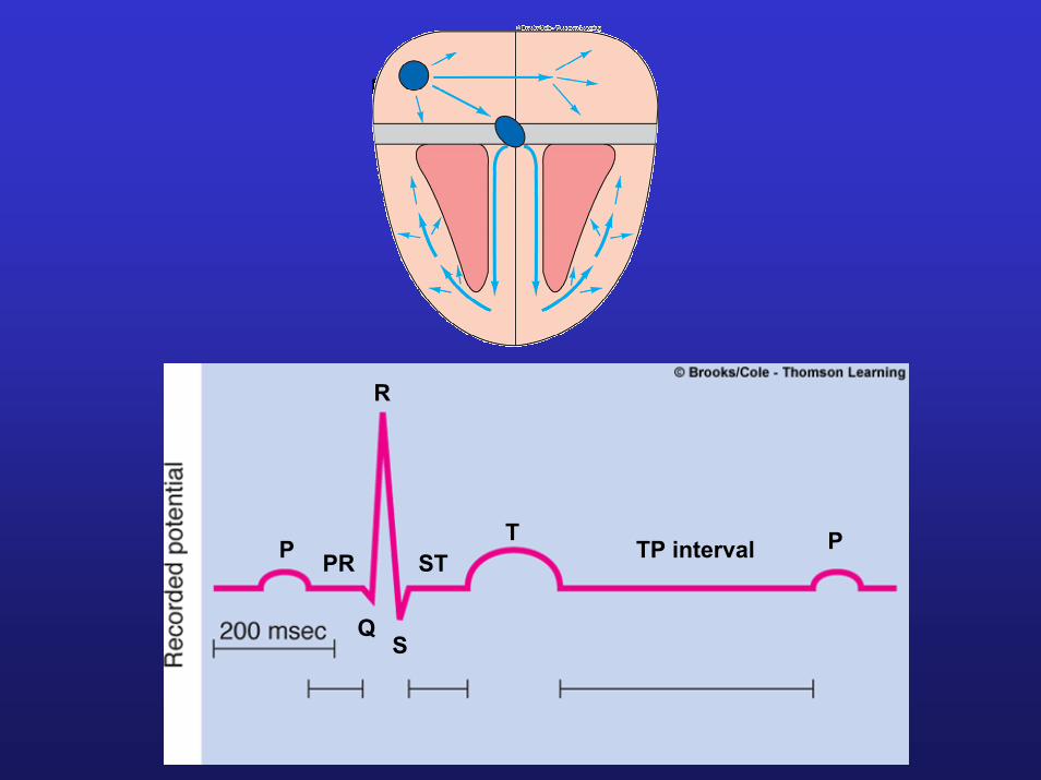
R
T
P
QS
PPR ST TP interval
R
T

NORMAL RATE AND RHYTHM
ABNORMALITIES IN RATE Tachycardia
Bradycardia-appositive of Tachycardia
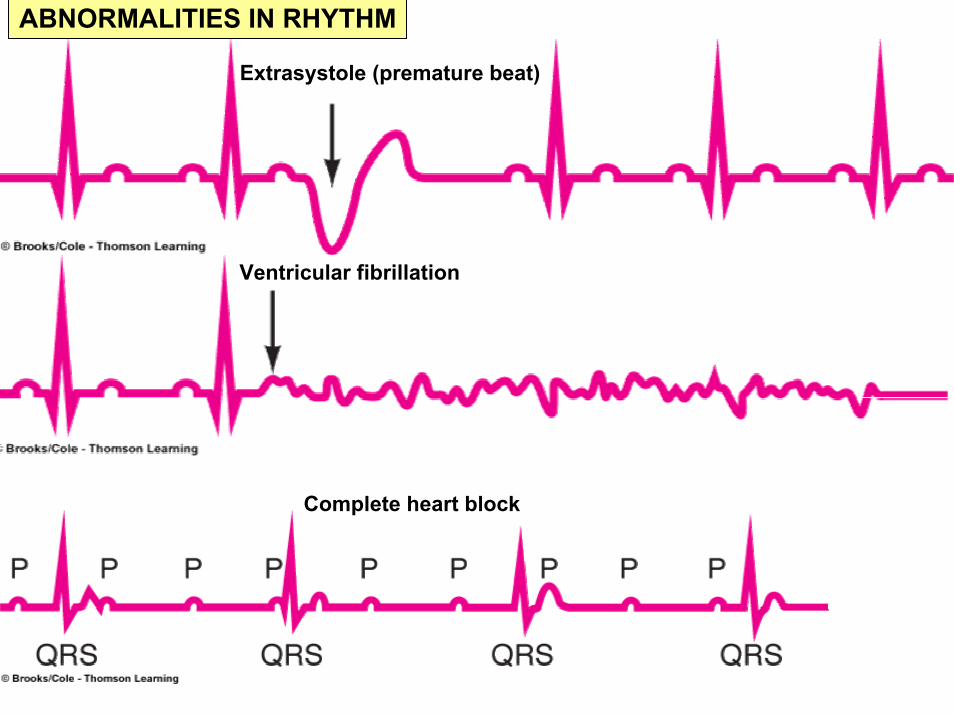
Extrasystole (premature beat)
Ventricular fibrillation
ABNORMALITIES IN RHYTHM
Complete heart block
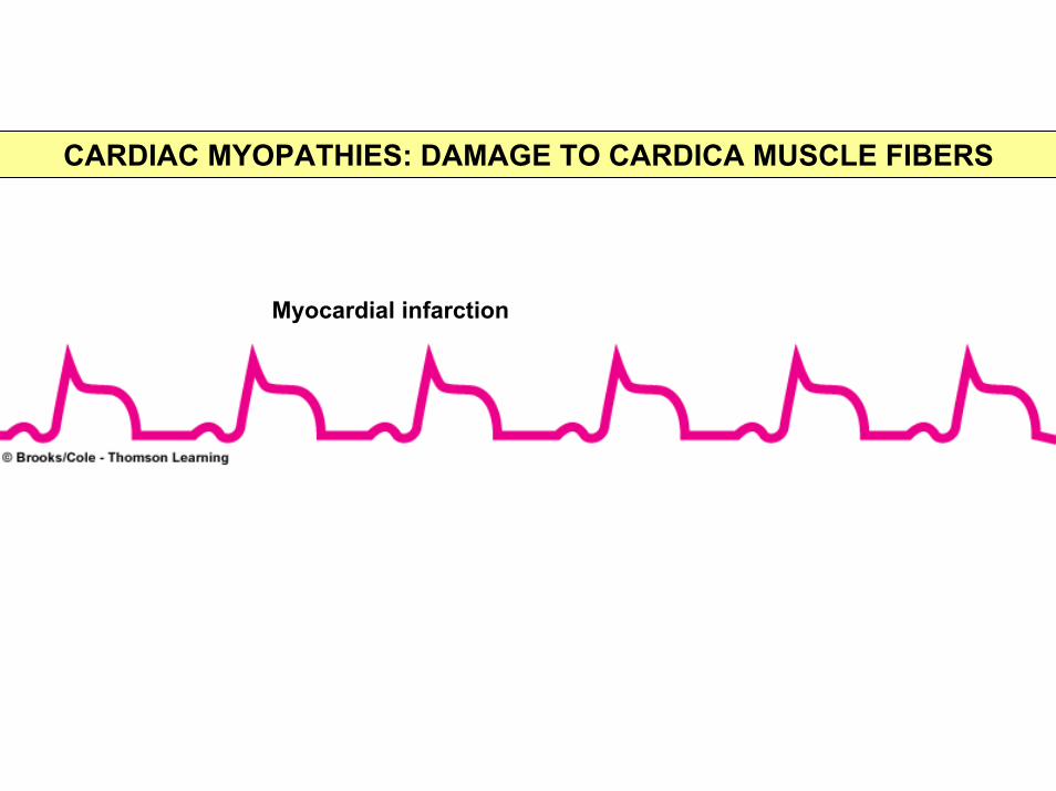
CARDIAC MYOPATHIES: DAMAGE TO CARDICA MUSCLE FIBERS
Myocardial infarction

Cardiac Cycle: Systole vs. Diastole
1) Ventricular filling
2) Isovolumetricventricular contraction
3) Ventricular ejection
4) Isovolumetricventricularrelaxation

Passive filling duringventricular and atrial diastole Atrial contraction
Rightatrium
Rightventricle
Leftatrium
Leftventricle
BA
Ventricular filling

Isovolumetric ventricular contraction
C
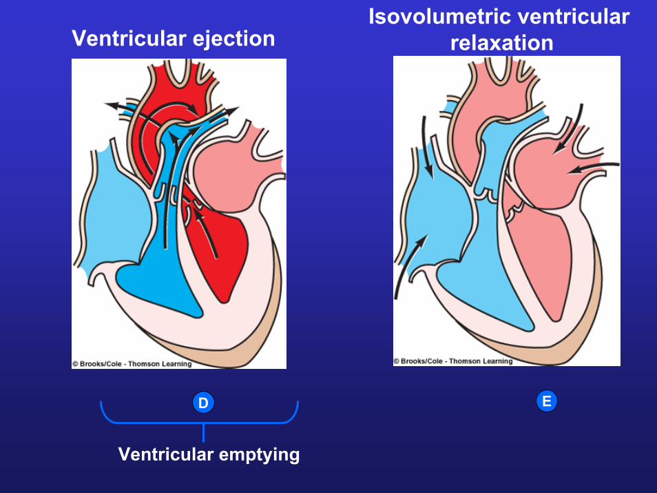
Isovolumetric ventricular relaxationVentricular ejection
Ventricular emptying
ED

Dicrotic Notch
DN represents the closure of the aortic valve during relaxation of ventricles
http://www.interactivephysiology.com/demo/systems/systems/cardio/index.html

Changes in Volume
Stroke volume~70 mL
http://www.interactivephysiology.com/demo/systems/systems/cardio/index.html
Cardiac Cycle
http://www.interactivephysiology.com/demo/systems/systems/cardio/index.html

Heart Sounds
http://www.interactivephysiology.com/demo/systems/systems/cardio/index.html

Laminar vs. Turbulent Flow
Laminar flow (does not create any sound)
Turbulent flow (can be heard)

Cardiac Output at Rest
Cardiac Output: VOLUME of blood pumped by each ventricle per minute
Cardiac Output = Heart Rate X Stroke Volume(CO) = (HR) X (SV)
IF: Average heart rate = 70 beats/minStroke volume = 70 mL
Cardiac Output at Rest ~ 5 L/min

Cardiac Output during ExerciseCardiac Output during Exercise ~ 25 L/min
How does the organism regulate cardiac output?
Heart rate
Parasympatheticactivity
Sympatheticactivity(and epinephrine)
Innervate atria via vagus nerve
Innervate atria and ventricles

Regulation of Cardiac Output
Sympathetic
Increase excitability- increase rate of depolarization -decrease AV nodal delay
Increase heart rate
Increase contractionof atrium and ventricles
Increase secretion of adrenaline from adrenal medulla
EyeNasal mucosa
Spinal nerves
Sympathetictrunk
Splanchinonerves
Liver
GallbladderPancreasAdrenal
glandKidney
Smallintestine
ColonRectumUrinary bladderGenitalia
Lung
Heart
Spinalnerves
Cranialnerves
Salivaryglands
Parotid gland
Trachea
Lacrimal gland
Stomach
Spleen
Parasympathetic
Decrease excitability-decrease rate of depolarization-decrease AV nodal delay
Decrease heart rate
Decrease contractionof atrium ONLY
No effect on adrenaline secretion

Sympathetic Regulation of Heart Rate
Thresholdpotential
Decrease K+ permeability

Parasympathetic Regulation of Heart Rate
Thresholdpotential
Increase K+ permeability

Right atrium Left atrium
SA node
AV node
Right ventricle Left ventricle
Sympathetic
Decrease AV nodal delay
Parasympathetic
Increase AV nodal delay

Regulation of Cardiac Output
Heart rate
Parasympatheticactivity
Sympatheticactivity(and epinephrine)
Innervate atria via vagus nerve
Innervate atria and ventricles

Regulation of Stroke VolumeStroke volume
Strength ofcardiac contraction
Extrinsiccontrol
Intrinsic control
End-diastolicvolume
Intrinsic control
Venous return
Sympathetic activity(and epinephrine)

Frank-Starling LawThe heart will pump out all the blood returned to it
Frank-Starling curve onsympathetic stimulation
Normal Frank-Starlingcurve

Effect of Sympathetic Stimulation on Stroke Volume
End-diastolic volume135 ml
Stroke volume70 ml
End-systolic volume65 ml
Normal SV

End-diastolic volume135 ml
Stroke volume100 ml
End-systolic volume35 ml
SV following sympathetic stimulation:
Due to increase Ca2+
influx during muscle fiber depolarization

End-diastolic volume175 ml
Stroke volume140 ml
End-systolic volume35 ml
SV following sympathetic stimulation and increase end-diastolic volume

Cardiac output
Heart rate Stroke volume
Parasympatheticactivity
Sympatheticactivity (andepinephrine)
End-diastolicvolume
Venous return
Extrinsiccontrol
Intrinsic control
Intrinsic control
Heart rate is determined by
the balance between
sympathetic and
parasympathetic stimulation
Notice that at rest
parasympathetic activation
have the dominant
effect

Heart FailureInability of the heart to contract
Normalstrokevolume
Decreasein strokevolume
Strokevolumewithuncompensatedheart failure Normal end-diastolic volume
Normal heart
Failing heart

Heart Failure
Caused by:
1) Damage heart muscle
2) Excess pumping against increased load (for example after hypertension or damaged valves)

Area of cardiacmuscle deprivedof blood supplyif coronary vesselis blocked at point
Area of cardiacmuscle deprivedof blood supplyif coronary vesselis blocked at point
Leftcoronaryartery
Rightcoronaryartery
Leftventricle
Rightventricle
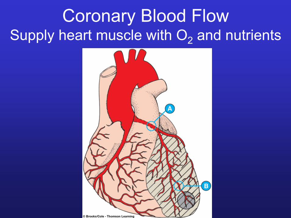
Coronary Blood FlowSupply heart muscle with O2 and nutrients

Coronary Blood Flow
Aortic pressure
Blood flow in leftcoronary artery
Systole Diastole

CARDIAC MYOPATHIES: DAMAGE TO CARDICA MUSCLE FIBERS
Myocardial infarction

Compensatory Measures Against Heart Failure
Increased blood volume by reducing water and Na+ secretion

Compensatory Measures Against Heart Failure: Sympathetic stimulation
Normal end-diastolic volume
Normal heart
Failing heart withsympatheticstimulation
Failing heartwithoutsympatheticstimulation
Normalstrokevolume
Increasein end-diastolicvolume

Interdependence of Blood Flow and Oxygen Need
Metabolic activity of cardiac muscle cells( oxygen need)
Adenosine
Vasodilation of coronary vessels
Blood flow to cardiac muscle cells
Oxygen available to meet oxygen need

Factors that Affect Blood FlowVascular SpamsAtherosclerosis
Thromboembolism

Atherosclerotic PlaqueCollagen-richsmooth musclecap of plaque
Normal bloodvessel wall
Lipid-rich coreof plaque
Endothelium
Plaque

Formation of Atherosclerotic Plaque
Blood vessel damage
Inflammatory response
Deposition and oxidation of low-density lipoprotein (bad cholesterol) (Prevented by Vit E, Vit C, beta-carotene)
Recruitment of macrophes and fibroblasts leading to formation of collagen cap
Ca2+ precipitation and hardening of blood vessels

Formation of ThrombusRupture of collagen cap
Platelet aggregation and formation of blood clot (thrombus)
Thrombus

ThrombormbolismLeading cause of strokes (brain) and myocardial ischemia (heart muscle)
Blood flow
Thrombus
Blood flow
Embolus

Consequences of Thrombormbolism
Blood flow
Embolus
Angina Pectoris or Hear Pain due to narrowing of coronary blood vessels
Heart Attack occurs when blood vessels are completely plugged