chapter 40 physiology, homeostasis, & temperature regulation
TRANSCRIPT
Chapter 40
Physiology, Homeostasis, & Temperature Regulation

Terms
Anatomy-structure Physiology-function Homeostasis-maintenance of a stable
internal environment Feedback loop

Parts of a feedback loop
Receptor Receives stimulus
Control Center (set point) CNS: brain or spinal cord
Effector Produces a response Usually a muscle or a gland

Feedback loops
Negative result is changed/opposed most are this type Ex: body temperature; blood pressure
Positive result is repeated/enhanced/reinforced only a few are this type Ex: blood clotting; childbirth

6 Organizational Levels
Chemicals (atoms, molecules,
macromolecules) Cellular Tissue Organ Organ System Organism


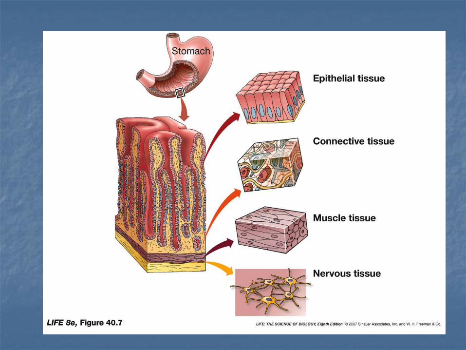
4 tissue types
Epithelial Connective Muscle Nervous

Epithelial
Avascular Has nerve supply Easily renewable Functions: protection, secretion,
absorption, excretion, sensory reception
Classified by cell shape and layer arrangement

Epithelial cell shape
Squamous-flat and thin Cuboidal-cube-shaped Columnar-tall and cylindrical Transitional-cell shape changes

Epithelial layer arrangement
Simple-single layer Stratified- two or more layers Pseudostratified-one layer that
appears to have several layers

Simple Squamous Epithelium
Structure: Single layer of flat thin cells
Function: diffusion
Location: Alveoli of lungs; capillaries; heart lining

Simple Cuboidal Epithelium
Structure: Single layer of cube-shaped cells
Function: Secretion and absorption
Location: Ducts of glands; ovarian surface

Simple Columnar Epithelium
Structure: Nonciliated: single layer of tall & narrow cells
without cilia Ciliated: single layer of tall & narrow cells with cilia
Function: Nonciliated: secretion (Goblet cells) and absorption Ciliated: movement of mucus
Location: Lines tracts with environmental openings

Pseudostratified Epithelium
Structure: 1 layer of tall & narrow cells that
appears to be more than 1--but it is not Ciliated (w/ cilia) and nonciliated (no
cilia) Function:
Secretion & movement of mucus Location:
Lines airways of upper respiratory tract

Pseudostratified - ???

Stratified Squamous Epithelium
Structure: 2 or more flat, thin layers
Function: protection
Location: Esophagus, tongue, vaginal lining,
epidermis of the skin

Stratified Cuboidal Epithelium
Structure: 2 or more layers of cube-shaped cells
Function: Protection; limited secretion and
absorption Location:
Sweat gland ducts

Stratified Columnar Epithelium
Structure: 2 or more layers of tall & narrow cells
Function: Protection & excretion
Location: Conjunctiva of eye; excretory ducts

Transitional Epithelium
Structure: Appearance of cells ranges from
squamous to cuboidal & columnar Function:
Allows for stretching without tearing (distensibility)
Location: Urinary bladder

Glandular Epithelium a/k/a Glands
Glands: consists of a cell or group of cells that secrete substances into ducts, onto surfaces, or into blood
2 types: Exocrine: secretion goes through a duct
Ex. Sweat, oil, saliva, pancreas Endocrine: secretion goes into blood
stream without passing through a duct Ex. Thyroid, pituitary, & pancreas

Connective Tissue
Most abundant and widely distributed tissue in the body
Consists of cells and a matrix (determines a tissues qualities; may be fluid, gel, fibers)
Has a nerve supply Highly vascular (except cartilage)

Mature Connective Tissue
Areolar Adipose Dense Regular Dense Irregular Elastic Cartilage Compact Bone Blood

Areolar Connective Tissue
Location Subcutaneous layer of skin
Function Strength Support elasticity

Adipose Connective Tissue
Location Around heart, kidneys, eyes, and in
yellow bone marrow Function
Energy reserve and protection

Dense Regular Connective Tissue
Location Tendons
Muscle to bone Ligaments
Bone to bone
Function attachment
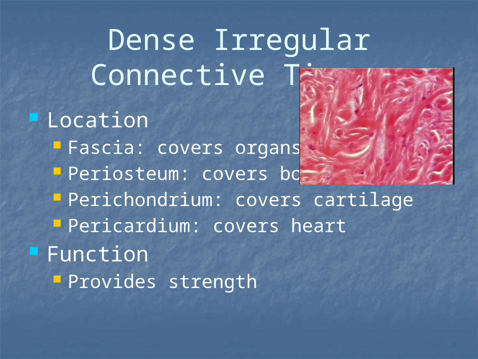
Dense Irregular Connective Tissue
Location Fascia: covers organs Periosteum: covers bones Perichondrium: covers cartilage Pericardium: covers heart
Function Provides strength
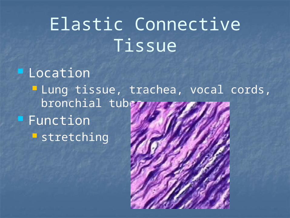
Elastic Connective Tissue
Location Lung tissue, trachea, vocal cords,
bronchial tubes Function
stretching

Cartilage Location
Nose, voice box, epiglottis, external ear, pubis symphysis
Function Support, cushioning, rigidity, flexibility

Compact Bone
Location Bones (osteocytes)
Function Support, protection, storage

Blood Location
Blood vessels, heart Function
Transport gases, immunity, clotting

Nervous Tissue
Tissue of the nervous system Basic functioning unit = neuron

The neuron
Cell body (soma) Dendrites (many) Axon (one)

Muscle Tissue
3 types Skeletal Smooth Cardiac

Skeletal Muscle
Attaches to bones of skeleton Striated (striped) in appearance voluntary

Smooth Muscle
Makes up walls of internal organs Nonstriated Involuntary

Cardiac Muscle
Found in heart Striated with intercalated discs involuntary

Membranes
Tissues that cover or line a part of the body
3 types Mucous Synovial Serous

Mucous Membranes
Line body cavities that open to the environment
Secrete mucus Ex: digestive system, reproductive
system, respiratory system

Synovial Membranes
Line the cavities of some joints Secrete synovial fluid Lubricates joints to prevent friction
during movement Bursae are often present here as well
bursitis

Serous Membranes Line body cavities that do not open
to the environment Secrete serous fluid 2 layers:
Parietal: lines cavity wall Visceral: covers organ(s)
3 locations: Lungs = pleura
pleurisy Heart = pericardium
pericarditis Abdomen = peritoneum
peritonitis

How Does Temperature Affect Living Systems?
Body temperature of some animals is coupled to environmental temperature.
In winter, a fish will acclimatize to colder water by expressing different isozymes.
Isozymes that are optimized at different temperatures can catalyze the same metabolic reaction.
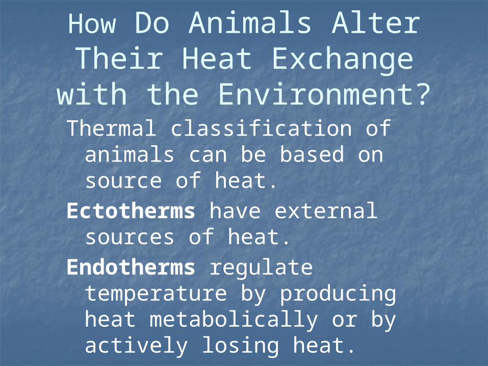
How Do Animals Alter Their Heat Exchange with the
Environment?Thermal classification of animals
can be based on source of heat.Ectotherms have external
sources of heat.Endotherms regulate
temperature by producing heat metabolically or by actively losing heat.
Heterotherms can behave either as an ectotherm or an endotherm.

Major differences between ectotherms and endotherms:
Resting metabolic rate Total energy expenditure when
at rest Response to changes in
environmental temperatures

Figure 40.9 Ectotherms and Endotherms React Differently to Environmental Temperatures (A)

Figure 40.9 Ectotherms and Endotherms React Differently to Environmental Temperatures (B)

An endotherm will increase its metabolic rate to maintain its body temperature in cold conditions.
Both endotherms and ectotherms may use behavioral regulation to maintain body temperature.
Example: moving into sun

Figure 40.10 Ectotherms and Endotherms Use Behavior to Regulate Body Temperature (Part 1)

Figure 40.10 Ectotherms and Endotherms Use Behavior to Regulate Body Temperature (Part 2)

40.3 How Do Animals Alter Their Heat Exchange with
the Environment?Both ectotherms and endotherms can alter heat exchange between their bodies and the environment.
Body temperature is determined by the balance between internal heat production and four types of heat exchange.

40.3 How Do Animals Alter Their Heat Exchange with
the Environment?Radiation: heat transfer via infrared radiation.
Conduction: heat transfer by direct contact.
Convection: heat transfer through a surrounding medium.
Evaporation: heat transfer through evaporation of water from a surface.

Figure 40.11 Animals Exchange Heat with the Environment

Blood flow to the skin helps endotherms and ectotherms maintain body temperature.
Increased blood flow to the skin increases heat loss and lowers body temperature.
Constriction of blood vessels to the skin results in less heat loss.

Figure 40.12 Some Ectotherms Regulate Blood Flow to the Skin (Part 1)

Figure 40.12 Some Ectotherms Regulate Blood Flow to the Skin (Part 2)

Fur on animals acts as insulation that retains body heat.
When animals are active and must lose excess heat, special blood vessels carry the heat to hairless skin surfaces.

Some ectotherms are able to raise their body temperature by producing heat:
Insects contract their flight muscles.
Honeybees regulate temperature as a group, adjusting individual heat and position in the cluster so that larvae are kept warm.

Endotherms can respond to changes in temperature by changing their metabolic rate—the rate at which they consume O2 and produce CO2.
In the thermoneutral zone the metabolic rate is low and independent of temperature.
The basal metabolic rate (BMR) is the metabolic rate of a resting animal at a temperature within the thermoneutral zone.

The basal metabolic rate (BMR) is correlated with body size and environmental temperature.
However, the BMR per gram of tissue increases as animals get smaller.
Example: a gram of mouse tissue uses energy at a rate 20 times greater than a gram of elephant tissue.

Figure 40.15 The Mouse-to-Elephant Curve
A curve showing BMR versus ambient temperature represents an integrated response of an animal.
The thermoneutral zone is bounded by a lower and an upper critical temperature.
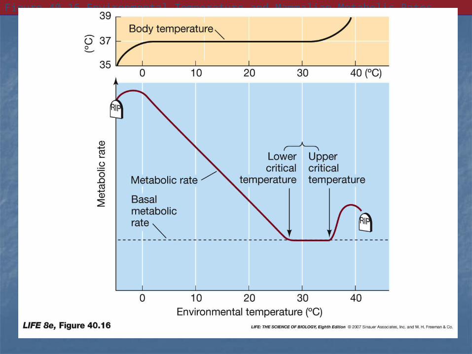
Figure 40.16 Environmental Temperature and Mammalian Metabolic Rates

How Do Mammals Regulate Their Body Temperatures?
Inside the thermoneutral zone an animal can adapt to changes without using much energy.
Outside the thermoneutral zone responses to temperature changes require bigger metabolic increases.

Endotherms respond to cold by producing heat and reducing heat loss.
Mammals produce heat in two ways:Shivering: skeletal muscles contract
and release energy from ATP as heat.Nonshivering: occurs in adipose tissue
called brown fat. The protein thermogenin causes heat release by altering ATP production.

Figure 40.17 Brown Fat

Reducing heat loss is important in cold climates.
Some cold-climate species have a smaller surface area than warm-climate relatives.
Rounder body shapes and shorter appendages reduce surface area-to-volume ratios.

Other adaptations to reducing heat loss include:
Increased thermal insulation with fur, feathers, or fat.
Ability to decrease blood flow to the skin by constricting blood vessels.
Use of countercurrent heat exchange in blood flow to appendages.

Figure 40.18 Adapting to Hot and Cold Climates

A rise in environmental temperature results in increased blood flow to the skin to dissipate heat.
If temperature exceeds the upper critical temperature, overheating is possible.
Evaporation of water through sweating or panting increases heat loss, but is an active process that also generates some heat.

The regulatory system that controls body temperature depends on feedback and acts as a thermostat.
In vertebrates a brain structure, the hypothalamus, is the major center of the thermostat.
The temperature of the hypothalamus can be the main feedback to the thermostat.

Cooling the hypothalamus can cause body temperature to rise by:
Constricting blood vessels to the skin
Increasing metabolic rateWarming the hypothalamus can
lower body temperature by: Dilating blood vessels to the
skin Sweating or panting

Figure 40.19 The Hypothalamus Regulates Body Temperature (Part 1)

Figure 40.19 The Hypothalamus Regulates Body Temperature (Part 2)

The temperature of the hypothalamus is a negative feedback signal; variability from its set point can trigger thermoregulatory responses.
Other factors can change hypothalamic set points:
Change in skin temperature Wakefulness or sleep Circadian rhythm: a daily internal
cycle
Fever is a rise in body temperature caused by pyrogens.
Exogenous pyrogens come from foreign substances; bacteria or viruses.
Endogenous pyrogens are produced by immune cells in response to infection.
Pyrogens cause a rise in the set point for metabolic heat production.

Hypothermia is a state of below-normal temperature.
Regulated hypothermia is a means of survival.
Small endotherms, like hummingbirds, can lower their temperature during inactive periods to conserve energy, known as daily torpor.
Long-lasting regulated hypothermia is called hibernation.

Figure 40.20 A Ground Squirrel Enters Repeated Bouts of Hibernation during Winter