chapter 3: tissue repair: regeneration, healing, & fibrosis. · 2020-01-22 · ecm components...
TRANSCRIPT
Chapter 3:
Tissue repair: Regeneration,
Healing, & Fibrosis.
Critical to the survival of an organism, is the ability to repair the damage caused by toxic insults & inflammation. In the following lectures, we will discuss the:
- Control of Cell Proliferation: The Cell-Cycle Proliferative Capacities of Tissues & Stem Cells;
- Nature & Signaling mechanisms of Growth factors receptors
- ECM roles & components;
- Cell & tissue regeneration;
- Repair by connective tissue;
- Cutaneous wound healing by 1st & 2nd intention;
- Pathologic aspects of repair & finally, an overview of repair processes
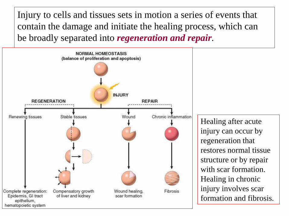
Injury to cells and tissues sets in motion a series of events that
contain the damage and initiate the healing process, which can
be broadly separated into regeneration and repair.
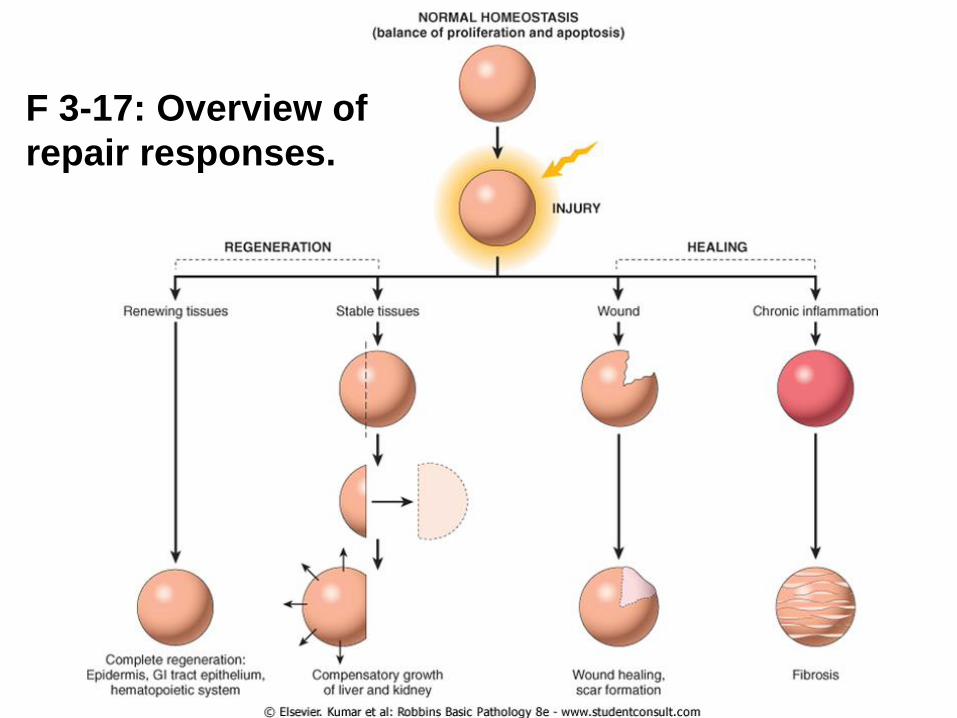
Healing after acute
injury can occur by
regeneration that
restores normal tissue
structure or by repair
with scar formation.
Healing in chronic
injury involves scar
formation and fibrosis.
Regeneration results in the complete restitution of lost or
damaged tissue; repair may restore some original structures but
can cause structural derangements.
Regeneration refers to the proliferation of cells and tissues to
replace lost structures.
Tissues with high proliferative capacity, such as the hematopoietic
system and the epithelia of the skin and GIT, renew themselves
continuously and can regenerate after injury, as long as the stem
cells of these tissues are not destroyed.
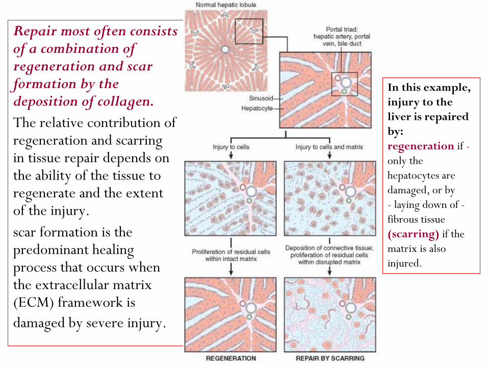
Repair most often consists of a combination of regeneration and scar formation by the deposition of collagen.
The relative contribution of regeneration and scarring in tissue repair depends on the ability of the tissue to regenerate and the extent of the injury.
scar formation is the predominant healing process that occurs when the extracellular matrix (ECM) framework is
damaged by severe injury.
In this example,
injury to the
liver is repaired
by:
-regeneration if
only the
hepatocytes are
damaged, or by
-- laying down of
fibrous tissue
(scarring) if the
matrix is also
injured.

Cryptogenic cirrhosis: X60: Liver section stained for reticulin, from a
patient die from liver failure. There are three regenerative liver
nodules (double arrow), separated by broad bands of reticulin fibers
(thick arrow), which is, normally, completely absent. An example of
healing by combine regeneration & fibrosis which follows injury to the liver
cells & stroma (commonly due to alcoholism or viral hepatitis), but in this
patient, the cause was unknown, i.e., cryptogenic.
3 Regenerative
liver nodules
Bands of
reticulin
fibers
Chronic inflammation that accompanies persistent injury also
stimulates scar formation because of local production of
growth factors and cytokines that promote fibroblast
proliferation and collagen synthesis.
is used to describe the extensive deposition FibrosisThe term
of collagen that occurs under these situations.
ECM components are essential for wound healing, because they
provide the framework for cell migration, maintain the correct
cell polarity for the re-assembly of multilayer structures, and
participate in the angiogenesis.
Furthermore, cells in the ECM (fibroblasts, macrophages, and
other cell types) produce growth factors, cytokines, and
chemokines that are critical for regeneration and repair.
If fibrosis develops in a tissue space occupied by an
organizationinflammatory exudate it is called
(e.g., organizing pneumonia, organizing pleurisy ).
the know, we have to To understand repair
, cell proliferation control of )1(
& how it is involved in repair, ECMfunctions of the )2(
in tissue homeostasis, and stem cellsthe roles of )3(
in the proliferation of different cell GFsthe roles of )4(
types involved in repair.
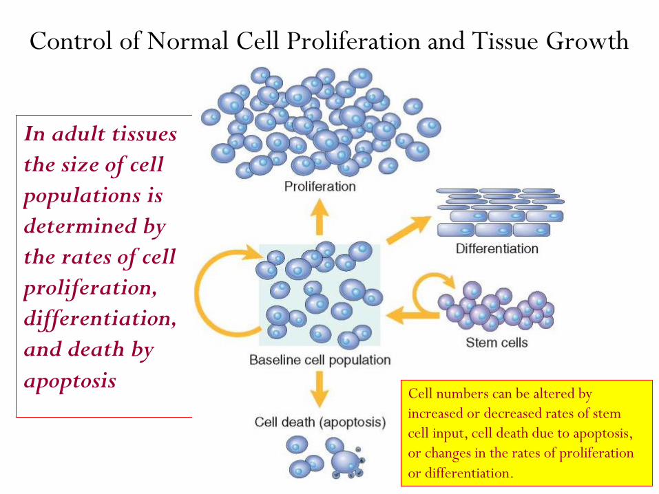
Control of Normal Cell Proliferation and Tissue Growth
In adult tissues
the size of cell
populations is
determined by
the rates of cell
proliferation,
differentiation,
and death by
apoptosis Cell numbers can be altered by
increased or decreased rates of stem
cell input, cell death due to apoptosis,
or changes in the rates of proliferation
or differentiation.
Several cell types proliferate during repair:
of the injured tissue (which attempt to remnantsThe )1(
restore normal structure e.g., liver cells)
to create new (ECs), endothelial cellsVascular )2(
vessels (angiogenesis) to provide nutrients needed for
the repair.
(the source of the fibrous tissue that forms fibroblasts )3(
the scar), to fill defects that cannot be corrected by
regeneration.
The impact of differentiation depends on the tissue under
which it occurs: in some tissues differentiated cells are not
replaced, while in others they die but are continuously
replaced by new cells generated from stem cells.
Cell proliferation can be stimulated by physiologic
and pathologic conditions.
Cell proliferation is largely controlled by signals (soluble or
contact-dependent) from the microenvironment that either
stimulate or inhibit proliferation.
An excess of stimulators or a deficiency of inhibitors
leads to net growth and, in the case of cancer,
uncontrolled growth.
Tissue Proliferative Activity
The tissues of the body are divided into three groups on the
basis of the proliferative activity of their cells:
1- Continuously dividing (labile tissues),
2- Quiescent (stable tissues), and
3- Nondividing (permanent tissues).

cells proliferate throughout In continuously dividing tissues -1
life, replacing those that are destroyed. In most of these tissues
, which have a adult stem cellsmature cells are derived from
tremendous capacity to proliferate and whose progeny may
differentiate into several kinds of cells.
normally have a low level of replication; Quiescent tissues -2
however, cells from these tissues can undergo rapid division in
response to stimuli and are thus capable of reconstituting the
tissue of origin.
Fibroblasts, endothelial cells, smooth muscle cells, chondrocytes,
and osteocytes are quiescent in adult mammals but proliferate in
response to injury. Fibroblasts in particular can proliferate
extensively, as in healing processes and fibrosis.

contain cells that have left the cell cycle and Nondividing tissues -3
cannot undergo mitotic division in postnatal life. To this group belong
neurons and skeletal and cardiac muscle cells.
If neurons in the central nervous system are destroyed, the tissue is
generally replaced by the proliferation of the central nervous
system–supportive elements, the glial cells. However, recent
results demonstrate that limited neurogenesis from stem cells may
occur in adult brains. skeletal muscle does have regenerative capacity,
through the differentiation of the satellite cells that are attached to the
endomysial sheaths.
Cardiac muscle has very limited, if any, regenerative capacity, and a
large injury to the heart muscle, as may occur in myocardial
infarction, is followed by scar formation.
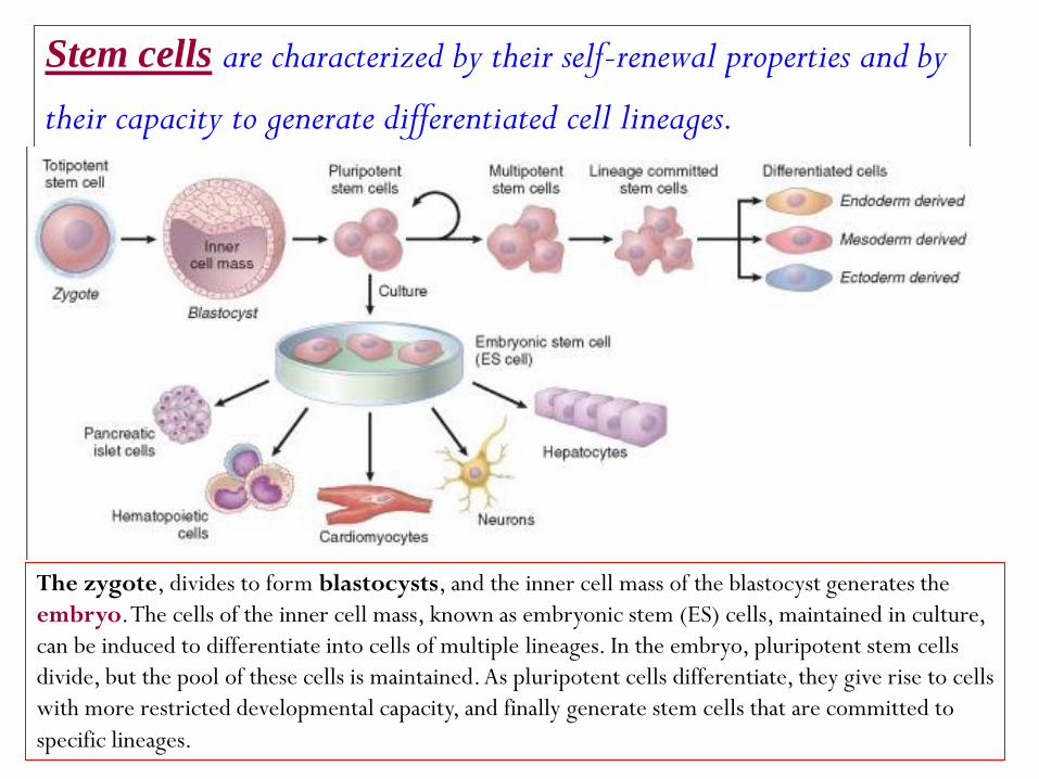
renewal properties and by -are characterized by their self Stem cells
their capacity to generate differentiated cell lineages.
The zygote, divides to form blastocysts, and the inner cell mass of the blastocyst generates the
embryo. The cells of the inner cell mass, known as embryonic stem (ES) cells, maintained in culture,
can be induced to differentiate into cells of multiple lineages. In the embryo, pluripotent stem cells
divide, but the pool of these cells is maintained. As pluripotent cells differentiate, they give rise to cells
with more restricted developmental capacity, and finally generate stem cells that are committed to
specific lineages.
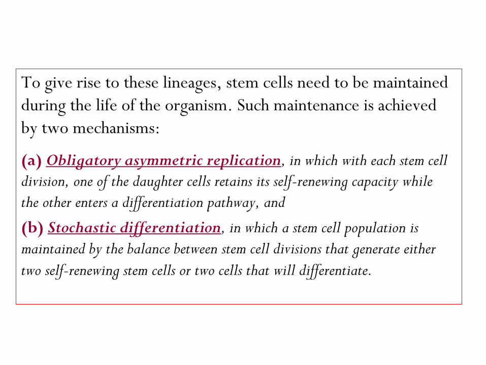
To give rise to these lineages, stem cells need to be maintained
during the life of the organism. Such maintenance is achieved
by two mechanisms:
, in which with each stem cell Obligatory asymmetric replication (a)
division, one of the daughter cells retains its self-renewing capacity while
the other enters a differentiation pathway, and
, in which a stem cell population is Stochastic differentiation (b)
maintained by the balance between stem cell divisions that generate either
two self-renewing stem cells or two cells that will differentiate.

In early stages of embryonic development, stem cells,
known as embryonic stem cells or ES cells, are
pluripotent, that is, they can generate all tissues of
the body.
Pluripotent stem cells give rise to multipotent stem
restricted developmentalcells, which have more
potential, and eventually produce differentiated
cells from the three embryonic layers.
or adult stem cells, stem cells (often referred to as In adults
somatic stem cells ) with a more restricted capacity to generate
different cell types have been identified in many tissues.
Somatic stem cells for the most part reside in special
microenvironments called niches, composed of mesenchymal,
endothelial, and other cell types. It is believed that niche cells generate or transmit stimuli that
regulate stem cell self-renewal and the generation of progeny cells.
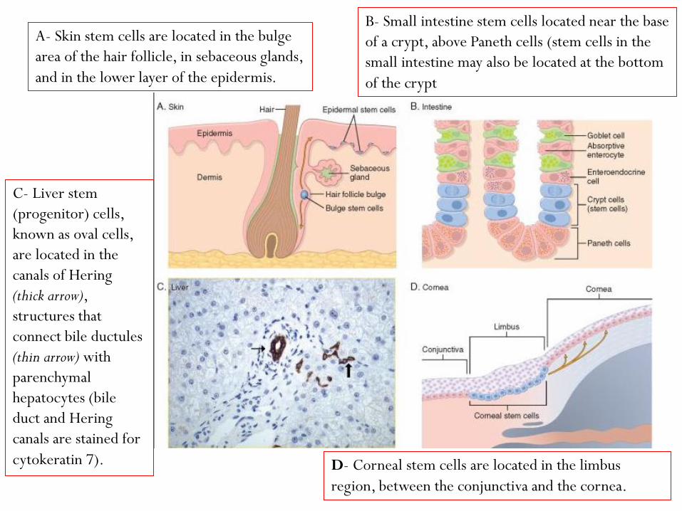
A- Skin stem cells are located in the bulge
area of the hair follicle, in sebaceous glands,
and in the lower layer of the epidermis.
B- Small intestine stem cells located near the base
of a crypt, above Paneth cells (stem cells in the
small intestine may also be located at the bottom
of the crypt
C- Liver stem
(progenitor) cells,
known as oval cells,
are located in the
canals of Hering
(thick arrow),
structures that
connect bile ductules
(thin arrow) with
parenchymal
hepatocytes (bile
duct and Hering
canals are stained for
cytokeratin 7). D- Corneal stem cells are located in the limbus
region, between the conjunctiva and the cornea.

Embryonic Stem Cells
The inner cell mass of blastocysts in early embryonic development
contains pluripotent stem cells known as ES cells. Cells isolated
from blastocysts can be maintained in culture as
undifferentiated cell lines or be induced to differentiate
into specific lineages such as heart and liver cells.
The study of ES cells has had an enormous impact on
biology and medicine:
1- ES cells have been used to study the specific signals and
differentiation steps required for the development of many
tissues.
2- ES cells made possible the production of knockout mice, an essential tool
to study the biology of particular genes and to develop models of human
disease, and more than 500 models of human diseases have been
created using these animals.
3- ES cells may in the future be used to repopulate damaged organs.
Reprogramming of Differentiated Cells: Induced
Pluripotent Stem Cells
Differentiated cells of adult tissues can be reprogrammed to become
pluripotent by transferring their nucleus to an enucleated oocyte.
The oocytes implanted into a surrogate mother can generate
cloned embryos that develop into complete animals.
This procedure, known as reproductive cloning, was successfully
demonstrated in 1997 by the cloning of Dolly the sheep.
There has been great hope that the technique of nuclear transfer
to oocytes may be used for therapeutic cloning in the treatment
of human diseases.
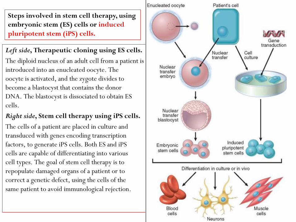
Left side, Therapeutic cloning using ES cells.
The diploid nucleus of an adult cell from a patient is
introduced into an enucleated oocyte. The
oocyte is activated, and the zygote divides to
become a blastocyst that contains the donor
DNA. The blastocyst is dissociated to obtain ES
cells.
Right side, Stem cell therapy using iPS cells.
The cells of a patient are placed in culture and
transduced with genes encoding transcription
factors, to generate iPS cells. Both ES and iPS
cells are capable of differentiating into various
cell types. The goal of stem cell therapy is to
repopulate damaged organs of a patient or to
correct a genetic defect, using the cells of the
same patient to avoid immunological rejection.
Steps involved in stem cell therapy, using
embryonic stem (ES) cells or induced
pluripotent stem (iPS) cells.
Adult (Somatic) Stem Cells
In the adult organism, stem cells are present in tissues that continuously
divide such as the bone marrow, the skin, and the lining of the GI tract.
Stem cells may also be present in organs such as liver, pancreas,
and adipose tissue, in which, under normal conditions, they do
not actively produce differentiated cell lineages.
Regardless of their proliferative activity, somatic stem cells
generate rapidly dividing cells known as transit amplifying cells.
These cells lose the capacity of self-perpetuation, and give rise
to cells with restricted developmental potential known as
progenitor cells.
A change in the differentiation of a cell from one type
to another is known as transdifferentiation, and the
capacity of a cell to transdifferentiate into diverse
lineages is referred to as developmental plasticity.
Hemopoietic stem cells (HSCs) maintained in
culture have been shown to transdifferentiate into
other cell types, such as hepatocytes and neurons.
Stem Cells in Tissue Homeostasis
. Bone marrow
The bone marrow contains HSCs and stromal cells (also known as
multipotent stromal cells, mesenchymal stem cells or MSCs).
Hematopoietic Stem Cells (HSCs) generate all of the blood cell lineages,
can reconstitute the bone marrow after depletion caused by disease
or irradiation, and are widely used for the treatment of
hematologic diseases. They can be collected directly from the bone
marrow, from umbilical cord blood, and from the peripheral
blood of individuals receiving cytokines such as granulocyte-
macrophage colony-stimulating factor, which mobilize HSCs.
Marrow Stromal Cells. MSCs are multipotent.
They have potentially important therapeutic applications,
because they can generate chondrocytes, osteoblasts,
adipocytes, myoblasts, and endothelial cell
precursors depending on the tissue to which they migrate.
MSCs migrate to injured tissues and generate stromal
cells or other cell lineages, but do not seem to
participate in normal tissue homeostasis.
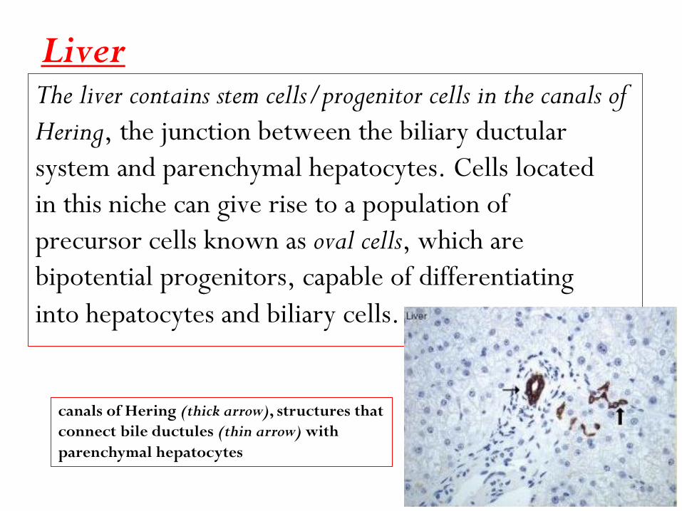
Liver The liver contains stem cells/progenitor cells in the canals of
Hering, the junction between the biliary ductular
system and parenchymal hepatocytes. Cells located
in this niche can give rise to a population of
precursor cells known as oval cells, which are
bipotential progenitors, capable of differentiating
into hepatocytes and biliary cells.
canals of Hering (thick arrow), structures that
connect bile ductules (thin arrow) with
parenchymal hepatocytes
Brain.
Neurogenesis from neural stem cells (NSCs) occurs in the brain
of adult rodents and humans. Thus, the long-established
dogma that no new neurons are generated in the
brain of normal adult mammals is now known to be
incorrect. NSCs (also known as neural precursor cells),
capable of generating neurons, astrocytes, and
oligodendrocytes, have been identified in two areas of
adult brains, the subventricular zone (SVZ) and the
dentate gyrus of the hippocampus
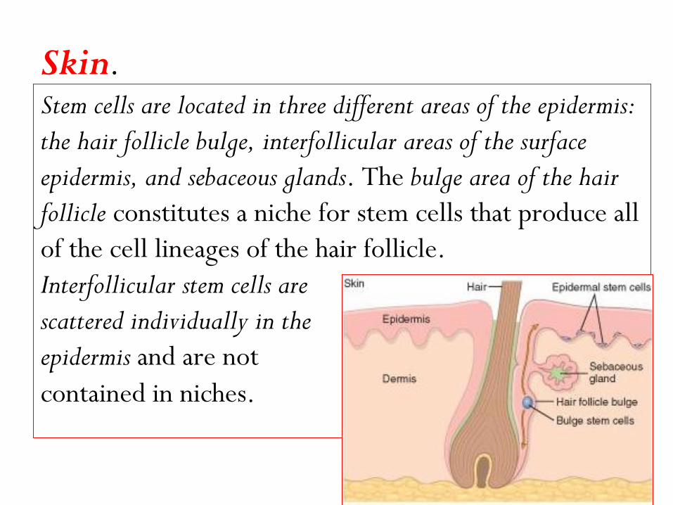
Skin. Stem cells are located in three different areas of the epidermis:
the hair follicle bulge, interfollicular areas of the surface
epidermis, and sebaceous glands. The bulge area of the hair
follicle constitutes a niche for stem cells that produce all
of the cell lineages of the hair follicle.
Interfollicular stem cells are
scattered individually in the
epidermis and are not
contained in niches.
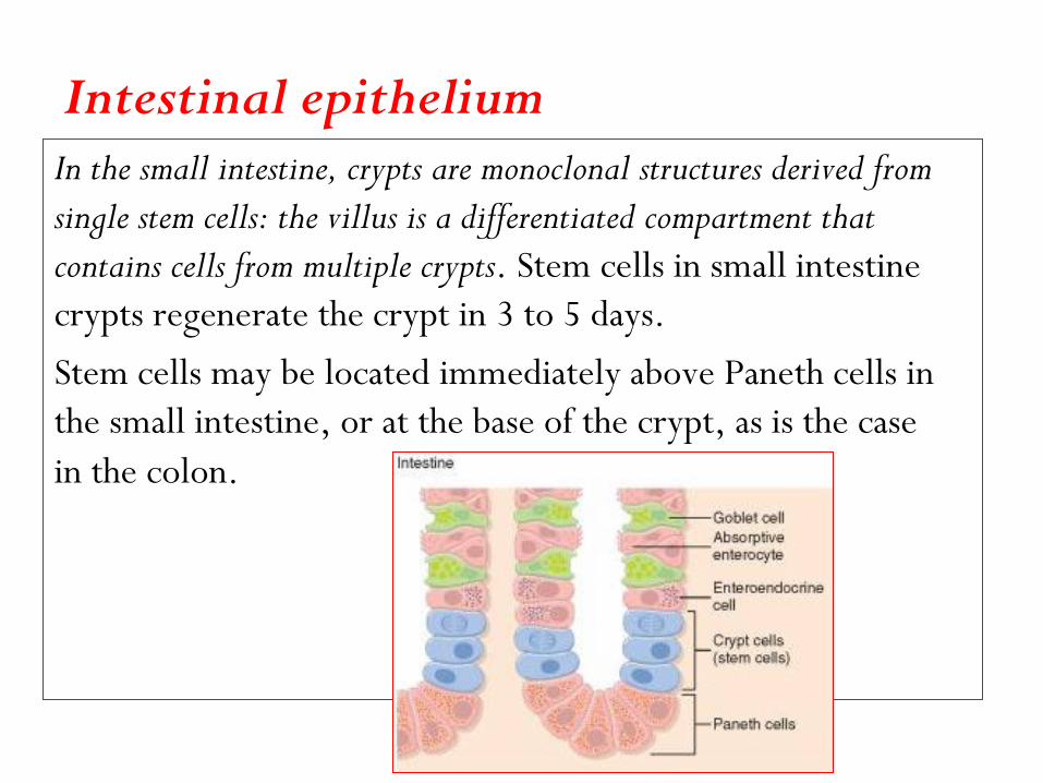
Intestinal epithelium In the small intestine, crypts are monoclonal structures derived from
single stem cells: the villus is a differentiated compartment that
contains cells from multiple crypts. Stem cells in small intestine
crypts regenerate the crypt in 3 to 5 days.
Stem cells may be located immediately above Paneth cells in
the small intestine, or at the base of the crypt, as is the case
in the colon.
Skeletal and cardiac muscle
Skeletal muscle myocytes do not divide, even after injury;
growth and regeneration of injured skeletal muscle occur by
replication of satellite cells. These cells, located
beneath the myocyte basal lamina, constitute a
reserve pool of stem cells that can generate
differentiated myocytes after injury. Active Notch
signaling, triggered by up-regulation of delta-like
(Dll) ligands, stimulates the proliferation of satellite
cells (Notch signaling is discussed later in “Mechanisms of Angiogenesis”).

Cornea The transparency of the cornea depends on the integrity of the
outermost corneal epithelium, which is maintained by limbal
stem cells (LSCs). These cells are located at the junction
between the epithelium of the cornea and the conjunctiva.
Hereditary or acquired conditions that result in LSC
deficiency and corneal opacification can be treated by limbal
transplantation or LSC grafting.

Cell Cycle and the Regulation of Cell
Replication
The replication of cells is stimulated by growth factors or by signaling
from ECM components through integrins.
The cell cycle consists of:
G1 (presynthetic),
S (DNA synthesis),
G2 (Premitotic), and
M (mitotic) phases.
Quiescent cells that have not entered the cell cycle are in the
G 0 state.
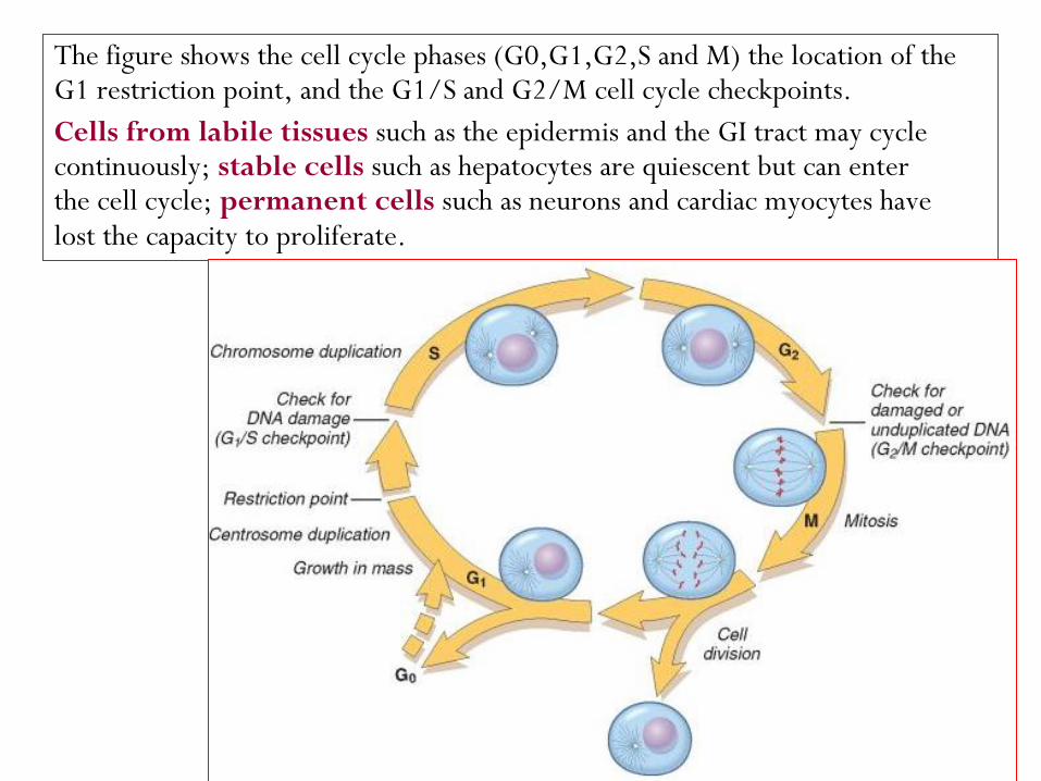
The figure shows the cell cycle phases (G0,G1,G2,S and M) the location of the G1 restriction point, and the G1/S and G2/M cell cycle checkpoints.
Cells from labile tissues such as the epidermis and the GI tract may cycle continuously; stable cells such as hepatocytes are quiescent but can enter the cell cycle; permanent cells such as neurons and cardiac myocytes have lost the capacity to proliferate.
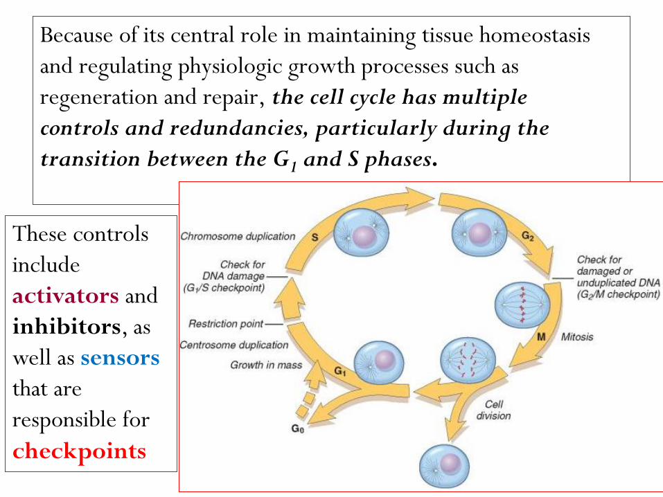
Because of its central role in maintaining tissue homeostasis
and regulating physiologic growth processes such as
regeneration and repair, the cell cycle has multiple
controls and redundancies, particularly during the
transition between the G1 and S phases.
These controls
include
activators and
inhibitors, as
well as sensors
that are
responsible for
checkpoints
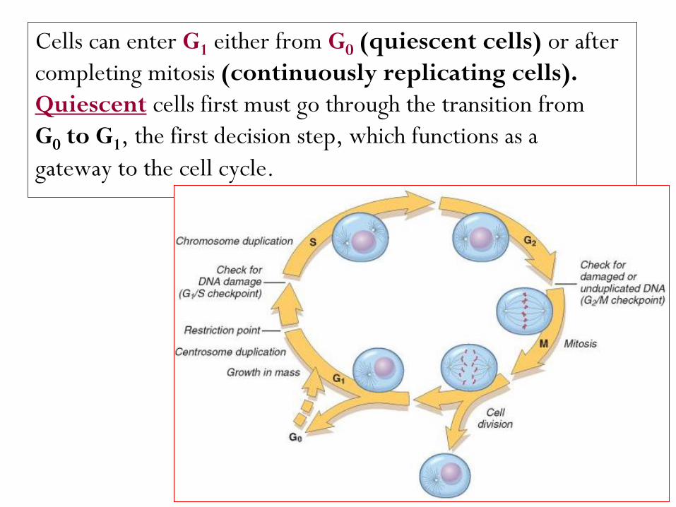
Cells can enter G1 either from G0 (quiescent cells) or after
completing mitosis (continuously replicating cells).
cells first must go through the transition from Quiescent
G0 to G1, the first decision step, which functions as a
gateway to the cell cycle.
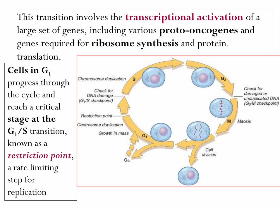
This transition involves the transcriptional activation of a
large set of genes, including various proto-oncogenes and
genes required for ribosome synthesis and protein.
translation.
Cells in G1
progress through
the cycle and
reach a critical
stage at the
G1/S transition,
known as a
restriction point,
a rate limiting
step for
replication

Upon passing this restriction point, normal cells become
irreversibly committed to DNA replication. Progression
through the cell cycle, particularly at the G1/S transition, is
tightly regulated by proteins called cyclins and associated
enzymes called cyclin-dependent kinases (CDKs).
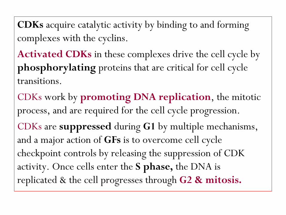
CDKs acquire catalytic activity by binding to and forming
complexes with the cyclins.
Activated CDKs in these complexes drive the cell cycle by
phosphorylating proteins that are critical for cell cycle
transitions.
CDKs work by promoting DNA replication, the mitotic
process, and are required for the cell cycle progression.
CDKs are suppressed during G1 by multiple mechanisms,
and a major action of GFs is to overcome cell cycle
checkpoint controls by releasing the suppression of CDK
activity. Once cells enter the S phase, the DNA is
replicated & the cell progresses through G2 & mitosis.
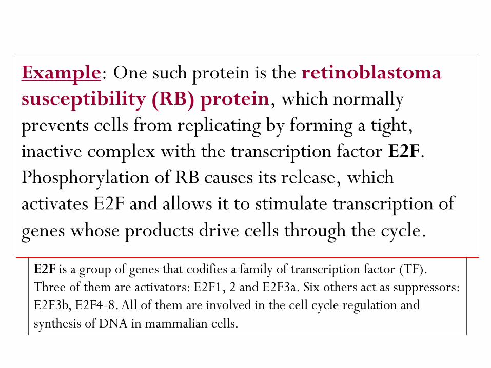
retinoblastoma : One such protein is the Example
susceptibility (RB) protein, which normally
prevents cells from replicating by forming a tight,
inactive complex with the transcription factor E2F.
Phosphorylation of RB causes its release, which
activates E2F and allows it to stimulate transcription of
genes whose products drive cells through the cycle.
E2F is a group of genes that codifies a family of transcription factor (TF).
Three of them are activators: E2F1, 2 and E2F3a. Six others act as suppressors:
E2F3b, E2F4-8. All of them are involved in the cell cycle regulation and
synthesis of DNA in mammalian cells.
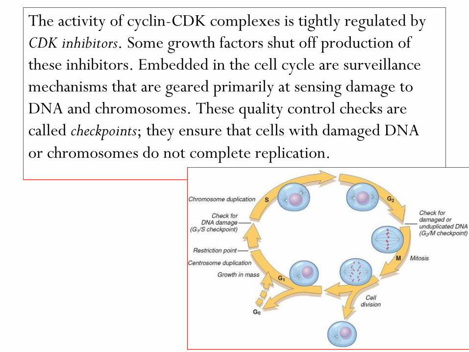
The activity of cyclin-CDK complexes is tightly regulated by
CDK inhibitors. Some growth factors shut off production of
these inhibitors. Embedded in the cell cycle are surveillance
mechanisms that are geared primarily at sensing damage to
DNA and chromosomes. These quality control checks are
called checkpoints; they ensure that cells with damaged DNA
or chromosomes do not complete replication.

The G1/S checkpoint monitors the integrity of DNA before
replication, whereas the G2/M checkpoint checks DNA
after replication and monitors whether the cell can safely
enter mitosis.
When cells sense DNA damage, checkpoint activation delays
the cell cycle and triggers DNA repair mechanisms.
If DNA damage is too severe to be repaired, the cells are
eliminated by apoptosis, or enter a nonreplicative state
called senescence, primarily through p53-dependent
mechanisms.
Growth Factors
The proliferation of many cell types is driven by polypeptides known as
growth factors. These factors, may also promote cell survival,
locomotion, contractility, differentiation, and angiogenesis,
activities that may be as important as their growth-
promoting effects.
All growth factors function as ligands that bind to
specific receptors, which deliver signals to the
target cells.
These signals stimulate the transcription of genes that may be
silent in resting cells, including genes that control cell cycle
entry and progression.
Epidermal Growth Factor (EGF) and
Transforming Growth Factor α (TGF-α).
(epidermal growth R: share a common α -EGF & TGF
) with intrinsic tyrosine kinase EGFR factor receptor, or
activity.
The EGFR is actually a family of receptors that respond to
EGF,TGF-α, & other ligands of the EGF family.
and hepatocytesfor mitogenicare α-Both EGF/TGF
including keratinocytes. ,epithelial cellsmost
In cutaneous wound healing, EGF is produced by
keratinocytes, macrophages, & other inflammatory cells.
The main EGFR called EGFR1 or ERB B1, which , lung & brain tumorsfrequently over expressed in
therapeutic target for the and is an important treatment of these tumors.
ERB B2 (also known as HER-2/NEU) has received great attention, because of its over-
in which, it is a ,breast cancersexpression in target for effective cancer control.
Hepatocyte Growth Factor (HGF).
The factor is often referred to as HGF/SF (scatter factor), but
in this chapter we will use the simpler notation, HGF.
HGF has mitogenic effects on hepatocytes and most epithelial
cells, including cells of the biliary epithelium, and epithelial
cells of the lungs, kidney, mammary gland, and skin.
It is produced by fibroblasts and most mesenchymal cells,
endothelial cells, and liver non-parenchymal cells.
It is produced as an inactive single-chain form (pro-HGF) that
is activated by serine proteases released in damaged tissues.
The receptor for HGF, c-MET, is often highly
expressed or mutated in human tumors, especially
in renal and thyroid papillary carcinomas.
Several HGF and c-MET inhibitors are presently being
evaluated in cancer therapy clinical trials.

Platelet-Derived Growth Factor (PDGF).
is a family of several closely related proteins, each consisting
of two chains. Three isoforms of PDGF (AA, AB, and BB)
are secreted as biologically active molecules. The more
recently identified isoforms PDGF-CC and PDGF-DD
require extracellular proteolytic cleavage to release the
active growth factor.
All PDGF isoforms exert their effects by binding to two cell
surface receptors, designated PDGFR α and β, which have
different ligand specificities.
PDGF is stored in platelet granules and is released on
platelet activation. It is produced by a variety of
cells, including activated macrophages, endothelial
cells, smooth muscle cells, and many tumor cells.
Vascular Endothelial Growth Factor (VEGF).
VEGFs are a family of homodimeric proteins that include
VEGF-A (referred throughout as VEGF), VEGF-B, VEGF-C,
VEGF-D, and PIGF (placental growth factor).
VEGF is a potent inducer of blood vessel formation in early
development (vasculogenesis) and has a central role in the
growth of new blood vessels (angiogenesis) in adults.
It promotes angiogenesis in chronic inflammation,
healing of wounds, and in tumors.
VEGF family members signal through three tyrosine kinase
receptors: VEGFR-1, VEGFR-2, and VEGFR-3.
VEGFR-2, located in endothelial cells and many other cell
types, is the main receptor for the vasculogenic and
angiogenic effects of VEGF.
The role of VEGFR-1 is less well understood, but it may
facilitate the mobilization of endothelial stem cells and has a
role in inflammation.
VEGF-C and VEGF-D bind to VEGFR-3 and act on lymphatic
endothelial cells to induce the production of lymphatic
vessels (lymphangiogenesis).
Fibroblast Growth Factor (FGF).
This is a family of growth factors containing more than
20 members, of which acidic FGF (aFGF, or FGF-1)
and basic FGF (bFGF, or FGF-2) are the best
characterized.
FGFs transduce signals through four tyrosine kinase
receptors (FGFRs 1–4). FGF-1 binds to all
receptors; FGF-7 is referred to as keratinocyte growth
factor or KGF.
FGFs contribute to wound healing responses, hematopoiesis,
angiogenesis, development, and other processes through
several functions:
1- Wound repair: FGF-2 and KGF (FGF-7) .
2- New blood vessel formation (angiogenesis): FGF-2, in particular.
3- Hematopoiesis: FGFs have been implicated in the differentiation
of specific lineages of blood cells and development of bone
marrow stroma.
4- Development: FGFs play a role in skeletal and cardiac muscle
development, lung maturation, and the specification of the
liver from endodermal cells.
Transforming Growth Factor β (TGF-β) and Related
Growth Factors.
TGF-β belongs to a superfamily of about 30 members that
includes three TGF-β isoforms (TGF-β1, TGF-β2, TGF-
β3) and factors with wide-ranging functions, such as BMPs,
activins, inhibins, and müllerian inhibiting substance.
TGF-β1 has the most widespread distribution in mammals
and will be referred to as TGF-β.
It is a homodimeric protein produced by a variety of different
cell types, including platelets, endothelial cells,
lymphocytes, and macrophages.
1- TGF-β is a growth inhibitor for most epithelial cells. It blocks the cell cycle
by increasing the expression of cell cycle inhibitors
2- TGF-β is a potent fibrogenic agent that stimulates fibroblast chemotaxis
and enhances the production of collagen, fibronectin, and
proteoglycans. It inhibits collagen degradation by decreasing matrix
proteases and increasing protease inhibitor activities.
3- TGF-β has a strong anti-inflammatory effect but may enhance some immune
functions. Knockout mice lacking the TGF-β1 gene in T cells have
defects in regulatory T cells leading to widespread inflammation with
abundant T-cell proliferation and CD4+ differentiation into TH1 and
TH2 helper cells.
Cytokines
Some of these proteins can also be considered as
growth factors, because they have growth-promoting
activities for a variety of cells.
Tumor necrosis factor (TNF) and IL-1 participate in
wound healing reactions, and TNF and IL-6 are
involved in the initiation of liver regeneration.
SIGNALING MECHANISMS IN CELL GROWTH
According to the source of the ligand and the location of its
receptors (i.e., in the same, adjacent, or distant cells),
three general modes of signaling, named autocrine,
paracrine, and endocrine, can be distinguished.
The binding of a ligand to its Receptor triggers a series of
events, by which extracellular signals are transduced into
the cell, leading to the stimulation or repression.
Signaling may occur (1) directly, in the same cell, (2) between
adjacent cells, or (3) over greater distances
Autocrine signaling, in which a soluble mediator acts
. on the cell that secretes itpredominantly
This pathway is important in the immune response (e.g
, lymphocyte proliferation induced by some cytokines)
.liver regeneration)(e.g and in compensatory epithelial hyperplasia
in the , in which, a substance affect cells Paracrine signaling
of the cell that released the agent. This vicinity immediate
pathway is important for (1) recruiting inflammatory cells
to the site of infection, & for (2) wound healing.
Endocrine signaling, in which a regulatory substance, such as a
acts on target , is released into the blood stream & hormone
cells at a distance.
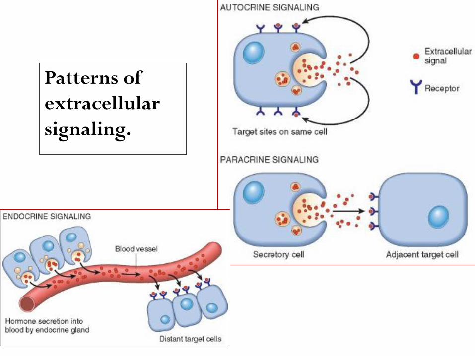
Patterns of
extracellular
signaling.
Receptors and Signal Transduction Pathways
The binding of a ligand to its receptor triggers a
series of events by which extracellular signals
are transduced into the cell resulting in changes
in gene expression.
Receptors are generally located on the surface of the
target cell but can also be found in the cytoplasm or
nucleus.
Receptors with intrinsic tyrosine kinase activity.
The ligands for receptors with tyrosine kinase activity include
most growth factors such as EGF, TGF-α, HGF, PDGF,
VEGF, FGF, c-KIT ligand, and insulin.
Binding of the ligand induces dimerization of the receptor,
tyrosine phosphorylation, and activation of the receptor
tyrosine kinase.
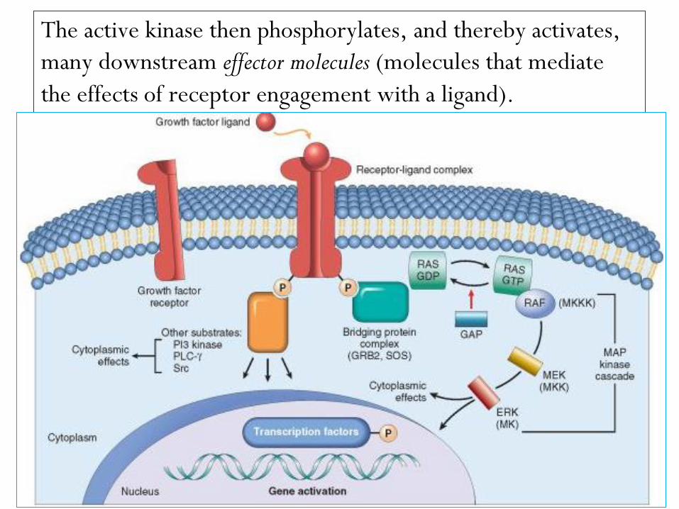
The active kinase then phosphorylates, and thereby activates,
many downstream effector molecules (molecules that mediate
the effects of receptor engagement with a ligand).

A prototypical adapter protein is GRB-2, which binds a guanosine
triphosphate–guanosine diphosphate (GTP-GDP) exchange factor called
SOS. SOS acts on the GTP-binding (G) protein RAS and catalyzes the
formation of RAS-GTP, which triggers the mitogen-activated protein
kinase (MAP kinase) cascade.
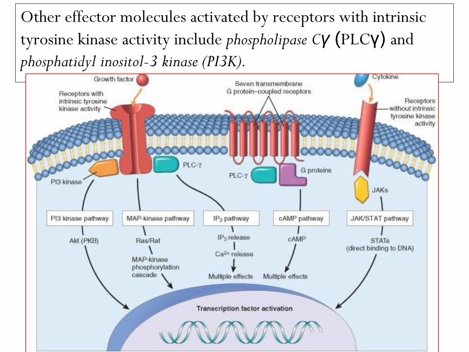
Other effector molecules activated by receptors with intrinsic
tyrosine kinase activity include phospholipase Cγ (PLCγ) and
phosphatidyl inositol-3 kinase (PI3K).
phospholipase Cγ (PLCγ) catalyzes the breakdown of
membrane inositol phospholipids into inositol 1,4,5-
triphosphate (IP3), which functions to increase
concentrations of calcium, an important effector
molecule, and diacylglycerol, which activates the serine-
threonine kinase protein kinase C that in turn activates
various transcription factors.
PI3K phosphorylates a membrane phospholipid, generating
products that activate the kinase Akt (also referred to as
protein kinase B), which is involved in cell proliferation
and cell survival through inhibition of apoptosis.
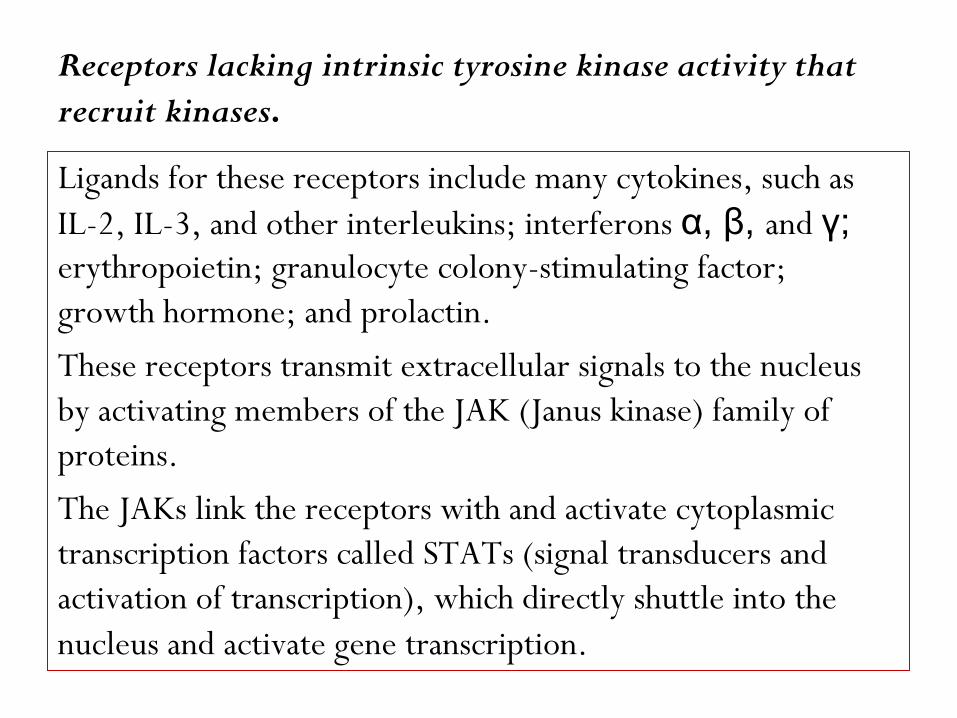
Receptors lacking intrinsic tyrosine kinase activity that
recruit kinases.
Ligands for these receptors include many cytokines, such as
IL-2, IL-3, and other interleukins; interferons α, β, and γ; erythropoietin; granulocyte colony-stimulating factor;
growth hormone; and prolactin.
These receptors transmit extracellular signals to the nucleus
by activating members of the JAK (Janus kinase) family of
proteins.
The JAKs link the receptors with and activate cytoplasmic
transcription factors called STATs (signal transducers and
activation of transcription), which directly shuttle into the
nucleus and activate gene transcription.
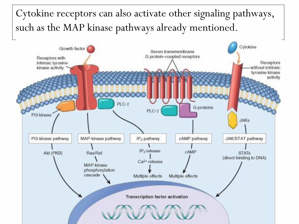
Cytokine receptors can also activate other signaling pathways,
such as the MAP kinase pathways already mentioned.
G protein–coupled receptors
Also called {seven transmembrane G-protein-coupled R}.
After ligand binding, the R associate with intracellular guanosine
triphosphate (GTP)-binding proteins (G-proteins), that
contain guanosine diphosphate (GDP).
Binding of the G proteins causes the exchange of GDP with
GTP, resulting in activation of the proteins.

Transduction pathways activated through G-protein-coupled
receptors are those involving:
(1) cyclic AMP (cAMP) as second messengers,
(2) the generation of inositol -1,4,5,-triphosphate (IP3),
which releases calcium from the ER.
These R constitute the largest family of plasma membrane R
(more than 1500 members have been identified) and
include those for: epinephrine, vasopressin, serotonin,
histamine, glucagon, and chemokines

Steroid hormone receptors.
These receptors are generally located in the nucleus and
function as ligand-dependent transcription factors.
The ligands diffuse through the cell membrane and bind the
inactive receptors, causing their activation.
The activated receptor then binds to specific DNA sequences known as
hormone response elements within target genes, or they
can bind to other transcription factors.
In addition to steroid hormones, other ligands that bind to
members of this receptor family include thyroid hormone,
vitamin D, and retinoids.

Transcription Factors
Many of the signal transduction systems used by
growth factors transfer information to the nucleus
and modulate gene transcription through the
activity of transcription factors.
Among the transcription factors that regulate cell
proliferation are products of several growth-
promoting genes, such as c-MYC and c-JUN, and of
cell cycle–inhibiting genes, such as p53.
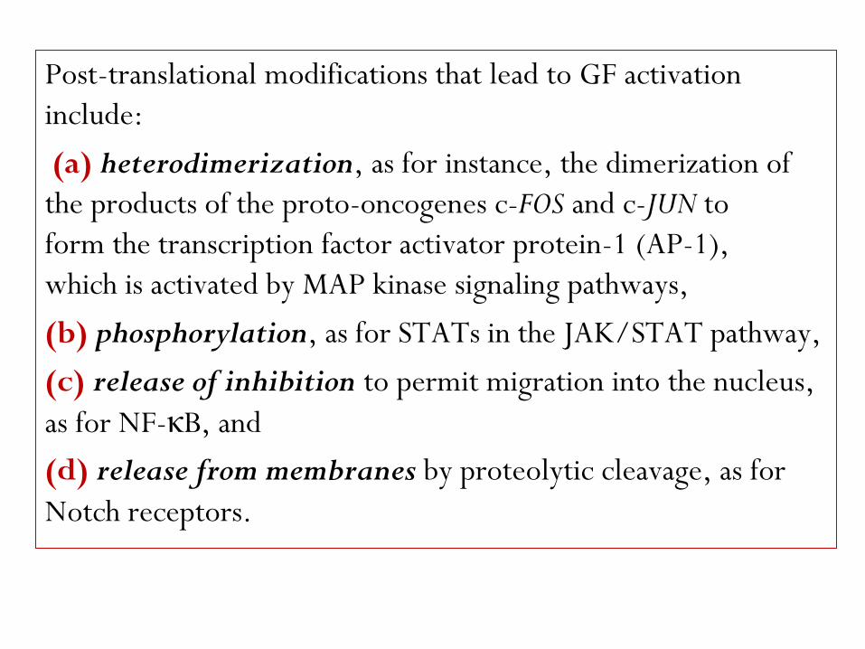
Post-translational modifications that lead to GF activation
include:
(a) heterodimerization, as for instance, the dimerization of
the products of the proto-oncogenes c-FOS and c-JUN to
form the transcription factor activator protein-1 (AP-1),
which is activated by MAP kinase signaling pathways,
(b) phosphorylation, as for STATs in the JAK/STAT pathway,
(c) release of inhibition to permit migration into the nucleus,
as for NF-κB, and
(d) release from membranes by proteolytic cleavage, as for
Notch receptors.
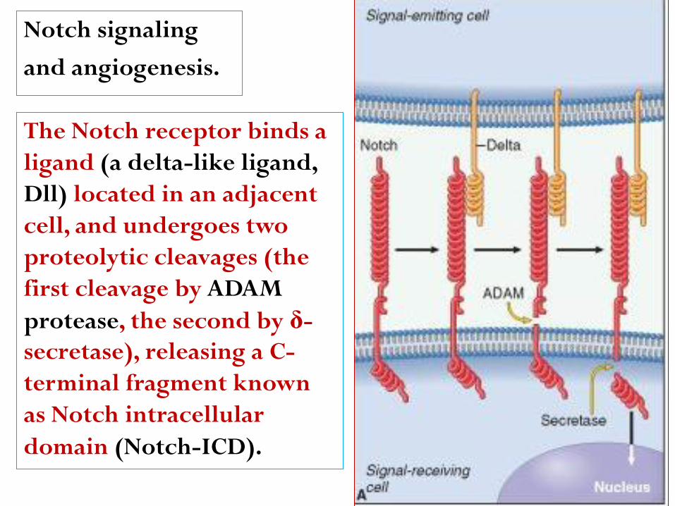
Notch signaling
and angiogenesis.
The Notch receptor binds a
ligand (a delta-like ligand,
Dll) located in an adjacent
cell, and undergoes two
proteolytic cleavages (the
first cleavage by ADAM
protease, the second by δ-
secretase), releasing a C-
terminal fragment known
as Notch intracellular
domain (Notch-ICD).

Notch signaling in
endothelial cells during
angiogenesis, triggered
by the binding of the
Dll4 ligand in a tip cell
to a Notch receptor in a
stalk cell. Notch-ICD
migrates into the
nucleus and activates
the transcription of
target genes.
Notch signaling
and angiogenesis.
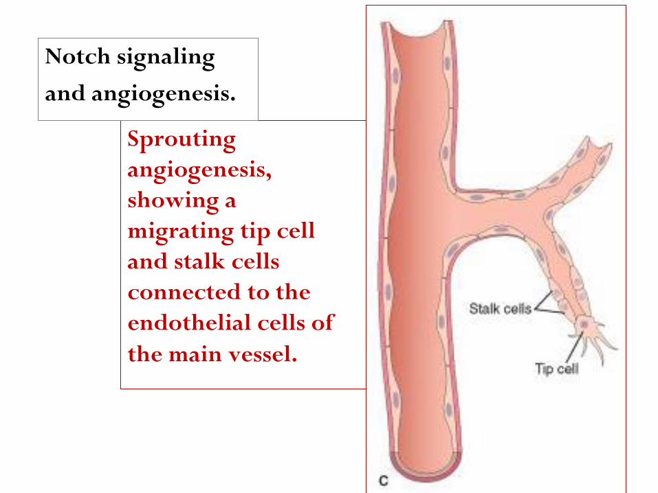
Sprouting
angiogenesis,
showing a
migrating tip cell
and stalk cells
connected to the
endothelial cells of
the main vessel.
Notch signaling
and angiogenesis.
Mechanisms of Tissue and Organ Regeneration
LIVER REGENERATION
In humans, resection of approximately 60% of the liver in
living donors results in the doubling of the liver remnant
in about one month. The portions of the liver that
remain after partial hepatectomy constitute an intact
“mini-liver” that rapidly expands and reaches the mass of
the original liver.
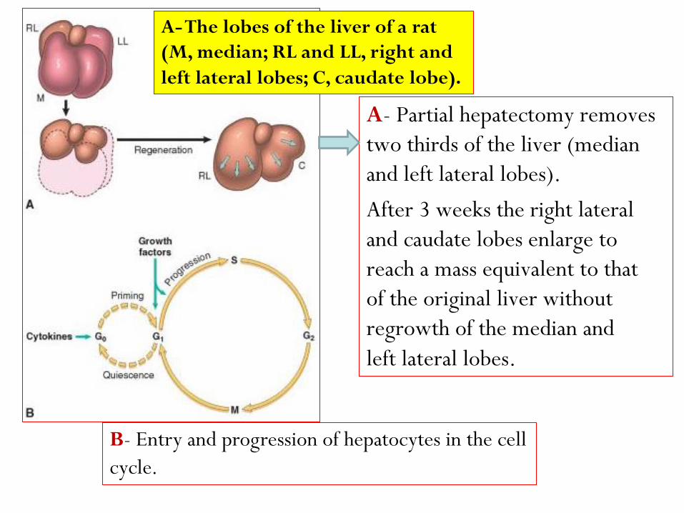
A- Partial hepatectomy removes
two thirds of the liver (median
and left lateral lobes).
After 3 weeks the right lateral
and caudate lobes enlarge to
reach a mass equivalent to that
of the original liver without
regrowth of the median and
left lateral lobes.
A- The lobes of the liver of a rat
(M, median; RL and LL, right and
left lateral lobes; C, caudate lobe).
B- Entry and progression of hepatocytes in the cell
cycle.
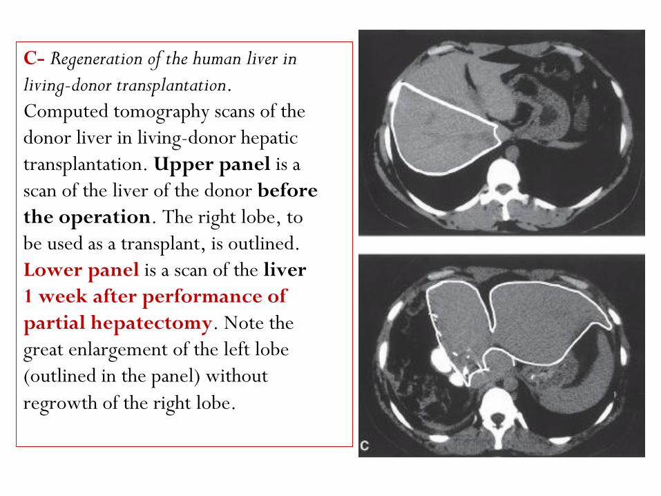
C- Regeneration of the human liver in
living-donor transplantation.
Computed tomography scans of the
donor liver in living-donor hepatic
transplantation. Upper panel is a
scan of the liver of the donor before
the operation. The right lobe, to
be used as a transplant, is outlined.
Lower panel is a scan of the liver
1 week after performance of
partial hepatectomy. Note the
great enlargement of the left lobe
(outlined in the panel) without
regrowth of the right lobe.
Restoration of liver mass is achieved without the regrowth of the lobes that
were resected at the operation.
Instead, growth occurs by enlargement of the lobes that remain
after the operation, a process known as compensatory growth
or compensatory hyperplasia.
Almost all hepatocytes replicate during liver regeneration after
partial hepatectomy. Because hepatocytes are quiescent cells, it
takes them several hours to enter the cell cycle, progress
through G1, and reach the S phase of DNA replication. The
wave of hepatocyte replication is synchronized and is followed
by synchronous replication of nonparenchymal cells (Kupffer
cells, endothelial cells, and stellate cells).

There is substantial evidence that hepatocyte proliferation in the
regenerating liver is triggered by the combined actions of cytokines
and polypeptide growth factors. With the exception of the
autocrine activity of TGF-α, hepatocyte replication is strictly
dependent on paracrine effects of growth factors and
cytokines such as HGF and IL-6 produced by hepatic
nonparenchymal cells.
There are two major restriction points for hepatocyte
replication: the G0/G1 transition that bring quiescent
hepatocytes into the cell cycle, and the G1/S transition
needed for passage through the late G1 restriction point.
Gene expression in the regenerating liver proceeds in phases,
starting with the immediate early gene response, which is a
transient response that corresponds to the G0/G1 transition.
More than 70 genes are activated during this response,
including the proto-oncogenes c-FOS and c-JUN, whose
products dimerize to form the transcription factor AP-1; c-
MYC, which encodes a transcription factor that activates many
different genes; and other transcription factors, such as NF-κB (nuclear factor kappa-light-chain-enhancer of activated B cells- protein complex that
controls transcription of DNA cytokine production and cell survival.), STAT-3,
and C/EBP (CCAAT-enhancer-binding proteins is a family of
transcription factors).
Quiescent hepatocytes become competent to enter the cell
cycle through a priming phase that is mostly mediated by the
cytokines TNF and IL-6, and components of the complement
system. Norepinephrine, serotonin, insulin, thyroid and
growth hormone, act as adjuvants for liver regeneration,
facilitating the entry of hepatocytes into the cell cycle.
Individual hepatocytes replicate once or twice during
regeneration and then return to quiescence in a strictly
regulated sequence of events, but the mechanisms of growth
cessation have not been established.
Growth inhibitors, such as TGF-β and activins, may
be involved in terminating hepatocyte replication,
but there is no clear understanding of their mode of
action. Intrahepatic stem or progenitor cells do not play a
role in the compensatory growth that occurs after partial
hepatectomy, and there is no evidence for hepatocyte
generation from bone marrow–derived cells during
this process. However, endothelial cells and other
nonparenchymal cells in the regenerating liver may
originate from bone marrow precursors.

Extracellular Matrix and Cell-Matrix
Interactions
Tissue repair and regeneration depend not only on the activity
of soluble factors, but also on interactions between cells
and the components of the extracellular matrix (ECM).
The ECM regulates the growth, proliferation, movement, and
differentiation of the cells living within it.
It is constantly remodeling, and its synthesis and degradation
accompanies morphogenesis, regeneration, wound healing,
chronic fibrotic processes, tumor invasion, and metastasis.
Its various functions include:
1- Mechanical support for cell anchorage and cell migration, and
maintenance of cell polarity.
2- Control of cell growth.
3- Maintenance of cell differentiation.
4- Scaffolding for tissue renewal.
5- Establishment of tissue microenvironments.
6- Storage and presentation of regulatory molecules.
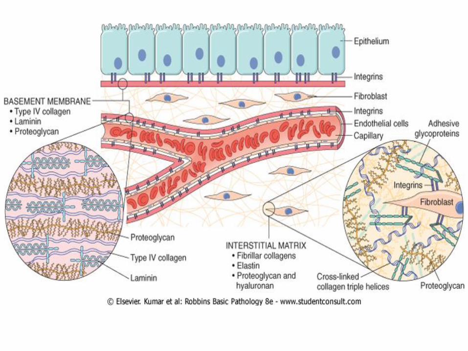
The ECM is composed of three groups of
macromolecules: fibrous structural proteins,
such as collagens and elastins that provide tensile
strength and recoil; adhesive glycoproteins that
connect the matrix elements to one another and to
cells; and proteoglycans and hyaluronan that
provide resilience and lubrication.
These molecules assemble to form two basic forms of ECM:
interstitial matrix and basement membranes.
is found in spaces between epithelial, The interstitial matrix
endothelial, and smooth muscle cells, as well as in connective
tissue. It consists mostly of fibrillar and nonfibrillar collagen,
elastin, fibronectin, proteoglycans, and hyaluronan.
are closely associated with cell Basement membranes
surfaces, and consist of nonfibrillar collagen (mostly type IV),
laminin, heparin sulfate, and proteoglycans.
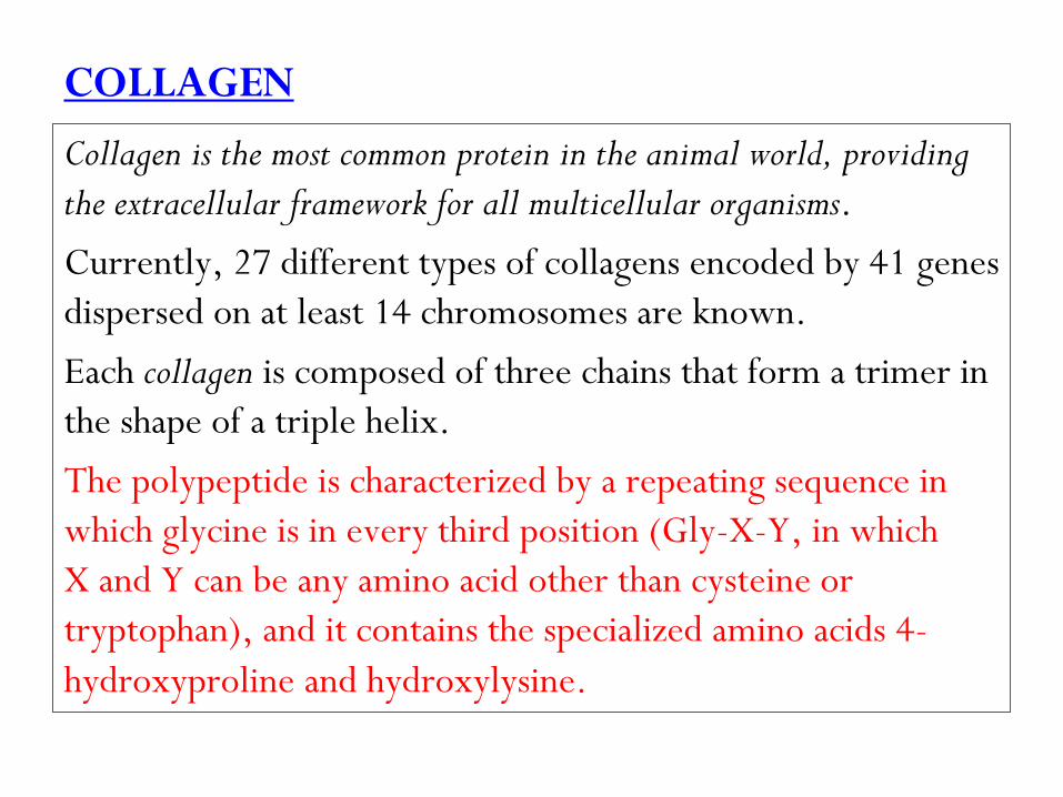
COLLAGEN
Collagen is the most common protein in the animal world, providing
the extracellular framework for all multicellular organisms.
Currently, 27 different types of collagens encoded by 41 genes
dispersed on at least 14 chromosomes are known.
Each collagen is composed of three chains that form a trimer in
the shape of a triple helix.
The polypeptide is characterized by a repeating sequence in
which glycine is in every third position (Gly-X-Y, in which
X and Y can be any amino acid other than cysteine or
tryptophan), and it contains the specialized amino acids 4-
hydroxyproline and hydroxylysine.
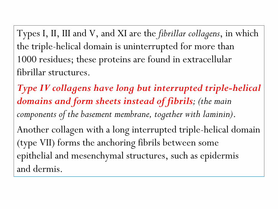
Types I, II, III and V, and XI are the fibrillar collagens, in which
the triple-helical domain is uninterrupted for more than
1000 residues; these proteins are found in extracellular
fibrillar structures.
Type IV collagens have long but interrupted triple-helical
domains and form sheets instead of fibrils; (the main
components of the basement membrane, together with laminin).
Another collagen with a long interrupted triple-helical domain
(type VII) forms the anchoring fibrils between some
epithelial and mesenchymal structures, such as epidermis
and dermis.

The messenger RNAs transcribed from fibrillar collagen genes
are translated into pre-pro-α chains that assemble in a type-
specific manner into trimers.
Hydroxylation of proline and lysine residues and lysine
glycosylation occur during translation. Three chains of a
particular collagen type assemble to form the triple helix
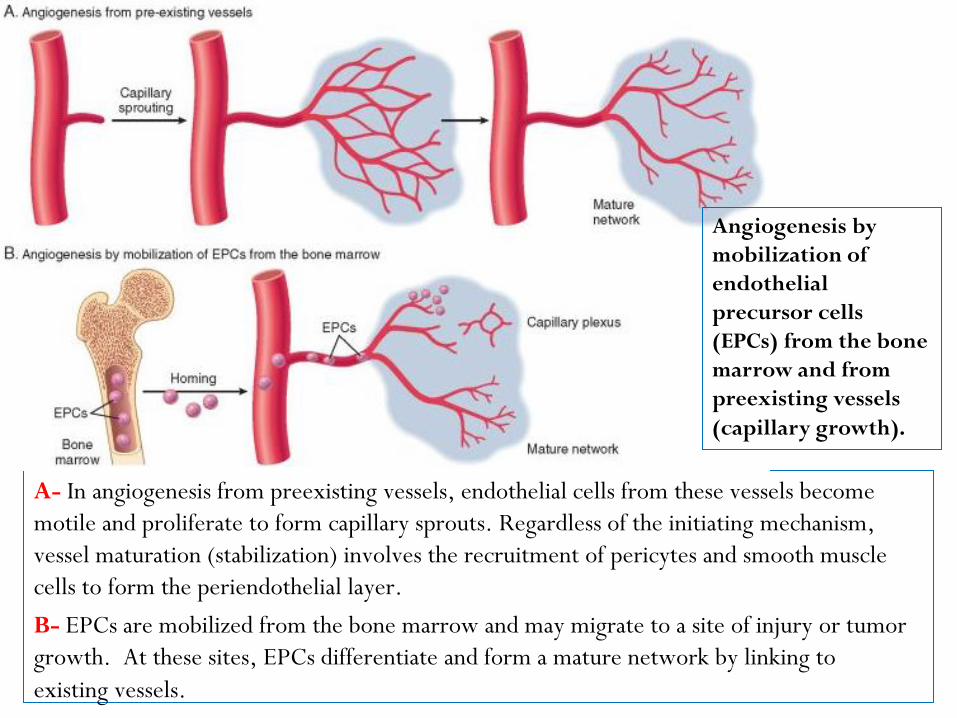
A- In angiogenesis from preexisting vessels, endothelial cells from these vessels become
motile and proliferate to form capillary sprouts. Regardless of the initiating mechanism,
vessel maturation (stabilization) involves the recruitment of pericytes and smooth muscle
cells to form the periendothelial layer.
B- EPCs are mobilized from the bone marrow and may migrate to a site of injury or tumor
growth. At these sites, EPCs differentiate and form a mature network by linking to
existing vessels.
Angiogenesis by
mobilization of
endothelial
precursor cells
(EPCs) from the bone
marrow and from
preexisting vessels
(capillary growth).
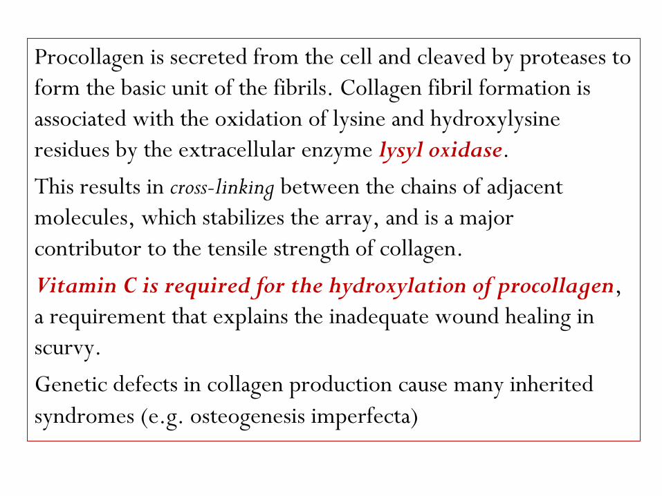
Procollagen is secreted from the cell and cleaved by proteases to
form the basic unit of the fibrils. Collagen fibril formation is
associated with the oxidation of lysine and hydroxylysine
residues by the extracellular enzyme lysyl oxidase.
This results in cross-linking between the chains of adjacent
molecules, which stabilizes the array, and is a major
contributor to the tensile strength of collagen.
Vitamin C is required for the hydroxylation of procollagen,
a requirement that explains the inadequate wound healing in
scurvy.
Genetic defects in collagen production cause many inherited
syndromes (e.g. osteogenesis imperfecta)
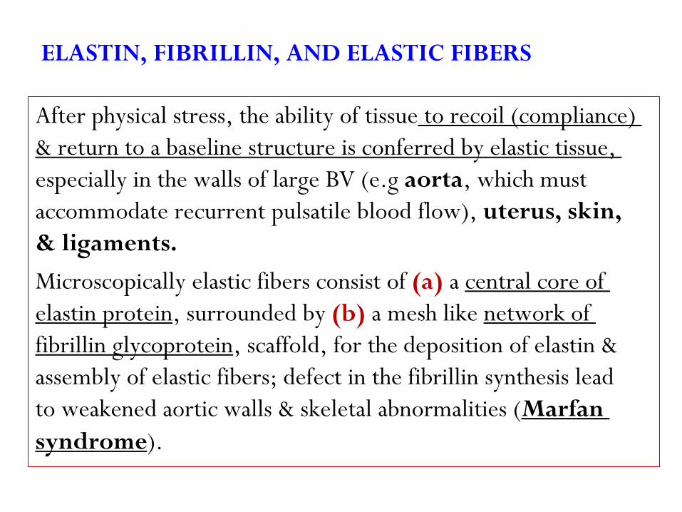
ELASTIN, FIBRILLIN, AND ELASTIC FIBERS
to recoil (compliance) After physical stress, the ability of tissue
& return to a baseline structure is conferred by elastic tissue,
especially in the walls of large BV (e.g aorta, which must
accommodate recurrent pulsatile blood flow), uterus, skin,
& ligaments.
central core of a (a)Microscopically elastic fibers consist of
network of a mesh like (b), surrounded by elastin protein
, scaffold, for the deposition of elastin & fibrillin glycoprotein
assembly of elastic fibers; defect in the fibrillin synthesis lead
Marfan to weakened aortic walls & skeletal abnormalities (
).syndrome
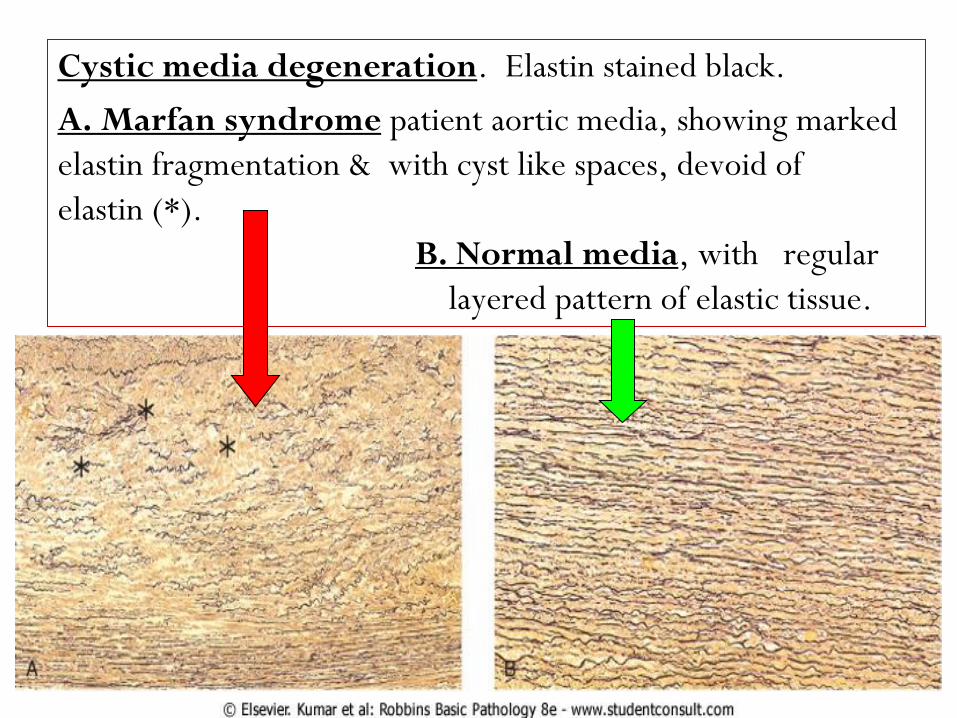
. Elastin stained black.Cystic media degeneration
patient aortic media, showing marked A. Marfan syndrome
elastin fragmentation & with cyst like spaces, devoid of
elastin (*).
, with regular B. Normal media
layered pattern of elastic tissue.
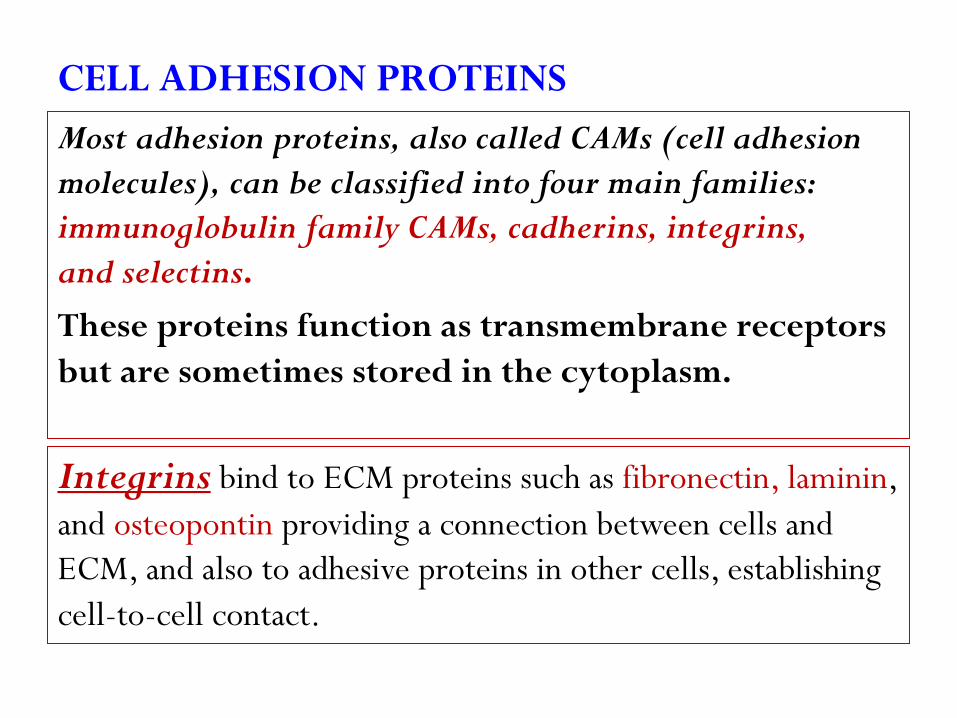
CELL ADHESION PROTEINS
Most adhesion proteins, also called CAMs (cell adhesion
molecules), can be classified into four main families:
immunoglobulin family CAMs, cadherins, integrins,
and selectins.
These proteins function as transmembrane receptors
but are sometimes stored in the cytoplasm.
, fibronectin, lamininbind to ECM proteins such as Integrinsand osteopontin providing a connection between cells and
ECM, and also to adhesive proteins in other cells, establishing
cell-to-cell contact.
is a large protein that binds to many molecules, Fibronectinsuch as collagen, fibrin, proteoglycans, and cell surface
receptors. It consists of two glycoprotein chains, held
together by disulfide bonds. Fibronectin messenger RNA
has two splice forms, giving rise to tissue fibronectin and
plasma fibronectin.
is the most abundant glycoprotein in the basement Lamininmembrane and has binding domains for both ECM and cell
surface receptors. In the basement membrane, polymers of
laminin and collagen type IV form tightly bound networks.
Laminin can also mediate the attachment of cells to connective
tissue substrates.
Cadherins and integrins link the cell surface with the
cytoskeleton through binding to actin and intermediate
filaments, providing a mechanism for the transmission of
mechanical force and the activation of intracellular signal
transduction pathways that respond to these forces.
The cytoskeletal proteins that co-localize with integrins at the
cell focal adhesion complex include talin, vinculin, and
paxillin. The integrin-cytoskeleton complexes function as
activated receptors and trigger a number of signal transduction
pathways, including the MAP (Mitogen-activated protein) kinase, PKC
(Protein kinase C), and PI3K (phosphatidylinositol-3-kinases) pathways,
which are also activated by growth factors.
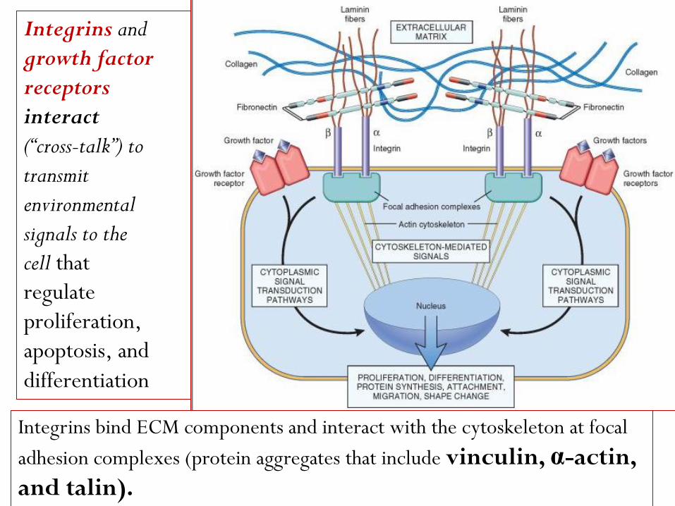
Integrins and
growth factor
receptors
interact
(“cross-talk”) to
transmit
environmental
signals to the
cell that
regulate
proliferation,
apoptosis, and
differentiation
Integrins bind ECM components and interact with the cytoskeleton at focal
adhesion complexes (protein aggregates that include vinculin, α-actin,
and talin).

Signaling from ECM components and growth factors is integrated by
the cell to produce various responses, including changes in cell
proliferation, locomotion, and differentiation.
This can initiate the
production of
intracellular
messengers or can
directly mediate
nuclear signals. Cell
surface receptors
for growth factors
may activate signal
transduction pathways
that overlap with those
activated by integrins.
dependent adherence protein):” This -(“calcium cadherinfamily contains almost 90 members, which participate in
interactions between cells of the same type, forming two
types of cell junctions called
(1) zonula adherens, small, spot-like junctions located near
the apical surface of epithelial cells, and
(2) desmosomes, stronger and more extensive junctions,
present in epithelial and muscle cells.
Cell-to-cell interactions mediated by cadherins and catenins
play a major role in regulating cell motility, proliferation,
and differentiation and account for the inhibition of cell
proliferation that occurs when cultured normal cells
contact each other (“contact inhibition”).
Diminished function of E-cadherin contributes to
certain forms of breast and gastric cancer.
(1) SPARC (secreted protein acidic and rich in cysteine), also
known as osteonectin, contributes to tissue remodeling in
response to injury and functions as an angiogenesis inhibitor;
(2) the thrombospondins, a family of large multifunctional
proteins, some of which, similar to SPARC, also inhibit
angiogenesis;
(3) osteopontin (OPN) is a glycoprotein that regulates
calcification, is a mediator of leukocyte migration involved in
inflammation, vascular remodeling, and fibrosis in various
organs; and
(4) the tenascin family, which consist of large multimeric
proteins involved in morphogenesis and cell adhesion.
Glycosaminoglycans (GAGS) and Proteoglycans
consist of long repeating polymers of specific GAGs
, GAGs hyaluronandisaccharides. With the exception of
are linked to a core protein, forming molecules called
proteoglycans. At most sites, ECM may contain several
different core proteins, each containing different GAGs.
Proteoglycans were originally described as ground substances
or mucopolysaccharides, whose main function was to organize
the ECM, but it is now recognized that these molecules
have diverse roles in regulating connective tissue structure
and permeability.
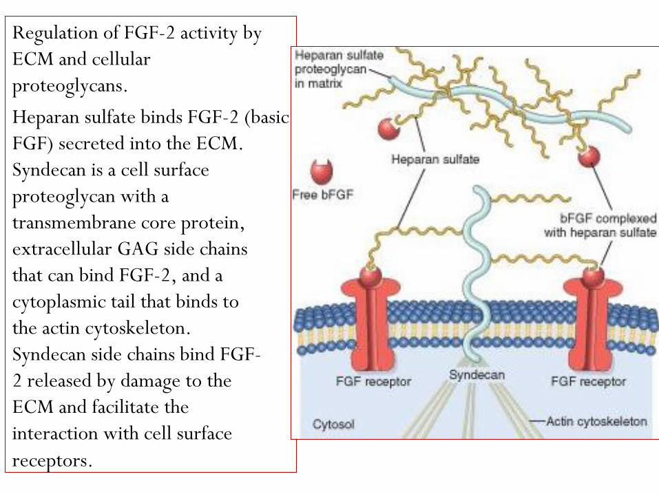
Regulation of FGF-2 activity by
ECM and cellular
proteoglycans.
Heparan sulfate binds FGF-2 (basic
FGF) secreted into the ECM.
Syndecan is a cell surface
proteoglycan with a
transmembrane core protein,
extracellular GAG side chains
that can bind FGF-2, and a
cytoplasmic tail that binds to
the actin cytoskeleton.
Syndecan side chains bind FGF-
2 released by damage to the
ECM and facilitate the
interaction with cell surface
receptors.

at Synthesis of hyaluronan -B
the inner surface of the
plasma membrane. The
molecule extends to the
extracellular space, while
still attached to hyaluronan
synthase.
C- Hyaluronan chains in
the extracellular space are
bound to the plasma
membrane through the
CD44 receptor. Multiple
proteoglycans may attach
to hyaluronan chains in the
ECM.

Proteoglycans can be integral membrane proteins and, through
their binding to other proteins and the activation of growth
factors and chemokines, act as modulators of inflammation,
immune responses, and cell growth and differentiation.
There are four structurally distinct families of GAGs:
heparan sulfate, chondroitin/dermatan sulfate, keratan
sulfate (are synthesized and assembled in the Golgi apparatus
and rough endoplasmic reticulum), and hyaluronan (HA)
(is produced at the plasma membrane by enzymes called
hyaluronan synthases and is not linked to a protein
backbone).
HA is a polysaccharide of the GAG family found in the ECM of many
tissues and is abundant in heart valves, skin and skeletal tissues,
synovial fluid, the vitreous of the eye, and the umbilical cord.
It is a huge molecule that consists of many repeats of a simple
disaccharide stretched end-to-end. It binds a large amount of
water (about 1000-fold its own weight), forming a viscous
hydrated gel that gives connective tissue the ability to resist
compression forces, and helps provide lubrication to many types
of connective tissue (cartilage in joints). Its concentration
increases in inflammatory diseases such as rheumatoid arthritis,
scleroderma, psoriasis, and osteoarthritis.
Enzymes called hyaluronidases fragment HA into lower
molecular weight molecules (LMW HA) that have different
functions than the parent molecule.
1- LMW HA produced by endothelial cells binds to the CD44
receptor on leukocytes, promoting the recruitment of
leukocytes to the sites of inflammation.
2- LMW HA molecules stimulate the production of
inflammatory cytokines and chemokines by white cells
recruited to the sites of injury.
Healing by Repair, Scar Formation and Fibrosis
If tissue injury is severe or chronic, and results in damage of
both parenchymal cells and the stromal framework of the
tissue, healing can not be accomplished by regeneration.
Under these conditions, the main healing process is repair by
deposition of collagen and other ECM components, causing
the formation of a scar.
Repair is a fibroproliferative response that “patches” rather than
restores the tissue. The term scar is most often used in
connection to wound healing in the skin, but is also used to
describe the replacement of parenchymal cells in any tissue by
collagen, as in the heart after myocardial infarction.

Repair by connective tissue deposition includes the
following basic features:
1- Inflammation.
2- Angiogenesis.
3- Migration and proliferation of fibroblasts.
4- Scar formation.
5- Connective tissue remodeling.
In most healing processes, a combination of repair and
regeneration occurs.
The relative contributions of repair and regeneration
are influenced by:
(1) The proliferative capacity of the cells of the tissue;
(2) The integrity of the extracellular matrix; and
(3) The resolution or chronicity of the injury and
inflammation.
Mechanisms of Angiogenesis
Angiogenesis is a fundamental process that affects physiologic
reactions (e.g. wound healing, regeneration, the
vascularization of ischemic tissues, and menstruation), and
pathologic processes, such as tumor development and
metastasis, diabetic retinopathy, and chronic inflammation.
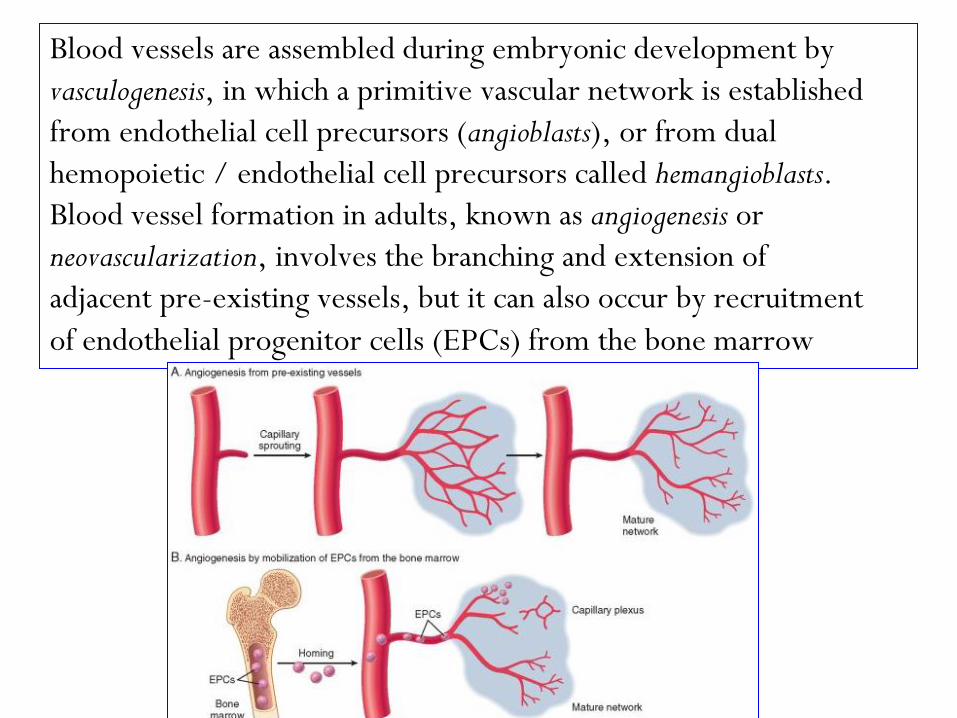
Blood vessels are assembled during embryonic development by
vasculogenesis, in which a primitive vascular network is established
from endothelial cell precursors (angioblasts), or from dual
hemopoietic / endothelial cell precursors called hemangioblasts.
Blood vessel formation in adults, known as angiogenesis or
neovascularization, involves the branching and extension of
adjacent pre-existing vessels, but it can also occur by recruitment
of endothelial progenitor cells (EPCs) from the bone marrow
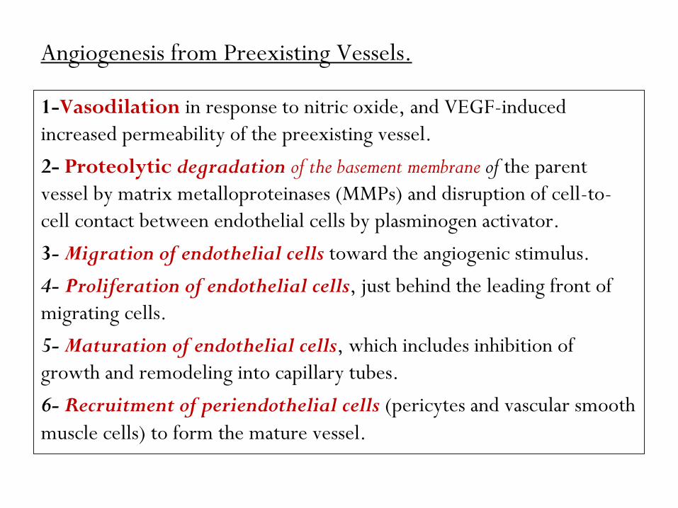
Angiogenesis from Preexisting Vessels.
1-Vasodilation in response to nitric oxide, and VEGF-induced
increased permeability of the preexisting vessel.
2- Proteolytic degradation of the basement membrane of the parent
vessel by matrix metalloproteinases (MMPs) and disruption of cell-to-
cell contact between endothelial cells by plasminogen activator.
3- Migration of endothelial cells toward the angiogenic stimulus.
4- Proliferation of endothelial cells, just behind the leading front of
migrating cells.
5- Maturation of endothelial cells, which includes inhibition of
growth and remodeling into capillary tubes.
6- Recruitment of periendothelial cells (pericytes and vascular smooth
muscle cells) to form the mature vessel.
Angiogenesis from Endothelial Precursor Cells (EPCs).
The nature of the homing mechanism is uncertain. These cells
express some markers of hematopoietic stem cells as well as
VEGFR-2, and vascular endothelial–cadherin (VE cadherin).
EPCs may contribute to the re-endothelization of vascular
implants and the neovascularization of ischemic organs,
cutaneous wounds, and tumors.
Growth Factors and Receptors Involved in Angiogenesis
is the most important growth factor in adult tissues undergoing VEGF
physiologic angiogenesis (e.g., proliferating endometrium) as
well as angiogenesis occurring in chronic inflammation,
wound healing, tumors, and diabetic retinopathy.
Of the various receptors for VEGF, VEGFR-2, a tyrosine
kinase receptor, is the most important in angiogenesis. It is
expressed by endothelial cells and their precursors, by other
cell types, and by many tumor cells.
VEGF induces the migration of EPCs in the bone marrow, and
enhances the proliferation and differentiation of these cells
at sites of angiogenesis. In angiogenesis originating from
preexisting local vessels, VEGF signaling stimulates the
survival of endothelial cells, their proliferation and their
motility, initiating the sprouting of new capillaries.
Endothelial cell proliferation, differentiation, and migration
can also be stimulated by FGF-2.
The Notch pathway, which promotes the proper
branching of new vessels and prevents excessive
angiogenesis by decreasing the responsiveness to VEGF.
Notch ligands and receptors are membrane-bound
molecules conserved between species.
In mammals there are five Notch ligands (Jagged 1 and 2,
and Delta-like ligand [Dll] 1, 3, and 4) and four
transmembrane receptors (Notch 1–4).
VEGF induces Delta-like ligand 4 in tip cells, while Notch1
and Notch4 are expressed in stalk cells.
The interaction between Delta-like ligand 4 and Notch
receptors in adjacent tip and stalk cells leads to a two-step
proteolytic cleavage of the receptor, releasing the Notch
intracellular domain, which translocates to the nucleus and
activates genes that dampen responsiveness to VEGF.
Blockade of Delta-like ligand 4 causes increased
proliferation of endothelial cells and capillary sprouting;
VEGF blockade has the opposite effects and also decreases
the survival of endothelial cells.
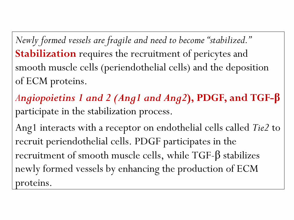
Newly formed vessels are fragile and need to become “stabilized.”
Stabilization requires the recruitment of pericytes and
smooth muscle cells (periendothelial cells) and the deposition
of ECM proteins.
Angiopoietins 1 and 2 (Ang1 and Ang2), PDGF, and TGF-β
participate in the stabilization process.
Ang1 interacts with a receptor on endothelial cells called Tie2 to
recruit periendothelial cells. PDGF participates in the
recruitment of smooth muscle cells, while TGF-β stabilizes
newly formed vessels by enhancing the production of ECM
proteins.
Agents or conditions that stimulate VEGF expression,
such as certain cytokines and growth factors (e.g.,
TGF-β, PDGF, TGF-α), and notably, tissue hypoxia,
can influence physiologic and pathologic
angiogenesis. VEGF transcription is regulated by the
transcription factor HIF, which is induced by hypoxia.
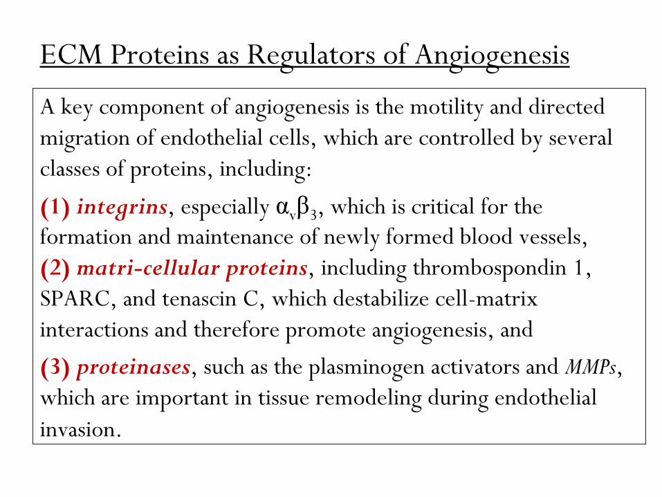
ECM Proteins as Regulators of Angiogenesis
A key component of angiogenesis is the motility and directed
migration of endothelial cells, which are controlled by several
classes of proteins, including:
(1) integrins, especially αvβ3, which is critical for the
formation and maintenance of newly formed blood vessels,
(2) matri-cellular proteins, including thrombospondin 1,
SPARC, and tenascin C, which destabilize cell-matrix
interactions and therefore promote angiogenesis, and
(3) proteinases, such as the plasminogen activators and MMPs,
which are important in tissue remodeling during endothelial
invasion.
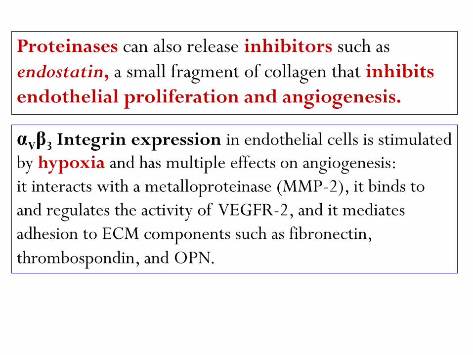
Proteinases can also release inhibitors such as
endostatin, a small fragment of collagen that inhibits
endothelial proliferation and angiogenesis.
αVβ3 Integrin expression in endothelial cells is stimulated
by hypoxia and has multiple effects on angiogenesis:
it interacts with a metalloproteinase (MMP-2), it binds to
and regulates the activity of VEGFR-2, and it mediates
adhesion to ECM components such as fibronectin,
thrombospondin, and OPN.
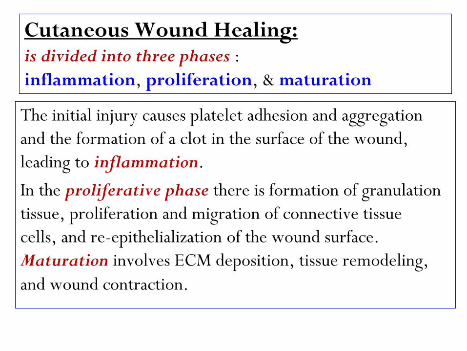
Cutaneous Wound Healing: is divided into three phases :
inflammation, proliferation, & maturation
The initial injury causes platelet adhesion and aggregation
and the formation of a clot in the surface of the wound,
leading to inflammation.
In the proliferative phase there is formation of granulation
tissue, proliferation and migration of connective tissue
cells, and re-epithelialization of the wound surface.
Maturation involves ECM deposition, tissue remodeling,
and wound contraction.
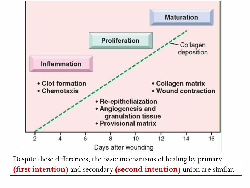
Despite these differences, the basic mechanisms of healing by primary
(first intention) and secondary (second intention) union are similar.
Healing by First Intention
One of the simplest examples of wound repair is the healing of a clean, uninfected surgical incision approximated
primary union . This is referred to as by surgical sutures or healing by first intention.
The incision causes only focal disruption (loss of continuity) of epithelial BM & death of relatively few epithelial & connective tissue cells.
As a result, epithelial regeneration predominates over fibrosis.
A small scar is formed , but there is minimal wound contraction. ,clotted blood-fills with fibrinThe narrow incisional space first
which is rapidly invaded by granulation tissue & covered by new epithelium.
are seen at the incision margin, neutrophils :hours24 Within
Basal cells at the cut edge of migrating toward the fibrin clot.
mitotic activity.begin to exhibit the epidermis
Within 24 to 48 hours: epithelial cells from both edges have
begun to migrate & proliferate along the dermis, depositing
BM components as they progress. The cells meet in the
thin but , yielding a scabbeneath the surface midline
continuous epithelial layer.
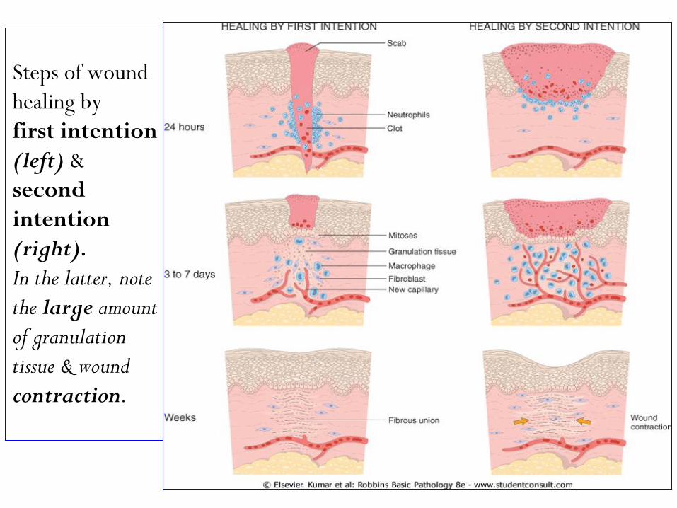
Steps of wound
healing by
first intention
(left) &
second
intention
(right).
In the latter, note
the large amount
of granulation
tissue & wound
contraction.
By day 3: neutrophils have been largely replaced by
macrophages, & granulation tissue progressively
invades the incision space.
Collagen fibers are now evident at the incision margins, but
these are vertically oriented & do not bridge the incision.
Epithelial cell proliferation continues, yielding a thickened
epidermal covering layer.
By day 5: angiogenesis reaches its peak as granulation
tissue fills the incisional space & Collagen fibrils become
more abundant & begin to bridge the incision.
The epidermis recovers its normal thickness as differentiation of
surface cells yields a mature epidermal architecture with
surface keratinization.
During the second week: There is continued collagen
accumulation & fibroblasts proliferation. The WBC infiltrate,
edema, & the increased vascularity are substantially
diminished.
, accomplished long process of "blanching“ (pallor) beginsThe
by: increased collagen deposition within the incisional scar &
the regression of vascular channels.
acellular the scar comprises ,By the end of the first month
devoid of inflammatory cells and connective tissue,
. epidermis covered by an essentially normal
(hair follicles, sweat & dermal appendagesHowever, the
sebaceous glands) which are destroyed in the line of incision
are permanently lost.
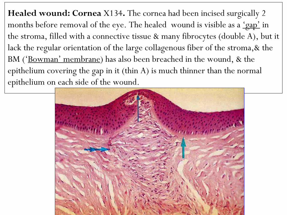
Healed wound: Cornea X134. The cornea had been incised surgically 2
months before removal of the eye. The healed wound is visible as a „gap‟ in
the stroma, filled with a connective tissue & many fibrocytes (double A), but it
lack the regular orientation of the large collagenous fiber of the stroma,& the
BM („Bowman‟ membrane) has also been breached in the wound, & the
epithelium covering the gap in it (thin A) is much thinner than the normal
epithelium on each side of the wound.
Healing by Secondary Intention
When cell or tissue loss is more extensive, as in Infarction,
Abscess, Ulcer, or Large wound, the reparative process is
more complex.
of parenchymal cells alone cannot restore the regeneration The
extensiveoriginal architecture, therefore, there is an
from the wound margins, of granulation tissue ingrowth
followed by ECM accumulation & scarring. This is called
.secondary union or healing by second intention
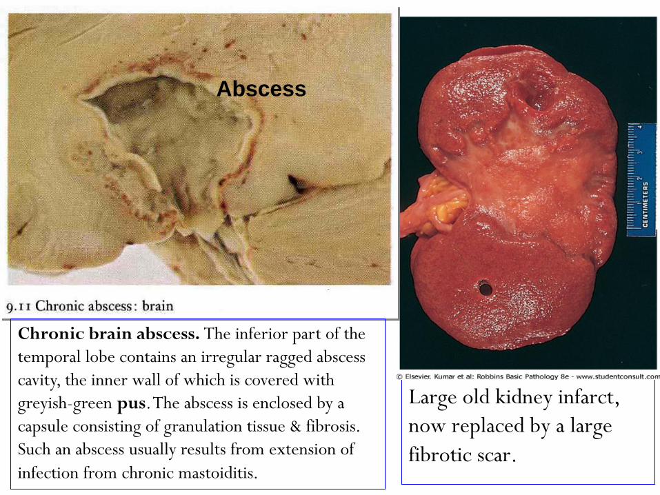
Large old kidney infarct,
now replaced by a large
fibrotic scar.
Abscess
Chronic brain abscess. The inferior part of the
temporal lobe contains an irregular ragged abscess
cavity, the inner wall of which is covered with
greyish-green pus. The abscess is enclosed by a
capsule consisting of granulation tissue & fibrosis.
Such an abscess usually results from extension of
infection from chronic mastoiditis.
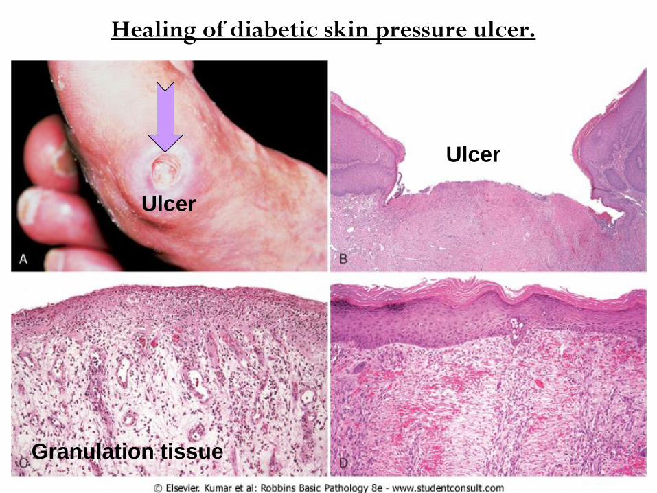
Healing of diabetic skin pressure ulcer.
Ulcer
Granulation tissue
Ulcer
from the primary in several aspects: DiffersSecondary union
(I) Large tissue defects, means a greater volume of necrotic
debris + exudate + fibrin that must be removed & more
intense inflammatory reaction needed, with greater
potential for secondary inflammation-mediated injury caused by
the:
causing cell injury & into ECM lysosomal enzymesrelease of (a)
matrix degradation, or
metabolic products, both ROS & AAactivated WBC release of (b)
of which are capable of causing tissue damage.
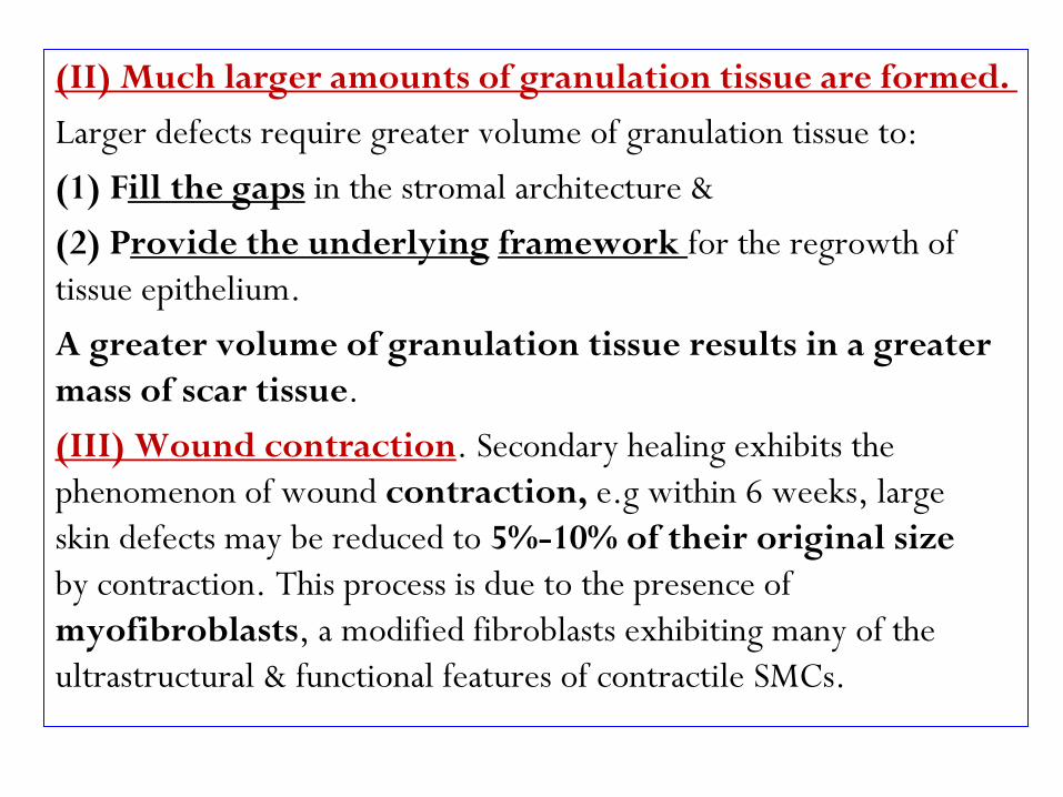
(II) Much larger amounts of granulation tissue are formed.
Larger defects require greater volume of granulation tissue to:
in the stromal architecture & ill the gapsF )1(
for the regrowth of framework rovide the underlying) P2(
tissue epithelium.
A greater volume of granulation tissue results in a greater
mass of scar tissue.
. Secondary healing exhibits the (III) Wound contraction
phenomenon of wound contraction, e.g within 6 weeks, large
skin defects may be reduced to 5%-10% of their original size
by contraction. This process is due to the presence of
myofibroblasts, a modified fibroblasts exhibiting many of the
ultrastructural & functional features of contractile SMCs.
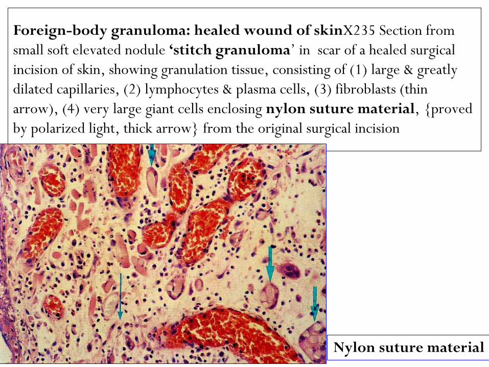
Foreign-body granuloma: healed wound of skinX235 Section from
small soft elevated nodule ‘stitch granuloma‟ in scar of a healed surgical
incision of skin, showing granulation tissue, consisting of (1) large & greatly
dilated capillaries, (2) lymphocytes & plasma cells, (3) fibroblasts (thin
arrow), (4) very large giant cells enclosing nylon suture material, {proved
by polarized light, thick arrow} from the original surgical incision
Nylon suture material
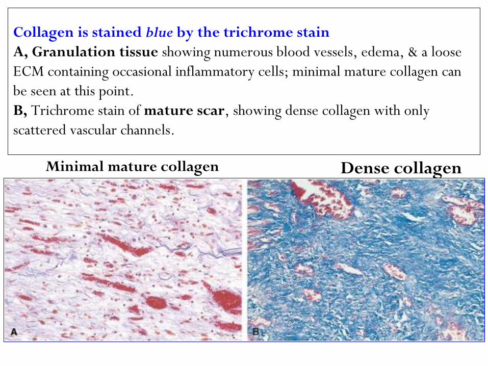
Collagen is stained blue by the trichrome stain
A, Granulation tissue showing numerous blood vessels, edema, & a loose
ECM containing occasional inflammatory cells; minimal mature collagen can
be seen at this point.
B, Trichrome stain of mature scar, showing dense collagen with only
scattered vascular channels.
Dense collagen Minimal mature collagen
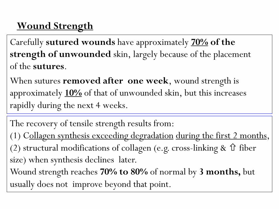
Wound Strength
of the %70have approximately sutured woundsCarefully
strength of unwounded skin, largely because of the placement
of the sutures.
When sutures removed after one week, wound strength is
increases of that of unwounded skin, but this %10approximately
rapidly during the next 4 weeks.
The recovery of tensile strength results from:
, months2 during the first ollagen synthesis exceeding degradation) C1(
(2) structural modifications of collagen (e.g. cross-linking & fiber
size) when synthesis declines later.
Wound strength reaches 70% to 80% of normal by 3 months, but
usually does not improve beyond that point.
In wound healing, normal cell growth & fibrosis may be altered by a variety of factors, frequently reducing the quality or adequacy of the reparative process:
Delay in wound healing may be caused by the following:
is the single most important cause of . Infection:1delay in healing, by prolonging the inflammation phase of the process, & potentially ↑ the local tissue injury.
has profound effects on wound healing, for .Nutrition:2example: protein deficiency & especially, vitamin C deficiency, inhibit collagen synthesis & retard healing.
Pathologic Aspects of Repair
-antidocumented -(steroids): have well. Glucocorticoids 3
effects, & their administration may result in inflammatory
. diminished fibrosis poor wound strength owing to
In some instances, however, the anti-inflammatory effects of
, corneal infectionse.g in desirable, glucocorticoids are
glucocorticoids are some times prescribed (along with
antibiotics) to reduce the likelihood of opacity that may
result from collagen deposition.
such as increased local pressure or . Mechanical factors 4torsion, may cause wounds to pull apart (separate), or
abdominal wound dehiscence after (e.g dehisce .lapratomy)
due either to , . Poor blood perfusion5
(a) atherosclerosis (which reduce arterial blood supply), or
(b) to obstructed venous drainage, e.g varicose veins, both impairs healing.
shot), -such as fragments of steel (eg gun . Foreign bodies6glass, wood, or even bone, impede (delay) healing.
factors in criticalThe type & the volume of tissue injured are a
healing:
can occur only in tissues Complete regeneration
even then, cells; labile & stablecomposed of
extensive injury will likely result in incomplete tissue
regeneration & at least partial loss of function.
must permanent cellsInjury to tissue composed of
with at most, attempts at scarringresult in inevitably
functional compensation by the remaining viable elements.
(e.g. healing of a MI).
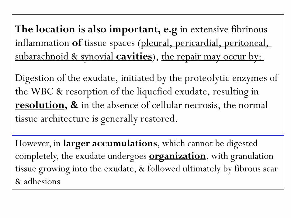
The location is also important, e.g in extensive fibrinous
pleural, pericardial, peritoneal, tissue spaces (of inflammation
the repair may occur by: ), cavitiessubarachnoid & synovial
Digestion of the exudate, initiated by the proteolytic enzymes of
the WBC & resorption of the liquefied exudate, resulting in
in the absence of cellular necrosis, the normal , &resolution
tissue architecture is generally restored.
However, in larger accumulations, which cannot be digested
, with granulation organizationcompletely, the exudate undergoes
tissue growing into the exudate, & followed ultimately by fibrous scar
& adhesions
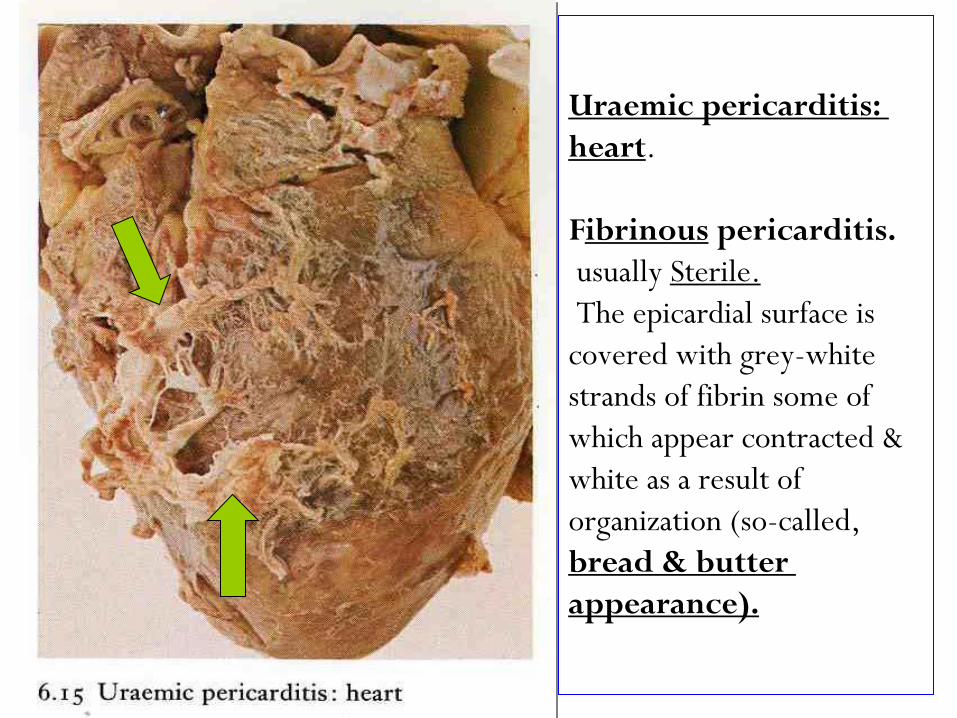
Uraemic pericarditis:
.heart
pericarditis. ibrinousF
Sterile.usually
The epicardial surface is
covered with grey-white
strands of fibrin some of
which appear contracted &
white as a result of
organization (so-called,
bread & butter
appearance).
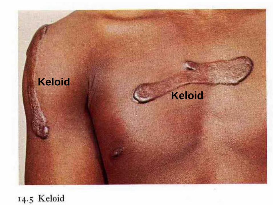
Aberrations (abnormalities) of cell growth & ECM production
may occur even in what begins as normal wound
healing, e.g., the accumulation of excessive amounts of
known as prominent, raised scarscan give rise to collagen
, more commonly seen in blacks . Keloids
excessive granulation Healing wounds may also generate
that protrudes above the level of the surrounding skin tissue
epithelialization. This is called-& in fact, hinders (prevent) re
, Thusexuberant granulation, or proud flesh.
restoration of epithelial continuity requires cautery
(burning) electrical, or chemical, or surgical resection of
the granulation tissue.
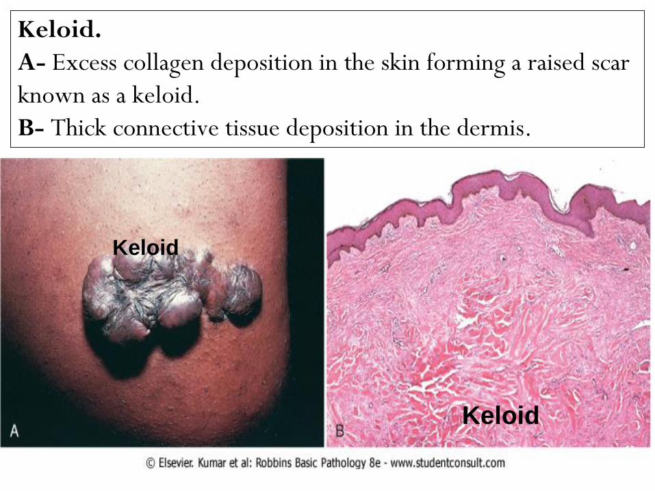
Keloid.
A- Excess collagen deposition in the skin forming a raised scar
known as a keloid.
B- Thick connective tissue deposition in the dermis.
Keloid
Keloid
Keloid
Keloid
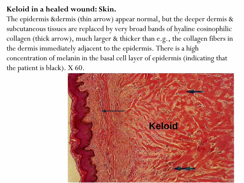
Keloid in a healed wound: Skin.
The epidermis &dermis (thin arrow) appear normal, but the deeper dermis &
subcutaneous tissues are replaced by very broad bands of hyaline eosinophilic
collagen (thick arrow), much larger & thicker than e.g., the collagen fibers in
the dermis immediately adjacent to the epidermis. There is a high
concentration of melanin in the basal cell layer of epidermis (indicating that
the patient is black). X 60.
Keloid
F 3-17: Overview of
repair responses.
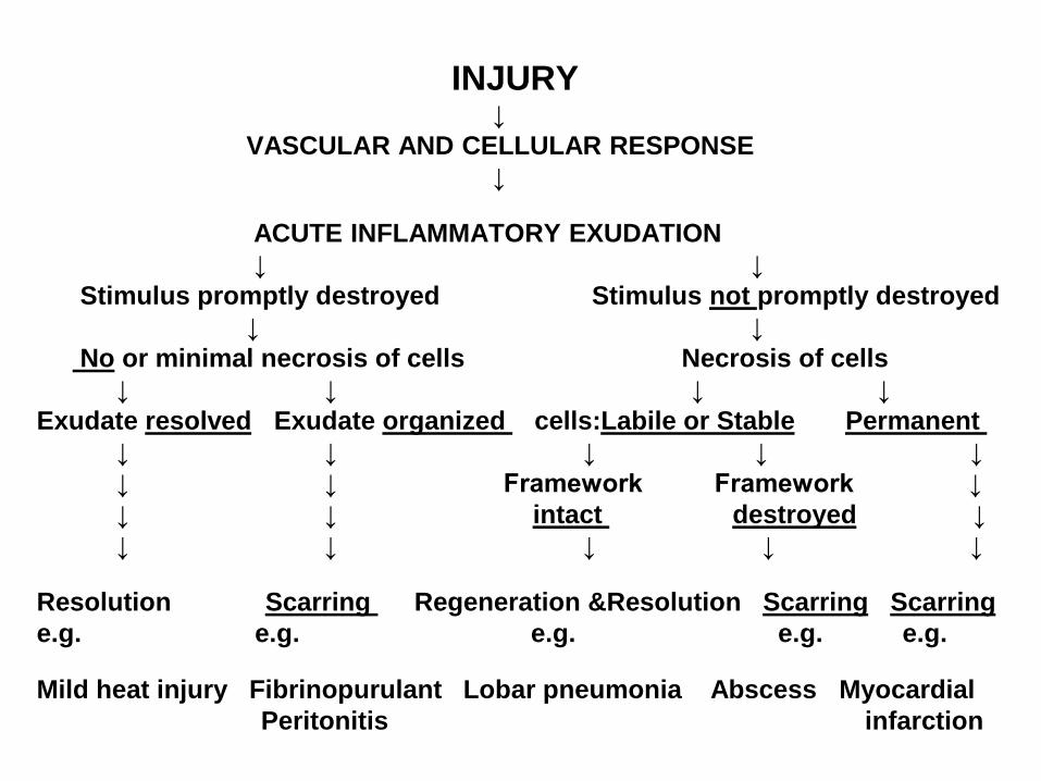
INJURY ↓
VASCULAR AND CELLULAR RESPONSE
↓
ACUTE INFLAMMATORY EXUDATION
↓ ↓
promptly destroyednot Stimulus promptly destroyed Stimulus
↓ ↓
or minimal necrosis of cells Necrosis of cells No
↓ ↓ ↓ ↓
Permanent Labile or Stablecells: organized Exudate resolvedExudate
↓ ↓ ↓ ↓ ↓
↓ ↓ Framework Framework ↓
↓ destroyed intact ↓ ↓
↓ ↓ ↓ ↓ ↓
Scarring ScarringRegeneration &Resolution Scarring Resolution
e.g. e.g. e.g. e.g. e.g.
Mild heat injury Fibrinopurulant Lobar pneumonia Abscess Myocardial
Peritonitis infarction
disabling fibrosis The mechanisms underlying the chronic inflammatory diseases such as: associated with
rheumatoid arthritis (RA), pulmonary fibrosis, & liver cirrhosis are essentially identical to those that are involved in normal wound healing
persistent stimulation of ,However, in these diseasesresults from chronic immune/autoimmune Fibrogenesis
reactions that sustain (maintain) the synthesis & secretion of 3 GFs (PDGF,TGF-β, FGF-2 basic), fibrogenic cytokines (IL-1&TNF), and proteases.
, normally collagenasesFor example, collagen degradation by important in wound remodeling, is responsible for much of the joint destruction seen in RA.
Remember:
Extensive regeneration, or compensatory hyperplasia
can occur only if the residual tissue is structurally &
functionally intact, as after partial surgical resection.
By contrast, if the tissue is damaged by infection or
inflammation, regeneration is incomplete & is
accompanied by scarring.