chapter 3 construction of a cdna library with genes
TRANSCRIPT

CHAPTER 3. CONSTRUCTION OF A BANANA CDNA LIBRARY
91
CHAPTER 3
CONSTRUCTION OF A CDNA LIBRARY WITH GENES
ASSOCIATED WITH TOLERANCE TO FUSARIUM
OXYSPORUM F. SP. CUBENSE IN CAVENDISH BANANAS

CHAPTER 3. CONSTRUCTION OF A BANANA CDNA LIBRARY
92
ABSTRACT
Identification of banana genes involved in the defence response against Fusarium
oxysporum f. sp. cubense (Foc) represents an important step towards understanding
disease resistance mechanisms in bananas. Suppression Subtractive Hybridisation
(SSH) was used to isolate differentially expressed genes in a tolerant banana cultivar
(GCTCV-218), compared to the susceptible cultivar Williams, in response to infection
by Foc, 6 hrs post inoculation. Southern Blot analysis and a PCR showing the
reduction of the housekeeping gene, actin, after the SSH, indicated that the subtraction
was efficient. A cDNA library containing 736 cDNA clones was constructed.
Sequencing results and BLASTX searches indicated that the cDNA library contained
cDNA fragments that are associated with defence responses in other plants. The
construction of a cDNA library enriched for differentially expressed transcripts is a
valuble first step supporting future studies concerning resistance to Foc in bananas.
CHAPTER 3. CONSTRUCTION OF A BANANA CDNA LIBRARY
93
INTRODUCTION
Bananas (Musa spp.) are one of the most important sources of human nutrition,
providing food and income to millions of people in the world (Jones, 2000). The crop
includes dessert and cooking bananas and is planted widely throughout the tropics and
subtropics. The most important dessert bananas are Cavendish (AAA) varieties, which
comprise almost 90% of the export market and 40% of all bananas cultivated (Jones,
2000). Because of the clonal nature of the plant, sustainable production of bananas is
threatened by diseases such as Fusarium wilt, caused by Fusarium oxysporum
Schechtend.:Fr. f.sp. cubense (E.F. Smith) Snyder & Hansen (Foc).
No effective control strategy exists for Fusarium wilt other than replacing susceptible
bananas with resistant banana varieties. For example, to continue banana production
in Central America during the 1960s, Cavendish varieties had to be introduced to
replace Gros Michel bananas that were highly susceptible to Foc race 1 (Stover,
1962). In the past three decades, however, a new race of the pathogen, Foc race 4,
has caused devastating losses to Cavendish bananas both in the tropics and subtropics
(Ploetz, 1994). Despite conventional and unconventional breeding efforts, no dessert
banana variety has been developed to replace Cavendish bananas in areas that are
severely affected by Fusarium wilt.
Resistance to Foc race 4 is found in wild banana varieties such as Calcutta 4 (AB)
(Jeger et al., 1995). This resistance has been introduced into banana hybrids in
breeding programmes such as Fundación Hondurereña de Investigacón Agrícolan
(FHIA) in Honduras (Moore et al., 1995; Jones, 2000). The introduction of resistance
into tetraploid hybrids involves the transfer of a set of chromosomes from Musa
balbisiana Colla, a starchy banana species known for its ability to tolerate biotic and
abiotic stresses (Jones, 2000). Tetraploid hybrids developed in banana breeding
programmes have seldom been accepted as a replacement for sweet triploid
Cavendish banana varieties, consisting exclusively of sets of chromosomes from M.
acuminata Colla (Jones, 2000). Other efforts to introduce resistance to Fusarium wilt
involve unconventional plant improvement efforts, such as induced mutations, the

CHAPTER 3. CONSTRUCTION OF A BANANA CDNA LIBRARY
94
production of somaclonal variants, and field selection of disease resistant plants
(Hwang and Tang, 1996).
Substantial progress in the development of Foc race 4-resistant Cavendish bananas
has been made at the Taiwan Banana Research Institute (TBRI) in Taiwan. The most
promising plant from this institute is GCTCV-218, a Cavendish selection with good
tolerance to Foc race 4 isolates belonging to vegetative compatibility groups (VCGs)
0120 (Chapter 2) and 0121 (Hwang and Ko, 2004). VCG0120 is widely distributed
throughout the world, but became particularly notorious for causing disease to
Cavendish bananas in the subtropics (Ploetz, 1994).
The search for genes conferring resistance to diseases and pests has become an
important objective towards developing genetically improved banana plants. At least
three different classes of genes play a role in the defence strategy of a plant
(Glazebrook et al., 1997). One of these comprises genes for constitutive (passive)
defence and is not directly involved in defence responses. These genes may play a
role in plant resistance by inhibiting pathogen entry by, for example, forming a thick
waxy cuticular layer that hinders penetration. Another class of genes are those that
serve in non-specific plant defence through the production of phytoalexins,
glucanases, chitinases, lignin, callose and enzymes for oxidative stress protection. In
addition, antimicrobial secondary metabolites and genes coding for thionins,
glutathionine S-transferases, lipoxygenases and phenylalanine ammonia-lyase (PAL)
are also induced (Glazebrook et al., 1997). Genes in these two classes are known as
minor genes for resistance and are present in all plants.
A third class of genes is required for race-specific resistance. These include major
resistance (R) genes and result in the inhibition of pathogen growth (Jørgensen, 1994).
A plant that possesses an R-gene has resistance to a specific pathogen containing the
corresponding avr-gene. A R-gene to Foc in banana has recently been identified in
M. acuminata Colla malaccensis Simmonds (Pereza-Escheverria et al., 2004), and
transgenic plants containing the RGC-2 gene are currently being evaluated to ascertain
whether this gene confers resistance to Foc.

CHAPTER 3. CONSTRUCTION OF A BANANA CDNA LIBRARY
95
Studying genes that are expressed in plants when they are infected by pathogens is an
important step towards understanding gene function and molecular mechanisms
underlying plant defence responses. These genes can be isolated and identified using
a highly effective and very powerful Polymerase Chain Reaction (PCR)-based
technique for generating enriched complimentary (c)DNA libraries, known as
Suppression Subtractive Hybridisation (SSH) (Diatchenko et al. 1996; Diatchenko et
al. 1999). SSH has numerous applications, mostly in clinical medical studies
(Carmeci et al., 1998; Kuang et al., 1998). Over the past few years,
SSH has also had an impact on agriculture, where it has been successfully applied for
gene discovery in plant-pathogen interactions (Birch et al., 1999; Beyer et al., 2001;
Hein et al., 2004; Lu et al., 2004).
SSH involves the hybridisation of cDNA, generated from mRNA that is transcribed
upon pathogen attack, from plants that contain the target genes (“tester”) and plants
that serve as reference material (“driver”). Gene sequences that are similar in the
“tester” and “driver” material will hybridise and be removed during the SSH reaction,
while those genes that are uniquely expressed in the “tester” but not in the “driver”
will not be hybridised. In the process, differentially expressed genes will be
selectively amplified by means of suppression PCR (Diatchenko et al., 1996;
Gurskaya et al., 1996).
Little is known regarding the molecular processes involved in resistance mechanisms,
metabolic pathways and downstream signalling of the banana-Foc interaction. An
analysis of pathogen-induced genes may lead to a better understanding of the
molecular processes involved in resistance and may further contribute to the
development of biotechnological strategies to fight the disease. The aim of this study,
therefore, was to use SSH to isolate genes that are differentially up-regulated in
GCTCV-218, but not in the susceptible Cavendish banana variety, Williams, upon
challenge with Foc ‘subtropical’ race 4 VCG 0120.

CHAPTER 3. CONSTRUCTION OF A BANANA CDNA LIBRARY
96
MATERIALS AND METHODS
Inoculation of Banana Plants with Foc
Plant material and growth conditions
Micropropagated Cavendish banana plantlets tolerant (GCTCV-218) and susceptible
(Williams) to Foc race 4 were obtained from Du Roi Laboratories in Letsitele, South
Africa. All plantlets were transplanted into plastic cups containing water, fertilised
regularly with a nutrient solution and maintained in a greenhouse at 18/25ºC with a
16-hrs natural sunlight/8-hrs dark photoperiod (Chapter 2). After 4 weeks the
plantlets developed a strong root structure, and were transferred to an aeroponic
system. The aeroponic system comprised five separated compartments that could
accommodate six plants per compartment. Strips of sponge were wrapped around the
pseudostems of each plant before they were stabilized in holes drilled through the
Perspex lids of each compartment, with their roots hanging above a fine mist sprayer.
The automated mist sprayer was set to irrigate the roots for 30 seconds every 2 min.
To allow the banana roots to recover from stress related to the planting procedure,
plants were kept in the aeroponic system for at least 10-14 days before inoculations
were carried out. All the plants were kept under the same light and temperature
conditions and were fertilised every 2 days.
Preparation of inoculum
Three highly virulent Foc isolates representing VCG 0120, CAV 045, 092 and 105,
were used to inoculate the roots of banana plantlets. Each isolate was first grown on
half strength potato dextrose agar (PDA) medium in 90-cm-diameter Petri dishes at
25oC for 7-10 days. Sterile distilled water (SDW) was then poured onto the
sporulating cultures, and the hyphae abraded with a sterile glass rod to loosen the
spores. The spore suspensions were then removed from the Petri dishes, mixed,
filtered through sterile cheesecloth, and diluted to a final concentration of 105
spores.ml-1.

CHAPTER 3. CONSTRUCTION OF A BANANA CDNA LIBRARY
97
Inoculation and sample collection
Once the banana plantlets developed into 15-cm plants, their roots were inoculated
with Foc. For inoculation, a small wound was made by first puncturing the healthy
root with a sterile needle. A single droplet of the Foc spore suspension was then
placed onto the wound, and the inoculation points sealed with Parafilm (Pechiney
Plastic Packaging, Menasha WI, USA) to ensure that the spores were not washed off
during irrigation. Six plants and six roots per plant were inoculated.
Root tissue for RNA extraction was collected 0, 3 and 6 hrs after inoculation from
three plants. The conidia of Foc germinate within in 3 hrs and by 6 hrs the plant
would most likely have responded in some way. To ensure that the root tissue was
fungus-free, samples were taken 1 cm away from the point of inoculation. Root tissue
from three different plants was pooled for each individual time point, placed in liquid
nitrogen, ground with a homogenizer (IKA A11 Basic analytical Mill, United
Scientific (Pty) Ltd., San Diego, USA) and stored at -80ºC. The remaining three
inoculated plants of both GCTCV-218 and Williams were maintained in the
greenhouse for six weeks, after which they were evaluated for disease development.
Disease rating was done using the rating scale for Fusarium wilt of banana (Carlier et
al., 2002).
Total RNA isolation
Total RNA was extracted from 100 mg banana roots per treatment, using the RNeasy
Plant Mini kit (Qiagen, Valencia, California, USA) following the manufacturer’s
instructions. Isolated RNA samples from the same time points were combined and
stored at –80ºC. The quantity of RNA was determined with a Nanodrop ND-100
Spectrophotometer (Nanodrop Technologies, Inc., Montchanin, USA). The RNA was
analyzed for the presence of distinct ribosomal bands and the absence of degraded
RNA by gel electrophoresis under non-denaturing conditions on 1% (w/v) agarose
gels. Total RNA samples were treated with RNase-free DNase I (Fermentas, Life
Sciences, Hanover, USA) to degrade single-stranded (ss) and double-stranded (ds)
DNA (Ausubel et al., 1999).
cDNA synthesis

CHAPTER 3. CONSTRUCTION OF A BANANA CDNA LIBRARY
98
Ds cDNA was synthesized from total RNA using a cDNA Synthesis System (Roche
Diagnostics, Mannheim, Germany) following the manufacturer’s instructions. First
strand synthesis was carried out in a reaction volume of 21 l, containing 20 g RNA,
2 l oligo dT15 primer (200 M) and RNase free water. The sample was incubated at
70ºC for 10 min and immediately placed on ice. The following components were
added: 8 l 5x Reverse Transcriptase-buffer, Avian Myeloblastosis Virus (AMV), 4
l 0.1 M dithiothreitol (DTT), 2 l AMV RT (25 U/l), 1 l RNase Inhibitor (25U/l)
and 4 l 10 mM dNTP-mix. After mixing, the sample was incubated at 42ºC for 60
min and immediately placed on ice to terminate the reaction. Second strand synthesis
followed immediately by taking 40 l of the cDNA from the first strand reaction, 30
l 5x second strand buffer, 1.5 l 10 mM dNTP-mix, 6.5 l second strand enzyme
blend (mixture of DNA polymerase I, Escherichia coli ligase and RNase H) and 72 l
redistilled water. The reaction was mixed gently and incubated at 16ºC for 2 hrs,
followed by the addition of 20 l (20 U) T4 DNA polymerase and incubation for a
further 5 min. The reaction was terminated by adding 17 l 0.2 M EDTA (pH 8.0).
Ds cDNA was purified using the Qiaquick PCR purification kit (Qiagen) according to
the manufacturer’s instructions.
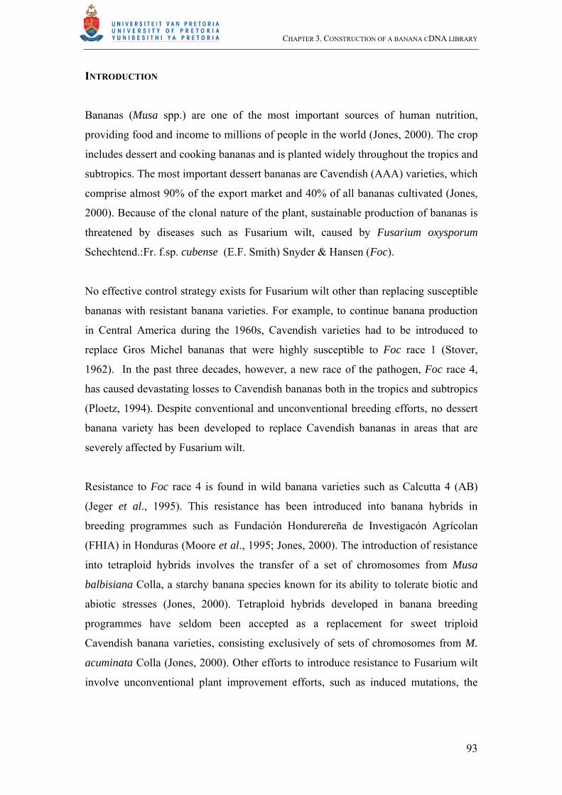
To determine whether cDNA was contaminated with genomic DNA, a cDNA sample
was screened for a small fragment of the banana actin gene by PCR-amplification,
using the primers ActinF (5’ACCGAAGCCCCTCTTAACCC-3’) and ActinR (5’-
GTATGGCTGACACCATCACC- 3’) (Fig. 1). PCR amplifications were carried out
using first strand cDNA and genomic DNA as templates. The PCR was conducted in
20 l and contained 10 mM Tris-HCl (pH 9), 1.5 mM MgCl2, 50 mM KCl, 2.5 mM
dNTP’s (Roche Diagnostics), 0.25 μM of each primer, 1 U Taq polymerase (Roche
Diagnostics) and 10 g DNA. The samples were heated at 94ºC for 2 min, then
cycled 30 times at 94ºC for 30 s, 55ºC for 30 s and 72ºC for 1 min, followed by a final
elongation step at 72ºC for 7 min. The PCR was conducted in a GeneAmp PCR
System 2700 (Applied Biosystems, Perkin Elmer, Ontario, Canada), and the PCR
products were separated by electrophoresis on a 2% agarose gel.
Suppression Subtractive Hybridisation (SSH)

CHAPTER 3. CONSTRUCTION OF A BANANA CDNA LIBRARY
99
cDNA derived from RNA extracted 3 and 6 hrs after inoculation of GCTCV-218 with
Foc was pooled and served as “tester”, and cDNA derived from RNA extracted from
Williams plants at the same time points after inoculation by Foc served as “driver”.
“Tester” and “driver” material was then subjected to SSH analysis (Diatchenko et al.
1996; 1999) using a PCR-Select cDNA Subtraction kit TM (Clontech, BD Biosciences,
Palo Alto, California) according to the manufacturer’s instructions, but with a ratio of
300:1 “tester” to “driver” material in the primary hybridisation.
Monitoring SSH efficiency
Reduction in actin levels. cDNA enriched for differentially expressed transcripts was
referred to as “subtracted tester” (ST), cDNA from infected tolerant and susceptible
banana plants was referred to as “unsubtracted tester” (UT) and unsubtracted “driver”
(UD), respectively. Reduction in actin levels during the SSH procedure was
determined by comparing cDNA levels in the ST with that of UT and UD. Ten g of
the ST, UT or UD was amplified with 0.33 l of the primer pair ActinF and ActinR
(15 M stock) in a 20 l PCR reaction. The reaction further contained 2.0 l PCR
buffer (10 mM Tris-HCl; 1.5 mM MgCl2; 50 mM KCl), 2.0 l dNTP mix (2.5 mM
stock) (Roche Diagnostics) and 0.2 l Taq polymerase (5 U/l) (Roche Diagnostics).
Primers were designed to amplify a cDNA fragment of the actin gene that flanked an
intron and did not contain an RsaI restriction site (Fig. 1). Each sample was denatured
at 94ºC for 2 min, then cycled 30 times at 94ºC for 30 sec, 55ºC for 30 sec, and 72ºC
for 1 min, before a final elongation step at 72ºC for 7 min. Amplification products (5
l) were collected after 20, 25 and 30 cycles and separated on a 2% agarose gel. The
amplified fragment was also sequenced to confirm that it was from the actin gene.
Presence of a banana defence-associated gene. Primers were designed from the
genebank nucleotide sequence of endochitinase (AF 416677) to amplify a cDNA
fragment of 149 bp that did not contain a RsaI restriction site. The presence of
endochitinase in the ST material was then determined by PCR amplification using 10
g of the ST, 2.0 l PCR buffer (10 mM Tris-HCl; 1.5 mM MgCl2; 50 mM KCl), 2.0
l dNTP mix (2.5 mM) (Roche Diagnostics), 0.33 l of EndochitF (5’-
GCCACCGTCAGAGGTTATACAG- 3’) and EndochitR (5’-
GACTATTAAGGGCTCCGTGGTT- 3’) (15 M stock) and 0.2 l Taq polymerase

CHAPTER 3. CONSTRUCTION OF A BANANA CDNA LIBRARY
100
(5U/l) (Roche Diagnostics). Samples were denatured at 94ºC for 2 min, then cycled
30 times at 94ºC for 30 sec, 59ºC for 30 sec and 72ºC for 1 min, followed by a final
elongation step at 72ºC for 7 min. PCR products were then visualised on a 2%
agarose gel and sequenced to confirm their identity.
Southern analysis
Adaptor sequences were removed by digesting 10 l each of ST, UT and UD cDNA
with RsaI (Roche Diagnostics). PCR products were then separated through a 1.5%
low melting point (LMP) agarose gel, excised and purified using a QIAquick® Gel
Extraction Kit (Qiagen). The ST, UT and UD cDNA (25-50 g) was diluted to 47.5
l with SDW, denatured at 95C for 3 min, and placed on ice for 2 min. Denatured
cDNA was labelled with Ready-To-Go TM labelling beads (-dCTP) (Amersham
Biosciences UK Limited, England) in a reaction mixture containing buffer, dATP,
dGTP, dTTP, FPLCpureTM Klenow fragment and random nonamers. Twenty five Ci
radioactive labelled [-32P] dCTP was added and the reaction mixture incubated at
37C for 20 min. Unincorporated radiolabeled nucleotides were removed using
MicroSpinTMG-50 Columns (Amersham Biosciences) using the manufacturer’s
instructions. Prior to use, the labelled ds DNA was denatured at 95C for 10 min
followed by incubation on ice for 3 min. The probe was centrifuged briefly and
immediately used for the hybridization
Southern-based screening was performed as described by Southern (1975). PCR
products from ST, UT and UD material were separated on three replicate 2% gels,
visualized under UV light and photographed, and 5 l of each sample was transferred
to a Hybond-N+ nylon transfer membrane (Amersham-Pharmacia). DNA transfer was
carried out via alkaline capillary blotting with transfer buffer (1.5 M NaCl, 0.5 M
NaOH) for 16 hrs. The membrane was rinsed in 2x Sodium Saline Citrate (SSC) (300
mM NaCl, 30 mM Na3citrate-2H20 (pH 7.0), air-dried and cross-linked on top of an
Ultraviolet Cross linker CL-508 (UVI-tec, St John’s Innovation Centre, Cambridge,
England) at 0.167 J (312 nm) for 2 min. Blots were wrapped in cling film and stored
at –20C. Hybridization of membranes was carried out as described by Ausubel
(1999) with 32P-radiolabelled PCR-amplified ST, UT and UD probes.

CHAPTER 3. CONSTRUCTION OF A BANANA CDNA LIBRARY
101
Subtracted library construction
To construct a cDNA library, SSH products were size fractioned on a 1.5% LMP
agarose gel and separated into fragments ranging from 150-400 bp and others ranging
from 400-700 bp. The fragments were then extracted from the agarose gel using a
Qiaquick Gel Extraction kit (Qiagen). Non-fractionated DNA was also purified
with the Qiaquick PCR purification kit (Qiagen) and included in the library. The
cDNA clones were all ligated with pGEM-T Easy vector (Promega, Madison, USA).
Ligation reactions were carried out in 10 l reaction volumes containing 10-50 g
insert DNA, 50 g pGEM-T Easy vector, 1x rapid ligation buffer and 3 Weiss units
T4-DNA-Ligase (Promega). Samples were incubated for 16 hrs at 4ºC and stored at -
20ºC.
Escherichia coli JM 109 competent cells (Promega) were transformed with cDNA
fragments by heat-shock as recommended by the manufacturer. Following the
transformation, cells were plated on Luria-Bertani (LB) agar plates (10 g l-1 peptone, 5
g l-1 yeast, 5 g l-1 NaCl, 15 g l-1 agar, 100 g ml-1 ampicillin, 60 g ml-1 X-gal and 60
g ml-1 Isopropyl--D-thiogalactopyranoside (IPTG) and incubated at 37ºC for 16-18
hrs. Identification of the recombinant clones was done by colour screening on
Xgal/IPTG indicator plates. A blue/white selection was used and 300 recombinant
(white) colonies were picked for each ligation reaction (non-fractionated, and size
fractionated). The library was replicated by transferring recombinant transformants to
1.5 ml tubes containing 700 l LB amended with 50µg/ml ampicillin and grown for
20-24 hrs at 37ºC on a rotary shaker (250 rpm), and 300 l 50% glycerol was added to
each tube prior to storing at -80ºC.
Colony PCR and sequencing of selected clones
Colony PCR was conducted on 15 randomly selected clones in 25 l reaction volumes
containing 2.5 l PCR buffer (10 mM Tris-HCl; 1.5 mM MgCl2; 50 mM KCl), 2.0 l
dNTP mix (2.5mM) (Roche Diagnostics), 0.5 l of each of the primers T7 (5’-
ATTATGCTGAGTGATATCCC-3’) and SP6 (10 M) (5’-
ATTTAGGTGACACTATAGAAT-3’), 0.5 l Taq DNA polymerase (5 U/l) (Roche

CHAPTER 3. CONSTRUCTION OF A BANANA CDNA LIBRARY
102
Diagnostics), 13.5 l SDW and 0.5 l of the bacterial colony grown in LB. PCR
conditions were as follows: Initial denaturation at 94ºC to avoid the preferential
cloning of highly abundant or small DNA molecules for 2 min, 25 cycles of 94ºC for
30 s, 50ºC for 30 s, and 72ºC for 1 min, and a final elongation step at 72ºC for 5 min.
PCR products were then separated by electrophoresis on a 1.5% agarose gel to
confirm successful insertion of cDNA fragments.
Cycle sequencing (Griffin and Griffin, 1993) was carried out in 10 l reaction
volumes containing 150 g template DNA (derived from colony PCR products), 2 l
Big Dye termination reaction mix (V3) (Applied Biosystems, Foster City, CA, USA),
2 l primer T7 (10 µM) and 4 l 5x dilution buffer (400 mM Tris-HCl pH 9.0, 10 mM
MgCl2). Samples were cycled 25 times at 96ºC for 10 sec, 50ºC for 5 sec and 60ºC
for 4 min. Sequenced products were diluted 1:20 and purified by adding 2 l 3 M
NaOAc (pH 4.6) and 50 l cold absolute ethanol to the reactions, followed by 10 min
incubation on ice and centrifugation at 10 000 rpm for 30 min at 4ºC. The supernatant
was removed and the product washed to remove salts and excess dye terminators,
using 250 l 70% ethanol. This was followed by centrifugation for 10 min at 10 000
rpm at 4ºC. Samples were dried in an incubator at 37ºC for 5 min and stored at –
20ºC. DNA sequences were analyzed on an ABI PRISM 377 DNA analyzer (Perkin
Elmer) at the DNA Sequencing Facility of the University of Pretoria, South Africa.
Vector and SSH adaptor sequences were removed manually using Vector NTI Suite
V.6 (InforMax, North Bethesda, USA) and database searching utilized BLAST
software (Altschul et al., 1990), available through the National Centre for
Biotechnology Information (NCBI) (website http://www.ncbi.nih.gov/BLAST).
Homologies were identified by BLASTX. The degree of sequence similarity between
banana cDNA clones and known sequences was represented by E-values.

CHAPTER 3. CONSTRUCTION OF A BANANA CDNA LIBRARY
103
RESULTS
Inoculation of Banana Plants with Foc
GCTCV-218 and Williams plants point-inoculated with Foc developed typical
internal Fusarium wilt symptoms after six weeks. The entire corm of susceptible
Williams plants showed brown-purple discoloration, while the corms of tolerant
GCTCV-218 plants only showed one or two brown specs. All three Williams plants
had a disease severity score of 5, while the three GCTCV-218 plants had disease
ratings of either 1 or 2. These results showed that the inoculation technique was
effective in causing disease.
RNA isolation
Yields of between 15-30 g total RNA for individual samples of GCTCV-218 and
Williams were extracted from banana roots. However, this concentration was reduced
substantially when RNA was passed through the Qiagen cleanup column, and
numerous extractions had to be done to obtain sufficient material for cDNA synthesis.
No degradation of RNA was visible for GCTCV-218 and Williams RNA samples at
0, 3 and 6 hrs after inoculation on 1% agarose gels, and two distinct bands were
present (Fig. 2).
cDNA synthesis
Successful synthesis of cDNA for both GCTCV-218 and Williams was confirmed by
PCR amplification from complementary and genomic DNA of the two banana
varieties. Amplification of genomic DNA with the Actin gene primer pair resulted in
a 260 bp fragment. This fragment is larger than the 170 bp fragment produced for first
strand cDNA (Fig. 3). The difference in size is a result of the absence of a 90-bp
intron sequence in cDNA, indicating that there was no genomic DNA present in the
cDNA samples. Sequencing confirmed that the amplified product was a fragment
from the actin gene.
Suppression Subtractive Hybridisation (SSH)

CHAPTER 3. CONSTRUCTION OF A BANANA CDNA LIBRARY
104
Reduction in actin levels. Amplification of the actin gene from ST material yielded no
visible PCR product, even after 30 PCR-cycles (Fig. 4). However, specific PCR-
products of 170 bp became visible after 20 cycles for the UT and UD, with intensities
increasing after further PCR-cycles. Assuming that PCR amplification is 100%
efficient during all amplification cycles and is, therefore, exponential, each cycle (x)
difference represents a 2x times less actin template in subtracted material compared to
unsubtracted material. In this subtraction procedure, ST would be expected to contain
at least 210 times less actin template than UT and UD. Although these figures are
theoretical, they emphasise the success of removing common sequences during the
SSH.
Presence of a banana defence-associated gene. A 149 bp fragment of endochitinase
was amplified in the ST material to show successful enrichment of a defence gene in
the subtracted material in response to pathogen infection (Fig. 5). Sequencing results
confirmed that the amplified fragment was from endochitinase.
Southern Blot analysis
Distinct bands were produced by the ST during PCR amplification compared to the
product “smear” produced for the UT and UD (Fig. 6), indicating that the ST had been
enriched for a number of differentially expressed products. During Southern analysis,
ST probes bound primarily to the ST PCR product, with substantially less
hybridization to UT and UD PCR products (Fig. 7). UT hybridised mainly to both UT
and UD, representing common genes in UT. Furthermore, very little UD probe
hybridized to the ST sample (Fig. 7), demonstrating that gene transcripts common to
both “driver” and “tester” had been removed by the subtraction, and thus implying an
enrichment for “tester” specific transcripts.
Subtracted library construction
A cDNA library of 736 banana clones expressed early after Foc infection was
constructed, with cDNA clones ranging in size from 150 to 700 bp. The non-
fractionated ST material yielded 250 clones. The fraction containing fragments
between 150 and 400 bp long yielded 240 clones, and the fraction containing
fragments between 400 and 700 bp long yielded 246 clones.

CHAPTER 3. CONSTRUCTION OF A BANANA CDNA LIBRARY
105
Colony PCR and sequencing
Colony PCR indicated that transformation of cDNA clones was successful and that
the majority of vectors had only single inserts that varied in size from 150 to 600 bp
(Fig. 8). Clones that had more than one insert were discarded from the library. A
small subset of 15 cDNA clones was sequenced to check the quality of the cDNA
library. Most cDNA clones that were sequenced (except clone 2-8) showed
significant similarities to plant genes and thus were assumed to be derived from the
host and not from the pathogen (Table 2).
Two of the 15 clones putatively identified by BLASTX searches showed homology to
peroxidases from rice, and one each to an unspecific monooxygenase cytochrome
P450, isoflavone reductase, Bowman Birk proteinase inhibitor, putative senescence
associated protein, auxin protein, cytosolic monohydroascorbate reductase, UTP-
glucose glucosyltransferase, reversibly glycosylated polypeptide and a putative
transcription factor. Two clones had no significant homology and two showed
homology to unknown proteins from barley (Table 2). Six of the 15 clones
sequenced, were grouped in the functional category associated with defence/stress
responses, two clones had sequences involved in metabolism and one clone had a
sequence that could play a role in transcription. The level of redundancy in the 15
sequenced clones was low; only two clones for a peroxidase and an unknown protein
occurred twice.
DISCUSSION
In this study, SSH was successfully utilised to isolate more than 700 cDNA clones in
the tolerant Cavendish banana cultivar, GCTCV-218, in response to infection by Foc.
A number of these clones showed significant sequence similarities to defence-
associated genes. The variety of putative gene functions that were assigned to the
banana clones selected for sequencing, provided confidence in the SSH library. The
isolation of theses genes at an early time point reveals that the tolerant banana,
GCTCV-218, recognises Foc and responds at the transcriptional level.

CHAPTER 3. CONSTRUCTION OF A BANANA CDNA LIBRARY
106
Two of the clones showed homology to a peroxidase gene from Oryza sativa L. This
is relevant because peroxidases have been implicated in several physiological
processes of importance in plant-pathogen interactions including lignification
(Lagrimini et al., 1987), cross-linking of cell-wall proteins (Bradley et al., 1992),
wound healing (Sherf et al., 1993) and papillae formation (Cadena-Gomez and
Nicholson, 1987). Enhanced peroxidase activity after pathogen infection has also
been correlated with resistance in many different host pathogen systems.
The BLASTX search showed a clone sharing similarity with an isoflavone reductase
from Pyrus communis L. This enzyme catalyzes the penultimate step in the synthesis
of isoflavonoid phytoalexins that play a role in plant defence (Ibrahim and Varin,
1993). One clone had homology to a Bowman-Birk type protease inhibitor from
kidney bean. The synthesis of theses inhibitors may be induced by pathogen infection
or wounding (Qi et al., 2005) and they have been linked with enhanced protection of
plants against insects and microorganisms (Deshimaru et al., 2004). The BLASTX
search also showed a clone with homology to cytochrome P450. This compound
plays a role in secondary metabolism by being involved in the phenylpropanoid
biosynthesis pathway (Dowd et al., 2004) that leads to lignin production.
Results of this study represent the first effort to isolate genes in banana with resistance
to Foc. Even though conventional breeding has been used by several banana
improvement programmes to introduce resistance to pathogens and pests (Hwang and
Tang, 1996), almost nothing is known about the genes underlying such resistance.
Although several technologies are available to discover these genes (Cochran et al.,
1983; Duguid and Dinauer, 1990; Liang et al., 1993; Lisitsyn and Wigler, 1993;
Velculescu et al., 1995; Schena et al., 1995), SSH used in this study has numerous
advantages above other gene isolation methods, and it was effective in isolating
differentially expressed genes from banana. An advantage of SSH is that it allows the
detection of low-abundance, differentially expressed cDNAs, such as those involved
in signalling and signal transduction (Birch et al., 2000).

CHAPTER 3. CONSTRUCTION OF A BANANA CDNA LIBRARY
107
The SSH procedure appeared to be efficiently executed. Small quantities (20 g) of
RNA from banana roots were sufficient to set up the SSH experiment. The isolation
of mRNA from total RNA yielded very little material (data not shown) and total RNA
was thus used for the procedure. This is important in an experiment where only a
limited amount of starting material can be obtained. Constitutively expressed genes,
such as the actin gene were removed during subtraction hybridisation, while induced
resistance genes, such as endochitinase, were found in the SSH final product.
Southern blot analysis further confirmed the subtraction efficiency of the SSH
technique. Southern blot data suggested that the subtracted “tester” had been enriched
for differentially expressed transcripts, and that “housekeeping” gene transcripts that
are common to both “driver” and “tester” had been removed by the subtraction.
From the limited number of clones sequenced in this study, it was clear that SSH was
effective in enriching for pathogen-induced transcripts from as early as 3 and 6 hrs
after inoculation with Foc. Rare transcripts, such as the putative transcription factor
isolated in this study, are often not detected by other methods such as differential
display (DD) (Liang and Pardee, 1992) reverse transcriptase (RT) PCR and cDNA-
amplified fragment length polymorphisms (AFLP) due to competition within the PCR
(Birch et al., 2000).
In most cases, the Clontech PCR-Select cDNA subtraction method greatly enriches
for differentially expressed genes. Nevertheless, the subtracted “tester” will still
contain some cDNAs that have escaped the subtraction and correspond to mRNAs
that are common to both “tester” and “driver” samples. This type of background
(false positives) may somewhat depend on RNA quality and purification and the
performance of the specific subtraction, but it mainly arises when very few mRNA
species are differentially expressed in “tester” and “driver” (Clontech PCR-Select
cDNA Subtraction Kit User Manual, 2000). It is important and essential to subject
the library to a differential screening method. Therefore, the cDNA library was
screened (Chapter 4) enabling us to select clones for a more comprehensive study.
The cDNA library constructed from genes differentially expressed in Cavendish
bananas after infection by Foc could be applied usefully in a number of ways. Firstly,

CHAPTER 3. CONSTRUCTION OF A BANANA CDNA LIBRARY
108
the identification of host genes involved in defence responses is one of the most
critical steps leading to the understanding of disease resistance mechanisms in plants
(Xiong et al., 2001). Secondly, the genes can be used as markers for resistance traits
in plant improvement programmes, and thirdly, the genes can be used for genetic
improvement of plants via transformation. The manipulation of defence-associated
genes and their signalling pathways by transgenic expression is a promising strategy
to improve disease resistance in plants (Martin, 1999).
This study is the first of its kind, utilising SSH for isolating defence associated genes
in banana against Foc. It could contribute significantly to the understanding of gene
expression profiles associated with defence mechanisms in monocotyledonous plants.

CHAPTER 3. CONSTRUCTION OF A BANANA CDNA LIBRARY
109
REFERENCES
Altschul, S.F., Gish, W., Miller, W., Myers, E.W. and Lipman, D.J. 1990. Basic local
alignment search tool. Journal of Molecular Biology 215: 403-410.
Ausubel, F.M., Brent, R., Kingston, R.E., Moore, D.D., Seidman, J.G. Smith, J.A.
Struhl, K., Albright, L.M. Coen, D.M. and Varki, A. 1999. Current Protocols in
Molecular Biology. 4 vols. 1. Molecular Biology-Technique. 2. Molecular Biology-
Laboratory manuals, John Wiley and Sons, Inc., England.
Beyer, K., Bincer, A., Boller, T. and Collinge, M. 2001. Identification of potato genes
induced during colonization by Phytophthora infestans. Molecular Plant Pathology 2:
125-134.
Birch, P.R.J., Avrova, A., Lyon, G.D., Duncan, J.M. and Toth, R.L. 1999. Isolation of
potato genes that are induced during an early stage of the hypersensitive response to
Phytophthora infestans. Molecular Plant-Microbe Interactions 12: 356-361.
Birch, P.R.J., Toth, I.K., Avrova, A.O., Dellagi, A., Lyon, G.D. and Pillay, D. 2000.
Recent advances in the identification of differentially expressed genes. South African
Journal of Science 96: 83-85.
Bradley, D.J., Kjellblom, P. and Lamb, C.J. 1992. Elicitor- and wound-induced
oxidative cross-linking of a proline rich plant cell protein: a novel, rapid defence
response. Cell 70: 21-30.
Cadena-Gomez, G. and Nicholson, R.L. 1987. Papillae formation and associated
peroxidase activity: a non-specific response to attempted fungal penetration of maize.
Physiological Molecular Plant Pathology 31: 51-67.
Carlier, J., De Waele, D. and Escalant, J.V. 2002. Global evaluation of Musa
germplasm for resistance to Fusarium wilt, Mycosphaerella leaf spot diseases and
nematodes. INIBAP Technical Guidelines 6, Montpellier, France.

CHAPTER 3. CONSTRUCTION OF A BANANA CDNA LIBRARY
110
Carmeci, C., Thompson, D.A., Kuang, W.W., Lightdale, N., Furthmayer, H. and
Weigel, R.J. 1998. Moesin expression is associated with the estrogen receptor-
negative breast cancer phenotype. Surgery 124: 211-217.
Cochran, B.H., Reffel, A.C. and Stiles, C.D. 1983. Molecular cloning of gene
sequences regulated by platelet-derived growth factor. Cell: 33: 939-947.
Clontech PCR-Select™. cDNA Subtraction Kit User Manual. 2000. Catalog #:
K1804-1. BD Biosciences, Clontech.
Deshimaru, M., Yoshimi, S., Shioi, S. and Terada, S. 2004. Multigene family for
Bowman-Birk type proteinase inhibitors of wild soja and soybean: The presence of
two BBI-A genes and pseudogenes. Bioscience, Biotechnology and Biochemistry 68:
1279-1286.
Diatchenko, L., Lau, Y.F., Campbell, A.P., Chenchik, A., Moqadam, F., Huang, B.,
Lukyanov, S., Lukyanov, K., Gurskaya, N., Sverdlov, E.D. and Siebert, P.D. 1996.
Suppression subtractive hybridization: a method for generating differentially
regulated or tissue-specific cDNA probes and libraries. Proceedings of the National
Academy of Sciences of the USA 93: 6025-6030.
Diatchenko, L., Lukyanov, S., Lau, Y.F. and Siebert, P.D. 1999. Suppression
subtractive hybridization: A versatile method for identifying differentially expressed
genes. Methods in Enzymology 303: 349-380.
Dowd, C., Wilson, I.W. and McFadden, H. 2004. Gene expression profile changes in
cotton root and hypocotyls tissues in response to infection with Fusarium oxysporum
f. sp. vasinfectum. Molecular Plant Microbe Interactions 17: 654-667.
Duguid, J.R. and Dinauer, M.C. 1990. Library subtraction of in vitro cDNA libraries
to identify differentially expressed genes in scrapie infection. Nucleic Acids Research
18: 2789-2792.

CHAPTER 3. CONSTRUCTION OF A BANANA CDNA LIBRARY
111
Glazebrook, J., Rogers, E.E. and Ausubel, F.M. 1997. Use of Arabidopsis for genetic
dissection of plant defence responses. Annual Review of Genetics 31: 547-569.
Griffin, H.G. and Griffin, A.M. 1993. DNA sequencing. Recent innovations and
future trends. Applied Biochemistry and Biotechnology 38: 147-159.
Gurskaya, N.G., Diatchenko, L., Chenchik, A., Siebert, P.D., Khaspekov, G.L.,
Lukyanov, K.A., Vagner, L.L., Ermolaeva, O.D., Lukyanov, S.A. and Sverdlov, E.D.
1996. Equalizing cDNA subtraction based on selective suppression of polymerase
chain reaction: cloning of Jurkat cell transcripts induced by phytohemaglutinin and
phorbol 12-myristate 13-acetate. Analytical Biochemistry 240: 90-97.
Hein, I., Campbell, E.I., Woodhead, M., Hedley, P.E., Young, V., Morris, W.L.,
Ramsay, L., Stockhaus, J., Lyon, G.D., Newton, A.C. and Birch, P.R.J. 2004.
Characterisation of early transcriptional changes involving multiple signalling
pathways in the Mla13 barley interaction with powdery mildew (Blumeria graminis f.
sp. hordei). Planta 218: 803-813.
Hwang, S. and Ko, W. 2004. Cavendish banana cultivars resistant to Fusarium wilt
acquired through somaclonal variation in Taiwan. Plant Disease 88: 580-588.
Hwang, S. and Tang, C. 1996. Somaclonal variation and its use for improving
Cavendish (AAA dessert) bananas in Taiwan. In: New frontiers in resistance breeding
for nematode, fusarium and sigatoka. (Eds. E. Frison, J.P. Horry and D. De Waele)
INIBAP, Montpellier, France.
Ibrahim, R.K. and Varin, L. 1993. Flavanoid enzymology. In: Methods in Plant
Biochemistry Vol. 9. (Ed. P.J. Lea) Academic Press, London.
Jeger, M.J., Eden-Green, S., Tresh, J.M., Johanson, A., Waller, J.M. and Brown, A.E.
1995. Banana diseases. In: Bananas and Plantains. (Ed. S. Gowen). Chapman and
Hall, London.

CHAPTER 3. CONSTRUCTION OF A BANANA CDNA LIBRARY
112
Jones, D.R. 2000. History of banana breeding. In: Diseases of banana, abacá and
enset. (Ed. D.R. Jones). CAB International, Wallingford, UK.
Jørgensen, J.H. 1994. Genetics of powdery mildew resistance in barley. Critical
Review in Plant Sciences 13: 97-119.
Kuang, W.W., Thompson, D.A., Moch, R.V. and Weigel, R.J. 1998. Differential
screening and suppression subtractive hybridisation identified genes differentially
expressed in an estrogen receptor-positive breast carcinoma cell line. Nucleic Acids
Research 26: 111-116.
Lagrimini, M.L., Burkhart, W., Moyer, M. and Rothstein, S. 1987. Molecular cloning
of complementary DNA encoding the lignin forming peroxidase from tobacco:
molecular analysis and tissue-specific expression. Proceedings of the National
Academy of Sciences of the USA 84: 7542-7546.
Liang, P. and Pardee, A.B. 1992. Differential display of eukaryotic messenger RNA
by means of the polymerase chain reaction. Science 257: 967-971.
Liang, P., Averboukh, L. and Pardee, A.B. 1993. Distribution and cloning of
eukaryotic mRNAs by means of differential display: refinements and optimization.
Nucleic Acids Research 21: 3269-3275.
Lisitsyn, N. and Wigler, M. 1993. Cloning the differences between two complete
genomes. Science 259: 946-951.
Lu, G., Jantasuryarat, C., Zhou, B. and Wang, G.L. 2004. Isolation and
characterization of novel defence response genes involved in compatible and
incompatible interaction between rice and Magnaporthe grisea. Theoretical and
Applied Genetics 108: 525-534.

CHAPTER 3. CONSTRUCTION OF A BANANA CDNA LIBRARY
113
Martin, G.B. 1999. Functional analysis of plant disease resistance genes and their
downstream effectors. Current Opinion in Plant Biology 2: 273-279.
Moore, N.Y., Bentley, S., Pegg, K.G. and Jones, D.R. 1995. Fusarium Wilt of
Bananas. Musa Disease Fact Sheet No. 5. INIBAP.
Pereza-Escheverria, S., Dale, J., Khanna, H., Smith, M. and Collet, C. 2004. Potential
resistance gene against Fusarium wilt race 4. 1st International Congress on Musa.
Harnessing research to improve livelihoods. Penang, Malaysia, 6-9 July 2004.
Ploetz, R.C. 1994. Panama disease: Return of the first banana menace.
InternationalJournal of Pest Management 40: 326-336.
Qi, R.-F., Song, Z.-W. and Chi, C.-W. 2005. Structural features and molecular
evolution of Bowman-Birk protease inhibitors and their potential application. Acta
Biochimica et Biophysica Sinica 37: 283-292.
Schena, M., Shalon, D., Davis, R.W. and Brown, P.O. 1995 Quantitative monitoring
of gene expression patterns with a complementary DNA microarray. Science 270:
467-470.
Sherf, B.A., Bajar, A.M. and Kolattukudy, P.E. 1993. Abolition of an inducible highly
anionic peroxidase activity in transgenic tomato. Plant Physiology 101: 201-208.
Southern, E.M. 1975. Detection of specific sequences among DNA fragments
separated by gel electrophoresis. Journal of Molecular Biology 98:503-517.
Stover, R.H. 1962. Fusarial Wilt (Panama Disease) of Bananas and other Musa
species. Phytopathology Paper 4. Commonwealth Mycological Institute, Kew,
Surrey, England.
Velculescu, V.E., Zhang, L., Vogelstein, B. and Kinzler, K.W. 1995. Serial analysis
of gene expression. Science 270: 484-487.

CHAPTER 3. CONSTRUCTION OF A BANANA CDNA LIBRARY
114
Xiong, L., Lee, M.-W., Qi, M. and Yang, Y. 2001. Identification of defence-related
rice genes by suppression subtractive hybridization and differential screening.
Molecular Plant-Microbe Interactions 14: 685-692.

CHAPTER 3. CONSTRUCTION OF A BANANA CDNA LIBRARY
115
5’
5’
3’
3’
GCAACTGTTTATGATANGGTTGAAGATCTGGCACCATACCTTCAACAATG 50 AGCTCCGTGTTGCTCCTGAGGAGCATCCTATTCTGCTCACCGAAGCCCCT 100 specific primer CTTAACCCAAAGACAAACAGGGAAAAGATGACCCAGATAATGTCTGAAAC 150 ACTINF TTTCATTGTACCAGCTATGTATGTTGCTATTCAGGGCGTCCTTTCGCTCT 200 ATGYWAGTGSCSGTACTAGTGGTTAGTATCTTGTGATCGCTTACCTGATG 250 TGRAGTTGGTCATTCTTTTTATTTTCAACTRAGATGCCCAAGYTAAATGC 300 TATTTTGACAGGCATTGTACTTGATTCTGGTGATGGTGTCAGCCATACCG 350 specific primer ACTINR TTCCTATCTATGAAGGATATGCTCTACCTCATGCCATTCTTCGTTTGGAT 400 CTTGCTGGTCGTGATCTCACGGATTGCCTGATGAANATCCTGACAGAGAG 450 GGGTTATTCATTCACTACTACTGCAGAACGGGAAATCGTAAGGGACATCA 500 AGGAGAARCTTGCCTACGTTGCTGKTGACTATGAACAGGAGCTGGATACT 550 GSCGAGACTAGCTCTGCTGTGGAGAAAASYWWKRAMCTTCCCGTTGGGCA 600 RGKTATCACGAWTGGGGCTNRARRWTYANGAGCCCCGGANGTGCTNTYCC 650 AGCCATCATTGATTGGCATGGAARCCNNTGGAGTTCATGAGACAACATAC 700 AACTCTATTATGAAGTGTGATGTGGATATCAGGAAAGATCTGTATGGCAA 750 TATTGTGCTTAGCGGTGGATCAGCAATGTTCCCTGGTATTGCCGATCGCA 800 TGAGCAAGGAGATCACAGCGCTTCCACCAAACAGCATGAAGATAAAGGTG 850 GTTGCCCCACCCGAAAGGAAATACAGCGTCTGGAT 885
Figure 1. Sequence of the cloned actin gene used for Actin gene-based PCRs.
Yellow: Actin Forward primer, Blue: Actin Reverse primer and Red: Intron (90 bp).

CHAPTER 3. CONSTRUCTION OF A BANANA CDNA LIBRARY
116
Figure 2. RNA samples from banana root material under non-denaturing
conditions. Lane 1 contains the 100 bp marker (Roche Diagnostics), lanes 2-4
GCTCV-218 RNA and lanes 5-7 Williams RNA.
1 2 3 4 5 6 7
1000 bp

CHAPTER 3. CONSTRUCTION OF A BANANA CDNA LIBRARY
117
Figure 3. Actin-based control for monitoring contamination of cDNA with
genomic DNA. PCR products from amplification (30 cycles) of genomic DNA (260
bp) (Lane 2), PCR products from second strand GCTCV-218 cDNA (Lanes 3 and 4)
and Williams cDNA (Lanes 5 and 6) (170 bp) using primer ActinF and ActinR,
separated by electrophoresis through a 2% (w/v) agarose gel. 100 bp molecular
marker (Roche Diagnostics) (Lane 1) was used to estimate the size of DNA
fragments.
200 bp
300 bp
1 2 3 4 5 6

CHAPTER 3. CONSTRUCTION OF A BANANA CDNA LIBRARY
118
Figure 4. PCR-based assessment of the decrease in actin cDNA levels following
Suppression Subtractive Hybridisation. Amplification of the banana actin gene
from subtracted “tester” (ST), unsubtracted “tester” (UT) and unsubtracted “driver”
(UD) cDNA samples. Lanes 1 and 6, 100 bp marker (Roche Diagnostics); lane 2,
water control; lanes 3-5, ST, UT and UD after 20 amplification cycles; lanes 7-9, ST,
UT and UD after 25 amplification cycles; lanes 10-12, ST, UT and UD after 30
amplification cycles.
1 2 3 4 5 6 7 8 9 10 11 12
20 Cycles 25 Cycles 30 Cycles

CHAPTER 3. CONSTRUCTION OF A BANANA CDNA LIBRARY
119
Figure 5. PCR-based assessment of the successful amplification of endochitinase
in subtracted “tester” cDNA following Suppression Subtractive Hybridisation.
Lane 1, 100 bp marker (Roche Diagnostics); lane 2, water control; lane 3
endochitinase fragment (149 bp).
1000 bp
100 bp
200 bp
1 2 3

CHAPTER 3. CONSTRUCTION OF A BANANA CDNA LIBRARY
120
Figure 6. Agarose gel electrophoresis of Suppression Subtractive Hybridisation
amplification products. Lane 1 and 14, 100 bp marker (Roche Diagnostics); lane 2,
6 and 10, subtracted “tester” PCR products; lane 3, 7 and 11, unsubtracted “tester”
PCR products; lane 4, 8 and 12, unsubtracted “driver” PCR products. Lanes 5, 9 and
13, water controls.
1 2 3 4 5 6 7 8 9 10 11 12 13 14
1000 bp

CHAPTER 3. CONSTRUCTION OF A BANANA CDNA LIBRARY
121
Figure 7. Southern analysis of subtracted “tester” (ST), unsubtracted “tester”
(UT) and unsubtracted “driver” (UD) cDNA amplification products.
Amplification products in Fig. 6 were transferred to a positively charged nylon
membrane and identical filters were independently hybridized with complex probes
derived from forward ST cDNA (Filter A), UT cDNA (Filter B) and UD cDNA (Filter
C). Lane 1, ST PCR products; lane 2, UT PCR products; lane 3, UD PCR products.
A B C
1 2 3 1 2 3 1 2 3

CHAPTER 3. CONSTRUCTION OF A BANANA CDNA LIBRARY
122
Figure 8. Colony PCR of nine selected Suppression Subtractive Hybridisation
cDNA clones indicating single inserts (except lane 6) of variable sizes between
150 and 600 bp. Lane 1 contains the 100 bp molecular marker (Roche Diagnostics)
used for size estimations.
1000 bp
1 2 3 4 5 6 7 8 9 10 11

CHAPTER 3. CONSTRUCTION OF A BANANA CDNA LIBRARY
123
Table 1. Primer and adaptor sequences used in the Suppression Subtractive
Hybridisation procedure.
Primer or Adaptor
name
Nucleotide sequence
PCR primer 1
Nested PCR primer 1
Nested PCR2
Adaptor 1
Adaptor 2R
5’-CTAATACGACTCACTATAGGGC-3’
5’-TCGAGCGGCCGCCCGGGCAGGT-3’
5’-AGCGTGGTCGCGGCCGAGGT-3’
5’-CTAATACGACTCACTATAGGGCTCGAGCGGCCGCCCGGGCAGGT-3’
3’-GATTATGCTGAGTGATATCCCGAGCTCGCCGGCGGGCCCGTCCA-5’
5’-CTAATACGACTCACTATAGGGCAGCGTGGTCGCGGCCGAGGT-3’
3’-GATTATGCTGAGTGATATCCCGTCGCACCAGCGCCGGCTCCA-5’

CHAPTER 3. CONSTRUCTION OF A BANANA CDNA LIBRARY
124
Table 2. Selected banana cDNA clones expressed in a tolerant banana, GCTCV-
218, after Fusarium oxysporum f. sp. cubense infection, following Suppression
Subtractive Hybridisation.
Clone
number
Protein similarity Origin of similar
sequence
BLASTX
Acc. No. of
similar sequence
E-
value
Putative
Function
2-25 Unspecific
monooxygenase,
cytochrome P450
Nicotiana tabacum L.
(tobacco)
pir T02995 7e-42
Defence
2-18 Isoflavone reductase
related protein
Pyrus communis L.
(pear)
gb AA24001.1 2e-70 Defence
1-1 Putative peroxidase Oryza sativa L. (rice) dbj BAB19339.1 6e-86 Defence
1-5 Putative peroxidase O. sativa emb CAE05415.1 2e-05 Defence/Stres
s
2-39 Bowman Birk type
proteinase inhibitor
Phaseolus vulgaris L.
(kidney bean)
spP01060IBB2_PH
AVU
2e-05 Defence/stres
s
3-146 Putative senescence
associated protein
Pisum sativa L. (pea) dbj BAB33421.1 4e-43 Defence/Stres
s
3-130 Aux/IAA protein Solanum tuberosum L.
(potato)
gb AAM29182.1 5e-21 Auxin-related
3-91 Cytosolic
monohydroascorbate
reductase
O. sativa dbj BAA77214.1 3e-04 Metabolism
3-26 UTP-Glucose
Glucosyltransferase
Arabidopsis thaliana
(L.) (thale cress)
pir T01732 3e-04 Metabolism
(primary)
2-2 Unknown protein Hordeum vulgare (L.)
(barley)
gb AAL77110.1 5e-55 Other
2-147 Reversibly
glycosylated
polypeptide
Gossypium hirsutum L.
(cotton)
emb CAC83750.1 1e-14 Other
1-97 No significant
homology
- - - Other
3-31 No significant
homology
- - Other
1-95 Unknown protein H. vulgare AF 474373_7 1e-49 Other
2-8 Putative transcription
factor
Periplaneta americana
L. (cockroach)
embCAB51041.1
(AJ243883)
2e-14 Transcription