chapter 24, part 2chapter 24 – digestion – part 2! 2! 4 small intestine histology! submucosa!...
TRANSCRIPT

Chapter 24 – Digestion – PART 2!
1!
Chapter 24, Part 2!The Digestive System!
2
SECTION 24-6!The small intestine digests and absorbs nutrients, and associated glandular organs assist with the digestive process
3
Small Intestine – Regions!
21 feet long in cadaver; 10 feet long in living person (What accounts for this difference?)!1. Duodenum (“12”, length of 12 finger widths)!• Entrance = pylorus!• Has Brunner’s (alkaline mucous) glands (Why
alkaline?)!2. Jejunum (“empty” in cadaver)!• No Brunner’s glands or Peyer’s patches!
3. Ileum (“twisted”)!• Many Peyer’s patches (M.A.L.T.)!• Opens into colon at ileocecal valve!

Chapter 24 – Digestion – PART 2!
2!
4
Small Intestine Histology!
Submucosa!
Lining is modified to increase surface area for secretion and absorption!
Compared to a simple tube:!1. Plica circulares (circular folds)!• Permanent folds of mucosa
and submucosa!• Folds contain submucosa
(identifies a plica in lab)!• Increase luminal surface
area by 3X!
5
Small Intestine Histology – 2!
2. Villi!• Folds of mucosa!• Fold contains lamina propria (not submucosa
– use for identification in lab)!• Contain lacteals!
Lymphatic capillaries!ü Transport chylomicrons (lipid +
protein) to blood!• Increase luminal surface area by 10X!
6
The Intestinal Wall – Villi Figure 24-17a and b!

Chapter 24 – Digestion – PART 2!
3!
7
Intestinal Villus Figure 24-17c and d!
Lamina propria!
8
Small Intestine Histology – 3!
3. Microvilli!• Folds of epithelial cell membrane!• Fold contains cytoplasm (not lamina propria or
submucosa) and actin filaments!• Form “brush border”!
Contains brush border enzymes - digestion!• Increase surface area by 20X!
Total increase in surface area (for digestion and absorption) compared to a simple, unmodified tube is: 3 x 10 x 20 = 600X !! !(i.e. Your small intestine would need to be 600 times longer to do the same amount of work.)!
9
a.k.a. crypts of Lieberkühn!• Epithelial cells lost to lumen replaced by
stem cells!• Some shed cells release brush border
enzymes into lumen!Enteroendocrine cells!• Gastrin and GIP!• Secretin!• Cholecystokinin!
Intestinal Glands!
Crypt

Chapter 24 – Digestion – PART 2!
4!
10
Intestinal Secretions!
About 1.8 l/day from!• Osmosis from mucosa to chyme in lumen!• Secretions of intestinal glands!
Buffer acidic chyme!Protection from digestive enzymes!
• Increase secretion in response to:!Local reflexes!Enterocrinin from neuroendocrine cells!
(↑ mucus production)!Vagus stimulation!
11
The Pancreas Figure 24-18!
12
Pancreas – Anatomy!
Heterocrine gland: both exocrine and endocrine!Head, body, tail, exocrine ducts!Pancreatic duct (of Wirsung) divides:!1. Pancreatic duct!• Joins common bile duct!• Becomes hepatopancreatic ampulla (of
Vater)!• Empties into duodenum!
2. Accessory duct (of Santorini)!• Opens into duodenum in < 10% of people!
Discussed in lab or later in notes!

Chapter 24 – Digestion – PART 2!
5!
13
Pancreas – Histology!
1. Exocrine portion = acini (“bunch of grapes”)!Secretes “pancreatic juice” (Not a good lab test
answer! – be more specific.)!• 1.2–1.5 l/day!
ü Digestive enzymes (see below)!ü Sodium bicarbonate (neutralizes
chyme)!2. Endocrine portion - islets of Langerhans!
• Glucagon, insulin, somatostatin, pancreatic polypeptide (Chapter 18)!
14
“Pancreatic Juice” (pH 7.5–8.8)!
Acinar cells!• Secrete digestive enzymes as proenzymes!
E.g. trypsin secreted as trypsinogen!Trypsinogen ----- enteropeptidase -----> trypsin!
(in brush border)!Trypsin activates chymotrypsin,
carboxypeptidase and elastase!Pancreatic duct cells!• Secrete ions including bicarbonate!• Water follows!
15
Pancreatic Enzymes (Discussed in detail later) !
Pancreatic alpha-amylase!• Carbohydrates!• Similar to salivary amylase!
Pancreatic lipase!• Lipids → fatty acids!
Pancreatic nucleases!• DNA, RNA!
Pancreatic proteolytic enzymes!• proteases, peptidases!

Chapter 24 – Digestion – PART 2!
6!
16
Gross Anatomy of the Liver Figure 24-19b!
• Largest visceral organ!• Falciform ligament separates R. and L. lobes!• Round ligament = remnant of fetal umbilical vein!• Liver cells = hepatocytes!
Discussed in lab or later in notes!
17
Gross Anatomy of the Liver – 2 Figure 24-19c!
Discussed in lab or later in notes!
18
Liver Histology Figure 24-20!
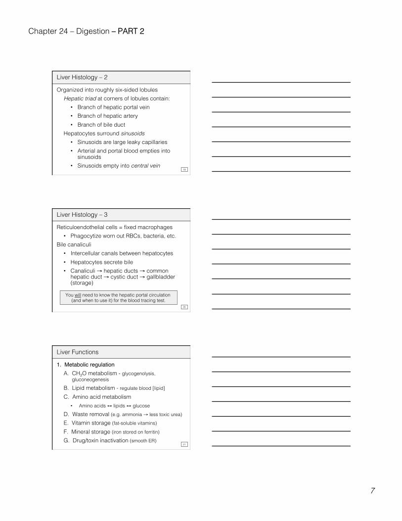
Chapter 24 – Digestion – PART 2!
7!
19
Liver Histology – 2!
Organized into roughly six-sided lobules!Hepatic triad at corners of lobules contain:!• Branch of hepatic portal vein!• Branch of hepatic artery!• Branch of bile duct!
Hepatocytes surround sinusoids!• Sinusoids are large leaky capillaries!• Arterial and portal blood empties into
sinusoids!• Sinusoids empty into central vein!
20
Liver Histology – 3!
Reticuloendothelial cells = fixed macrophages!• Phagocytize worn out RBCs, bacteria, etc.!
Bile canaliculi!• Intercellular canals between hepatocytes!• Hepatocytes secrete bile!• Canaliculi → hepatic ducts → common
hepatic duct → cystic duct → gallbladder (storage)!
You will need to know the hepatic portal circulation (and when to use it) for the blood tracing test.!
21
Liver Functions!
1. Metabolic regulationA. CH2O metabolism - glycogenolysis,
gluconeogenesis!B. Lipid metabolism - regulate blood [lipid]!C. Amino acid metabolism!• Amino acids ↔ lipids ↔ glucose!
D. Waste removal (e.g. ammonia → less toxic urea)!E. Vitamin storage (fat-soluble vitamins)!F. Mineral storage (iron stored on ferritin)!G. Drug/toxin inactivation (smooth ER)!

Chapter 24 – Digestion – PART 2!
8!
22
Liver Functions – 2!
2. Hematological regulationA. Phagocytosis and antigen presentation
(Kupffer cells)!B. Synthesize plasma proteins (hepatocytes)!C. Remove circulating hormones!D. Remove circulating antibodies!E. Remove (or store) toxins!
3. Synthesis and excretion of bile
23
3. Bile Functions!
• 800–1000 ml produced per day by hepatocytes!• pH 7.6–8.6!Contains:!
• Water, bile salts, bile acids, cholesterol, lecithin, pigments, ions!
Functions:!A. Digestive!• Bile salts emulsify fats (do NOT themselves
digest fats)!• Increases surface area for digestion by
lipases!• Solubilizes cholesterol!
24
Bile Functions – 2!
Bile salts!• Bile acids (derived from cholesterol combined
with sodium or amino acids!• Acids = Bilcholic, deoxycholic,
chenodeoxycholic, and lithocholic acids (just FYI, do not memorize)

Chapter 24 – Digestion – PART 2!
9!
25
Bile Functions – 3!
B. Excretory!• Bile pigment = modified bilirubin (from heme
of hemoglobin)!• Bilirubin (liver) → (small intestine to large
intestine) bilirubin → urobilinogen → stercobilin (excreted in feces) !
C. 90% of bile salts reabsorbed by ileum!
26
The Gallbladder Figure 24-21!
Discussed in lab or later in notes!
27
Gallbladder – 2!
Functions:!1. Bile storage (holds 40–70 ml)!2. Bile modification!• Reabsorbs water, concentrates bile!
3. Bile release!• Presence of lipids in small intestine
causes CCK release!• Hepatopancreatic sphincter (of Oddi)
relaxes!• Gallbladder contracts !

Chapter 24 – Digestion – PART 2!
10!
28
Major Duodenal Hormones Figure 24-22!
29
Activities of Major Digestive Tract Hormones!Figure 24-23!
Food in stomach!
Chyme in duodenum!
Material arrives in jejunum!
30
SECTION 24-7!The large intestine is divided into three parts with regional specialization!

Chapter 24 – Digestion – PART 2!
11!
31
The Large Intestine Figure 24-23!
Discussed in lab or later in notes!
32
Large Intestine!
Functions:!1. Reabsorption of water - forming feces!2. Absorption of vitamins (e.g., vitamin K - some
produced by bacteria)!3. Storage of feces!
Anatomy:!• About 5 feet long!• Mesocolon attaches it to posterior abdominal
wall!• Ileocecal valve (sphincter) connects with
small intestine!
33
Large Intestine Anatomy – 2!
Cecum opens into colon!• Blind pouch!• Appendix attached!
Rectum/Anus!• Temporary storage of feces!
Anus has stratified squamous EPI!• Internal anal sphincter!
Smooth muscle = involuntary!Stretch initiates urge to defecate!
• External anal sphincter!Skeletal muscle = voluntary!

Chapter 24 – Digestion – PART 2!
12!
34
Large Intestine Histology!
Mucosa!• No villi or permanent folds!• Has intestinal (mucous) glands!• Epithelial cells!
a) Absorptive cells (water)!b) Goblet cells (lubricating mucus)!c) Do NOT produce digestive enzymes!
• Lymph nodules (lots)!
35
Large Intestine Histology – 2!
Submucosa!• Many lymphoid nodules!
Muscularis externa!• 3 longitudinal smooth muscle bands =
taeniae coli!Contractions produce haustra (“pouches”)!
Serosa!• Has epiploic (fatty) appendages!
36
The Large Intestine Figure 24-25!No villi!Many goblet cells!
Discussed in lab or later in notes!

Chapter 24 – Digestion – PART 2!
13!
37
Large Intestine Physiology!
1. Absorption!A. Water!B. Others!• Electrolytes!• Vitamins (K, biotin, B5)!
Much produced by bacteria!• Bile salts (especially cecum)!• Urobilinogen (excreted in urine)!
38
Large Intestine Physiology – 2!
2. Movements!Gastroileal and gastroenteric reflexes → ileum!
A. Peristalsis in small intestine!• Move materials into cecum!
B. Haustral churning or segmentation movements!• Chyme forced from one haustrum to the next!
C. Mass movements (mass peristalsis)!• Stimulus = stretch of stomach or duodenum!• Strong contractions in transverse colon
onward!• Move contents into rectum!
39
Defecation Reflex!
Mass movements push contents into rectum:!1. Rectum distended!2. Stretch receptors activated!3. Reflexes initiated!
A. Myenteric plexus stimulated!• Increase peristalsis in rectum!
B. Parasympathetic neurons stimulated in spinal cord!• Increase peristalsis in rest of colon!• Relax internal anal sphincter!

Chapter 24 – Digestion – PART 2!
14!
40
Defecation Reflex – 2!
C. Somatic motor neurons stimulated!• Contract external anal sphincter!
(Can be voluntarily relaxed)!
41
The Defecation Reflex – 3 Figure 24-26!
42
The point is to get materials that can be used:!• For ATP synthesis (catabolism) or!• To make new organic molecules (anabolism)!
SECTION 24-8!Digestion is the mechanical and chemical alteration of food that allows the absorption and use of nutrients!

Chapter 24 – Digestion – PART 2!
15!
43
Digestion and Absorption Figure 24-27!
Great Figure!
44
Carbohydrate Digestion and Absorption!
A. Salivary and pancreatic enzymes!Salivary amylase and pancreatic α-amylase!
• Optimum pH 6.5–7.5!1. Salivary amylase!Starches → disaccharides and trisaccharides!• Inactivated by stomach acid!
2. Pancreatic α-amylase!• Starches → disaccharides and
trisaccharides!
45
Carbohydrate Digestion and Absorption – 2!
B. Brush border enzymes!• Are integral proteins on intestinal microvilli!• Some function in lumen after epithelial cells
are shed!• Produce monosaccharides!
Maltase: maltose → 2 glucose!Sucrase: sucrose → glucose + fructose!Lactase: lactose → glucose + galactose!

Chapter 24 – Digestion – PART 2!
16!
46
Carbohydrate Digestion and Absorption – 3!
C. Absorption of monosaccharides !Transport requires carrier proteins!1. Facilitated diffusion and cotransport into cells!
a) Facilitated diffusion!• [higher] to [lower] with carrier!• No ATP required!
47
Carbohydrate Digestion and Absorption – 4!
b) Cotransport = secondary active transport!• E.g. Na+ pumped out of cell (Na+/K+
ATPase) into lumen!• Na+ and glucose bind to carrier at
lumen!• Both enter cell down Na+ concentration
gradient!• Na+ pumped back out!
2. Facilitated diffusion into blood!
48
Carbohydrases Table 24-1!

Chapter 24 – Digestion – PART 2!
17!
49
Lipid Digestion and Absorption!
A. Lingual lipase and pancreatic lipase (with bile salts)!Triglycerides → monoglycerides + fatty acids!Bile salts in small intestine’s lumen:!• Derived from cholesterol!• Both hydrophilic and hydrophobic parts!• Emulsify fats → ↑ surface area for attack!• Coat monoglycerides + fatty acids → !
Form micelles!
50
Bile-Salt Micelle!
In lumen of intestine
51
Lipid Digestion and Absorption – 2!
B. Micelle contacts epithelial cell!• Lipid diffuses into cell!• New triglycerides formed!• Triglycerides coated with protein → !
Form chylomicrons!C. Chylomicrons leave base of cell by exocytosis!• Enter lacteals (lymphatics) in intestinal villus!• Transported to circulation!

Chapter 24 – Digestion – PART 2!
18!
52
Lipases and Nucleases Table 24-1!
53
Protein Digestion and Absorption!
1. Stomach - pepsin!• Proteins to peptides!
2. Small intestine!A. Enzymes in lumen from pancreas!
e.g. trypsinogen ⎯ enteropeptidase → trypsin!Trypsin, chymotrypsin, elastase• Break only specific types of peptide bonds
and produce polypeptides!Carboxypeptidase• Lops off any terminal amino acid and
produces free amino acids!
54
Protein Digestion and Absorption – 2!
B. Brush border enzymes!• Exopeptidases and dipeptidases!
Polypeptides → amino acids!C. Facilitated diffusion and cotransport into cells!D. Facilitated diffusion and cotransport into
blood!

Chapter 24 – Digestion – PART 2!
19!
55
Proteases Table 24-1!
?!
56
Secretion and Absorption of Water Figure 24-28!
Small intestine:9000 - 1200 ml = 7800 ml reabsorbed
Large intestine:1400 - 150 ml = 1250 ml reabsorbed
57
Secretion and Absorption of Water!
Food, drink 2000 ml!Saliva 1500 ml!Gastric secretion 1500 ml!Intestinal secretion 2000 ml!Bile 1000 ml!Pancreatic secretion 1000 ml!Colon mucus 200 ml!
9200 ml!Lost in feces: 150 ml!
Most reabsorption in small intestine (water follows salt)!

Chapter 24 – Digestion – PART 2!
20!
58
SECTION 24-9!Many age-related changes affect digestion and absorption!
59
Effects of Aging!
1. Decreased rate of stem cell division!2. Decreased smooth muscle tone!3. Cumulative damage over time!• Tooth loss, EtOH effects on liver!
4. Increased incidence of cancers!• Affects epithelial stem cells!• Colon, esophagus, throat, buccal cavity!
5. Increased susceptibility to dehydration!• Osmoreceptors not as effective!