chapter 24 ear reconstruction - wiley-blackwell · if bandaging is undertaken, both ears will...
TRANSCRIPT

374
Background
The ears are paired sensory organs comprising theauditory system, involved in the detection of sound,and the vestibular system, involved with maintain-ing body balance/equilibrium. The ear dividesanatomically and functionally into three regions: theexternal ear (Figure 24.1), the middle ear, and theinner ear. All three regions are involved in hearing.Only the inner ear functions in the vestibular system.
The external ear (or pinna, the part that is visible),is composed of cartilage and skin, and has a rela-tively poor blood supply. The external ear serves toprotect the tympanic membrane (the eardrum), andto collect and direct sound waves through the earcanal to the eardrum. About 2.7–3cm long, the canalcontains modified sweat glands that secrete cerumen,or earwax. Excess amounts of cerumen can blocksound transmission.
The external size of the ear is 85% grown by theage of four years, and by year six the costal cartilageis suggested to be matured sufficiently to provide areconstruction size and form similar to that of anadult.
The aetiology of ear abnormalities is consideredunder two headings – congenital and acquired. Boxes24.1 and 24.2 provide examples of both.
Chapter 24
Ear Reconstruction
Reconstructing the external ear
Such is the anatomical complexity of the ear and thenose, their recreation in order to appear normal andaesthetically acceptable to the patient, are stated tobe two of the most difficult and challenging of allreconstructive procedures1–9.
Ear reconstruction is a procedure to correct a malformation of the ear resulting from a congenitalor acquired condition1–9. The operation may be asingle procedure, or multistaged with a range ofapproaches. In using the patient’s own tissuesand/or adjuncts, multiple procedures may berequired to reconstruct a single or matching set ofears.
Some reconstructive procedures in children (e.g.congenital conditions described, such as deformitiesof the helix or antihelix (Figure 24.2), ‘bat’, shell, ormalformed ears (Figures 24.2–24.4), are planned to be fully completed prior to the child commencingschool, corresponding to the child’s growth, toprevent teasing from other children, and to minimisetime away from school.
Box 24.1 Congenital malformations.
Atresia partial or complete absence may be seenas part of syndromic craniofacial mal-formations
Microtia abnormally small – incidence about1–7000, predominantly on the right side(may be seen as part of syndromic craniofacial malformations)
Macrotia abnormally enlargedBat/shell ears protruding ears or the absence of the
antihelix fold (surgical procedure oftenreferred to as an otoplasty)1–9.
Box 24.2 Acquired malformations – trauma and disease.
• Trauma – haematoma due to accident or assault, dogor human bite, burns, cauliflower ear (repeateddamage, as in boxing, promoting thick fibrous tissue),motor vehicle accidents
• Benign cysts, benign tumours related to sun damage,keloid scars related to ear piercing
• Malignant skin tumours (70% due to sun damage,some pathology basal cell carcinoma but mainly squamous cell carcinoma with high metastaticspread, more common in males, occasionallymelanoma1–5).
SRP24 6/10/05 4:52 PM Page 374

Ear Reconstruction
375
Definitions of parts shown above
1. Helix – the in-curve rim of the external ear2. Antihelix – a landmark of the outer ear3. Lobule – a landmark of the outer ear. The very bottom part of the outer ear4. Crest of helix – a landmark of the outer ear5. External auditory meatus – or external auditory canal. The auditory canal is the channel through which the sounds are led from the outside to the middle ear
Figure 24.1 Anatomy ofthe ear.
Figure 24.2 Malformation of the upper rim of the ear.
Figure 24.3 Unilateral protruding (shell type) ear.
SRP24 6/10/05 4:52 PM Page 375

376
Reconstructive Plastic Surgical Nursing
Figure 24.4 Unilateral protruding (shell type) ear.
Box 24.3 Elective ear reconstruction approaches– examples.
• Use of the patient’s own local/regional tissue (e.g.skin, cartilage, bone) with or without the use of tissueexpansion
• Costal cartilage from a rib is stated to continue to bethe most reliable source of tissue for use as an under-lying framework for skin cover
• Tissue expansion followed by insertion of a custommoulded Silastic® implant, placed under expandedskin
• Attaching a custom designed prosthetic ear by usingclips/studs/magnets (Brånemark® technique), or sur-gical glue (see Chapter 25).
Box 24.4 Reconstruction following trauma.
• Following animal or human bite, anti-tetanus andantibiotic cover is required
• Primary or secondary surgical repair by suturing• Microvascular reconstruction following trauma
(Figure 21.10)• Elective reconstruction following the healing process.
Box 24.5 Surgical options and risk managementissues.
• Split to full thickness skin grafts – rarely used• Local advancement skin flaps may be used for recon-
struction following skin cancer5
• Tissue expansion (potential for over expansion withresultant avascular necrosis and prothesis extrusion)(see Chapter 22)
• Combined skin flaps and tissue expansion• Tubed pedicles (potential for failure to vascularise,
infection)• Silastic prosthetic implants (potential for rejection,
infection)• Costal cartilage grafts (potential for pneumothorax
during harvesting)• Brånemark® reconstruction (potential for excess
wound granulation at titanium implant sites) (seeChapter 25).
Box 24.6 Preferred surgical outcomes of earreconstruction.
Corrective to correct malformations of the ear thatresult from birth defects, trauma ortumours
Restorative to restore optimal structural stability thatassist in optimal function
Aesthetic to restore, or improve, the visual aestheticsof the ear.
Such is the anatomical complexity of the ear that itsreconstruction, other than for a protruding ear(s) inthe child or adult, may involve multistaged and/ormicrosurgical procedures or combinations1–10.
SRP24 6/10/05 4:52 PM Page 376

Ear Reconstruction
377
Review
Perioperative management – children, Chapter 16.
Box 24.7 Preoperative preparation.
• Nurse clinicians charged with the care of patientspostoperatively should be clear regarding the extentof the surgery to be undertaken for children, as theparents may not have completely understood theextent of the donor sites required
• Clinicians should enquire from the surgeon the extentof the surgery and what potential clinical complica-tions may occur postoperatively
• Enquiry should include any donor sites, drains, woundcare, dressings, and potential complications thatrequire a high level of postoperative monitoring/observation and discharge management10
• The absence or malformation of an ear can be emotionally traumatic at any age, and the risks ofeven minimal failure must be well understood by allconcerned
• Pre-surgical preparation is extremely important withpsychological considerations high on the list11,12
• Some procedures may be recognised as being more toappease the parents rather than the child. Children intheir very early years may not appreciate the long-term implications that may occur in respect of theirbody image
• The patient’s hair should be washed preoperativelyand long hair should be tied back.
Box 24.8 Intraoperative period – bat/shell ear reconstruction.
• Prior to surgery, surgical soap or Vaseline® may beapplied to keep stray hairs away from the operationsite. This makes early hair washing and the removalof old blood remaining postoperatively much easier
• In most instances local anaesthetic (lidocaine) andadrenaline is injected to reduce intra-operative bleed-ing, and provide postoperative comfort management
• This also separates the skin off the cartilage, assistingin the surgical dissection.
Box 24.9 Wound dressings following surgery.
• Wound dressings will reflect the simplicity/complexityof the procedure undertaken
• Postoperative dressing application is undertaken toprotect the avascular cartilage from excess compres-sion and secondary injury (e.g. haematoma) (Figure24.5)
• The potential complications related to dressing compression, requires special care to be taken whenwound dressings are applied to the ear
• Vaseline® gauze, acriflavine impregnated wool, ornormal soft wool is usually used to pack ‘dead space’in the front of the ear and behind the ear, mouldingthe exact ear shape desired (Figure 24.5)
• Providing dressing uniformity in height, and contour,with the surrounding skull, assists in exerting equalcompression as bandages are applied
• Any form of soft wool type dressing is preferable togauze, as the roughness of the gauze is likely to causeskin irritation, causing children to attempt to prema-turely remove the dressing
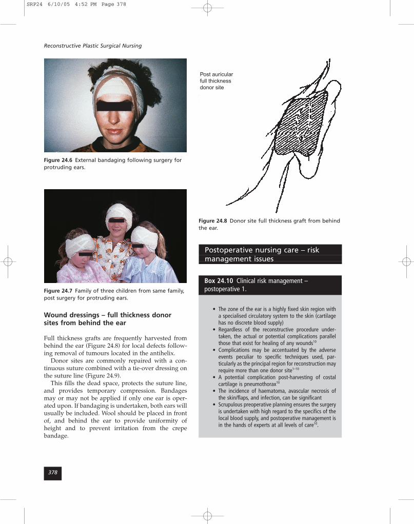
• The head may remain bandaged for up to two weekspostoperatively, or according to the surgeon’s prefer-ence6,7,9 (Figures 24.6 & 24.7)
• One research paper has shown that for the majorityof patients having an aesthetic otoplasty, bandagingfor two days may be sufficient13
• Hair washing and the application of ‘branded’ sportshead bands, ski caps, knitted hats or elasticised,tubular, low compression bandages are becomingmore commonly used for children and adult patientswho are considered co-operative.
Figure 24.5 Internal packing of ear contours(eliminating dead spaces) as part of dressing the ear post surgery.
SRP24 6/10/05 4:52 PM Page 377
378
Wound dressings – full thickness donorsites from behind the ear
Full thickness grafts are frequently harvested frombehind the ear (Figure 24.8) for local defects follow-ing removal of tumours located in the antihelix.
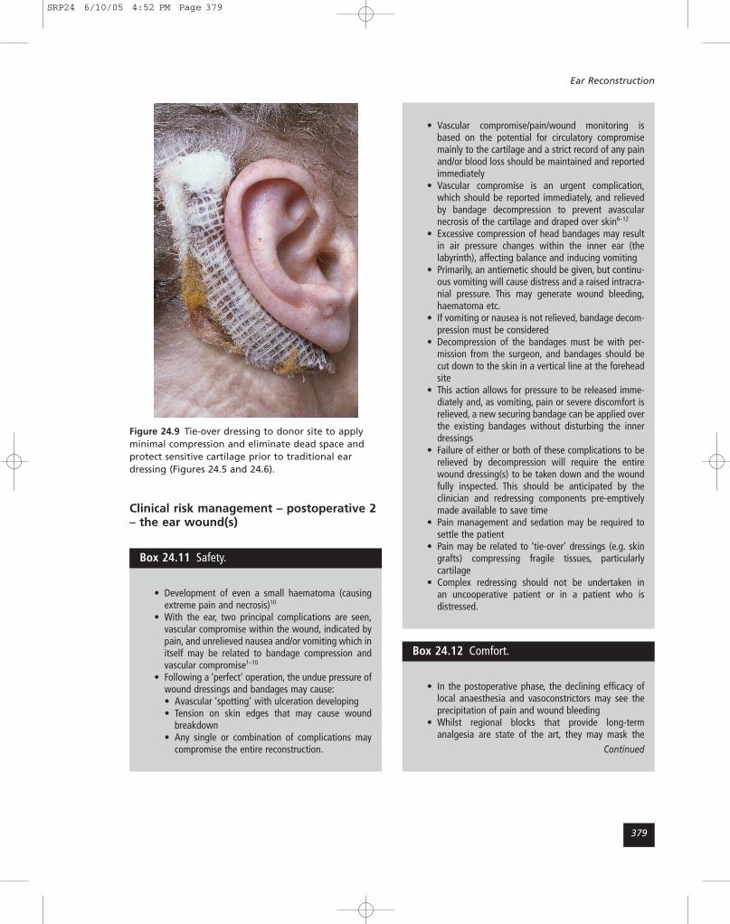
Donor sites are commonly repaired with a con-tinuous suture combined with a tie-over dressing onthe suture line (Figure 24.9).
This fills the dead space, protects the suture line,and provides temporary compression. Bandagesmay or may not be applied if only one ear is oper-ated upon. If bandaging is undertaken, both ears willusually be included. Wool should be placed in frontof, and behind the ear to provide uniformity ofheight and to prevent irritation from the crepebandage.
Reconstructive Plastic Surgical Nursing
Figure 24.6 External bandaging following surgery forprotruding ears.
Figure 24.7 Family of three children from same family,post surgery for protruding ears.
Post auricularfull thicknessdonor site
Figure 24.8 Donor site full thickness graft from behindthe ear.
Postoperative nursing care – riskmanagement issues
Box 24.10 Clinical risk management –postoperative 1.
• The zone of the ear is a highly fixed skin region witha specialised circulatory system to the skin (cartilagehas no discrete blood supply)
• Regardless of the reconstructive procedure under-taken, the actual or potential complications parallelthose that exist for healing of any wounds10
• Complications may be accentuated by the adverseevents peculiar to specific techniques used, par-ticularly as the principal region for reconstruction mayrequire more than one donor site1–10
• A potential complication post-harvesting of costalcartilage is pneumothorax10
• The incidence of haematoma, avascular necrosis ofthe skin/flaps, and infection, can be significant
• Scrupulous preoperative planning ensures the surgeryis undertaken with high regard to the specifics of thelocal blood supply, and postoperative management isin the hands of experts at all levels of care10.
SRP24 6/10/05 4:52 PM Page 378
Ear Reconstruction
379
Clinical risk management – postoperative 2– the ear wound(s)
Figure 24.9 Tie-over dressing to donor site to applyminimal compression and eliminate dead space andprotect sensitive cartilage prior to traditional eardressing (Figures 24.5 and 24.6).
Box 24.11 Safety.
• Development of even a small haematoma (causingextreme pain and necrosis)10
• With the ear, two principal complications are seen,vascular compromise within the wound, indicated bypain, and unrelieved nausea and/or vomiting which initself may be related to bandage compression andvascular compromise1–10
• Following a ‘perfect’ operation, the undue pressure ofwound dressings and bandages may cause:• Avascular ‘spotting’ with ulceration developing• Tension on skin edges that may cause wound
breakdown• Any single or combination of complications may
compromise the entire reconstruction.
• Vascular compromise/pain/wound monitoring isbased on the potential for circulatory compromisemainly to the cartilage and a strict record of any painand/or blood loss should be maintained and reportedimmediately
• Vascular compromise is an urgent complication,which should be reported immediately, and relievedby bandage decompression to prevent avascularnecrosis of the cartilage and draped over skin6–12
• Excessive compression of head bandages may resultin air pressure changes within the inner ear (thelabyrinth), affecting balance and inducing vomiting
• Primarily, an antiemetic should be given, but continu-ous vomiting will cause distress and a raised intracra-nial pressure. This may generate wound bleeding,haematoma etc.
• If vomiting or nausea is not relieved, bandage decom-pression must be considered
• Decompression of the bandages must be with per-mission from the surgeon, and bandages should becut down to the skin in a vertical line at the foreheadsite
• This action allows for pressure to be released imme-diately and, as vomiting, pain or severe discomfort isrelieved, a new securing bandage can be applied overthe existing bandages without disturbing the innerdressings
• Failure of either or both of these complications to berelieved by decompression will require the entirewound dressing(s) to be taken down and the woundfully inspected. This should be anticipated by the clinician and redressing components pre-emptivelymade available to save time
• Pain management and sedation may be required tosettle the patient
• Pain may be related to ‘tie-over’ dressings (e.g. skingrafts) compressing fragile tissues, particularly cartilage
• Complex redressing should not be undertaken in an uncooperative patient or in a patient who is distressed.
Box 24.12 Comfort.
• In the postoperative phase, the declining efficacy oflocal anaesthesia and vasoconstrictors may see theprecipitation of pain and wound bleeding
• Whilst regional blocks that provide long-term analgesia are state of the art, they may mask the
Continued
SRP24 6/10/05 4:52 PM Page 379

380
Reconstructive Plastic Surgical Nursing
development of a haematoma or any bleeding – any strikethrough bleeding should be reported immediately
• The nurse clinician should review analgesia given inthe post anaesthesia care unit and provide for anal-gesia to be given orally (or rectally, if appropriate, inchildren) as ordered strictly four-hourly for the first 72hours14,15
• Pain not relieved by analgesia should be reportedimmediately
• This pain may be accompanied by nausea and vomit-ing (see above under Safety)
• Children need to be free of pain and nausea, warmand comforted with a sense of safety, as crying andirritability can raise the blood pressure and increasethe tension on the wound(s) and potential for bleeding14,15
• In both children and adults, additional wounds maybe present
• Pain may be quite significant from donor sites such asthe hip (movement) or chest wound sites (breathing),and provision for comfort measures should be madefor these or other wounds
• Following complex multisited procedures, narcoticanalgesia may be required initially for one to twodoses, and, for some patients, a short term narcoticinfusion may be useful14,15
• Pain may result from blood that has leaked into theear labyrinth
• The ear should be checked with an auriscope andcarefully cleaned with saline soaked cotton buds
• Temporomandibular joint pain may result from chil-dren or adults clenching or grinding their teeth – oralanalgesia and/or low dose diazepam may be requiredto relieve the spasm and resultant pain.
Review
Setting goals of care and preferred clinical andpatient outcomes, Chapter 2, Boxes 2.7–2.9. Adaptas appropriate to the patient’s clinical needs.
Box 24.13 Wound management – generalnursing care.
• Assess for the presence of other wounds including, forexample, wound drainage sites if skin, cartilage, costalcartilage or bone is used, and that additional drainagebottles are present
• If costal cartilage has been harvested the potential forpneumothorax is significant and the patient may havea chest tube inserted to assist in ventilation of thelung
• For procedures that include insertion of tissueexpander(s), see Chapter 22
• Following aesthetic otoplasty or other similar proce-dures, dressings may be changed on day one postoperatively to assess for haematoma or malposi-tioning, which is easier to correct in the early stages6–9
• Nurse clinicians removing the primary and secondarydressings should be extremely careful at all stages toensure that bleeding or pain and that dislodgementof dressings does not occur
• For congenital malformations, and skin tumours of theear, skin grafts or flaps may be used, and primarydressings may also be undertaken on day 1 postoperatively1
• Some procedures may be left open without dressings,particularly tubed pedicles, which require vascularmonitoring
• For dressings left intact for a longer period, it is impor-tant to observe for bleeding through the bandages, orpoorly-filled drainage tubes or collecting bottles, aschildren cannot support excessive blood loss
• The removal of dressings postoperatively requires thepatient to be in a reclining position and the bandagescut as for decompression (middle of the foreheaddown to the skin)
• Each layer is removed individually and gently. If nofurther dressings are required, the patient may gohome and gently shower and wash the hair underwarm (not hot) water
• Hair drying should be undertaken by gentle towelling– not rubbing – or using hair dryer on cool – not hot
• Emphasis is put on the need for protection and safetyof the ears(s), as cartilage is slow to heal and the pro-cedure may fail
• Headbands should remain on overnight to protectagainst secondary injury to the ear
• A soft diet is required for the first 7–10 days as exces-sive or hard chewing will cause discomfort to ears, asthe temporomandibular joints are very close11.
SRP24 6/10/05 4:52 PM Page 380

Ear Reconstruction
381
References
(1) Atlantic Coast Ear Specialists. Anatomical tour of the ear.http://www.earaces.com/anatomy.htm
(2) Biavati M.J. & Leach J.L. Ear reconstruction.http://www.emedicine.com/ent/topic79.htm
(3) Davison S., Thomassen J. & Cohen M. Ear reconstruc-tion and salvage. http://www.emedicine.com/plastic/topic213.htm
(4) Tennessee Craniofacial Centre. Microtia reconstruction.http://www.erlanger.org/craniofacial/book/Ear/Ear_2.htm (excellent – photos – surgery)
(5) Kavanagh, K.T. Scalp advancement flap for ear recon-struction in a patient skin cancer. http://www.entusa.com/ear_reconstruction.htm (excellent)
(6) Aston S.J., Beasley R.W. & Thorne C.N.M. Grabb andSmith’s plastic surgery, 5th edn. New York: Lippincott &Raven Publishers, 1997.
(7) McCarthy J.G. (ed.) Plastic surgery, volumes 1–8.Philadelphia: W.B. Saunders, Harcourt Brace, 1990.
(8) Ausher B.M., Erikson E. & Wilkins E.G. Plastic surgery:indications, operations and outcomes. St Louis: Mosby,2000.
(9) Ruberg R.L. & Smith D.J. Plastic surgery: a core curriculum. St. Louis: Mosby, 1994.
(10) Morris A. McG., Stevenson J.H. & Watson A.C.H. Com-plications of plastic surgery. Baillière Tindall: London,1989.
(11) Goodman T.A. (ed.) Core curriculum for plastic and recon-structive surgical nursing, 2nd edn. Pitman, N.J.: ASPSNAmerican Society of Plastic Surgery Nurses, 1996.
(12) Fortunato N. & McCullough S. Plastic and recon-structive surgery. Mosby’s Perioperative Series. St. Louis:Mosby, 1998.
(13) Barltey K.V. How long should ears be bandaged afterotoplasty? Journal of Laryngology and Otolaryngology,1998 (June); 112(6):531–2.
(14) Keuren K.V. & Eland J.A. Perioperative pain manage-ment in children. Nursing Clinics of North America, 1997(March); 32(1):31–44.
(15) Mooney K.M. Perioperative management of the pae-diatric patient. Plastic Surgical Nursing, 1997(summer);17(2):69–75.
Recommended websites
Photo galleries of pre-operative, surgical procedure andpost surgery outcomeshttp://www.plasticsurgery.org/index.cfmhttp://www.earreconstruction.com/surgical-
technique.htmlhttp://www.plasticsurgerydoctors.com/procedures/
plasticsurgeryprocedureindex.htmlhttp://www.plasticsurgery.com.au/index.shtml
Box 24.14 Independence
• Most procedures are undertaken as day or short stayprocedures
• Early mobilisation is usually possible but this is depen-dant on the extent of donor sites that may compro-mise full mobility for a few days
• Decisions will be based on an overall assessment ofthe safety of the wounds and the patient
• Children will require diversionary activities and shouldbe persuaded towards quietness and protection of thewounds – structured play, not rough and tumblegames.
Box 24.15 Discharge management.
The patient should be given the following verbal andwritten advice:
• Do not remove dressings unless otherwise instructed• The ears must remain free from any undue pressure
at all times• Do not consciously lie on the ear(s)• Quiet activity – resting as much as possible for the
first few days or until instructed• Wear knitted ski hat or beanie over dressings in bed
at night to secure the bandages• Soft diet – no straining• Report any excessive pain, unusual odour or bleeding
to your surgeon or nearest emergency room• Do not get bandages wet, and if this does occur return
to the surgeon’s rooms for changing• When dressings are removed, wear a favourite tennis
headband, knitted ski hat or beanie at night for aboutfour weeks to ensure ears do not accidentally foldforward – cartilage heals slowly and is easily frac-tured.
Review
Auditing preferred clinical and patient outcomes,Chapter 2, Boxes 2.10–2.12.
SRP24 6/10/05 4:52 PM Page 381