chapter 2 genes and genetic disease. genetic perspective microscopic studies – 1800s – nucleus...
TRANSCRIPT

Chapter 2
Genes and Genetic Disease
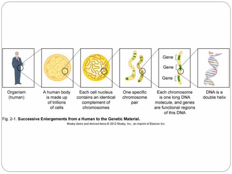
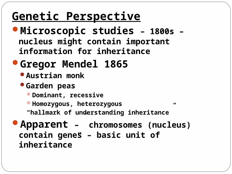
Genetic PerspectiveMicroscopic studies – 1800s –
nucleus might contain important information for inheritance
Gregor Mendel 1865Austrian monkGarden peas
Dominant, recessiveHomozygous, heterozygous “hallmark of understanding inheritance”
Apparent –” chromosomes (nucleus) contain genes – basic unit of inheritance”

Gregor Mendel 1822-1884

St. Thomas Abbey Brno Czech Rep.
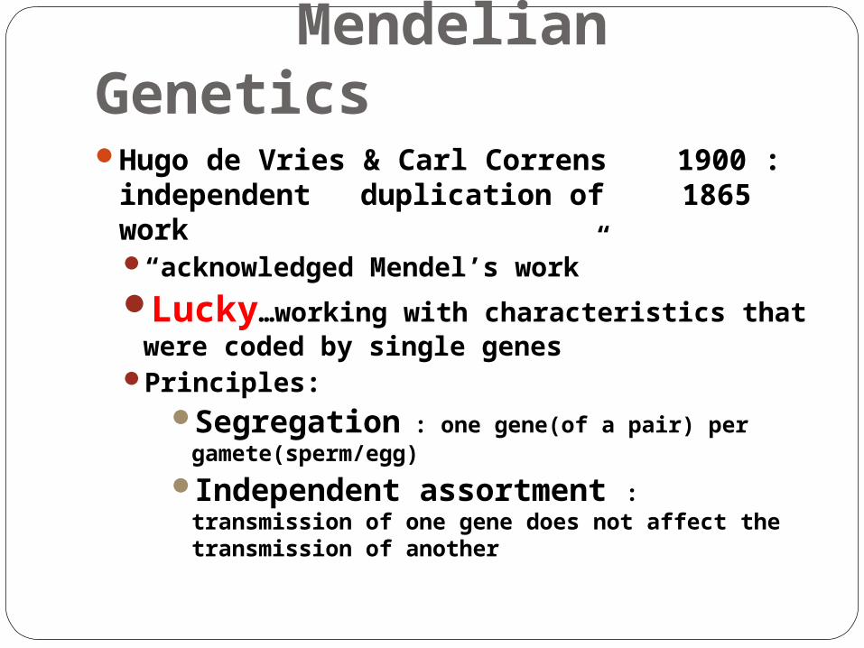
Mendelian GeneticsHugo de Vries & Carl Correns 1900 :
independent duplication of 1865 work
“acknowledged Mendel’s work”Lucky…working with characteristics that
were coded by single genesPrinciples:
Segregation : one gene(of a pair) per gamete(sperm/egg)
Independent assortment : transmission of one gene does not affect the transmission of another
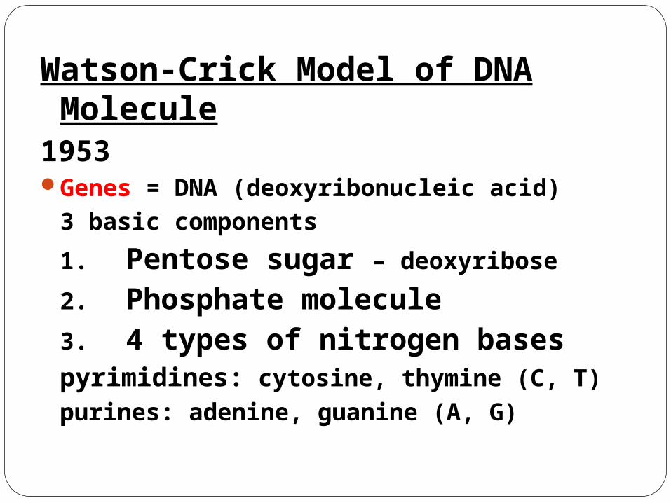
Watson-Crick Model of DNA Molecule
1953Genes = DNA (deoxyribonucleic acid)
3 basic components
1. Pentose sugar – deoxyribose
2. Phosphate molecule3. 4 types of nitrogen bases
pyrimidines: cytosine, thymine (C, T)
purines: adenine, guanine (A, G)
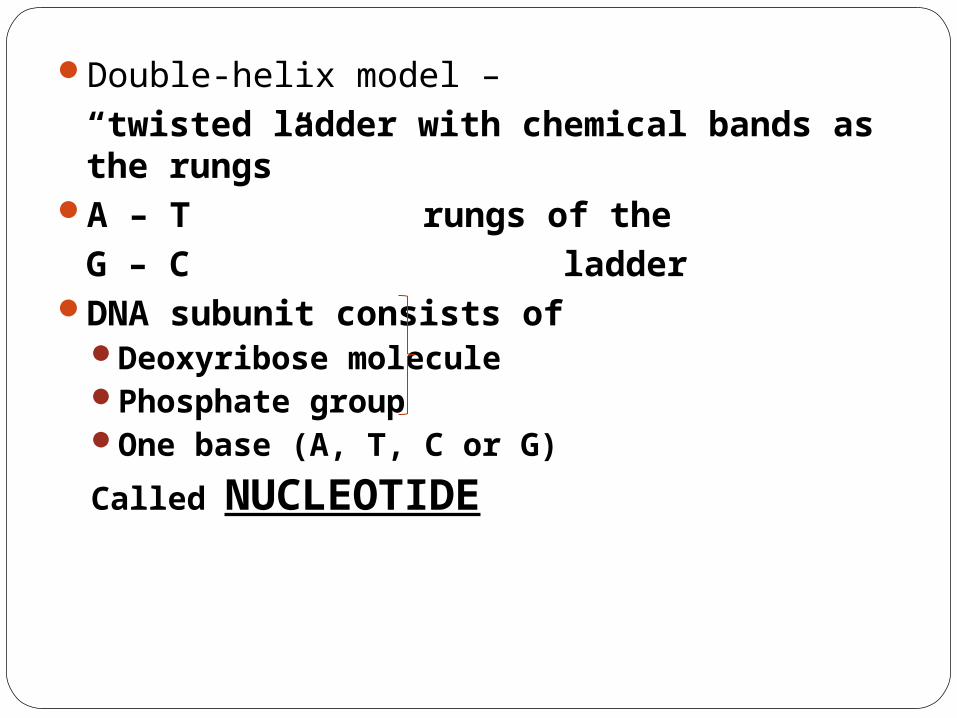
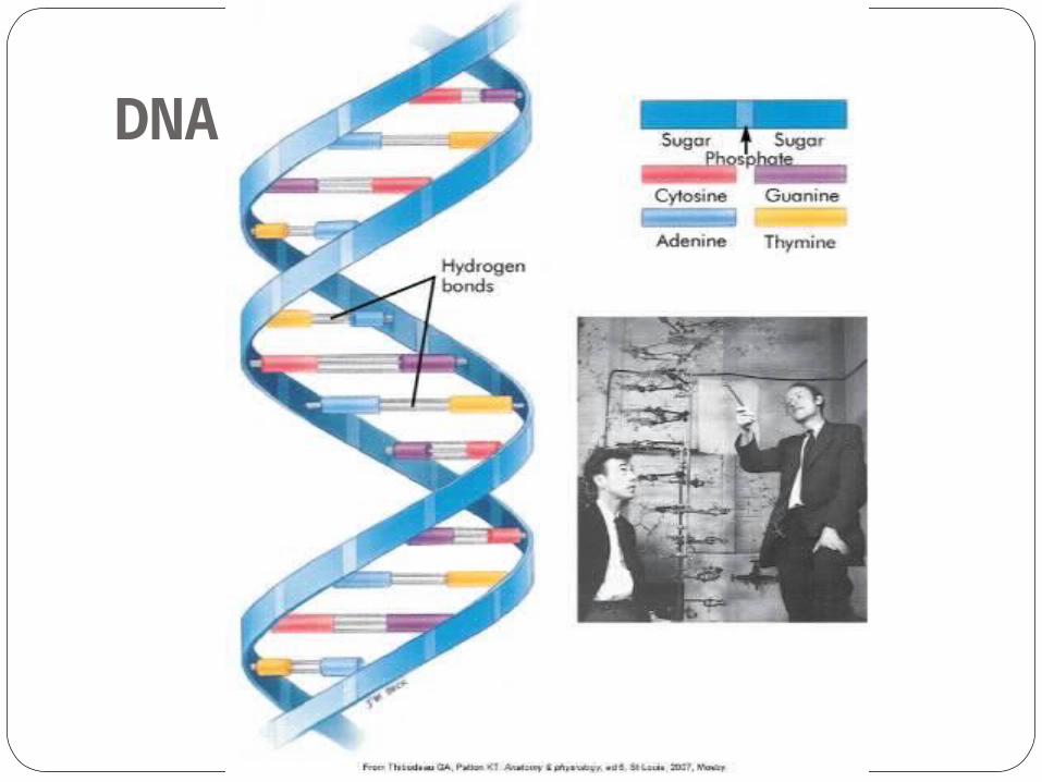
Double-helix model –“twisted ladder with chemical bands as the rungs”
A – T rungs of theG – C ladder
DNA subunit consists ofDeoxyribose moleculePhosphate groupOne base (A, T, C or G)
Called NUCLEOTIDE
DNA
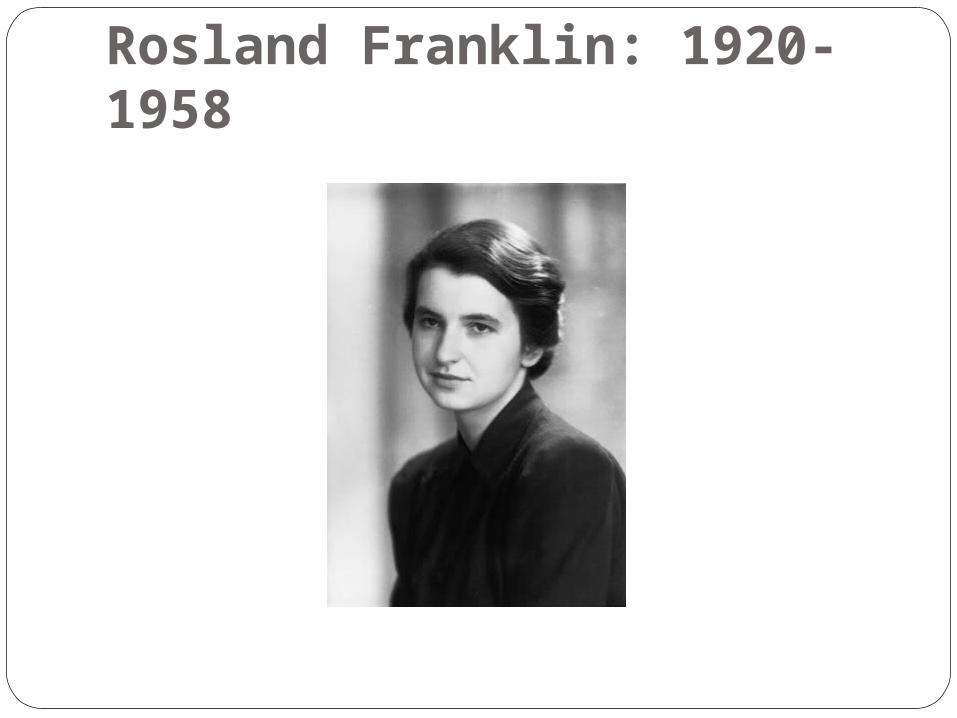
Rosland Franklin: 1920-1958

DNA as the Genetic CodeProteins – structural, functional (receptors
& enzymes)Amino acids → polypeptides → one/more
→ protein (tissues, enzymes, receptors)20 different amino acids
4 bases (A-T, C-G) – specify which amino acid is placed into the polypeptideGroup of 3 bases – each amino acid termed
CODON
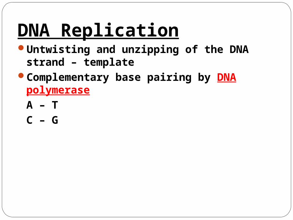
DNA ReplicationUntwisting and unzipping of the DNA
strand – templateComplementary base pairing by DNA
polymeraseA – TC – G
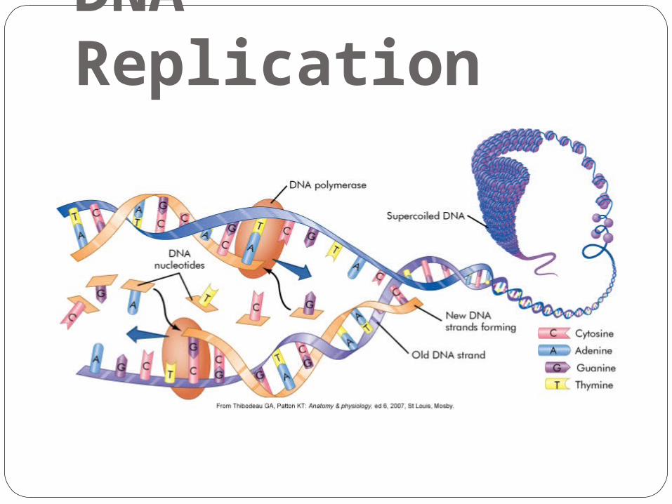
DNA Replication

MutationAny inherited alteration of genetic material
“Chromosome aberrations”
Base pair substitution (missense/stop) A-T………..G-C
Frame shift mutation – deletion or insertion
ATGCTACG……AT_CTACG or ATG G CTACG
SO - insert the wrong amino acid(s) into the polypeptide chain(s) → abnormal proteins (MUTATION)
Mutations Spontaneous – absence of known
mutagen
Hot spots – chromosome areas with ↑ rates cytosine followed by guanine → large percentage of disease – causing mutations
Radiation & chemicals - ↑ frequency

Genes to ProteinsDogma: Transcription Translation
DNA RNA Proteins
NUCLEUS CYTOPLASM
RNA – 2 differences1.- ribose sugar: added Oxygen2.- uracil, rather than thymine
A – UC – G
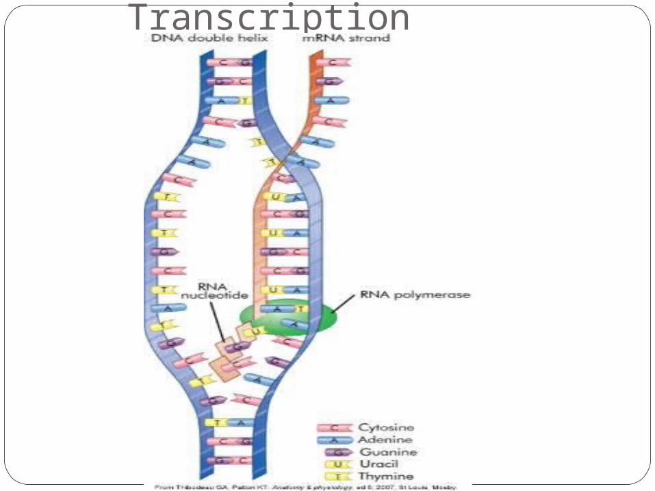
TranscriptionMessenger RNA – synthesized from DNA
template (RNA polyerase) single strand of DNA
mRNA → cytoplasm
Transcription

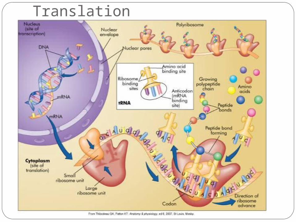
Translation*RNA directs synthesis of polypeptides at the
ribosometRNA **contains a sequence of nucleotides
(anticodon) complementary to the triad of nucleotides on the mRNA strand (codon)
mRNA = UGC… tRNA = ACG remember:RNA- U:A, C:G
DNA- T:A, C:G
*Nobel Prize Chemistry 2009: Ramakrishnan, Steitz, Yonath “structure of ribosomes”…new antibiotics!
**Transfer RNA
Translation

ChromosomesAbnormalities – leading cause mental
retardation & miscarriageSomatic cells
46 chromosomes (23 pairs) – Diploid
Gametes23 chromosomes (1 member of pair) - Haploid
ChromosomesMeiosis – haploid cells from diploid
cells :sperms & eggs (reduction division)
Mitosis – forms somatic cells :new cells
Figure 2-9

KaryotypeOrdered display of chromosomes

Chromosome Aberrations Euploid cells
Contains a multiple of the normal number of chromosomes (23)
Haploid and diploid
Polyploid cellsTriploid – 3 copies of 23(haploid) → 69
chromosomesTetraploid – 4 copies 23(haploid) → 92
chromosomes
Chromosome Aberrations Aneuploidy
Somatic cell does not contain a multiple of 23 chromosomes
3 copies of one → trisomy (may survive)1 copy only → monosomy (lethal)
“More is better”

Chromosome Aberrations Disjunction – normal separation of
chromosomes during cell division
Non-disjunction – failure of homologous chromosomes to separate – meiosis / mitosisUsual cause of aneuploidy

ChromosomesAutosomes
First 22 of the 23 pairsTwo members are identical and said to be
homologousSex chromosomes
Remaining pairFemales – XX – homologousMales – XY – non-homologous

Nondisjunction

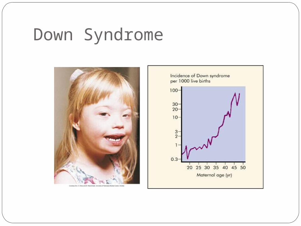
Autosomal AneuploidyDown Syndrome
Trisomy 211:800 live birthsMentally retarded, low nasal bridge,
epicanthal folds, protruding tongue, poor muscle tone
Risk ↑ with maternal age > 35
Down Syndrome
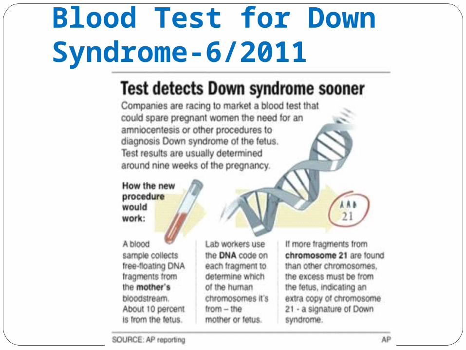
Blood Test for Down Syndrome-6/2011
Sex Chromosome Aneuploidy1 in 500 males / 1 in 900 females
Females trisomy X
Females single X – total 45 chromosomesTurner Syndrome
Males – two x and one Y (47 chromosomes)Klinefelter Syndrome
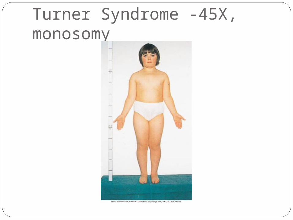
Turner Syndrome -45X, monosomy

Klinefelter Syndrome-47,XXY
Abnormalities of Chromosome Structure
Breakage – repair – may alter structure
Loss – Cri du chat syndrome – deletion short arm #5 → low birth weight mental retardation and microcephaly
Duplication – less seriousInversion – balanced – no apparent effectTranslocation – interchange of material
between two non-homologous chromosomesRobertsonian – fusion at centromere → single
chromosome
Abnormalities of Chromosome Structure
Fragile sites – areas that develop breaks or gaps
Fragile X syndrome – long arm X chromosomeMental retardationMale (XY) verses female (XX)

Genetics Gregor Mendel – 1865
Austrian monkGarden peasMendelian traits
Genetics“trait caused by a single gene →
mendelian trait”Locus – gene location on a chromosomeAllele – different form of a particular
gene at the given locus Example: Hgb A verses Hgb S
Polymorphism – two or more alleles at a locus

Genetics“humans are diploid – one chromosome
from mom one from dad – 23 + 23 = 46”Homozygous – loci on a pair of
chromosomes have identical genesExample: O blood type (OO)
Heterozygous – loci on a pair of chromosomes have different genesExample: AB blood type (A & B genes on a pair of loci)
Genetics Genotype – genetic makeup of the
organism
Phenotype – observable, detectable or outward appearance of the genetics of an organismExample: A blood type – could be AA or AO
A – phenotypeAA/AO – genotype

GeneticsDominant/Recessive – two alleles are
found together, observable allele is dominant, other allele is recessive and not observable
A – large letter = dominanta – small letter = recessiveAlleles can be co-dominant
Example: AB blood type
GeneticsCarrier - one that has a disease gene but
is phenotypically normalto demonstrate a recessive disease the
pair of recessive genes must be inherited Example: Dd Heterozygous – carrier
dd Homozygous _ disease
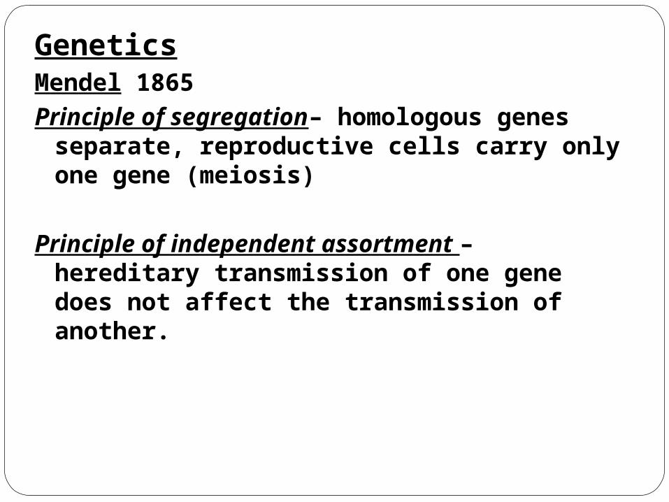
GeneticsMendel 1865Principle of segregation– homologous
genes separate, reproductive cells carry only one gene (meiosis)
Principle of independent assortment – hereditary transmission of one gene does not affect the transmission of another.

Chromosome Theory of Inheritance
“Single Gene Disease – 4 mode of inheritance”
1) Autosomal dominant2) Autosomal recessive3) X-linked dominant4) X-linked recessive
PedigreesUsed to study specific genetic disorders
within familiesBegin with proband

Pedigrees

Single Gene Disorders Autosomal dominant – rare <
1:500Normal parent x affected heterozygote “Rare”
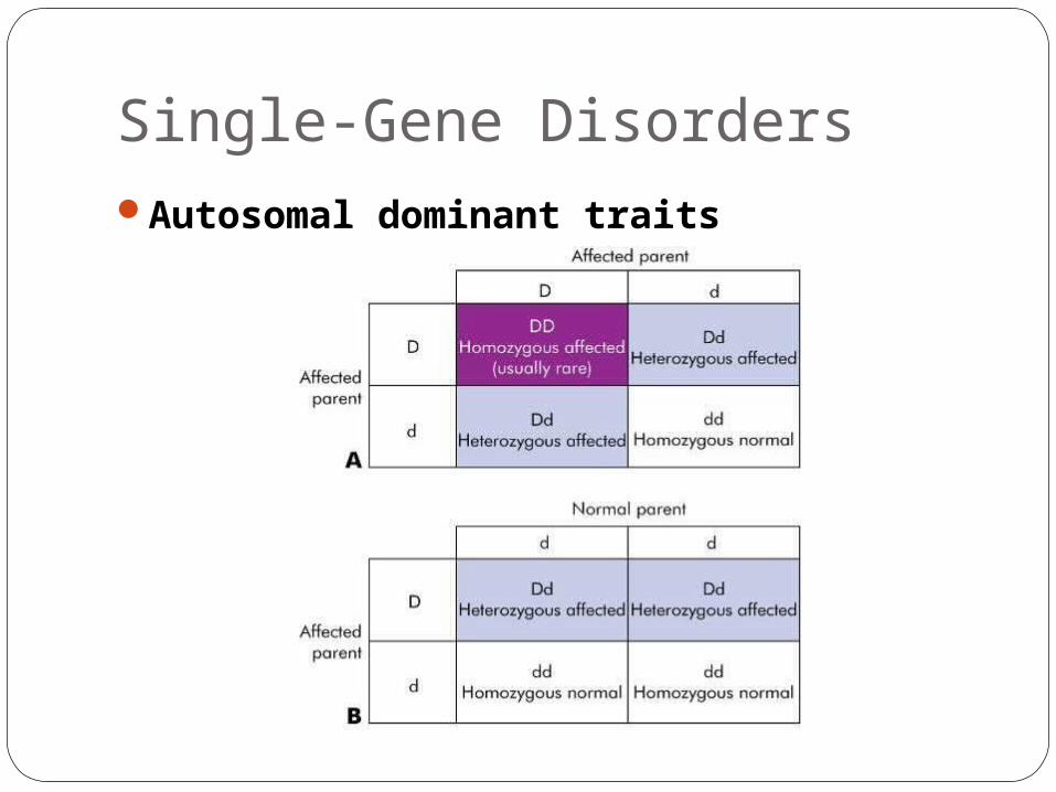
Single-Gene DisordersAutosomal dominant traits

Single-Gene DisordersAutosomal dominant trait pedigree

Single Gene DisordersRecurrence risk
AD – one parent with disease, one without risk :50% risk

Single Gene DisordersAutosomal dominant
Achondroplasia 4p16.3…FGFR3 Gene …80% new mutations
Marfan syndrome 15q 15-21Neurofibromatosis 17q11BrachydactylyNoonan syndromeHuntington disease

Wizard of Oz
ReviewDelayed age of onsetPenetranceexpressivity
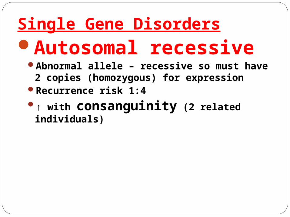
Single Gene DisordersAutosomal recessive
Abnormal allele – recessive so must have 2 copies (homozygous) for expression
Recurrence risk 1:4↑ with consanguinity (2 related
individuals)
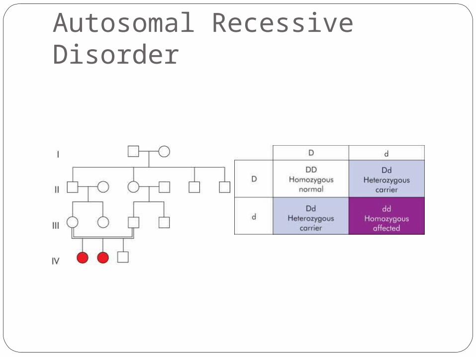
Autosomal Recessive Disorder
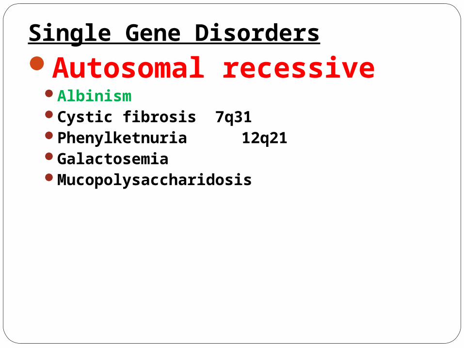
Single Gene DisordersAutosomal recessive
AlbinismCystic fibrosis 7q31Phenylketnuria 12q21GalactosemiaMucopolysaccharidosis
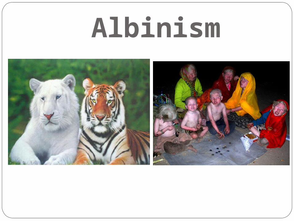
Albinism
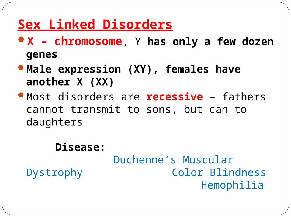
Sex Linked DisordersX – chromosome, Y has only a few dozen
genesMale expression (XY), females have
another X (XX)Most disorders are recessive – fathers cannot
transmit to sons, but can to daughters
Disease:
Duchenne’s Muscular DystrophyColor BlindnessHemophilia
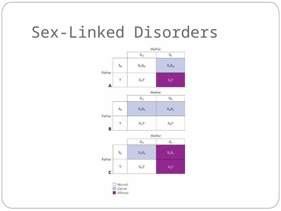
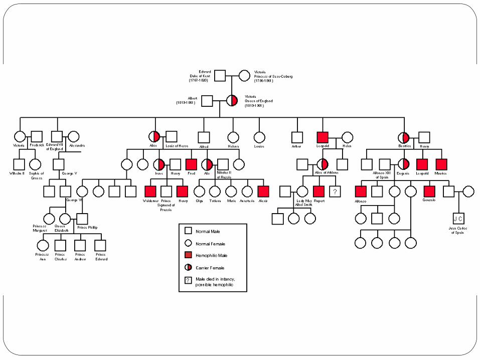
Sex-Linked Disorders

Review – Multi-factorial Inheritance
DiseasesCleft lip & palateNeural tube defectsClubfootSome congenital heart disease
Figure 2-31 Example of Diseases: A Gene Map