chapter 2 chymotrypsin-like proteases as key regulators of the
TRANSCRIPT
Chapter 2
Chymotrypsin-like proteases as key regulators of the positive-sense RNA virus life cycle
Chapter 2
22
Proteases: Introduction
Proteins are the major building blocks of life and for that reason protein synthesis is one of the crucial processes in the life cycle of every organism. However, the linking of individual amino acids to form a functional protein leaves little room for error; substitution of one amino acid for another may already have severe effects on protein function. At the same time, cleavage by specialized enzymes called proteases can be used to activate a protein, a process that may be almost as important as the correct synthesis of the polypeptide chain. For example, our own blood clotting mechanism is regulated by proteases through the step-by-step activation of coagulation factors, which often themselves are proteases. In many other processes, ranging from food digestion to embryonic development, in all kinds of life forms correct protein processing is equally important. Even various viruses, which are by some definitions not even considered to be alive, employ proteases for the release and activation of proteins to start their replication.
Proteases (or peptidases) catalyze the hydrolysis of peptide bonds and are often classified according to their mode of action. They are either exo-acting peptide bond hydrolases (exopeptidases) that cleave amino acids from the carboxyl or amino terminus of proteins, or endopeptidases, also referred to as proteases, that can hydrolyze internal peptide bonds (Barrett & McDonald, 1986). Currently six catalytic types of proteases (for sake of simplicity hereafter called classes) are recognized on the basis of their active site nucleophiles: serine, cysteine, aspartic, threonine, glutamic acid and metalloproteases (Barrett et al., 2004; Rawlings et al., 2008). The mechanism used to cleave a peptide bond is in principle the same for all classes of proteases. It involves a nucleophilic attack of either an amino acid residue (serine, cysteine and threonine proteases) or a water molecule (aspartic acid, metallo- and glutamic acid proteases) on the carbonyl group of a peptide bond, assisted by the donation of a proton to the peptide bond’s nitrogen (Barrett et al.,2004; Rawlings et al., 2008).
Proteases within the same class are further grouped into clans. A clan usually consists of several protease families each comprising an evolutionary lineage that is characterized by the conserved tertiary structure, active site residues and sequence motifs around the catalytic residues (Barrett & Rawlings, 1995; Rawlings et al., 2008).
Serine proteases
One of the best studied classes of proteases is that of the serine proteases (for a review see (Hedstrom, 2002)). In this class two families can be distinguished; one is represented by cellular chymotrypsin (Fig. 2A) and the other by bacterial subtilisin, each belonging to a separate clan, PA(S) and SB, respectively (Barrett et al., 2004). These families share a similar active site configuration but bear no other structural resemblance to each other and are most likely an example of convergent evolution (Neurath, 1984). The active site of these serine proteases contains a His-Asp-Ser motif (His-57, Asp-102, Ser-195 in chymotrypsin, trypsin and elastase; His-64, Asp-32, Ser-221 in subtilisin) that is believed to be responsible for catalyzing the hydrolysis of peptide bonds. Chymotrypsin, for example, enhances the rate of peptide bond hydrolysis by a factor of at least 109 (Nelson & Cox, 2004).
The His-Asp-Ser motif is also referred to as “the catalytic triad” and provides an active nucleophile in the form of a polarized serine. A biological catalyst must be able to
Chapter 2
23
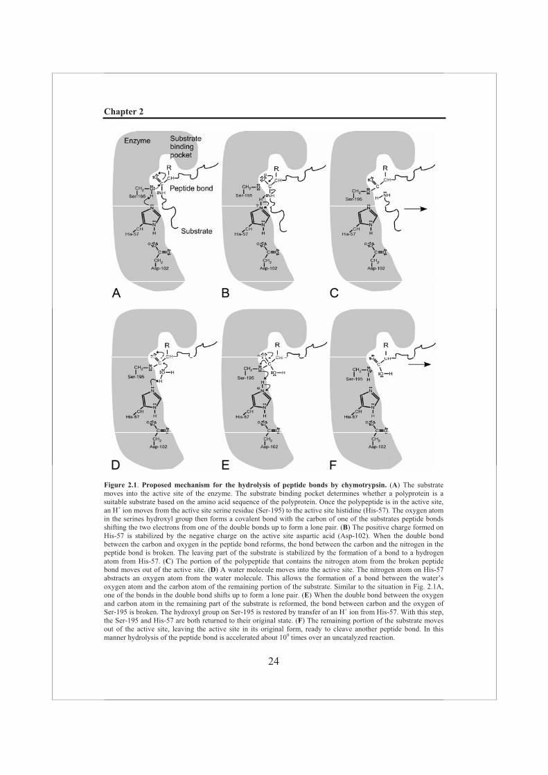
function at a pH of about 7, but at neutral pH the hydroxyl group of a serine residue is normally protonated and therefore not a very good nucleophile. In the active site of chymotrypsin the hydroxyl group of Ser-195 is hydrogen-bonded to His-57, which is in turn hydrogen-bonded to Asp-102 (Fig. 2.1). When a peptide substrate binds to chymotrypsin, a subtle change in conformation compresses the hydrogen bond between His-57 and Asp-102, resulting in a stronger interaction, allowing the histidine to act as an enhanced general base to abstract the proton from the Ser-195 hydroxyl group. This prevents the development of a very unstable positive charge on the serine, thus increasing its nucleophilicity and consequently making it highly reactive (Fig. 2.1A-C). The reaction described above is the first step in catalysis and results in a short-lived acyl-enzyme intermediate between the substrate and the catalytic serine residue, which proceeds through a negatively charged tetrahedral transition state intermediate (Birktoft & Blow, 1972) to subsequent cleavage of the peptide bond (Fig. 2.1A-C). The negative charge on the oxygen (now called oxyanion) is stabilized by the so-called “oxyanion hole”, a pocket in the enzyme in which main chain amide groups form hydrogen-bonds with the oxyanion. During the second step (or deacylation), the acyl-enzyme intermediate is hydrolyzed by a water molecule (Fig. 2.1D-F). The incoming water molecule is deprotonated by general base catalysis of His-57, generating a strongly nucleophilic hydroxide ion. After the attack of hydroxide on the ester bond of the acyl-enzyme intermediate, the peptide is released. Subsequently, His-57, which now acts as a general acid, accepts a proton from the hydroxyl group of the reactive serine and the system is turned to its original state, ready to cleave a new peptide bond (Hedstrom, 2002; Nelson & Cox, 2004).
Serine proteases of the chymotrypsin family participate in a wide range of biological reactions and are found in both prokaryotic and eukaryotic organisms as well as in viruses. About 30 years ago, a virus-encoded cysteine protease activity was first demonstrated for the encephalomyocarditis virus (EMCV) (Pelham, 1978), one of the prototypic picornaviruses. It was subsequently mapped on the virus genome in a region now known as 3C. The enzyme was purified and the protease was functionally characterized and shown to be conserved in other picornaviruses and some other virus families. A few years later, this protease was recognized to be related to chymotrypsin-like proteases (Gorbalenya et al., 1986; Bazan & Fletterick, 1988; Gorbalenya et al., 1989a). After the identification of a chymotrypsin-like fold in these viral cysteine proteases by X-ray crystallography (Allaire et al., 1994; Matthews et al., 1994; Bergmann et al., 1997), the common ancestry of this group of viral cysteine proteases and the canonical chymotrypsin-like serine proteases was no longer a matter of debate. As a result, they are assigned to a separate clan PA(C). In fact, it has been suggested that the primordial ancestor of all chymotrypsin and chymotrypsin-like proteases was a cysteine protease (Gorbalenya et al.,1986; Brenner, 1988). Moreover, based on a database search of known protein structures similar to NK-lysin (a single-domain protein that consists of only one ß-barrel, but with a similar fold to the two ß-barrels of the chymotrypsin-like proteases), it was speculated that the hepatitis A virus (HAV) chymotrypsin-like protease may be the closest known relative of this primordial ancestor (Liepinsh et al., 1997).
Chapter 2
24
Figure 2.1. Proposed mechanism for the hydrolysis of peptide bonds by chymotrypsin. (A) The substrate moves into the active site of the enzyme. The substrate binding pocket determines whether a polyprotein is a suitable substrate based on the amino acid sequence of the polyprotein. Once the polypeptide is in the active site, an H+ ion moves from the active site serine residue (Ser-195) to the active site histidine (His-57). The oxygen atom in the serines hydroxyl group then forms a covalent bond with the carbon of one of the substrates peptide bonds shifting the two electrons from one of the double bonds up to form a lone pair. (B) The positive charge formed on His-57 is stabilized by the negative charge on the active site aspartic acid (Asp-102). When the double bond between the carbon and oxygen in the peptide bond reforms, the bond between the carbon and the nitrogen in the peptide bond is broken. The leaving part of the substrate is stabilized by the formation of a bond to a hydrogen atom from His-57. (C) The portion of the polypeptide that contains the nitrogen atom from the broken peptide bond moves out of the active site. (D) A water molecule moves into the active site. The nitrogen atom on His-57 abstracts an oxygen atom from the water molecule. This allows the formation of a bond between the water’s oxygen atom and the carbon atom of the remaining portion of the substrate. Similar to the situation in Fig. 2.1A, one of the bonds in the double bond shifts up to form a lone pair. (E) When the double bond between the oxygen and carbon atom in the remaining part of the substrate is reformed, the bond between carbon and the oxygen of Ser-195 is broken. The hydroxyl group on Ser-195 is restored by transfer of an H+ ion from His-57. With this step, the Ser-195 and His-57 are both returned to their original state. (F) The remaining portion of the substrate moves out of the active site, leaving the active site in its original form, ready to cleave another peptide bond. In this manner hydrolysis of the peptide bond is accelerated about 109 times over an uncatalyzed reaction.

Chapter 2
25
Virus-encoded proteases in the viral life cycle
After the initial observation of a virus-encoded proteolytic activity for EMCV (see previous paragraph) (Pelham, 1978), the involvement of viral proteases in gene expression and protein maturation was described for many other viruses, with most virus-encoded proteases found to date being employed by positive-sense RNA viruses. These viruses often express part of their genome as a large polyprotein that needs to be processed into the smaller functional subunits (reviewed in (Krausslich & Wimmer, 1988; Hellen et al., 1989; Dougherty & Semler, 1993)). In this manner, virus-encoded proteases, sometimes assisted by cellular proteases, control in essence viral replication. In turn, their activity or substrate specificity may be controlled by binding to virus proteins (co-factors) (Tomei et al., 1996; Wassenaar et al., 1997; Banerjee et al., 2004). In addition, proteases may have specific cellular protein targets, thereby modulating host-cell functions presumably to promote viral replication. The precise role of these cleavages in the viral life cycle is a topic of intense study of many research groups. In the next paragraphs, the chymotrypsin-like (3C, 3C-like and 2A proteases) (Fig. 2.2) and the processes they control are reviewed for the animal positive-sense RNA viruses, including picornaviruses, caliciviruses and nidoviruses.
The chymotrypsin-like proteases in the picornavirus life cycle
Classification, nomenclature and genome organization of picornaviruses
The viruses that belong to the Picornaviridae (a contraction of the prefix “pico” meaning “very small” and RNA) all have an icosahedral virion with a diameter of about 30 nanometer (nm). The picornavirus family is currently divided in nine genera: aphthovirus,cardiovirus, enterovirus, erbovirus, hepatovirus, kobuvirus, parechovirus, teschovirus and rhinovirus (Pringle, 1999; King et al., 2000; Stanway et al., 2005) and comprises a prevalent group of viruses infecting mainly vertebrates (Christian et al., 2000). Members of most genera are able to infect humans and the spectrum of picornavirus-related disease ranges from viral meningitis (an infection of the thin lining covering the brain and spinal cord), myocarditis (inflammation of the heart), poliomyelitis (also known as polio or infantile paralysis) to viral respiratory infections, like the common cold (Hollinger & Emerson, 2001; Pallansch & Roos, 2001).
The picornavirus genome consists of a single positive-sense RNA molecule of between 7.2 kilobases (kb) (human rhinovirus 14 (HRV14)) to 8.5 kb (foot-and-mouth disease virus (FMDV)) and possesses a number of features that are conserved across the whole group. Untranslated regions (UTRs) are present at both the 5' end (600-1200 nucleotides (nt)) and the 3' end (50-100 nt) of the genome, which are also modified. The 3' end is polyadenylated and a small basic protein VPg (~23 amino acids) is covalently attached to the genomic 5' end. The 5' UTR includes a secondary structure known as the IRES (internal ribosome entry site). The rest of the genome encompasses a single open reading frame (ORF) that encodes a large precursor polyprotein of between 2100-2400 amino acids. This polyprotein commonly contains the following domain organization: NH2-L-VP4-VP2-VP3-VP1-2A-2B-2C-3A-3B-3C-3D-COOH, with L being absent in some viruses. Upon virion assembly the paralogous proteins, VP3, VP2 and VP1 form the T=3 virions. All other (“nonstructural”) proteins except for VP4 are not part of virions; they are primarily involved in the replicative process, but may also control virion biogenesis.
Chapter 2
26
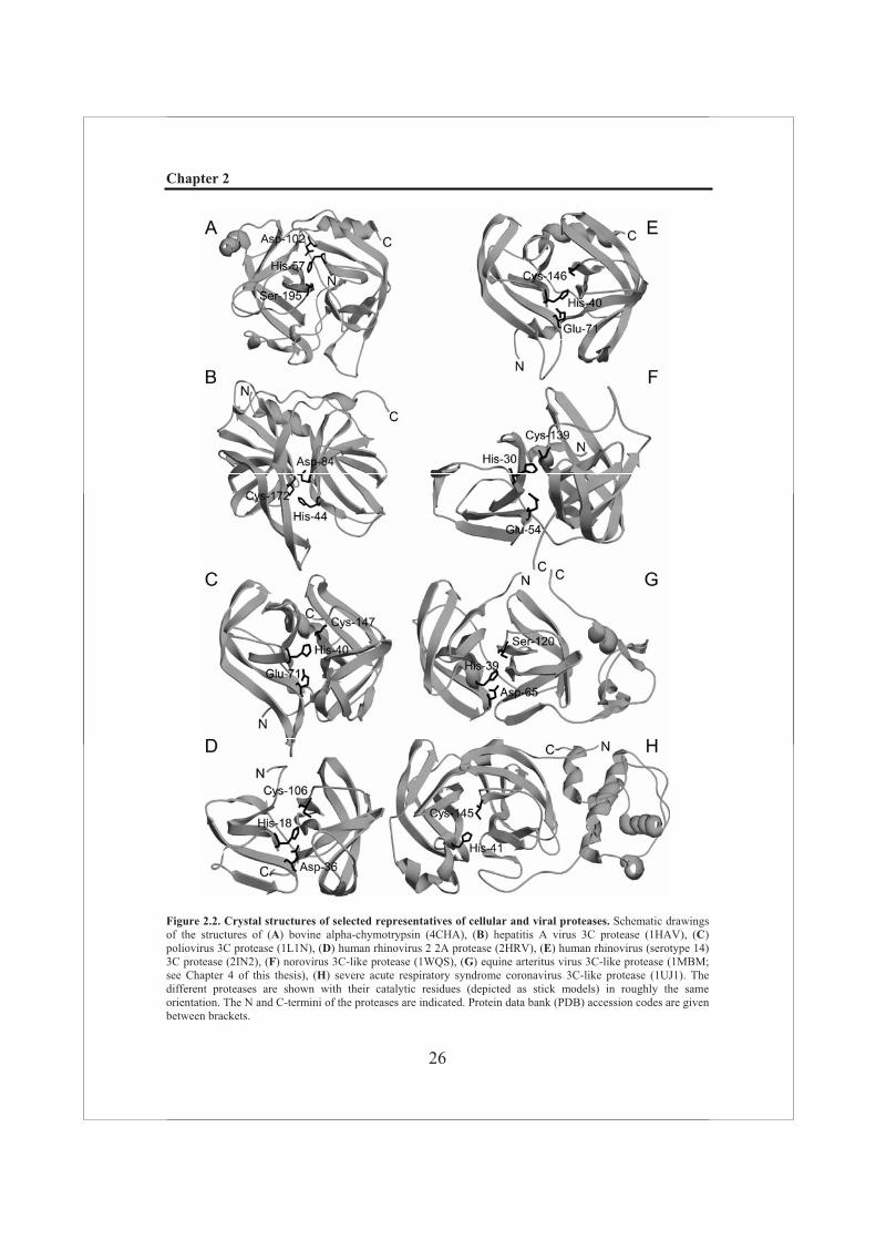
Figure 2.2. Crystal structures of selected representatives of cellular and viral proteases. Schematic drawings of the structures of (A) bovine alpha-chymotrypsin (4CHA), (B) hepatitis A virus 3C protease (1HAV), (C)poliovirus 3C protease (1L1N), (D) human rhinovirus 2 2A protease (2HRV), (E) human rhinovirus (serotype 14) 3C protease (2IN2), (F) norovirus 3C-like protease (1WQS), (G) equine arteritus virus 3C-like protease (1MBM; see Chapter 4 of this thesis), (H) severe acute respiratory syndrome coronavirus 3C-like protease (1UJ1). The different proteases are shown with their catalytic residues (depicted as stick models) in roughly the same orientation. The N and C-termini of the proteases are indicated. Protein data bank (PDB) accession codes are given between brackets.
Chapter 2
27
Proteases and proteolytic processing in picornaviruses
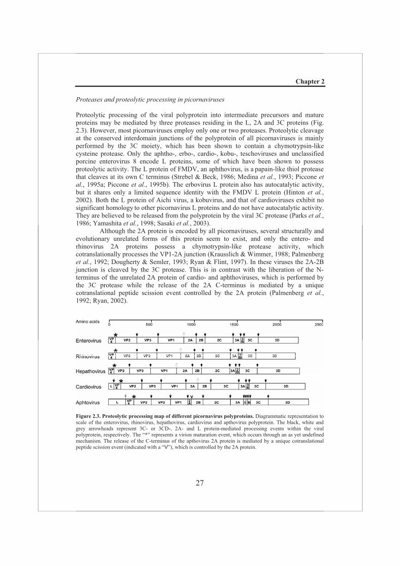
Proteolytic processing of the viral polyprotein into intermediate precursors and mature proteins may be mediated by three proteases residing in the L, 2A and 3C proteins (Fig. 2.3). However, most picornaviruses employ only one or two proteases. Proteolytic cleavage at the conserved interdomain junctions of the polyprotein of all picornaviruses is mainly performed by the 3C moiety, which has been shown to contain a chymotrypsin-like cysteine protease. Only the aphtho-, erbo-, cardio-, kobu-, teschoviruses and unclassified porcine enterovirus 8 encode L proteins, some of which have been shown to possess proteolytic activity. The L protein of FMDV, an aphthovirus, is a papain-like thiol protease that cleaves at its own C terminus (Strebel & Beck, 1986; Medina et al., 1993; Piccone etal., 1995a; Piccone et al., 1995b). The erbovirus L protein also has autocatalytic activity, but it shares only a limited sequence identity with the FMDV L protein (Hinton et al.,2002). Both the L protein of Aichi virus, a kobuvirus, and that of cardioviruses exhibit no significant homology to other picornavirus L proteins and do not have autocatalytic activity. They are believed to be released from the polyprotein by the viral 3C protease (Parks et al.,1986; Yamashita et al., 1998; Sasaki et al., 2003).
Although the 2A protein is encoded by all picornaviruses, several structurally and evolutionary unrelated forms of this protein seem to exist, and only the entero- and rhinovirus 2A proteins possess a chymotrypsin-like protease activity, which cotranslationally processes the VP1-2A junction (Krausslich & Wimmer, 1988; Palmenberg et al., 1992; Dougherty & Semler, 1993; Ryan & Flint, 1997). In these viruses the 2A-2B junction is cleaved by the 3C protease. This is in contrast with the liberation of the N-terminus of the unrelated 2A protein of cardio- and aphthoviruses, which is performed by the 3C protease while the release of the 2A C-terminus is mediated by a unique cotranslational peptide scission event controlled by the 2A protein (Palmenberg et al.,1992; Ryan, 2002).
Figure 2.3. Proteolytic processing map of different picornavirus polyproteins. Diagrammatic representation to scale of the enterovirus, rhinovirus, hepathovirus, cardiovirus and apthovirus polyprotein. The black, white and grey arrowheads represent 3C- or 3CD-, 2A- and L protein-mediated processing events within the viral polyprotein, respectively. The “*” represents a virion maturation event, which occurs through an as yet undefined mechanism. The release of the C-terminus of the apthovirus 2A protein is mediated by a unique cotranslational peptide scission event (indicated with a “V”), which is controlled by the 2A protein.
Chapter 2
28
Structural aspects of the picornavirus 2A and 3C proteases
As mentioned above, both the 3C and the 2A proteases of picornaviruses are cysteine proteases. However, bioinformatics analysis predicted that these two proteases have backbone folds that are similar to those of cellular chymotrypsin, a serine protease, rather than of other (cellular) cysteine proteases, even though the overall sequence identity between the viral proteases and chymotrypsin is very low (Gorbalenya et al., 1986; Bazan & Fletterick, 1988; Gorbalenya et al., 1989a). The 2A and 3C proteases contain the Gly-X-Cys-Gly motif (where X represents any amino acid) that is reminiscent of the Gly-Asp-Ser-Gly motif in the active site of the chymotrypsin-like serine proteases. Crystal structures of these proteases from different picornaviruses (HRV2 and HRV14, hepatitis A virus (HAV), FMDV and poliovirus (PV)) confirmed that the 2A and 3C proteases are chymotrypsin-like cysteine proteases (Allaire et al., 1994; Matthews et al., 1994; Bergmann et al., 1997; Mosimann et al., 1997; Petersen et al., 1999; Seipelt et al., 1999; Birtley et al., 2005). These structural analyses revealed also that the 2A protease differs from all known chymotrypsin-like proteases in that its N-terminal domain is not a -barrel, but rather a four-stranded antiparallel -sheet. In addition, a tightly bound Zn2+ ion in the C-terminal domain of the 2A protease was discovered (Yu & Lloyd, 1992). The Zn2+ ion is tetrahedrally coordinated by the side chains of three cysteine residues and one histidine residue, which are highly conserved among the 2A proteases. Structural and biochemical analyses suggest that Zn2+ binding may be important for the stability of the enzyme, possibly to compensate for the instability caused by the small N-terminal domain (Petersen et al., 1999). 2A proteases can be very small; the 2A protease of HRV2 for example consists of only 142 residues and this makes it the smallest enzyme known in this family.
The catalytic triad of the HRV2 2A protease consists of His-18, Asp-35, and Cys-106 (Petersen et al., 1999), which is also supported by mutagenesis studies of other closely related proteases (Yu & Lloyd, 1991; Sommergruber et al., 1997). Besides interacting with His-18, the third triad member Asp-35 is also involved in a large network of hydrogen bonding interactions. It has been proposed that the cysteine and histidine residues form a thiolate imidazolium ion pair, similar to that of papain (Sarkany et al., 2000). The substrate preference of the 2A protease is mostly defined by residues at the P4, P2, and P1’ positions (using the cleavage site nomenclature of Schechter and Berger, (Schechter & Berger, 1967)) (Liebig et al., 1991; Wang et al., 1998). A threonine residue at P2 is strongly preferred for cleavage by the protease, and a model of the enzyme/substrate complex suggests this residue may hydrogen-bond with Ser-83 of the protease (Petersen et al.,1999). In comparison, the S1 pocket appears to be rather open and can accommodate a variety of side chains (Sommergruber et al., 1992; Petersen et al., 1999). It is believed that the 2A-driven processing of the viral polyprotein occurs in cis, hence releasing its own N-terminus from the polyprotein (Toyoda et al., 1986).
The catalytic triad of the 3C protease of HRV14 consists of residues His-40, Glu-71, and Cys-146. In the HAV 3C protease, Cys-172 is the first catalytic residue and Asp-84 is equivalent to Glu-71 in HRV14, but its side chain points away from the second member of the catalytic triad, the His-44 residue, which renders it useless in catalysis (Allaire et al.,1994). It has been suggested that instead of Asp-84, Tyr-143 may function as the third member of the triad in the HAV 3C protease (Allaire et al., 1994; Bergmann et al., 1997). The natural substrates of 3C proteases generally have a glutamine residue at the P1 position, a glycine residue at the P1’ position and a small aliphatic side chain at P4 (Seipelt et al., 1999). The 3C crystal structures suggest that the P1 glutamine side chain interacts
Chapter 2
29
with conserved Thr-142 and His-161 residues in the S1 pocket of the HRV14 3C protease (Allaire et al., 1994; Matthews et al., 1994), confirming earlier predictions derived from comparative sequence analysis (Gorbalenya et al., 1986; Bazan & Fletterick, 1988; Gorbalenya et al., 1989a). In vitro assays using recombinant 3C proteases showed that the P5 through P2’ residues of the substrate are absolutely required for recognition of the cleavage site (Cordingley et al., 1989; Long et al., 1989). As both the N- and C-termini of the protease are far from the active site in the solved structures, the 3C protease is likely to function exclusively in trans when cleaving the viral polyprotein.
The picornavirus 3C and 2A proteases and cellular substrates
In addition to their role in the maturation of the viral polyprotein, the picornavirus 3C and 2A proteases are known to target cellular substrates. It is thought that the dramatic translation inhibition in e.g. PV-infected cells is induced by 3C- and 2A-mediated cleavage of host proteins involved in transcription, translation, and cytoskeletal integrity. A number of RNA polymerase transcription factors including TATA-binding protein (TBP) (Clark etal., 1993; Das & Dasgupta, 1993; Yalamanchili et al., 1996), Octamer binding protein (Oct-1) (Yalamanchili et al., 1997c) and cyclic AMP-responsive element binding protein (CREB) (Yalamanchili et al., 1997b) are cleaved by the PV 3C protease, as well as transcription factor IIIC (TFIIIC) (Clark & Dasgupta, 1990; Clark et al., 1991; Rubinstein et al., 1992; Porter, 1993; Shen et al., 1996), TATA-binding associated factor 110 (TAF110) (Banerjee et al., 2005), polyA-binding protein (PABP) (Joachims et al., 1999), and the cytoskeletal protein MAP-4 (microtubule-associated protein 4) (Joachims et al.,1995). Weidman et al. reported the degradation of transcriptional activator p53 by the 3C protease in vivo and in vitro, however, unlike for other transcription factors, p53 degradation may be indirect (Weidman et al., 2001). The 3C protease of FMDV has been reported to induce proteolytic cleavage of host cell histone H3 (Falk et al., 1990) and to cleave the translation initiation factor eIF4G (Belsham et al., 2000; Strong & Belsham, 2004). In contrast to e.g. coxsackieviruses, translation initiation factor eIF4G is not a substrate of the HAV 3C protease (Zhang et al., 2007).
The picornavirus 2A protease is also known to cleave proteins that are involved in host cell transcription, e.g. TBP (Yalamanchili et al., 1997a), translation initiation factor eIF4G (Gradi et al., 1998; Gradi et al., 2003), PABP (Kerekatte et al., 1999; Kuyumcu-Martinez et al., 2002) and the cytoskeletal protein dystrophin (Badorff et al., 2000).
It is likely that the shut-off of host cell transcription plays an important role in the replication of picornaviruses. Strong evidence for this hypothesis was recently presented by Kundu et al. A cell line resistant to PV 3C protease cleavage of TBP displayed smaller plaques and lower viral yields compared to the wild-type cell line (Kundu et al., 2005). Still, the exact mechanisms by which PV and most other picornaviruses mediate nearly complete translation inhibition in host cells remain largely elusive. In PV-infected Hela cells partial translation inhibition was originally thought to result from cleavage of translation initiation factor eIF4GI by the viral 2A protease, although more recently strong evidence was presented that also cellular proteases activated during infection play a role in eIF4GI cleavage (Zamora et al., 2002). However, cleavage of eIF4GI was shown to be only partially responsible for the translation shutoff (Bonneau & Sonenberg, 1987; Irurzun et al.,1995), thus additional events are required to bring about complete host cell translation shutoff (Perez & Carrasco, 1992). For example, the discovery of the cleavage of a
Chapter 2
30
functional homologue of eIF4GI, termed eIF4GII, correlated better with the temporal inhibition of translation in both PV- and HRV-infected cells (Gradi et al., 1998). An additional factor may be the cleavage of PABP, since both the 2A protease and 3C protease cleave PABP during enterovirus infection (Joachims et al., 1999; Kerekatte et al., 1999; Kuyumcu-Martinez et al., 2002).
In addition to its proteolytic activity, the ability to specifically bind viral RNA is unique to picornaviral protease 3C (and its precursors) and the role of the 3C-RNA interaction in regulating viral RNA synthesis has been demonstrated for PV (Andino et al.,1990; Andino et al., 1993). For PV, HRV and HAV secondary structures formed at the 5´ end of their genomes were identified as specific RNA targets for binding by viral proteases (Andino et al., 1990; Andino et al., 1993; Walker et al., 1995; Kusov & Gauss-Muller, 1997; Gamarnik & Andino, 1998). For PV, it was proposed that the stable 3C precursor known as 3CD (see also below) is involved in the switch from translation to RNA synthesis by binding to the 5´ end of the viral genome (Gamarnik & Andino, 1998). Although the interaction with viral RNA has been studied in some detail for 3C of various picornaviruses, the precise molecular requirements for this function still remain to be addressed (Jewell et al., 1992; Schultheiss et al., 1995; Walker et al., 1995; Kusov et al.,1997; Kusov & Gauss-Muller, 1997).
Modulation of the picornavirus 3C and 2A proteases by cofactors
Polyprotein precursors or processing intermediates often have functions in replication that are distinct from those of the mature cleavage products. An example of a molecule exhibiting such differential functions is the 3CD product of picornaviruses, containing RNA binding and protease activities that reside in its 3C moiety and the silent RNA-dependent RNA polymerase in the 3D domain. In viral RNA replication, 3CD forms a ternary ribonucleoprotein (RNP) complex with the 5’-terminal sequences of genomic RNA (the 5’ cloverleaf structure) and a cellular RNA-binding protein termed poly(rC)-binding protein 2 (PCBP2) or the viral protein 3AB (Andino et al., 1990; Andino et al., 1993; Parsley et al., 1997), but also exhibits protease activity towards all 3C cleavage sites in the polyprotein. Moreover, biochemical studies on PV 3C and 3CD enzymes showed that processing of the viral capsid precursor is in fact more efficiently mediated by 3CD than by 3C (Parsley et al., 1999). 3CD is also able to trans-cleave 3CD molecules more efficiently than is 3C, and it processes sites within the P3 precursor more rapidly (Jore et al., 1988; Ypma-Wong et al., 1988a; Ypma-Wong et al., 1988b; Andino et al., 1993; Harris et al.,1994; Parsley et al., 1999). There were no differences found between 3C and 3CD in the processing of a nonstructural polyprotein precursor, 2C3AB (Parsley et al., 1999). Yet, the exact biochemical roles of specific 3D amino acid sequences and domains for 3CD protease activity are poorly understood. Possibly the structural domains within the 3D portion of the 3CD contribute to the enhanced activity of this protease toward 3C cleavage sites residing in the P1 precursor (Jore et al., 1988; Ypma-Wong et al., 1988a; Marcotte et al., 2007).
Infection of mammalian cells with PV results in the direct cleavage of several transcription factors by the 3C protease (see also the previous paragraph). It is possible that 3C or a precursor of 3C enters the nucleus of infected cells to shut-off host cell transcription. Although diffusion of 3C into the nucleus, when present at sufficiently high concentrations, can not be ruled out, another explanation seems more likely. Recently, a single basic type of nuclear localization signal (NLS) was identified in the 3D domain
Chapter 2
31
(Sharma et al., 2004b). Possibly, 3C enters the nucleus in the form of its precursor, 3CD, which then generates 3C by auto-proteolysis leading to cleavage of transcription factors. However, the presence of the NLS alone was not sufficient for nuclear entry of 3C/3CD; other cofactors may be required or upon PV infection additional alterations in the nuclear membrane are induced which enable successful nuclear translocation of 3C/3CD (Sharma et al., 2004b).
Another virus-encoded protein that may regulate polyprotein processing is the 2C protein, that is highly conserved among picornaviruses (Argos et al., 1984) and has been implicated in a number of functions during viral replication such as uncoating (Li & Baltimore, 1990), host cell membrane rearrangement (Cho et al., 1994), RNA replication (reviewed in (Wimmer et al., 1993)), and encapsidation (Vance et al., 1997), although the exact role of 2C in these processes is not fully understood. It was demonstrated that the purified 2C protein is capable of inhibiting the activity of both the 3C and 2A proteases in PV-infected cells. PV infection of HeLa cell lines that expressed 2C in an inducible fashion resulted in a processing pattern consistent with slower processing of a number of PV precursor polypeptides (Banerjee et al., 2004). Possibly, 2C downregulates 3C activity by physically interacting with it, which was demonstrated by co-immunoprecipitation experiments (Banerjee et al., 2004). Mutations in the amphipathic helix of 2C (Paul et al.,1994), which was proposed to be responsible for its membrane binding properties, resulted in abnormalities in polyprotein processing of the P2 and P3 region by the 3C protease, which confirms a possible regulatory role for 2C in 3C-mediated polyprotein processing (Teterina et al., 2006).
The chymotrypsin-like protease in the calicivirus life cycle
Classification, nomenclature and genome organization of caliciviruses
The family Caliciviridae is composed of small (30 to 40 nm), nonenveloped, icosahedral viruses with a linear, single-stranded, positive-sense RNA genome of between 7.3 and 8.3 kb. The RNA is polyadenylated at its 3’ end and has a virus-encoded protein (VPg) covalently linked to its 5’ end (Ehresmann & Schaffer, 1977; Meyers et al., 1991; Herbert et al., 1997; Dunham et al., 1998; Sosnovtsev & Green, 2000). Common features of this family include the presence of a single major structural protein that forms the capsid and the presence of cup-shaped depressions on the surface of the virion. Hence the name Caliciviridae was chosen for this virus family, referring to the Latin word “calix”, which means “cup” or “chalice”. Caliciviruses infect a broad range of animals, including reptiles, cattle, rabbits, pigs, cats and marine mammals, but also chimpanzees and humans. A calicivirus infection may cause various disease syndromes, like respiratory disease in cats, epidemic gastroenteritis in humans, or an often fatal hemorrhagic disease in rabbits. Currently, four genera are recognized within this family, based on differences in genome organization and sequence diversity of the polymerase and capsid genes (Green et al.,2000). These four genera are: lagovirus (mainly infecting members of the order Lagomorpha, e.g.. hares and rabbits), vesivirus (which cause vesicular lesions), norovirus (formerly known as “Norwalk-like virus”; first isolated in Norwalk, USA), and sapovirus (formerly known as “Sapporo-like virus”; first isolated in Sapporo, Japan), for which respectively, rabbit hemorrhagic disease virus (RHDV), feline calicivirus (FCV), Norwalk virus (NoV), and Sapporo virus (SaV) have been assigned as the prototype species (Green
Chapter 2
32
et al., 2001; Mayo, 2002). However, the recent characterization of the unclassified bovine enteric virus Newbury agent-1 (Newbury-1) suggests that the current classification should be revised and endorses a fifth genus in the Caliciviridae (Smiley et al., 2002; Oliver et al.,2006).
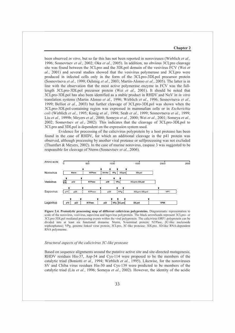
The calicivirus genome is organized into two or three, sometimes partially overlapping, open reading frames (ORFs), depending on the genus and genogroup. For example, viruses in the genera sapovirus and lagovirus have at least two major ORFs (ORF1 and ORF2), but depending on the genogroup (GI-IV), the genome may contain two ORFs (sapovirus GII and GIII) or three ORFs (sapovirus GI, GIV, and GV) (Noel et al.,1997; Numata et al., 1997; Guo et al., 1999; Robinson et al., 2002). Also the genomes of the noroviruses and the vesiviruses are organized into three ORFs. ORF2 encodes the major capsid protein VP1 and in noroviruses ORF3 has been shown to encode a minor structural protein, VP2 (Liu et al., 1996; Katayama et al., 2002; Farkas et al., 2004). In all caliciviruses ORF1 encodes a ~200 kDa nonstructural polyprotein (excluding the in-frame capsid protein sequences for the genera lagovirus and sapovirus), which is processed by a single virus-encoded protease encoded in the 3’-terminal half of ORF1.
From N- to C-terminus, the calicivirus ORF1 polyprotein can be divided into at least six functional domains: the N-terminal protein (Nterm) (Ettayebi & Hardy, 2003); the 2C-like nucleoside triphosphatase (NTPase) (Pfister & Wimmer, 2001); the 3A-like protein; the genome linked virus protein (VPg) (Daughenbaugh et al., 2003); the 3C-like protease (3CLpro) (Someya et al., 2005); and the 3D-like RNA-dependent RNA polymerase (3DLpol) (Liu et al., 1999b; Green et al., 2001; Pletneva et al., 2001; Ng et al.,2004). The arrangement of the three conserved nonstructural proteins in the order: nucleoside triphosphatebinding protein, chymotrypsin-like cysteine protease and RNA-dependent RNA polymerase is a feature which the caliciviruses have in common with the picornaviruses and related plant viruses (potyviruses, comoviruses, and nepoviruses) and therefore they are grouped within the picornavirus-like supergroup (Goldbach & Wellink, 1988).
Protease and proteolytic processing in caliciviruses
Sequence comparisons between caliviviruses and picornaviruses predicted that the calicivirus protease belongs to the group of 3C-like cysteine proteases (3CLpros). The activity of the 3CLpro of human and animal calicivirus strains has been analyzed by expression in bacteria, rabbit reticulocyte lysates and in mammalian cells (Boniotti et al.,1994; Wirblich et al., 1995; Liu et al., 1996; Martin Alonso et al., 1996; Seah et al., 1999; Sosnovtseva et al., 1999). The first full calicivirus proteolytic cleavage map (obtained using in vitro methods) was presented for the lagovirus RHDV (Wirblich et al., 1995; Wirblich etal., 1996) and detailed cleavage maps are also available for the vesivirus FCV (Sosnovtseva et al., 1999; Sosnovtsev et al., 2002) and the norovirus Southampton virus (SV) (Liu et al.,1996; Liu et al., 1999b). Several of the individual protease cleavage sites of SV have been independently confirmed in other norovirus strains (Seah et al., 1999; Someya et al., 2000; Hardy et al., 2002).
Although the caliciviruses have a largely conserved polyprotein domain organization, they differ most pronouncedly in the N-terminal part of their polyprotein and there are some differences in polyprotein processing (Fig. 2.4). For three caliciviruses (RHDV, FCV and human SaV Mc10) 3CLpro-induced cleavage of Nterm (Fig. 2.4) has
Chapter 2
33
been observed in vitro, but so far this has not been reported in noroviruses (Wirblich et al.,1996; Sosnovtsev et al., 2002; Oka et al., 2005). In addition, no obvious 3CLpro cleavage site was found between the 3CLpro and the 3DLpol domain of the vesivirus FCV (Wei etal., 2001) and several studies showed that the vesivirus polymerase and 3CLpro were produced in infected cells only in the form of the 3CLpro-3DLpol precursor protein (Sosnovtseva et al., 1999; Oehmig et al., 2003; Martin-Alonso et al., 2005). The latter is in line with the observation that the most active polymerase enzyme in FCV was the full-length 3CLpro-3DLpol precursor protein (Wei et al., 2001). It should be noted that 3CLpro-3DLpol has also been identified as a stable product in RHDV and NoV in in vitrotranslation systems (Martin Alonso et al., 1996; Wirblich et al., 1996; Sosnovtseva et al.,1999; Belliot et al., 2003) but further cleavage of 3CLpro-3DLpol was shown when the 3CLpro-3DLpol-containing region was expressed in mammalian cells or in Escherichia coli (Wirblich et al., 1995; Konig et al., 1998; Seah et al., 1999; Sosnovtseva et al., 1999; Liu et al., 1999b; Meyers et al., 2000; Someya et al., 2000; Wei et al., 2001; Someya et al.,2002; Sosnovtsev et al., 2002). This indicates that the cleavage of 3CLpro-3DLpol to 3CLpro and 3DLpol is dependent on the expression system used.
Evidence for processing of the calicivirus polyprotein by a host protease has been found in the case of RHDV, for which an additional cleavage in the p41 protein was observed, although processing by another viral protease or selfprocessing was not excluded (Thumfart & Meyers, 2002). In the case of murine norovirus, caspase 3 was suggested to be responsible for cleavage of Nterm (Sosnovtsev et al., 2006).
Figure 2.4. Proteolytic processing map of different calicivirus polyproteins. Diagrammatic representation to scale of the norovirus, vesivirus, sapovirus and lagovirus polyprotein. The black arrowheads represent 3CLpro- or 3CLpro-3DLpol mediated processing events within the viral polyprotein. The calicivirus ORF1 polyprotein can be divided into at least six functional domains: Nterm, N-terminal protein; NTPase, 2C-like nucleoside triphosphatase; VPg, genome linked virus protein; 3CLpro, 3C-like protease; 3DLpro, 3D-like RNA-dependent RNA polymerase.
Structural aspects of the calicivirus 3C-like protease
Based on sequence alignments around the putative active site and site-directed mutagenesis, RHDV residues His-37, Asp-54 and Cys-114 were proposed to be the members of the catalytic triad (Boniotti et al., 1994; Wirblich et al., 1995). Likewise, for the noroviruses SV and Chiba virus residues His-30 and Cys-139 were predicted to be members of the catalytic triad (Liu et al., 1996; Someya et al., 2002). However, the identity of the acidic
Chapter 2
34
residue that completes the catalytic triad has remained controversial. In NoV, two possible candidates were identified: Glu-54 and Asp-67 (Someya et al., 2002). Mutagenesis studies of NoV 3CLpro by Hardy et al. (2002) suggested that indeed an acidic residue (Glu-54) is required for proteolysis of the polyprotein (Hardy et al., 2002), which was later confirmed by analysis of the crystal structure of the NoV 3CLpro (Zeitler et al., 2006). On the other hand, crystallographic analysis of the Chiba virus protease by Nakamura et al. (Nakamura et al., 2005) suggested that Glu-54 is not essential for protease activity, similar to the situation in the case of the coronavirus main protease (see later in this chapter). Despite these differences and the low levels of amino acid sequence similarity between the 3C-like protease domains of the SaV Mc10, FCV F4, RHDV FRG and NoV Chiba virus strains, the overall structures were predicted to be similar (Oka et al., 2007).
Cleavage site preferences for the 3CLpro in caliciviruses are poorly understood. The 3CLpros of RHDV and FCV preferentially cleave sites containing glycine, alanine, threonine or serine residues at the P1’ position and glutamic acid at the P1 position (Wirblich et al., 1995; Sosnovtseva et al., 1999). Bacterial expression studies identified cleavages at Glu-Gly or Glu-Ala dipeptides in SV ORF1 (Liu et al., 1999a) and processing at Glu-Gly and Glu-Ala in a eukaryotic system was confirmed by expression of the polyprotein of the genogroup II Camberwell strain in COS cells (Seah et al., 1999). As an exception, two sites of primary cleavage for the SV strain have glutamine residues at P1, as analyzed by translation of the ORF1 polyprotein in reticulocyte lysates (Liu et al., 1996) and also cleavage at a Gln-Gly dipeptide in RHDV has been reported (Meyers et al., 2000).
Although primary cleavage sites for the caliciviruses have been identified, there is little data on the substrate requirements in terms of residues flanking the scissile bonds. For example, upon mutagenesis of the P2 position of the cleavage site for the 3CLpro of RHDV, several replacements were tolerated (Wirblich et al., 1995; Hardy et al., 2002). At least one residue, His-157, that is part of the S1 site in noroviruses is important to substrate binding and its replacement with any other tested residue severely reduced activity (Someya et al., 2002).
Modulation of the calicivirus 3C-like protease by cofactors
In caliciviruses, as in the picornaviruses, functional processing intermediates containing protease and polymerase moieties (3CLpro-3DLpol) were identified (Sosnovtseva et al.,1999; Belliot et al., 2003). Accordingly, the calicivirus 3CLpro may be modulated by the polymerase moiety. Indeed, the 3CLpro-3DLpol covalent complex of NoV was shown to possess a protease activity that differed from the mature protease in its ability to function in trans on a p20-VPg-3CLpro-3DLpol precursor (Belliot et al., 2003; Sosnovtsev et al.,2006; Scheffler et al., 2007).
The calicivirus 3C-like protease and cellular substrates
Thus far, very limited studies have been reported regarding the involvement of the calicivirus 3CLpro in functions other than viral polyprotein processing. However, using recombinant 3CLpros from NoV strain MD145-12 and the vesivirus FCV, cleavage of PABP, present in either HeLa S10 cytoplasmic extracts or in isolated ribosome fractions, was observed in in vitro cleavage reactions. The NoV 3CLpro PABP cleavage products

Chapter 2
35
were indistinguishable from those generated by the PV 3C protease cleavage, while the FCV 3CLpro products differed in size. The effect of PABP cleavage by the NoV 3CLpro was analyzed in HeLa cell translation extracts, and the presence of 3CLpro inhibited translation of both endogenous and exogenous mRNAs. Since the caliciviruses apparently do not encode a picornavirus 2A-like protease, the ability of the recombinant NoV and FCV 3CLpro to cleave HeLa eIF4G was examined. However, no cleavage of HeLa eIF4GI was observed for either protease (Kuyumcu-Martinez et al., 2004).
Although the calicivirus 3CLpro resembles the picornavirus 3C protease in a number of ways, a counterpart to the highly conserved amino acid sequence motif KFRDI in the interdomain junction forming the RNA binding site of 3C, is not evident in the NoV 3CLpro sequence. The absence of this motif coincides with a unique organization of the 5’-UTR in caliciviruses. Thus, the calicivirus protease may not bind the 5’-UTR RNA, a feature that has been implicated in the switch from translation to RNA synthesis in picornaviruses (see above).
The chymotrypsin-like protease in the nidovirus life cycle
Classification, nomenclature and genome organization of nidoviruses
The order Nidovirales is comprised of several groups of enveloped, positive-sense RNA viruses, which were found to cluster in phylogenetic analyses of key replicative enzymes like the viral RNA-dependent RNA polymerase (RdRp) and helicase, suggesting that these proteins are evolutionarily related (Gorbalenya et al., 1989c; Snijder et al., 1990; den Boon et al., 1991; Cavanagh, 1997; de Vries et al., 1997; Cowley et al., 2000; Gonzalez et al.,2003; Snijder et al., 2005; Spaan et al., 2005; Gorbalenya et al., 2006). The name of the order was based on a common feature of these viruses, the generation in infected cells of a nested set of 3’-coterminal mRNAs (“nidus” means “nest” in Latin) (Cavanagh, 1997; Snijder et al., 2005; Spaan et al., 2005). At present, the order comprises the families Coronaviridae (consisting of the genera Coronavirus and Torovirus), Roniviridae andArteriviridae (Bredenbeek et al., 1990; Snijder et al., 1990; Cavanagh, 1997; de Vries et al., 1997; Cowley et al., 2000; Gorbalenya, 2001; Siddell et al., 2005; Snijder et al., 2005; Spaan et al., 2005). The genus Coronavirus has been subdivided into three groups, which were originally based on serological relationships, a division that was subsequently supported by genetic studies. Recently, it was proposed to re-define the genera Coronavirusand Torovirus as two subfamilies within the Coronaviridae family or two families within the Nidovirales order, and to convert the current three informal Coronavirus groups into three genera within the coronavirus subfamily/family (Cavanagh, 1997; Gonzalez et al.,2003; Gorbalenya et al., 2004; Spaan et al., 2005; Walker et al., 2005). In addition, the establishment of a new nidovirus genus named Bafinivirus (referring to the bacilliform morphology of this cluster of fish nidoviruses) was proposed after phylogenetic analysis of helicase and polymerase core domains of white bream virus (WBV). A preliminary characterization of this virus isolated from fish identified toroviruses (followed by coronaviruses) as the closest known relatives of WBV (Granzow et al., 2001; Schutze etal., 2006).
Coronaviruses were named after the array of large spikes on the viral envelope that resembles a crown (“corona” means “crown” in Latin) and was first observed by electron microscopy upon negative staining of avian infectious bronchitis virus (IBV) (Fig. 1.2)
Chapter 2
36
(Berry et al., 1964). Other examples of well studied coronaviruses are the human coronavirus 229E (HCoV-229E), porcine transmissible gastroenteritis virus (TGEV), murine coronavirus (MHV) and the recently discovered severe acute respiratory syndrome coronavirus (SARS-CoV). The toroviruses (e.g. equine torovirus (EToV)), which were named after their tubular nucleocapsid that may bend into an open torus (Fig. 1.2), and roniviruses (“roni” stands for rod-shaped nidovirus) have been studied only to a limited extent. Viruses that belong to the Arteriviridae family are the prototypic equine arteritis virus (EAV), porcine reproductive and respiratory syndrome virus (PRRSV), lactate dehydrogenase-elevating virus (LDV) and simian hemorrhagic fever virus (SHFV).
Nidovirus infections are mostly associated with respiratory and/or enteric disorders, although other organs (e.g. the central nervous system) can also be involved. Their outcome may range from an asymptomatic, persistent carrier-state to a lethal hemorrhagic fever. Members of the Coronaviridae family were mainly known to cause respiratory and enteric infections in humans and domestic animals e.g. cattle, dogs, cats and birds (Wege et al., 1982; Siddell & Snijder, 1998), but following the 2003 SARS-CoV epidemic virus discovery projects identified many novel coronaviruses in other species, including a variety of coronaviruses occurring in bats (Wang et al., 2006). In the family Roniviridae, the gill-associated virus (GAV) (Cowley et al., 2000) infects the black tiger prawn (Penaeus monodon) and thusfar viruses of the Arteriviridae family have been found to infect only mammals e.g. swine, horses, mice, and monkeys.
The nidovirus genome is a single-stranded positive-sense RNA molecule (between 12 and 31 kb) that contains a 5’ cap structure, a 3’ poly(A) tail and untranslated regions of variable size at its 5’ and 3’ termini. A number of open reading frames (ORFs), which varies between individual nidoviruses, encode the proteins needed for genome replication and virion formation. The two large, 5'-proximal and partially overlapping open reading frames, ORF1a and ORF1ab, encode the protein functions needed for viral RNA synthesis. Expression of ORF1b, which encodes key replicase functions like the RdRp and helicase, involves a ribosomal frameshift occurring just upstream of the ORF1a termination codon (Brierley et al., 1987; den Boon et al., 1991; Brierley, 1995; Cowley et al., 2000; Baranov et al., 2005), resulting in the synthesis of polyprotein 1a (pp1a) and the C-terminally extended pp1ab. This ribosomal frameshift site includes a “slippery” heptanucleotide sequence, at which the ribosome makes a 1 frameshift. This site is conserved among corona-, toro- and arteriviruses, but not in roniviruses which may have evolved a different sequence to perform the same function (Brierley et al., 1987; den Boon et al., 1991; Brierley, 1995; Cowley et al., 2000; Baranov et al., 2005). The polyprotein encoded by nidovirus ORF1a is characterized by the presence of the 3CLpro and the three transmembrane (TM) domains (TM1-TM2-3CLpro-TM3) (Fig. 2.5) (Gorbalenya et al.,1989b). These conserved hydrophobic domains, two of which typically flank the 3CLpro at either side (Snijder & Meulenberg, 1998; Gorbalenya, 2001; Ziebuhr, 2005), are thought to anchor the nidovirus replication complex to intracellular membranes (van der Meer et al.,1998; Prentice et al., 2004). The Exo and MT domains, which are lacking in arteriviruses, have been hypothesized to play a key role in the evolution and maintenance of large nidovirus genomes (Gorbalenya et al., 2006). The Hel domain is preceded by a zinc binding domain (ZBD) (not indicated) and appears to be required for the multiple enzymatic activities of the Hel domain, including its nucleoside triphosphate hydrolase (NTPase), RNA 5’-triphosphatase and nucleic acid duplex unwinding (Heusipp et al., 1997; Seybert etal., 2000; Bautista et al., 2002; Tanner et al., 2003; Ivanov & Ziebuhr, 2004; Ivanov et al.,2004b; Seybert et al., 2005). In arteriviruses, the ZBD-Hel domain has been implicated in
Chapter 2
37
both genomic and subgenomic RNA synthesis (van Dinten et al., 1997; van Dinten et al.,2000; Seybert et al., 2005). The uridylate-specific endoribonuclease (N) domain (Snijder etal., 2003; Bhardwaj et al., 2004; Ivanov et al., 2004a; Gioia et al., 2005; Ricagno et al.,2006) is essential for RNA synthesis and/or the production of virus progeny in corona- and arteriviruses (Ivanov et al., 2004a; Posthuma et al., 2006). In both SARS-CoV and HCoV-229E, it has been shown to efficiently cleave double-stranded RNA at specific uridylate-containing sequences.
Proteases and protelytic processing in nidoviruses
Processing of the two replicase polyproteins pp1a and pp1ab is mediated by two to four viral proteases that are encoded in ORF1a (Fig. 2.5) and ultimately yields twelve mature cleavage products in case of the “small-genome” arteriviruses, whereas for the “large-genome” corona-, toro- and roniviruses up to 16 products are generated (Ziebuhr et al.,2000). The C-terminal half of pp1a and the ORF1b-encoded part of pp1ab are processed by a chymotrypsin-like protease that thus directly regulates expression of the RdRp and helicase and has therefore been coined the nidovirus “main protease” (reviewed by (Ziebuhr et al., 2000)). The main protease cleaves at least eight sites in arterivirus replicases and up to twelve in coronavirus replicases. Processing is thought to occur both in cis and in trans. So-called “accessory proteases” reside in the N-proximal domains of the replicase and they release themselves autocatalytically from pp1a and pp1ab (Baker et al.,1989; Snijder et al., 1992) (reviewed by (Ziebuhr et al., 2000); see also (Draker et al.,2006)). Despite their low sequence similarity (Gorbalenya et al., 1991; den Boon et al.,1995), they all belong to the same large superfamily of papain-like cysteine proteases (PLpros) ((Gorbalenya & Snijder, 1996) and references herein).
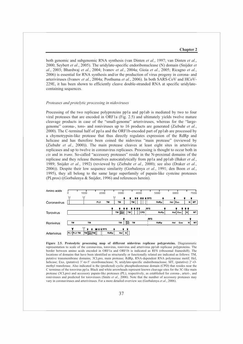
Figure 2.5. Proteolytic processing map of different nidovirus replicase polyproteins. Diagrammatic representation to scale of the coronavirus, torovirus, ronivirus and arterivirus pp1ab replicase polyproteins. The border between amino acids encoded in ORF1a and ORF1b is indicated as RFS (ribosomal frameshift). The locations of domains that have been identified as structurally or functionally related are indicated as follows: TM, putative transmembrane domains; 3CLpro, main protease; RdRp, RNA-dependent RNA polymerase motif; Hel, helicase; Exo, (putative) 3’-to-5’ exoribonuclease; N, uridylate-specific endoribonuclease; MT, (putative) 2’-O-methyl transferase. Also indicated is the (predicted) cyclic phosphodiesterase domain (CPD) that resides near the C terminus of the torovirus pp1a. Black and white arrowheads represent known cleavage sites for the 3C-like main protease (3CLpro) and accessory papain-like proteases (PL), respectively, as established for corona-, arteri-, and roniviruses and predicted for toroviruses (Smits et al., 2006). Note that the number of accessory proteases may vary in coronaviruses and arteriviruses. For a more detailed overview see (Gorbalenya et al., 2006).
Chapter 2
38
Structural aspects of the nidovirus chymotrypsin-like protease
Originally, comparative sequence analysis with picornavirus 3C proteins and the 3C-like proteins of related viruses identified a protease domain in the first reported coronavirus replicase polyproteins sequence, that of IBV. Its catalytic system was proposed to resemble that of the cysteine chymotrypsin-like protease and to involve a His-Asp/Glu-Cys catalytic triad (Gorbalenya et al., 1989c). Likewise, in arteriviruses a putative chymotrypsin-like protease was identified, which was predicted to employ a nucleophilic serine (den Boon etal., 1991). Although, in accordance with these predictions, mutagenesis studies with three different coronaviruses provided evidence for the protease and the presumed catalytic histidine and cysteine residues (Liu & Brown, 1995; Lu et al., 1995; Ziebuhr et al., 1995; Tibbles et al., 1996; Seybert et al., 1997; Ziebuhr et al., 1997; Hegyi et al., 2002), no such evidence was obtained for the presumed third member (Asp/Glu) of the catalytic triad. This prediction was subsequently revised to postulate that coronaviruses might lack the acidic counterpart of the catalytic Asp/Glu of 3C and 3C-like proteases. Recently, based on crystal structures of several coronavirus proteases, it was shown that the catalytic centre of the coronavirus chymotrypsin-like protease indeed contains only a catalytic His/Cys dyad (Anand et al., 2002; Anand et al., 2003; Yang et al., 2003).
Based on sequence alignments and mutagenesis studies, also the roni- and torovirus chymotrypsin-like proteases are believed to employ a catalytic dyad of His/Cys and His/Ser, respectively (Snijder et al., 1996; Ziebuhr et al., 1997; Anand et al., 2002; Barrette-Ng et al., 2002; Anand et al., 2003; Ziebuhr et al., 2003; Draker et al., 2006; Smits et al., 2006). The members of the arterivirus chymotrypsin-like protease catalytic centre were shown to be His/Asp/Ser (Snijder et al., 1996; Barrette-Ng et al., 2002).
Since not only the structure but also the substrate specificity of the nidovirus main proteases resembles that of the picornavirus 3C protease they are also referred to as 3C-like proteases. They share a preference for a small residue (alanine, serine or glycine) at the P1’ position of the cleavage site, whereas the P1 position is occupied by a glutamine residue (coronaviruses) or a glutamic acid (arteriviruses) (Snijder et al., 1996; Wassenaar et al.,1997; van Dinten et al., 1999). The use of other residues at the P1’ position, e.g. an asparagine residue as found in coronaviruses at the nsp8/9 site, can significantly reduce the cleavage efficiency (Ziebuhr & Siddell, 1999; Hegyi & Ziebuhr, 2002; Fan et al., 2004). The preference for glutamine at the P1 position of coronavirus 3CLpro substrates may be based on its ability to interact with the imidazole of His-162 at the bottom of the S1 subsite, as illustrated for the coronavirus TGEV (Anand et al., 2003). At the P2 position of coronavirus 3CLpro substrates a leucine residue is strongly preferred, although other hydrophobic residues are occasionally found at this position. The conservation of a small residue at the P4 position of coronavirus 3CLpro substrates can be explained by a relatively clogged up S4 subsite (Anand et al., 2003).
The crystal structures of both corona- and arterivirus 3CLpros (as will be described in detail in Chapter 4) confirmed their predicted two ß-barrel fold, but unlike the picornavirus 3C proteases the nidovirus enzymes possess an additional C-terminal domain. The C-terminal domain III is unique for nidoviruses (with the single exception of potyvirus 3CLpros) but differs both in size and structure between arteriviruses and coronaviruses (Anand et al., 2002; Barrette-Ng et al., 2002; Anand et al., 2003; Yang et al., 2003). This extension (called domain III in coronaviruses) mainly consists of -helices (Anand et al.,2002; Anand et al., 2003; Yang et al., 2003) and is about twice the size of the corresponding domain of the arterivirus nsp4 protease which comprises ~49 residues and
Chapter 2
39
consists of a combination of -strands and -helices (Barrette-Ng et al., 2002). Because the crystal structures of the 3CLpro of TGEV and SARS-CoV revealed the formation of protease dimers (Anand et al., 2002; Anand et al., 2003; Yang et al., 2003) and the residues at the dimer interface are conserved in coronaviruses, it has been proposed that a dimer may be the biologically functional form of the enzyme. In the dimer, the N-terminal amino acids (also called the N-finger) have many specific interactions with domains II and III of the parental monomer and domain III of the other monomer. It is unclear whether the interactions between the N-terminus and domains II and III are the consequence or the basis of dimerization, but the bulk of experimental data suggests that only the dimeric form of the SARS main protease is active (Fan et al., 2004; Shi et al., 2004; Chen et al., 2005; Hsu etal., 2005a; Hsu et al., 2005b; Chen et al., 2006; Graziano et al., 2006; Shi & Song, 2006).
Modulation of the nidovirus protease by cofactors
Experiments studying the processing of EAV pp1a indicated that fully processed nsp2 was required for processing of the nsp4/5 site by the EAV 3CLpro present in nsp4 (Wassenaar et al., 1997), but not for activity of the protease towards other cleavage sites. This suggests that nsp2 acts as a cofactor in the processing of the nsp4/5 junction and may be needed for the proper processing of other (pp1ab) sites. In addition, nsp5 may also modulate the 3CLpro activity as part of the proteolytically active nsp4-5 precursor, since cleavage of the nsp5/6 and nsp6/7 junctions was especially prominent when the nsp4/5 site was not cleaved (Wassenaar et al., 1997). Using a transient expression system, simultaneous expression of an EAV nsp6-8 substrate and nsp4 or nsp4-5 also revealed prominent differences in cleavage of the nsp6/7 and nsp7/8 junctions (van Aken et al., unpublished results). These observations somewhat resemble those made for the interaction of the PV 3C protease with the C-terminal 3D polymerase moiety in the 3CD precursor (see above).
The nidovirus main protease possesses a C-terminal domain that is unique for 3C and 3CLpros (see previous paragraph and also Chapter 4). However, the arterivirus C-terminal domain seems to exert a different role in the proteolytic activity of the nidovirus main protease than the corresponding domain in coronaviruses. The latter is thought to play an important structural role in the dimerization process of the coronavirus main protease, a dramatic loss of catalytic activity was observed upon deletion of residues 1 to 5 or the complete domain III (Anand et al., 2002; Hsu et al., 2005b), but in arteriviruses this domain was shown to be dispensable for catalytic activity (as will be described in Chapter 6).
Additional functions of the nidovirus 3C-like protease
Thus far no reports have been made about the involvement of the nidovirus 3CLpro in additional functions other than viral polyprotein processing.
Chapter 2
40
RNA virus-encoded proteases as antiviral targets
Traditionally, antiviral strategies have predominantly relied on the prevention of viral infections by inducing pre-existing immunity through the use of vaccines. This approach has been successful in protection against several important viruses, e.g smallpox virus, poliomyelitis virus and hepatits A virus. However, vaccines offer rather moderate protection for elderly people, infants and immunocompromised individuals. Furthermore, for many viruses a vaccine is not yet available or the currently available vaccine production system is just too slow to respond adequately in the case of a new or unforeseen outbreak. Moreover, RNA viruses may evolve extremely fast due to their error-prone replication and their genomes may undergo frequent recombination or reassortment resulting in antigenic drift and/or shift. Such new virus strains may not be recognized by the human immune system, and may cause unpredictable and severe epidemics or even pandemics, like influenza outbreaks. For example, the yearly regional outbreaks and the 1918 year pandemic of influenza A are believed to have been caused by strains that emerged as a result of antigenic drift and antigenic shift of the genome, respectively. In general, populations of RNA viruses are genetically heterogeneous and constitute large genetic reservoirs which include variants with drug-resistant phenotypes. These intrinsic properties of RNA viruses have serious consequences for vaccination as well as antiviral treatment. Hence, there is not only a need for improvement of existing antiviral agents but also for the development of drugs against new targets. Viral enzymes are attractive targets because they generally perform functions that are vital for the viral replication cycle. The drug itself should be stable under in vivo conditions and combine properties like a high potency with an acceptable toxicity, easy administration, easy penetration of the blood-brain barrier, and, if possible, it should be cost effective. However, only a limited number of potential antiviral agents meet all these requirements. Many initially promising drugs turned out to have severe side effects in clinical trials and especially accomplishing the proper delivery of the drugs has been shown to be extremely difficult.
Logical stages of the RNA virus life cycle to target are attachment and entry, replication, assembly, and release. During the past 20 years, only for a limited number of viruses antiviral research has resulted in the development of drugs directed against enzymes or processes that are crucial for their life cycle. Nonetheless, several drugs against RNA viruses have been approved for use in humans, e.g. amantadine for influenza A (Nahata, 1987) and for HIV the entry inhibitor enfuvirtide (Burton, 2003; Robertson, 2003) and nucleoside analogs (e.g. azidothymidine (AZT)) and nonnucleoside reverse transcriptase inhibitors, which have proven successful in clinical trials (Sharma et al., 2004a). However, polymerase inhibitors are still not available for many other medically important viruses, and viral resistance to the existing drugs has become a serious problem. Viral proteases also represent an attractive target for the development of novel antiviral agents. They generally have distinct substrate specificities and the active-site regions usually are highly conserved, making it likely that a drug that targets the active site of a protease will inhibit most serotypes. Another property of viral proteases critical in the development of antiviral drugs is that they have little sequence similarity to cellular proteins, even to those that share the same fold. This property makes it less likely that compounds that are specific for the viral enzymes will have undesirable cross-reactivity against homologous cellular enzymes.
Peptides normally cleaved by a protease are commonly used as lead compounds in protease inhibitor drug design. They may serve as inhibitors by replacing the scissile bond in the natural substrate peptide with a noncleavable bond, as has been demonstrated for, e.g.

Chapter 2
41
HCV (Ingallinella et al., 2000), or through product inhibition. Many protease-catalyzed reactions are naturally inhibited by their cleavage products, as has been observed, e.g. for the HCV NS3 protease (Steinkuhler et al., 1998). To date with the aid of X-ray crystallography, in silico modelling and bio-informatics, and high throughput screening assays, protease inhibitor discovery has progressed into mechanism-based drug design (Matthews et al., 1999; Wlodawer, 2002; Chrusciel & Strohbach, 2004; Yang et al., 2005). Using this approach, several inhibitors of viral proteases ranging from chemical compounds to plant proteins have been discovered.
The 2003 outbreak of SARS-CoV has reminded us once again that viral infections are still a major concern for public health worldwide. It has also demonstrated how, in a relatively short period of time and based on the characterization of related animal viruses, the concerted efforts of the scientific community could produce antiviral strategies to combat a previously unknown emerging RNA virus. Within a year after the outbreak, potential antiviral drugs against SARS-CoV were developed using lead compounds developed to inhibit the related 3C proteases of picornaviruses (Anand et al., 2003). However, as mentioned above, due to their relatively high mutation rate, RNA viruses will develop resistance, regardless of the compound’s potency, and will always be a complication to reckon with. As a result, researchers are forced to continuously develop new drugs or therapies. With the development of highly active anti-retroviral therapy, commonly referred to as HAART (Ikuta et al., 2000), a very effective therapy has become available for the treatment of HIV-1 infected patients. These therapies combine antiviral drugs with different modes of action and protein targets (e.g. protease and polymerase inhibitors), to reduce the chances of the emergence of drug-resistant mutants. Future research should make clear whether such therapies may work for other viral infections and underlines the need to continue to define and characterize additional virus-encoded proteins as targets for antiviral therapy.
Chapter 2
42
Reference List
1. Allaire, M., Chernaia, M. M., Malcolm, B. A. & James, M. N. (1994).Picornaviral 3C cysteine proteinases have a fold similar to chymotrypsin-like serine proteinases. Nature 369, 72-76
2. Anand, K., Palm, G. J., Mesters, J. R., Siddell, S. G., Ziebuhr, J. & Hilgenfeld, R. (2002). Structure of coronavirus main proteinase reveals combination of a chymotrypsin fold with an extra alpha-helical domain. EMBO J21, 3213-3224
3. Anand, K., Ziebuhr, J., Wadhwani, P., Mesters, J. R. & Hilgenfeld, R. (2003).Coronavirus main proteinase (3CLpro) structure: basis for design of anti-SARS drugs. Science 300, 1763-1767
4. Andino, R., Rieckhof, G. E., Achacoso, P. L. & Baltimore, D. (1993).Poliovirus RNA synthesis utilizes an RNP complex formed around the 5'-end of viral RNA. EMBO J 12, 3587-3598
5. Andino, R., Rieckhof, G. E. & Baltimore, D. (1990). A functional ribonucleoprotein complex forms around the 5' end of poliovirus RNA. Cell 63,369-380
6. Argos, P., Kamer, G., Nicklin, M. J. & Wimmer, E. (1984). Similarity in gene organization and homology between proteins of animal picornaviruses and a plant comovirus suggest common ancestry of these virus families. Nucleic Acids Res 12,7251-7267
7. Badorff, C., Berkely, N., Mehrotra, S., Talhouk, J. W., Rhoads, R. E. & Knowlton, K. U. (2000). Enteroviral protease 2A directly cleaves dystrophin and is inhibited by a dystrophin-based substrate analogue. J Biol Chem 275, 11191-11197
8. Baker, S. C., Shieh, C. K., Soe, L. H., Chang, M. F., Vannier, D. M. & Lai, M. M. (1989). Identification of a domain required for autoproteolytic cleavage of murine coronavirus gene A polyprotein. J Virol 63, 3693-3699
9. Banerjee, R., Weidman, M. K., Echeverri, A., Kundu, P. & Dasgupta, A.(2004). Regulation of poliovirus 3C protease by the 2C polypeptide. J Virol 78,9243-9256
10. Banerjee, R., Weidman, M. K., Navarro, S., Comai, L. & Dasgupta, A. (2005).Modifications of both selectivity factor and upstream binding factor contribute to poliovirus-mediated inhibition of RNA polymerase I transcription. J Gen Virol 86,2315-2322
11. Baranov, P. V., Henderson, C. M., Anderson, C. B., Gesteland, R. F., Atkins, J. F. & Howard, M. T. (2005). Programmed ribosomal frameshifting in decoding the SARS-CoV genome. Virology 332, 498-510
12. Barrett, A. J. & McDonald, J. K. (1986). Nomenclature: protease, proteinase and peptidase. Biochem J 237, 935
13. Barrett, A. J., Rawlings, N. D. & Woessner. J. F. (2004). Handbook of proteolytic enzymes. London: Academic Press.
14. Barrett, A. J. & Rawlings, N. D. (1995). Families and clans of serine peptidases. Arch Biochem Biophys 318, 247-250
15. Barrette-Ng, I. H., Ng, K. K. S., Mark, B. L., van Aken, D., Cherney, M. M., Garen, C., Kolodenko, Y., Gorbalenya, A. E., Snijder, E. J. & James, M. N.
Chapter 2
43
G. (2002). Structure of arterivirus nsp4 - The smallest chymotrypsin-like proteinase with an alpha/beta C-terminal extension and alternate conformations of the oxyanion hole. J Biol Chem 277, 39960-39966
16. Bautista, E. M., Faaberg, K. S., Mickelson, D. & McGruder, E. D. (2002).Functional properties of the predicted helicase of porcine reproductive and respiratory syndrome virus. Virology 298, 258-270
17. Bazan, J. F. & Fletterick, R. J. (1988). Viral cysteine proteases are homologous to the trypsin-like family of serine proteases: structural and functional implications. Proc Natl Acad Sci U S A 85, 7872-7876
18. Belliot, G., Sosnovtsev, S. V., Mitra, T., Hammer, C., Garfield, M. & Green, K. Y. (2003). In vitro proteolytic processing of the MD145 norovirus ORF1 nonstructural polyprotein yields stable precursors and products similar to those detected in calicivirus-infected cells. J Virol 77, 10957-10974
19. Belsham, G. J., McInerney, G. M. & Ross-Smith, N. (2000). Foot-and-mouth disease virus 3C protease induces cleavage of translation initiation factors eIF4A and eIF4G within infected cells. J Virol 74, 272-280
20. Bergmann, E. M., Mosimann, S. C., Chernaia, M. M., Malcolm, B. A. & James, M. N. (1997). The refined crystal structure of the 3C gene product from hepatitis A virus: specific proteinase activity and RNA recognition. J Virol 71,2436-2448
21. Berry, D. M., Cruickshank, J. G., Chu, H. P. & Wells, R. J. (1964). The structure of infectious bronchitis virus. Virology 23, 403-407
22. Bhardwaj, K., Guarino, L. & Kao, C. C. (2004). The severe acute respiratory syndrome coronavirus Nsp15 protein is an endoribonuclease that prefers manganese as a cofactor. J Virol 78, 12218-12224
23. Birktoft, J. J. & Blow, D. M. (1972). Structure of crystalline -chymotrypsin. V. The atomic structure of tosyl- -chymotrypsin at 2 A resolution. J Mol Biol 68, 187-240
24. Birtley, J. R., Knox, S. R., Jaulent, A. M., Brick, P., Leatherbarrow, R. J. & Curry, S. (2005). Crystal structure of foot-and-mouth disease virus 3C protease. New insights into catalytic mechanism and cleavage specificity. J Biol Chem 280,11520-11527
25. Boniotti, B., Wirblich, C., Sibilia, M., Meyers, G., Thiel, H. J. & Rossi, C.(1994). Identification and characterization of a 3C-like protease from rabbit hemorrhagic disease virus, a calicivirus. J Virol 68, 6487-6495
26. Bonneau, A. M. & Sonenberg, N. (1987). Proteolysis of the p220 component of the cap-binding protein complex is not sufficient for complete inhibition of host cell protein synthesis after poliovirus infection. J Virol 61, 986-991
27. Bredenbeek, P. J., Snijder, E. J., Noten, F. H., den Boon, J. A., Schaaper, W. M., Horzinek, M. C. & Spaan, W. J. (1990). The polymerase gene of corona- and toroviruses: evidence for an evolutionary relationship. Adv Exp Med Biol 276,307-316
28. Brenner, S. (1988). The molecular evolution of genes and proteins: a tale of two serines. Nature 334, 528-530
29. Brierley, I. (1995). Ribosomal frameshifting viral RNAs. J Gen Virol 76 ( Pt 8),1885-1892

Chapter 2
44
30. Brierley, I., Boursnell, M. E., Binns, M. M., Bilimoria, B., Blok, V. C., Brown, T. D. & Inglis, S. C. (1987). An efficient ribosomal frame-shifting signal in the polymerase-encoding region of the coronavirus IBV. EMBO J 6, 3779-3785
31. Burton, A. (2003). Enfuvirtide approved for defusing HIV. Lancet Infect Dis 3,260
32. Cavanagh, D. (1997). Nidovirales: a new order comprising Coronaviridae and Arteriviridae. Arch Virol 142, 629-633
33. Chen, H., Wei, P., Huang, C., Tan, L., Liu, Y. & Lai, L. (2006). Only one protomer is active in the dimer of SARS 3C-like proteinase. J Biol Chem 281,13894-13898
34. Chen, S., Chen, L., Tan, J., Chen, J., Du, L., Sun, T., Shen, J., Chen, K., Jiang, H. & Shen, X. (2005). Severe acute respiratory syndrome coronavirus 3C-like proteinase N terminus is indispensable for proteolytic activity but not for enzyme dimerization. Biochemical and thermodynamic investigation in conjunction with molecular dynamics simulations. J Biol Chem 280, 164-173
35. Cho, M. W., Teterina, N., Egger, D., Bienz, K. & Ehrenfeld, E. (1994).Membrane rearrangement and vesicle induction by recombinant poliovirus 2C and 2BC in human cells. Virology 202, 129-145
36. Christian, P., E. B. Carstens, L. L. Domier, K. M. Johnson, N. Nakashima, P. Scottie & F. van der Wilk (2000). In Virus Taxonomy. 7th Report of the ICTV, pp. 678-683. Edited by M. H. V. van Regenmortel, C. M. Fauquet, D. H. L. Bishop, E. B. Carstens, M. K. Estes, S. M. Lemon, J. Maniloff, M. A. Mayo, D. J. McGeoch, C. R. Pringle & R. B. Wickner. New York: Academic Press.
37. Chrusciel, R. A. & Strohbach, J. W. (2004). Non-peptidic HIV protease inhibitors. Curr Top Med Chem 4, 1097-1114
38. Clark, M. E. & Dasgupta, A. (1990). A transcriptionally active form of TFIIIC is modified in poliovirus-infected HeLa cells. Mol Cell Biol 10, 5106-5113
39. Clark, M. E., Hammerle, T., Wimmer, E. & Dasgupta, A. (1991). Poliovirus proteinase 3C converts an active form of transcription factor IIIC to an inactive form: a mechanism for inhibition of host cell polymerase III transcription by poliovirus. EMBO J 10, 2941-2947
40. Clark, M. E., Lieberman, P. M., Berk, A. J. & Dasgupta, A. (1993). Direct cleavage of human TATA-binding protein by poliovirus protease 3C in vivo and in vitro. Mol Cell Biol 13, 1232-1237
41. Cordingley, M. G., Register, R. B., Callahan, P. L., Garsky, V. M. & Colonno, R. J. (1989). Cleavage of small peptides in vitro by human rhinovirus 14 3C protease expressed in Escherichia coli. J Virol 63, 5037-5045
42. Cowley, J. A., Dimmock, C. M., Spann, K. M. & Walker, P. J. (2000). Gill-associated virus of Penaeus monodon prawns: an invertebrate virus with ORF1a and ORF1b genes related to arteri- and coronaviruses. J Gen Virol 81, 1473-1484
43. Das, S. & Dasgupta, A. (1993). Identification of the cleavage site and determinants required for poliovirus 3CPro-catalyzed cleavage of human TATA-binding transcription factor TBP. J Virol 67, 3326-3331
44. Daughenbaugh, K. F., Fraser, C. S., Hershey, J. W. & Hardy, M. E. (2003).The genome-linked protein VPg of the Norwalk virus binds eIF3, suggesting its role in translation initiation complex recruitment. EMBO J 22, 2852-2859

Chapter 2
45
45. de Vries, A. A. F., Horzinek, M. C., Rottier, P. J. M. & de Groot, R. J. (1997).The genome organization of the Nidovirales: similarities and differences between arteri-, toro-, and coronaviruses. Seminars in Virology 8, 33-47
46. den Boon, J. A., Faaberg, K. S., Meulenberg, J. J., Wassenaar, A. L. M., Plagemann, P. G. W., Gorbalenya, A. E. & Snijder, E. J. (1995). Processing and evolution of the N-terminal region of the arterivirus replicase ORF1a protein: identification of two papainlike cysteine proteases. J Virol 69, 4500-4505
47. den Boon, J. A., Snijder, E. J., Chirnside, E. D., de Vries, A. A. F., Horzinek, M. C. & Spaan, W. J. M. (1991). Equine arteritis virus is not a togavirus but belongs to the coronaviruslike superfamily. J Virol 65, 2910-2920
48. Dougherty, W. G. & Semler, B. L. (1993). Expression of virus-encoded proteinases: functional and structural similarities with cellular enzymes. Microbiol Rev 57, 781-822
49. Draker, R., Roper, R. L., Petric, M. & Tellier, R. (2006). The complete sequence of the bovine torovirus genome. Virus Res 115, 56-68
50. Dunham, D. M., Jiang, X., Berke, T., Smith, A. W. & Matson, D. O. (1998).Genomic mapping of a calicivirus VPg. Arch Virol 143, 2421-2430
51. Ehresmann, D. W. & Schaffer, F. L. (1977). RNA synthesized in calicivirus-infected cells is atypical of picornaviruses. J Virol 22, 572-576
52. Ettayebi, K. & Hardy, M. E. (2003). Norwalk virus nonstructural protein p48 forms a complex with the SNARE regulator VAP-A and prevents cell surface expression of vesicular stomatitis virus G protein. J Virol 77, 11790-11797
53. Falk, M. M., Grigera, P. R., Bergmann, I. E., Zibert, A., Multhaup, G. & Beck, E. (1990). Foot-and-mouth disease virus protease 3C induces specific proteolytic cleavage of host cell histone H3. J Virol 64, 748-756
54. Fan, K., Wei, P., Feng, Q., Chen, S., Huang, C., Ma, L., Lai, B., Pei, J., Liu, Y., Chen, J. & other authors. (2004). Biosynthesis, purification, and substrate specificity of severe acute respiratory syndrome coronavirus 3C-like proteinase. JBiol Chem 279, 1637-1642
55. Farkas, T., Zhong, W. M., Jing, Y., Huang, P. W., Espinosa, S. M., Martinez, N., Morrow, A. L., Ruiz-Palacios, G. M., Pickering, L. K. & Jiang, X. (2004).Genetic diversity among sapoviruses. Arch Virol 149, 1309-1323
56. Gamarnik, A. V. & Andino, R. (1998). Switch from translation to RNA replication in a positive-stranded RNA virus. Genes Dev 12, 2293-2304
57. Gioia, U., Laneve, P., Dlakic, M., Arceci, M., Bozzoni, I. & Caffarelli, E.(2005). Functional characterization of XendoU, the endoribonuclease involved in small nucleolar RNA biosynthesis. J Biol Chem 280, 18996-19002
58. Goldbach, R. & Wellink, J. (1988). Evolution of plus-strand RNA viruses. Intervirology 29, 260-267
59. Gonzalez, J. M., Gomez-Puertas, P., Cavanagh, D., Gorbalenya, A. E. & Enjuanes, L. (2003). A comparative sequence analysis to revise the current taxonomy of the family Coronaviridae. Arch Virol 148, 2207-2235
60. Gorbalenya, A. E. (2001). Big nidovirus genome. When count and order of domains matter. Adv Exp Med Biol 494, 1-17
61. Gorbalenya, A. E., Blinov, V. M. & Donchenko, A. P. (1986). Poliovirus-encoded proteinase 3C: a possible evolutionary link between cellular serine and cysteine proteinase families. FEBS Lett 194, 253-257
Chapter 2
46
62. Gorbalenya, A. E., Donchenko, A. P., Blinov, V. M. & Koonin, E. V. (1989a). Cysteine proteases of positive strand RNA viruses and chymotrypsin-like serine proteases. A distinct protein superfamily with a common structural fold. FEBS Lett 243, 103-114
63. Gorbalenya, A. E., Donchenko, A. P., Koonin, E. V. & Blinov, V. M. (1989b). N-terminal domains of putative helicases of flavi- and pestiviruses may be serine proteases. Nucleic Acids Res 17, 3889-3897
64. Gorbalenya, A. E., Enjuanes, L., Ziebuhr, J. & Snijder, E. J. (2006). Nidovirales: evolving the largest RNA virus genome. Virus Res 117, 17-37
65. Gorbalenya, A. E., Koonin, E. V., Donchenko, A. P. & Blinov, V. M. (1989c). Coronavirus genome: prediction of putative functional domains in the non-structural polyprotein by comparative amino acid sequence analysis. Nucleic Acids Res 17, 4847-4861
66. Gorbalenya, A. E., Koonin, E. V. & Lai, M. M. (1991). Putative papain-related thiol proteases of positive-strand RNA viruses. Identification of rubi- and aphthovirus proteases and delineation of a novel conserved domain associated with proteases of rubi-, alpha- and coronaviruses. FEBS Lett 288, 201-205
67. Gorbalenya, A. E. & Snijder, E. J. (1996). Viral cysteine proteases. Persp Drug Discov Design 6, 64-86
68. Gorbalenya, A. E., Snijder, E. J. & Spaan, W. J. (2004). Severe acute respiratory syndrome coronavirus phylogeny: toward consensus. J Virol 78, 7863-7866
69. Gradi, A., Svitkin, Y. V., Imataka, H. & Sonenberg, N. (1998). Proteolysis of human eukaryotic translation initiation factor eIF4GII, but not eIF4GI, coincides with the shutoff of host protein synthesis after poliovirus infection. Proc Natl Acad Sci U S A 95, 11089-11094
70. Gradi, A., Svitkin, Y. V., Sommergruber, W., Imataka, H., Morino, S., Skern, T. & Sonenberg, N. (2003). Human rhinovirus 2A proteinase cleavage sites in eukaryotic initiation factors (eIF) 4GI and eIF4GII are different. J Virol 77, 5026-5029
71. Granzow, H., Weiland, F., Fichtner, D., Schutze, H., Karger, A., Mundt, E., Dresenkamp, B., Martin, P. & Mettenleiter, T. C. (2001). Identification and ultrastructural characterization of a novel virus from fish. J Gen Virol 82, 2849-2859
72. Graziano, V., McGrath, W. J., DeGruccio, A. M., Dunn, J. J. & Mangel, W. F. (2006). Enzymatic activity of the SARS coronavirus main proteinase dimer. FEBS Lett 580, 2577-2583
73. Green, K. Y., M. S. Balayan, T. Berke, I. N. Clarke, M. K. Estes, D. O. Matson, S. Nakata, J. D. Neill, M. J. Studdert & H. J. Thiel (2000). In Virus Taxonomy. 7th Report of the ICTV, pp. 725-734. Edited by M. H. V. van Regenmortel, C. M. Fauquet, D. H. L. Bishop, E. B. Carstens, M. K. Estes, S. M. Lemon, J. Maniloff, M. A. Mayo, D. J. McGeoch, C. R. Pringle & R. B. Wickner. New York: Academic Press.
74. Green, K. Y., R. M. Chanock & A. Z. Kapikian (2001). In Fields Virology, 4th edn, pp. 841-847. Edited by D. M. Knipe & P. M. Howley. Philadelphia: Lippincott, Williams & Wilkins.
Chapter 2
47
75. Guo, M., Chang, K. O., Hardy, M. E., Zhang, Q., Parwani, A. V. & Saif, L. J.(1999). Molecular characterization of a porcine enteric calicivirus genetically related to Sapporo-like human caliciviruses. J Virol 73, 9625-9631
76. Hardy, M. E., Crone, T. J., Brower, J. E. & Ettayebi, K. (2002). Substrate specificity of the Norwalk virus 3C-like proteinase. Virus Res 89, 29-39
77. Harris, K. S., Xiang, W., Alexander, L., Lane, W. S., Paul, A. V. & Wimmer, E. (1994). Interaction of poliovirus polypeptide 3CDpro with the 5' and 3' termini of the poliovirus genome. Identification of viral and cellular cofactors needed for efficient binding. J Biol Chem 269, 27004-27014
78. Hedstrom, L. (2002). Serine protease mechanism and specificity. Chem Rev 102,4501-4524
79. Hegyi, A., Friebe, A., Gorbalenya, A. E. & Ziebuhr, J. (2002). Mutational analysis of the active centre of coronavirus 3C-like proteases. J Gen Virol 83, 581-593
80. Hegyi, A. & Ziebuhr, J. (2002). Conservation of substrate specificities among coronavirus main proteases. J Gen Virol 83, 595-599
81. Hellen, C. U. T., Krausslich, H. G. & Wimmer, E. (1989). Proteolytic processing of polyproteins in the replication of RNA viruses. Biochemistry 28,9881-9890
82. Herbert, T. P., Brierley, I. & Brown, T. D. (1997). Identification of a protein linked to the genomic and subgenomic mRNAs of feline calicivirus and its role in translation. J Gen Virol 78 ( Pt 5), 1033-1040
83. Heusipp, G., Harms, U., Siddell, S. G. & Ziebuhr, J. (1997). Identification of an ATPase activity associated with a 71-kilodalton polypeptide encoded in gene 1 of the human coronavirus 229E. J Virol 71, 5631-5634
84. Hinton, T. M., Ross-Smith, N., Warner, S., Belsham, G. J. & Crabb, B. S.(2002). Conservation of L and 3C proteinase activities across distantly related aphthoviruses. J Gen Virol 83, 3111-3121
85. Hollinger, F. B. & S. U. Emerson (2001). In Fields Virology, 4th edn, pp. 799-940. Edited by B. N. Fields, D. M. Knipe, P. M. Howley, D. N. Griffin, R. A. Lamb, M. A. Martin, B. Roizman & S. E. Straus. Philadelphia: Lippincott, Williams & Wilkins.
86. Hsu, M. F., Kuo, C. J., Chang, K. T., Chang, H. C., Chou, C. C., Ko, T. P., Shr, H. L., Chang, G. G., Wang, A. H. & Liang, P. H. (2005a). Mechanism of the maturation process of SARS-CoV 3CL protease. J Biol Chem 280, 31257-31266
87. Hsu, W. C., Chang, H. C., Chou, C. Y., Tsai, P. J., Lin, P. I. & Chang, G. G.(2005b). Critical assessment of important regions in the subunit association and catalytic action of the severe acute respiratory syndrome coronavirus main protease. J Biol Chem 280, 22741-22748
88. Ikuta, K., Suzuki, S., Horikoshi, H., Mukai, T. & Luftig, R. B. (2000). Positive and negative aspects of the human immunodeficiency virus protease: development of inhibitors versus its role in AIDS pathogenesis. Microbiol Mol Biol Rev 64,725-745
89. Ingallinella, P., Bianchi, E., Ingenito, R., Koch, U., Steinkuhler, C., Altamura, S. & Pessi, A. (2000). Optimization of the P'-region of peptide inhibitors of hepatitis C virus NS3/4A protease. Biochemistry 39, 12898-12906
Chapter 2
48
90. Irurzun, A., Sanchez-Palomino, S., Novoa, I. & Carrasco, L. (1995). Monensin and nigericin prevent the inhibition of host translation by poliovirus, without affecting p220 cleavage. J Virol 69, 7453-7460
91. Ivanov, K. A., Hertzig, T., Rozanov, M., Bayer, S., Thiel, V., Gorbalenya, A. E. & Ziebuhr, J. (2004a). Major genetic marker of nidoviruses encodes a replicative endoribonuclease. Proc Natl Acad Sci U S A 101, 12694-12699
92. Ivanov, K. A., Thiel, V., Dobbe, J. C., van der, M. Y., Snijder, E. J. & Ziebuhr, J. (2004b). Multiple enzymatic activities associated with severe acute respiratory syndrome coronavirus helicase. J Virol 78, 5619-5632
93. Ivanov, K. A. & Ziebuhr, J. (2004). Human coronavirus 229E nonstructural protein 13: characterization of duplex-unwinding, nucleoside triphosphatase, and RNA 5'-triphosphatase activities. J Virol 78, 7833-7838
94. Jewell, D. A., Swietnicki, W., Dunn, B. M. & Malcolm, B. A. (1992). Hepatitis A virus 3C proteinase substrate specificity. Biochemistry 31, 7862-7869
95. Joachims, M., Harris, K. S. & Etchison, D. (1995). Poliovirus protease 3C mediates cleavage of microtubule-associated protein 4. Virology 211, 451-461
96. Joachims, M., Van Breugel, P. C. & Lloyd, R. E. (1999). Cleavage of poly(A)-binding protein by enterovirus proteases concurrent with inhibition of translation in vitro. J Virol 73, 718-727
97. Jore, J., De Geus, B., Jackson, R. J., Pouwels, P. H. & Enger-Valk, B. E.(1988). Poliovirus protein 3CD is the active protease for processing of the precursor protein P1 in vitro. J Gen Virol 69, 1627-1636
98. Katayama, K., Shirato-Horikoshi, H., Kojima, S., Kageyama, T., Oka, T., Hoshino, F., Fukushi, S., Shinohara, M., Uchida, K., Suzuki, Y. & other authors. (2002). Phylogenetic analysis of the complete genome of 18 Norwalk-like viruses. Virology 299, 225-239
99. Kerekatte, V., Keiper, B. D., Badorff, C., Cai, A., Knowlton, K. U. & Rhoads, R. E. (1999). Cleavage of Poly(A)-binding protein by coxsackievirus 2A protease in vitro and in vivo: another mechanism for host protein synthesis shutoff? J Virol73, 709-717
100. King, A. M. Q., F. Brown, P. Christian, T. Hovi, T. Hyypia, N. J. Knowles, S. M. Lemon, P. D. Minor, A. C. Palmenberg, T. Skern & G. Stanway (2000). In Virus Taxonomy. 7th Report of the ICTV, pp. 657-678. Edited by M. H. V. van Regenmortel, C. M. Fauquet, D. H. L. Bishop, E. B. Carstens, M. K. Estes, S. M. Lemon, J. Maniloff, M. A. Mayo, D. J. McGeoch, C. R. Pringle & R. B. Wickner. New York: Academic Press.
101. Konig, M., Thiel, H. J. & Meyers, G. (1998). Detection of viral proteins after infection of cultured hepatocytes with rabbit hemorrhagic disease virus. J Virol 72,4492-4497
102. Krausslich, H. G. & Wimmer, E. (1988). Viral Proteinases. Annu Rev Biochem57, 701-754
103. Kundu, P., Raychaudhuri, S., Tsai, W. & Dasgupta, A. (2005). Shutoff of RNA polymerase II transcription by poliovirus involves 3C protease-mediated cleavage of the TATA-binding protein at an alternative site: incomplete shutoff of transcription interferes with efficient viral replication. J Virol 79, 9702-9713
104. Kusov, Y. Y. & Gauss-Muller, V. (1997). In vitro RNA binding of the hepatitis A virus proteinase 3C (HAV 3Cpro) to secondary structure elements within the 5' terminus of the HAV genome. RNA 3, 291-302
Chapter 2
49
105. Kusov, Y. Y., Morace, G., Probst, C. & Gauss-Muller, V. (1997). Interaction of hepatitis A virus (HAV) precursor proteins 3AB and 3ABC with the 5' and 3' termini of the HAV RNA. Virus Res 51, 151-157
106. Kuyumcu-Martinez, M., Belliot, G., Sosnovtsev, S. V., Chang, K. O., Green, K. Y. & Lloyd, R. E. (2004). Calicivirus 3C-like proteinase inhibits cellular translation by cleavage of poly(A)-binding protein. J Virol 78, 8172-8182
107. Kuyumcu-Martinez, N. M., Joachims, M. & Lloyd, R. E. (2002). Efficient cleavage of ribosome-associated poly(A)-binding protein by enterovirus 3C protease. J Virol 76, 2062-2074
108. Li, J. P. & Baltimore, D. (1990). An intragenic revertant of a poliovirus 2C mutant has an uncoating defect. J Virol 64, 1102-1107
109. Liebig, H. D., Skern, T., Luderer, M., Sommergruber, W., Blaas, D. & Kuechler, E. (1991). Proteinase trapping: screening for viral proteinase mutants by alpha complementation. Proc Natl Acad Sci U S A 88, 5979-5983
110. Liepinsh, E., Andersson, M., Ruysschaert, J. M. & Otting, G. (1997). Saposin fold revealed by the NMR structure of NK-lysin. Nat Struct Biol 4, 793-795
111. Liu, B. L., Clarke, I. N. & Lambden, P. R. (1996). Polyprotein processing in Southampton virus: identification of 3C-like protease cleavage sites by in vitro mutagenesis. J Virol 70, 2605-2610
112. Liu, B. L., Lambden, P. R., Gunther, H., Otto, P., Elschner, M. & Clarke, I. N. (1999a). Molecular characterization of a bovine enteric calicivirus: relationship to the Norwalk-like viruses. J Virol 73, 819-825
113. Liu, B. L., Viljoen, G. J., Clarke, I. N. & Lambden, P. R. (1999b).Identification of further proteolytic cleavage sites in the Southampton calicivirus polyprotein by expression of the viral protease in E. coli. J Gen Virol 80, 291-296
114. Liu, D. X. & Brown, T. D. (1995). Characterisation and mutational analysis of an ORF 1a-encoding proteinase domain responsible for proteolytic processing of the infectious bronchitis virus 1a/1b polyprotein. Virology 209, 420-427
115. Long, A. C., Orr, D. C., Cameron, J. M., Dunn, B. M. & Kay, J. (1989). A consensus sequence for substrate hydrolysis by rhinovirus 3C proteinase. FEBS Lett 258, 75-78
116. Lu, Y., Lu, X. & Denison, M. R. (1995). Identification and characterization of a serine-like proteinase of the murine coronavirus MHV-A59. J Virol 69, 3554-3559
117. Marcotte, L. L., Wass, A. B., Gohara, D. W., Pathak, H. B., Arnold, J. J., Filman, D. J., Cameron, C. E. & Hogle, J. M. (2007). Crystal structure of poliovirus 3CD protein: virally encoded protease and precursor to the RNA-dependent RNA polymerase. J Virol 81, 3583-3596
118. Martin Alonso, J. M., Casais, R., Boga, J. A. & Parra, F. (1996). Processing of rabbit hemorrhagic disease virus polyprotein. J Virol 70, 1261-1265
119. Martin-Alonso, J. M., Skilling, D. E., Gonzalez-Molleda, L., del Barrio, G., Machin, A., Keefer, N. K., Matson, D. O., Iversen, P. L., Smith, A. W. & Parra, F. (2005). Isolation and characterization of a new Vesivirus from rabbits. Virology 337, 373-383
120. Matthews, D. A., Dragovich, P. S., Webber, S. E., Fuhrman, S. A., Patick, A. K., Zalman, L. S., Hendrickson, T. F., Love, R. A., Prins, T. J., Marakovits, J. T. & other authors. (1999). Structure-assisted design of mechanism-based irreversible inhibitors of human rhinovirus 3C protease with potent antiviral
Chapter 2
50
activity against multiple rhinovirus serotypes. Proc Natl Acad Sci U S A 96,11000-11007
121. Matthews, D. A., Smith, W. W., Ferre, R. A., Condon, B., Budahazi, G., Sisson, W., Villafranca, J. E., Janson, C. A., McElroy, H. E., Gribskov, C. L. & other authors. (1994). Structure of human rhinovirus 3C protease reveals a trypsin-like polypeptide fold, RNA-binding site, and means for cleaving precursor polyprotein. Cell 77, 761-771
122. Mayo, M. A. (2002). Virus taxonomy - Houston 2002. Arch Virol 147, 1071-1076 123. Medina, M., Domingo, E., Brangwyn, J. K. & Belsham, G. J. (1993). The two
species of the foot-and-mouth disease virus leader protein, expressed individually, exhibit the same activities. Virology 194, 355-359
124. Meyers, G., Wirblich, C. & Thiel, H. J. (1991). Genomic and subgenomic RNAs of rabbit hemorrhagic disease virus are both protein-linked and packaged into particles. Virology 184, 677-686
125. Meyers, G., Wirblich, C., Thiel, H. J. & Thumfart, J. O. (2000). Rabbit hemorrhagic disease virus: genome organization and polyprotein processing of a calicivirus studied after transient expression of cDNA constructs. Virology 276,349-363
126. Mosimann, S. C., Cherney, M. M., Sia, S., Plotch, S. & James, M. N. (1997).Refined X-ray crystallographic structure of the poliovirus 3C gene product. J Mol Biol 273, 1032-1047
127. Nahata, M. C. (1987). Clinical use of antiviral drugs. Drug Intell Clin Pharm 21,399-405
128. Nakamura, K., Someya, Y., Kumasaka, T., Ueno, G., Yamamoto, M., Sato, T., Takeda, N., Miyamura, T. & Tanaka, N. (2005). A norovirus protease structure provides insights into active and substrate binding site integrity. J Virol 79, 13685-13693
129. Nelson, D. L. & Cox, M. M. (2004). Lehninger Principles of Biochemistry, 4th
edn. New York: W. H. Freeman. 130. Neurath, H. (1984). Evolution of proteolytic enzymes. Science 224, 350-357 131. Ng, K. K., Pendas-Franco, N., Rojo, J., Boga, J. A., Machin, A., Alonso, J. M.
& Parra, F. (2004). Crystal structure of norwalk virus polymerase reveals the carboxyl terminus in the active site cleft. J Biol Chem 279, 16638-16645
132. Noel, J. S., Liu, B. L., Humphrey, C. D., Rodriguez, E. M., Lambden, P. R., Clarke, I. N., Dwyer, D. M., Ando, T., Glass, R. I. & Monroe, S. S. (1997).Parkville virus: a novel genetic variant of human calicivirus in the Sapporo virus clade, associated with an outbreak of gastroenteritis in adults. J Med Virol 52, 173-178
133. Numata, K., Hardy, M. E., Nakata, S., Chiba, S. & Estes, M. K. (1997).Molecular characterization of morphologically typical human calicivirus Sapporo. Arch Virol 142, 1537-1552
134. Oehmig, A., Buttner, M., Weiland, F., Werz, W., Bergemann, K. & Pfaff, E.(2003). Identification of a calicivirus isolate of unknown origin. J Gen Virol 84,2837-2845
135. Oka, T., Katayama, K., Ogawa, S., Hansman, G. S., Kageyama, T., Ushijima, H., Miyamura, T. & Takeda, N. (2005). Proteolytic processing of sapovirus ORF1 polyprotein. J Virol 79, 7283-7290
Chapter 2
51
136. Oka, T., Yamamoto, M., Yokoyama, M., Ogawa, S., Hansman, G. S., Katayama, K., Miyashita, K., Takagi, H., Tohya, Y., Sato, H. & other authors. (2007). Highly conserved configuration of catalytic amino acid residues among calicivirus-encoded proteases. J Virol 81, 6798-6806
137. Oliver, S. L., Asobayire, E., Dastjerdi, A. M. & Bridger, J. C. (2006). Genomic characterization of the unclassified bovine enteric virus Newbury agent-1 (Newbury1) endorses a new genus in the family Caliciviridae. Virology 350, 240-250
138. Pallansch, M. A. & R. P. Roos (2001). In Fields Virology, 4th edn, pp. 723-776. Edited by B. N. Fields, D. M. Knipe, P. M. Howley, D. N. Griffin, R. A. Lamb, M. A. Martin, B. Roizman & S. E. Straus. Philadelphia: Lippincott, Williams & Wilkins.
139. Palmenberg, A. C., Parks, G. D., Hall, D. J., Ingraham, R. H., Seng, T. W. & Pallai, P. V. (1992). Proteolytic processing of the cardioviral P2 region: primary 2A/2B cleavage in clone-derived precursors. Virology 190, 754-762
140. Parks, G. D., Duke, G. M. & Palmenberg, A. C. (1986). Encephalomyocarditis virus 3C protease: efficient cell-free expression from clones which link viral 5' noncoding sequences to the P3 region. J Virol 60, 376-384
141. Parsley, T. B., Cornell, C. T. & Semler, B. L. (1999). Modulation of the RNA binding and protein processing activities of poliovirus polypeptide 3CD by the viral RNA polymerase domain. J Biol Chem 274, 12867-12876
142. Parsley, T. B., Towner, J. S., Blyn, L. B., Ehrenfeld, E. & Semler, B. L. (1997).Poly (rC) binding protein 2 forms a ternary complex with the 5'-terminal sequences of poliovirus RNA and the viral 3CD proteinase. RNA 3, 1124-1134
143. Paul, A. V., Molla, A. & Wimmer, E. (1994). Studies of a putative amphipathic helix in the N-terminus of poliovirus protein 2C. Virology 199, 188-199
144. Pelham, H. R. (1978). Translation of encephalomyocarditis virus RNA in vitro yields an active proteolytic processing enzyme. Eur J Biochem 85, 457-462
145. Perez, L. & Carrasco, L. (1992). Lack of direct correlation between p220 cleavage and the shut-off of host translation after poliovirus infection. Virology189, 178-186
146. Petersen, J. F., Cherney, M. M., Liebig, H. D., Skern, T., Kuechler, E. & James, M. N. (1999). The structure of the 2A proteinase from a common cold virus: a proteinase responsible for the shut-off of host-cell protein synthesis. EMBO J 18, 5463-5475
147. Pfister, T. & Wimmer, E. (2001). Polypeptide p41 of a Norwalk-like virus is a nucleic acid-independent nucleoside triphosphatase. J Virol 75, 1611-1619
148. Piccone, M. E., Rieder, E., Mason, P. W. & Grubman, M. J. (1995a). The foot-and-mouth disease virus leader proteinase gene is not required for viral replication. J Virol 69, 5376-5382
149. Piccone, M. E., Zellner, M., Kumosinski, T. F., Mason, P. W. & Grubman, M. J. (1995b). Identification of the active-site residues of the L proteinase of foot-and-mouth disease virus. J Virol 69, 4950-4956
150. Pletneva, M. A., Sosnovtsev, S. V. & Green, K. Y. (2001). The genome of hawaii virus and its relationship with other members of the caliciviridae. VirusGenes 23, 5-16
151. Porter, A. G. (1993). Picornavirus nonstructural proteins: emerging roles in virus replication and inhibition of host cell functions. J Virol 67, 6917-6921
Chapter 2
52
152. Posthuma, C. C., Nedialkova, D. D., Zevenhoven-Dobbe, J. C., Blokhuis, J. H., Gorbalenya, A. E. & Snijder, E. J. (2006). Site-directed mutagenesis of the Nidovirus replicative endoribonuclease NendoU exerts pleiotropic effects on the arterivirus life cycle. J Virol 80, 1653-1661
153. Prentice, E., McAuliffe, J., Lu, X., Subbarao, K. & Denison, M. R. (2004).Identification and characterization of severe acute respiratory syndrome coronavirus replicase proteins. J Virol 78, 9977-9986
154. Pringle, C. R. (1999). Virus taxonomy at the 11th International Congress of Virology, Sydney, Australia, 1999. Arch Virol 144, 2065-2070
155. Rawlings, N. D., Morton, F. R., Kok, C. Y., Kong, J. & Barrett, A. J. (2008).MEROPS: the peptidase database. Nucleic Acids Res 36, D320-D325
156. Ricagno, S., Egloff, M. P., Ulferts, R., Coutard, B., Nurizzo, D., Campanacci, V., Cambillau, C., Ziebuhr, J. & Canard, B. (2006). Crystal structure and mechanistic determinants of SARS coronavirus nonstructural protein 15 define an endoribonuclease family. Proc Natl Acad Sci U S A 103, 11892-11897
157. Robertson, D. (2003). US FDA approves new class of HIV therapeutics. Nat Biotechnol 21, 470-471
158. Robinson, S., Clarke, I. N., Vipond, I. B., Caul, E. O. & Lambden, P. R.(2002). Epidemiology of human Sapporo-like caliciviruses in the South West of England: molecular characterisation of a genetically distinct isolate. J Med Virol67, 282-288
159. Rubinstein, S. J., Hammerle, T., Wimmer, E. & Dasgupta, A. (1992). Infection of HeLa cells with poliovirus results in modification of a complex that binds to the rRNA promoter. J Virol 66, 3062-3068
160. Ryan, M. D. (2002). In Molecular Biology of Picornaviruses, pp. 213-223. Edited by B. L. Semler & E. Wimmer. Washington DC: ASM Press.
161. Ryan, M. D. & Flint, M. (1997). Virus-encoded proteinases of the picornavirus super-group. J Gen Virol 78 ( Pt 4), 699-723
162. Sarkany, Z., Skern, T. & Polgar, L. (2000). Characterization of the active site thiol group of rhinovirus 2A proteinase. FEBS Lett 481, 289-292
163. Sasaki, J., Nagashima, S. & Taniguchi, K. (2003). Aichi virus leader protein is involved in viral RNA replication and encapsidation. J Virol 77, 10799-10807
164. Schechter, I. & Berger, A. (1967). On the size of the active site in proteases. I. Papain. Biochem Biophys Res Commun 27, 157-162
165. Scheffler, U., Rudolph, W., Gebhardt, J. & Rohayem, J. (2007). Differential cleavage of the norovirus polyprotein precursor by two active forms of the viral protease. J Gen Virol 88, 2013-2018
166. Schultheiss, T., Sommergruber, W., Kusov, Y. & Gauss-Muller, V. (1995).Cleavage specificity of purified recombinant hepatitis A virus 3C proteinase on natural substrates. J Virol 69, 1727-1733
167. Schutze, H., Ulferts, R., Schelle, B., Bayer, S., Granzow, H., Hoffmann, B., Mettenleiter, T. C. & Ziebuhr, J. (2006). Characterization of White bream virus reveals a novel genetic cluster of nidoviruses. J Virol 80, 11598-11609
168. Seah, E. L., Marshall, J. A. & Wright, P. J. (1999). Open reading frame 1 of the Norwalk-like virus Camberwell: completion of sequence and expression in mammalian cells. J Virol 73, 10531-10535
Chapter 2
53
169. Seipelt, J., Guarne, A., Bergmann, E., James, M., Sommergruber, W., Fita, I. & Skern, T. (1999). The structures of picornaviral proteinases. Virus Res 62, 159-168
170. Seybert, A., Posthuma, C. C., van Dinten, L. C., Snijder, E. J., Gorbalenya, A. E. & Ziebuhr, J. (2005). A complex zinc finger controls the enzymatic activities of nidovirus helicases. J Virol 79, 696-704
171. Seybert, A., van Dinten, L. C., Snijder, E. J. & Ziebuhr, J. (2000). Biochemical characterization of the equine arteritis virus helicase suggests a close functional relationship between arterivirus and coronavirus helicases. J Virol 74, 9586-9593
172. Seybert, A., Ziebuhr, J. & Siddell, S. G. (1997). Expression and characterization of a recombinant murine coronavirus 3C-like proteinase. J Gen Virol 78 ( Pt 1),71-75
173. Sharma, P. L., Nurpeisov, V., Hernandez-Santiago, B., Beltran, T. & Schinazi, R. F. (2004a). Nucleoside inhibitors of human immunodeficiency virus type 1 reverse transcriptase. Curr Top Med Chem 4, 895-919
174. Sharma, R., Raychaudhuri, S. & Dasgupta, A. (2004b). Nuclear entry of poliovirus protease-polymerase precursor 3CD: implications for host cell transcription shut-off. Virology 320, 195-205
175. Shen, Y., Igo, M., Yalamanchili, P., Berk, A. J. & Dasgupta, A. (1996). DNA binding domain and subunit interactions of transcription factor IIIC revealed by dissection with poliovirus 3C protease. Mol Cell Biol 16, 4163-4171
176. Shi, J. & Song, J. (2006). The catalysis of the SARS 3C-like protease is under extensive regulation by its extra domain. FEBS J 273, 1035-1045
177. Shi, J., Wei, Z. & Song, J. (2004). Dissection study on the severe acute respiratory syndrome 3C-like protease reveals the critical role of the extra domain in dimerization of the enzyme: defining the extra domain as a new target for design of highly specific protease inhibitors. J Biol Chem 279, 24765-24773
178. Siddell, S. G. & E. J. Snijder (1998). In Topley and Wilson's Microbiology and Microbial Infections, pp. 463-484. Edited by B. W. J. Mahy & L. Collier. London: Edward Arnold.
179. Siddell, S. G., J. Ziebuhr & E. J. Snijder (2005). In Topley and Wilson's microbiology and microbial infections, Virology volume, 10th edn, pp. 823-856. Edited by B. W. Mahy & V. ter Meulen. London: Hodder Arnold.
180. Smiley, J. R., Chang, K. O., Hayes, J., Vinje, J. & Saif, L. J. (2002).Characterization of an enteropathogenic bovine calicivirus representing a potentially new calicivirus genus. J Virol 76, 10089-10098
181. Smits, S. L., Snijder, E. J. & de Groot, R. J. (2006). Characterization of a torovirus main proteinase. J Virol 80, 4157-4167
182. Snijder, E. J., Bredenbeek, P. J., Dobbe, J. C., Thiel, V., Ziebuhr, J., Poon, L. L., Guan, Y., Rozanov, M., Spaan, W. J. M. & Gorbalenya, A. E. (2003).Unique and conserved features of genome and proteome of SARS-coronavirus, an early split-off from the coronavirus group 2 lineage. J Mol Biol 331, 991-1004
183. Snijder, E. J., den Boon, J. A., Bredenbeek, P. J., Horzinek, M. C., Rijnbrand, R. & Spaan, W. J. M. (1990). The carboxyl-terminal part of the putative Berne virus polymerase is expressed by ribosomal frameshifting and contains sequence motifs which indicate that toro- and coronaviruses are evolutionarily related. Nucleic Acids Res 18, 4535-4542
Chapter 2
54
184. Snijder, E. J. & Meulenberg, J. J. M. (1998). The molecular biology of arteriviruses. J Gen Virol 79, 961-979
185. Snijder, E. J., S. G. Siddell & A. E. Gorbalenya (2005). In Topley and Wilson's microbiology and microbial infections, Virology volume, 10th edn, pp. 390-404. Edited by B. W. Mahy & V. ter Meulen. London: Hodder Arnold.
186. Snijder, E. J., Wassenaar, A. L. M., van Dinten, L. C., Spaan, W. J. M. & Gorbalenya, A. E. (1996). The arterivirus nsp4 protease is the prototype of a novel group of chymotrypsin-like enzymes, the 3C-like serine proteases. J Biol Chem 271, 4864-4871
187. Snijder, E. J., Wassenaar, A. L. M. & Spaan, W. J. M. (1992). The 5' end of the equine arteritis virus replicase gene encodes a papainlike cysteine protease. J Virol66, 7040-7048
188. Someya, Y., Takeda, N. & Miyamura, T. (2000). Complete nucleotide sequence of the chiba virus genome and functional expression of the 3C-like protease in Escherichia coli. Virology 278, 490-500
189. Someya, Y., Takeda, N. & Miyamura, T. (2002). Identification of active-site amino acid residues in the Chiba virus 3C-like protease. J Virol 76, 5949-5958
190. Someya, Y., Takeda, N. & Miyamura, T. (2005). Characterization of the norovirus 3C-like protease. Virus Res 110, 91-97
191. Sommergruber, W., Ahorn, H., Zophel, A., Maurer-Fogy, I., Fessl, F., Schnorrenberg, G., Liebig, H. D., Blaas, D., Kuechler, E. & Skern, T. (1992).Cleavage specificity on synthetic peptide substrates of human rhinovirus 2 proteinase 2A. J Biol Chem 267, 22639-22644
192. Sommergruber, W., Seipelt, J., Fessl, F., Skern, T., Liebig, H. D. & Casari, G.(1997). Mutational analyses support a model for the HRV2 2A proteinase. Virology 234, 203-214
193. Sosnovtsev, S. V., Belliot, G., Chang, K. O., Prikhodko, V. G., Thackray, L. B., Wobus, C. E., Karst, S. M., Virgin, H. W. & Green, K. Y. (2006). Cleavage map and proteolytic processing of the murine norovirus nonstructural polyprotein in infected cells. J Virol 80, 7816-7831
194. Sosnovtsev, S. V., Garfield, M. & Green, K. Y. (2002). Processing map and essential cleavage sites of the nonstructural polyprotein encoded by ORF1 of the feline calicivirus genome. J Virol 76, 7060-7072
195. Sosnovtsev, S. V. & Green, K. Y. (2000). Identification and genomic mapping of the ORF3 and VPg proteins in feline calicivirus virions. Virology 277, 193-203
196. Sosnovtseva, S. A., Sosnovtsev, S. V. & Green, K. Y. (1999). Mapping of the feline calicivirus proteinase responsible for autocatalytic processing of the nonstructural polyprotein and identification of a stable proteinase-polymerase precursor protein. J Virol 73, 6626-6633
197. Spaan, W. J. M., D. Cavanagh, R. J. de Groot, L. Enjuanes, A. E. Gorbalenya, E. J. Snijder & P. J. Walker (2005). In Virus Taxonomy, 8th Report of the ICTV, pp. 937-945. Edited by C. M. Fauqet, M. A. Mayo, J. Maniloff, U. Desselberger & L. A. Ball. London: Elsevier/Academic Press.
198. Stanway, G., F. Brown, P. Christian, T. Hovi, T. Hyypiä, A.M.Q. King, N.J. Knowles, S.M. Lemon, P.D. Minor, M.A. Pallansch, A.C. Palmenberg & T. Skern (2005). In Virus Taxonomy. 8th Report of the ICTV, pp. 757-778. Edited by C. M. Fauquet, M. A. Mayo, J. Maniloff, U. Desselberger & L. A. Ball. London: Elsevier/Academic Press.
Chapter 2
55
199. Steinkuhler, C., Biasiol, G., Brunetti, M., Urbani, A., Koch, U., Cortese, R., Pessi, A. & De Francesco, R. (1998). Product inhibition of the hepatitis C virus NS3 protease. Biochemistry 37, 8899-8905
200. Strebel, K. & Beck, E. (1986). A second protease of foot-and-mouth disease virus. J Virol 58, 893-899
201. Strong, R. & Belsham, G. J. (2004). Sequential modification of translation initiation factor eIF4GI by two different foot-and-mouth disease virus proteases within infected baby hamster kidney cells: identification of the 3Cpro cleavage site. J Gen Virol 85, 2953-2962
202. Tanner, J. A., Watt, R. M., Chai, Y. B., Lu, L. Y., Lin, M. C., Peiris, J. S., Poon, L. L., Kung, H. F. & Huang, J. D. (2003). The severe acute respiratory syndrome (SARS) coronavirus NTPase/helicase belongs to a distinct class of 5' to 3' viral helicases. J Biol Chem 278, 39578-39582
203. Teterina, N. L., Gorbalenya, A. E., Egger, D., Bienz, K., Rinaudo, M. S. & Ehrenfeld, E. (2006). Testing the modularity of the N-terminal amphipathic helix conserved in picornavirus 2C proteins and hepatitis C NS5A protein. Virology344, 453-467
204. Thumfart, J. O. & Meyers, G. (2002). Rabbit hemorrhagic disease virus: identification of a cleavage site in the viral polyprotein that is not processed by the known calicivirus protease. Virology 304, 352-363
205. Tibbles, K. W., Brierley, I., Cavanagh, D. & Brown, T. D. (1996).Characterization in vitro of an autocatalytic processing activity associated with the predicted 3C-like proteinase domain of the coronavirus avian infectious bronchitis virus. J Virol 70, 1923-1930
206. Tomei, L., Failla, C., Vitale, R. L., Bianchi, E. & De Francesco, R. (1996). A central hydrophobic domain of the hepatitis C virus NS4A protein is necessary and sufficient for the activation of the NS3 protease. J Gen Virol 77, 1065-1070
207. Toyoda, H., Nicklin, M. J., Murray, M. G., Anderson, C. W., Dunn, J. J., Studier, F. W. & Wimmer, E. (1986). A second virus-encoded proteinase involved in proteolytic processing of poliovirus polyprotein. Cell 45, 761-770
208. van der Meer, Y., van Tol, H., Locker, J. K. & Snijder, E. J. (1998). ORF1a-encoded replicase subunits are involved in the membrane association of the arterivirus replication complex. J Virol 72, 6689-6698
209. van Dinten, L. C., den Boon, J. A., Wassenaar, A. L. M., Spaan, W. J. M. & Snijder, E. J. (1997). An infectious arterivirus cDNA clone: identification of a replicase point mutation that abolishes discontinuous mRNA transcription. Proc Natl Acad Sci U S A 94, 991-996
210. van Dinten, L. C., Rensen, S., Gorbalenya, A. E. & Snijder, E. J. (1999).Proteolytic processing of the open reading frame 1b-encoded part of arterivirus replicase is mediated by nsp4 serine protease and Is essential for virus replication. J Virol 73, 2027-2037
211. van Dinten, L. C., van Tol, H., Gorbalenya, A. E. & Snijder, E. J. (2000). The predicted metal-binding region of the arterivirus helicase protein is involved in subgenomic mRNA synthesis, genome replication, and virion biogenesis. J Virol74, 5213-5223
212. Vance, L. M., Moscufo, N., Chow, M. & Heinz, B. A. (1997). Poliovirus 2C region functions during encapsidation of viral RNA. J Virol 71, 8759-8765
Chapter 2
56
213. Walker, P. A., Leong, L. E. & Porter, A. G. (1995). Sequence and structural determinants of the interaction between the 5'-noncoding region of picornavirus RNA and rhinovirus protease 3C. J Biol Chem 270, 14510-14516
214. Walker, P. J., J. R. Bonami, V. Boonsaeng, P. S. Chang, J. A. Cowley, L. Enjuanes, T. W. Flegel, D. V. Lightner, P. C. Loh, E. J. Snijder & K. Tang (2005). In Virus Taxonomy, 8th Report of the ICTV, pp. 975-979. Edited by C. M. Fauquet, M. A. Mayo, J. Maniloff, U. Desselberger & L. A. Ball. London: Elsevier/Academic Press.
215. Wang, L. F., Shi, Z., Zhang, S., Field, H., Daszak, P. & Eaton, B. T. (2006).Review of bats and SARS. Emerg Infect Dis 12, 1834-1840
216. Wang, Q. M., Johnson, R. B., Sommergruber, W. & Shepherd, T. A. (1998).Development of in vitro peptide substrates for human rhinovirus-14 2A protease. Arch Biochem Biophys 356, 12-18
217. Wassenaar, A. L. M., Spaan, W. J. M., Gorbalenya, A. E. & Snijder, E. J.(1997). Alternative proteolytic processing of the arterivirus replicase ORF1a polyprotein: evidence that NSP2 acts as a cofactor for the NSP4 serine protease. JVirol 71, 9313-9322
218. Wege, H., Siddell, S. & ter, M., V. (1982). The biology and pathogenesis of coronaviruses. Curr Top Microbiol Immunol 99, 165-200
219. Wei, L., Huhn, J. S., Mory, A., Pathak, H. B., Sosnovtsev, S. V., Green, K. Y. & Cameron, C. E. (2001). Proteinase-polymerase precursor as the active form of feline calicivirus RNA-dependent RNA polymerase. J Virol 75, 1211-1219
220. Weidman, M. K., Yalamanchili, P., Ng, B., Tsai, W. & Dasgupta, A. (2001).Poliovirus 3C protease-mediated degradation of transcriptional activator p53 requires a cellular activity. Virology 291, 260-271
221. Wimmer, E., Hellen, C. U. T. & Cao, X. M. (1993). Genetics of Poliovirus. Annual Review of Genetics 27, 353-436
222. Wirblich, C., Sibilia, M., Boniotti, M. B., Rossi, C., Thiel, H. J. & Meyers, G.(1995). 3C-like protease of rabbit hemorrhagic disease virus: identification of cleavage sites in the ORF1 polyprotein and analysis of cleavage specificity. J Virol69, 7159-7168
223. Wirblich, C., Thiel, H. J. & Meyers, G. (1996). Genetic map of the calicivirus rabbit hemorrhagic disease virus as deduced from in vitro translation studies. JVirol 70, 7974-7983
224. Wlodawer, A. (2002). Rational approach to AIDS drug design through structural biology. Annu Rev Med 53, 595-614
225. Yalamanchili, P., Banerjee, R. & Dasgupta, A. (1997a). Poliovirus-encoded protease 2APro cleaves the TATA-binding protein but does not inhibit host cell RNA polymerase II transcription in vitro. J Virol 71, 6881-6886
226. Yalamanchili, P., Datta, U. & Dasgupta, A. (1997b). Inhibition of host cell transcription by poliovirus: cleavage of transcription factor CREB by poliovirus-encoded protease 3Cpro. J Virol 71, 1220-1226
227. Yalamanchili, P., Harris, K., Wimmer, E. & Dasgupta, A. (1996). Inhibition of basal transcription by poliovirus: a virus- encoded protease (3Cpro) inhibits formation of TBP-TATA box complex in vitro. J Virol 70, 2922-2929
228. Yalamanchili, P., Weidman, K. & Dasgupta, A. (1997c). Cleavage of transcriptional activator Oct-1 by poliovirus encoded protease 3Cpro. Virology239, 176-185
Chapter 2
57
229. Yamashita, T., Sakae, K., Tsuzuki, H., Suzuki, Y., Ishikawa, N., Takeda, N., Miyamura, T. & Yamazaki, S. (1998). Complete nucleotide sequence and genetic organization of Aichi virus, a distinct member of the Picornaviridae associated with acute gastroenteritis in humans. J Virol 72, 8408-8412
230. Yang, H., Xie, W., Xue, X., Yang, K., Ma, J., Liang, W., Zhao, Q., Zhou, Z., Pei, D., Ziebuhr, J. & other authors. (2005). Design of wide-spectrum inhibitors targeting coronavirus main proteases. PLoS Biol 3, e324
231. Yang, H., Yang, M., Ding, Y., Liu, Y., Lou, Z., Zhou, Z., Sun, L., Mo, L., Ye, S., Pang, H. & other authors. (2003). The crystal structures of severe acute respiratory syndrome virus main protease and its complex with an inhibitor. Proc Natl Acad Sci U S A 100, 13190-13195
232. Ypma-Wong, M. F., Dewalt, P. G., Johnson, V. H., Lamb, J. G. & Semler, B. L. (1988a). Protein 3Cd Is the Major Poliovirus Proteinase Responsible for Cleavage of the P1 Capsid Precursor. Virology 166, 265-270
233. Ypma-Wong, M. F., Filman, D. J., Hogle, J. M. & Semler, B. L. (1988b).Structural domains of the poliovirus polyprotein are major determinants for proteolytic cleavage at Gln-Gly pairs. J Biol Chem 263, 17846-17856
234. Yu, S. F. & Lloyd, R. E. (1991). Identification of essential amino acid residues in the functional activity of poliovirus 2A protease. Virology 182, 615-625
235. Yu, S. F. & Lloyd, R. E. (1992). Characterization of the roles of conserved cysteine and histidine residues in poliovirus 2A protease. Virology 186, 725-735
236. Zamora, M., Marissen, W. E. & Lloyd, R. E. (2002). Multiple eIF4GI-specific protease activities present in uninfected and poliovirus-infected cells. J Virol 76,165-177
237. Zeitler, C. E., Estes, M. K. & Venkataram Prasad, B. V. (2006). X-ray crystallographic structure of the Norwalk virus protease at 1.5-A resolution. JVirol 80, 5050-5058
238. Zhang, B., Morace, G., Gauss-Muller, V. & Kusov, Y. (2007). Poly(A) binding protein, C-terminally truncated by the hepatitis A virus proteinase 3C, inhibits viral translation. Nucleic Acids Res 35, 5975-5984
239. Ziebuhr, J. (2005). The coronavirus replicase. Curr Top Microbiol Immunol 287,57-94
240. Ziebuhr, J., Bayer, S., Cowley, J. A. & Gorbalenya, A. E. (2003). The 3C-like proteinase of an invertebrate nidovirus links coronavirus and potyvirus homologs. J Virol 77, 1415-1426
241. Ziebuhr, J., Herold, J. & Siddell, S. G. (1995). Characterization of a human coronavirus (strain 229E) 3C-like proteinase activity. J Virol 69, 4331-4338
242. Ziebuhr, J., Heusipp, G. & Siddell, S. G. (1997). Biosynthesis, purification, and characterization of the human coronavirus 229E 3C-like proteinase. J Virol 71,3992-3997
243. Ziebuhr, J. & Siddell, S. G. (1999). Processing of the human coronavirus 229E replicase polyproteins by the virus-encoded 3C-like proteinase: identification of proteolytic products and cleavage sites common to pp1a and pp1ab. J Virol 73,177-185
244. Ziebuhr, J., Snijder, E. J. & Gorbalenya, A. E. (2000). Virus-encoded proteinases and proteolytic processing in the Nidovirales. J Gen Virol 81, 853-879