chapter 19 disorders of cardiac function essentials of pathophysiology
TRANSCRIPT
CHAPTER 19
DISORDERS OF CARDIAC FUNCTION
CHAPTER 19
DISORDERS OF CARDIAC FUNCTION
Essentials of Pathophysiology
PRE-LECTURE QUIZ
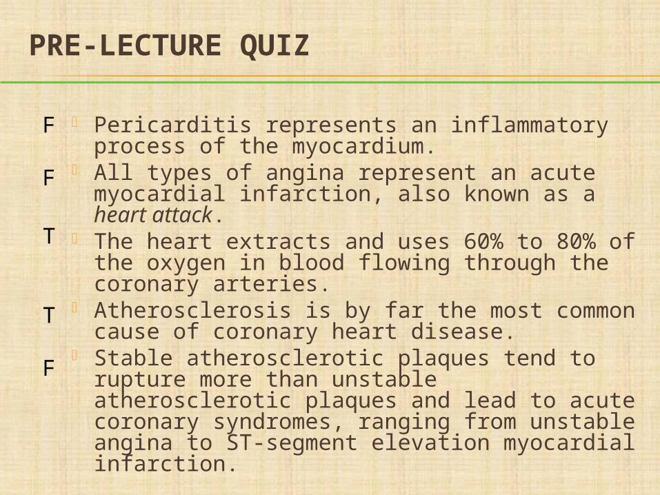
Pericarditis represents an inflammatory process of the myocardium.
All types of angina represent an acute myocardial infarction, also known as a heart attack.
The heart extracts and uses 60% to 80% of the oxygen in blood flowing through the coronary arteries.
Atherosclerosis is by far the most common cause of coronary heart disease.
Stable atherosclerotic plaques tend to rupture more than unstable atherosclerotic plaques and lead to acute coronary syndromes, ranging from unstable angina to ST-segment elevation myocardial infarction.
F
F
T
T
F
PRE-LECTURE QUIZ
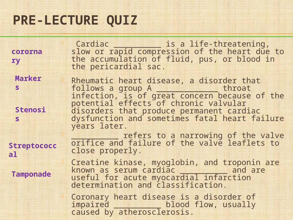
Cardiac __________ is a life-threatening, slow or rapid compression of the heart due to the accumulation of fluid, pus, or blood in the pericardial sac.
Rheumatic heart disease, a disorder that follows a group A _____________ throat infection, is of great concern because of the potential effects of chronic valvular disorders that produce permanent cardiac dysfunction and sometimes fatal heart failure years later.
__________ refers to a narrowing of the valve orifice and failure of the valve leaflets to close properly.
Creatine kinase, myoglobin, and troponin are known as serum cardiac __________ and are useful for acute myocardial infarction determination and classification.
Coronary heart disease is a disorder of impaired __________ blood flow, usually caused by atherosclerosis.
Tamponade
Stenosis
Streptococcal
Markers
corornary
DISORDERS THAT AFFECT THE WHOLE HEART Pericardial disorders
Coronary heart disease
Myocardial diseases
These disorders can cause symptoms of both right- and left-sided heart failure
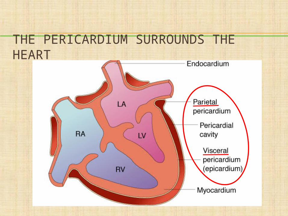
THE PERICARDIUM SURROUNDS THE HEART
PERICARDITIS
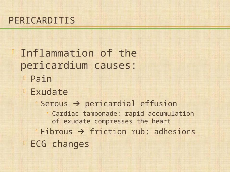
Inflammation of the pericardium causes: Pain Exudate
º Serous pericardial effusion Cardiac tamponade: rapid accumulation of
exudate compresses the heart
º Fibrous friction rub; adhesions ECG changes
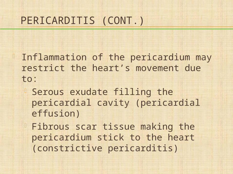
PERICARDITIS (CONT.)
Inflammation of the pericardium may restrict the heart’s movement due to: Serous exudate filling the pericardial
cavity (pericardial effusion) Fibrous scar tissue making the
pericardium stick to the heart (constrictive pericarditis)
CONSEQUENCES OF
PERICARDIAL EFFUSION
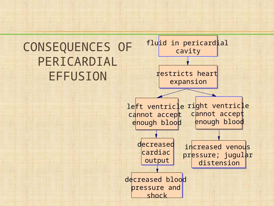
fluid in pericardial cavity
restricts heart expansion
left ventricle cannot accept enough blood
decreased cardiac output
decreased blood pressure and
shock
right ventricle cannot accept enough blood
increased venous pressure; jugular
distension
QUESTION
What is the immediate treatment for severe cardiac tamponade?
a. Oxygenb. Cardiac drugsc. Surgeryd. Pericardiocentesis (removal of fluid
from the sac with a needle)
PERICARDITIS
Inflammation of the pericardium causes: Pain Exudate
º Serous pericardial effusion Cardiac tamponade: rapid accumulation of
exudate compresses the heart
º Fibrous friction rub; adhesions ECG changes
PERICARDITIS (CONT.)
Inflammation of the pericardium may restrict the heart’s movement due to: Serous exudate filling the pericardial cavity
(pericardial effusion) Fibrous scar tissue making the pericardium
stick to the heart (constrictive pericarditis)
CONSEQUENCES OF
PERICARDIAL EFFUSION
fluid in pericardial cavity
restricts heart expansion
left ventricle cannot accept enough blood
decreased cardiac output
decreased blood pressure and
shock
right ventricle cannot accept enough blood
increased venous pressure; jugular
distension
QUESTION
What is the immediate treatment for severe cardiac tamponade?
a. Oxygenb. Cardiac drugsc. Surgeryd. Pericardiocentesis(removal of fluid from the sac with a
needle)
ANSWER
d. Pericardiocentesis (removal of fluid from the sac with a needle)
Rationale: In severe cardiac tamponade, there is so much fluid in the pericardial sac compressing the heart that its function declines rapidly. The fluid must be removed quickly by inserting a needle into the pericardial space and aspirating the accumulated fluid.
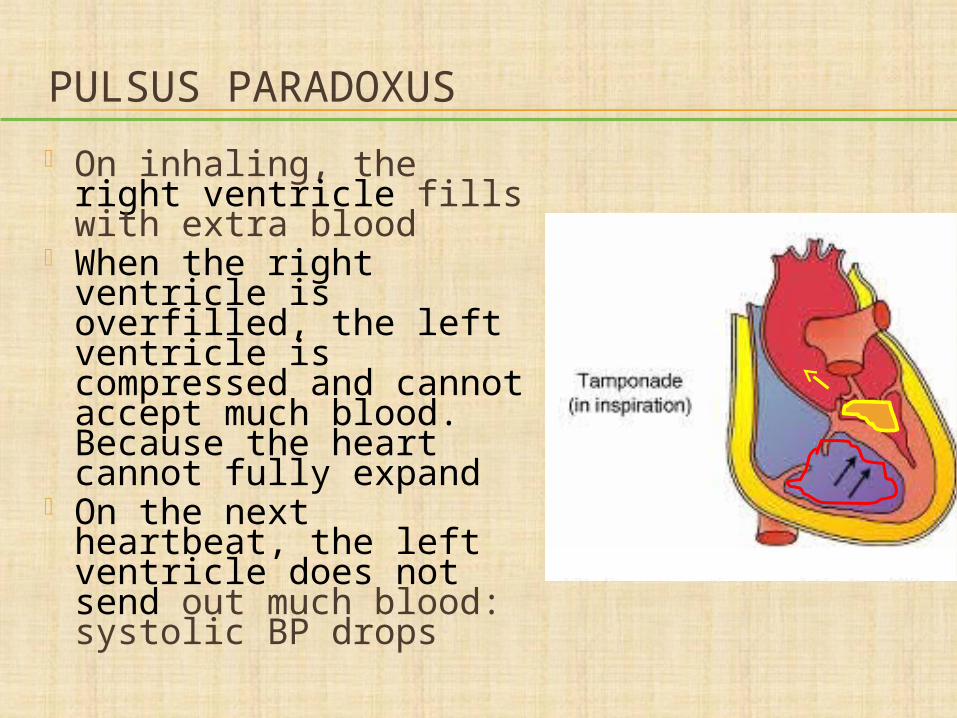
PULSUS PARADOXUS
On inhaling, the right ventricle fills with extra blood
When the right ventricle is overfilled, the left ventricle is compressed and cannot accept much blood. Because the heart cannot fully expand
On the next heartbeat, the left ventricle does not send out much blood: systolic BP drops
CORONARY HEART DISEASE
Atherosclerosis blocks coronary arteries Ischemia may cause:
Angina Heart attack Cardiac arrhythmias Conduction deficits Heart failure Sudden death

Plaque/Thrombus Formation
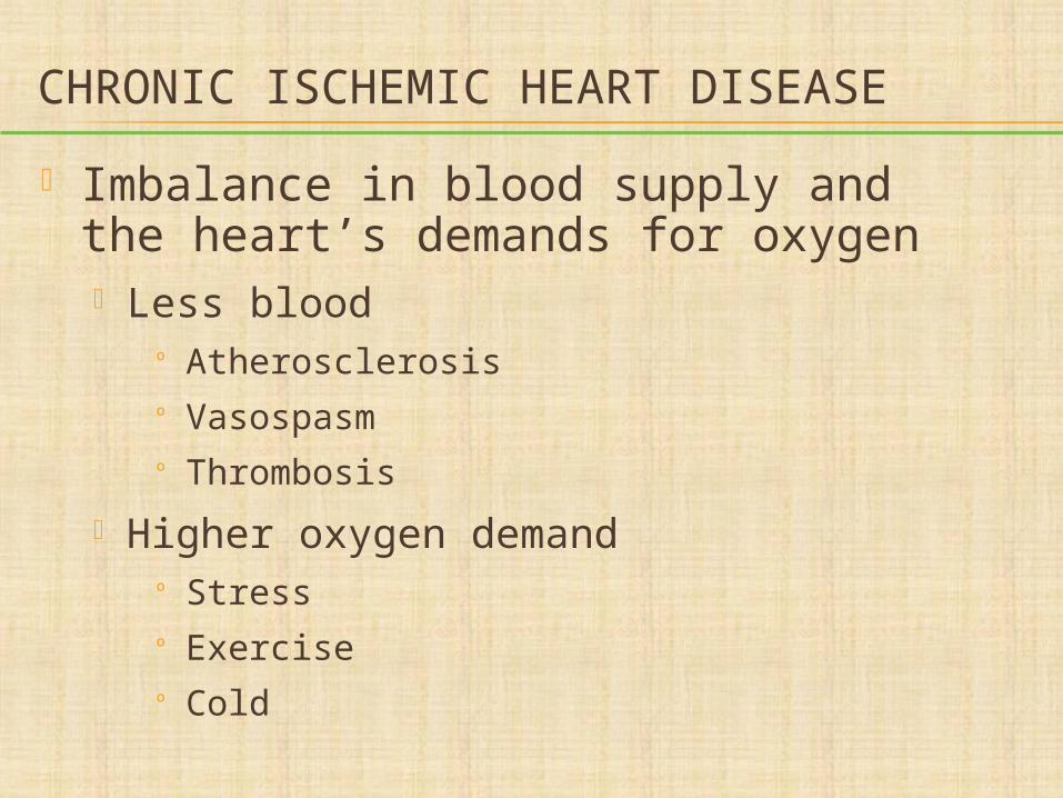
CHRONIC ISCHEMIC HEART DISEASE
Imbalance in blood supply and the heart’s demands for oxygen Less blood
º Atherosclerosisº Vasospasmº Thrombosis
Higher oxygen demandº Stressº Exerciseº Cold
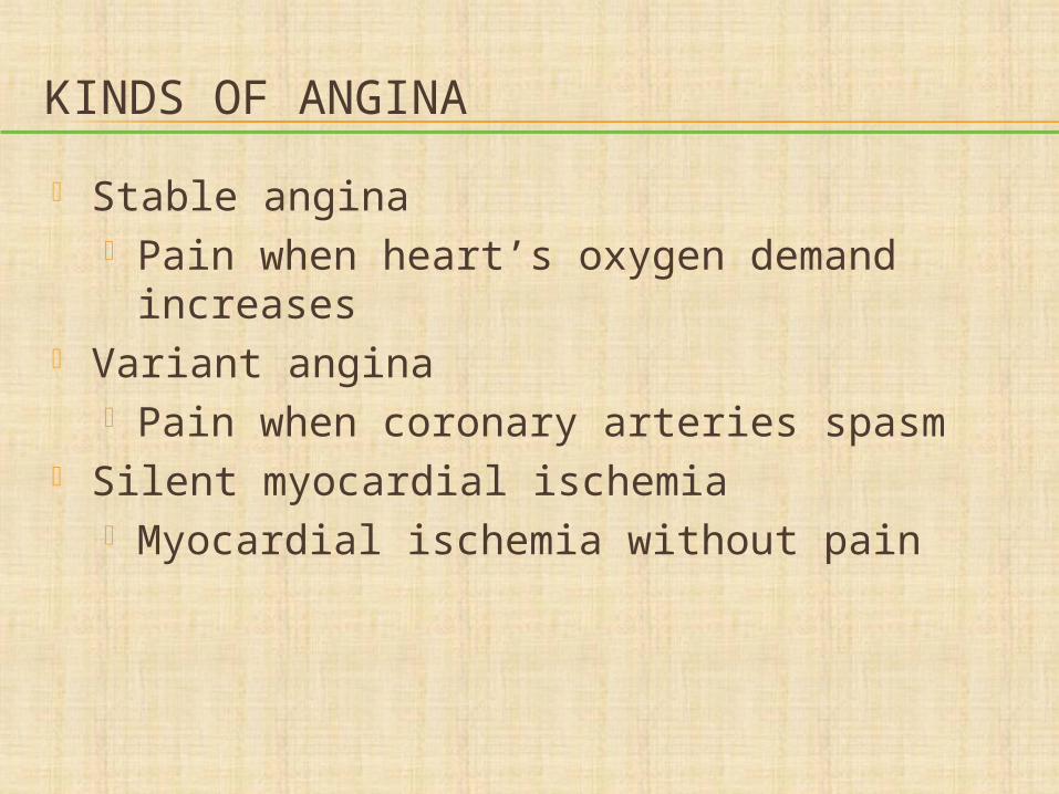
KINDS OF ANGINA
Stable angina Pain when heart’s oxygen demand
increases Variant angina
Pain when coronary arteries spasm Silent myocardial ischemia
Myocardial ischemia without pain
ACUTE CORONARY SYNDROMES
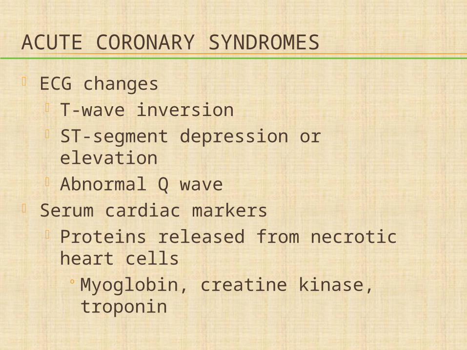
ECG changes T-wave inversion ST-segment depression or elevation Abnormal Q wave
Serum cardiac markers Proteins released from necrotic heart
cellsº Myoglobin, creatine kinase,
troponin
QUESTION
Tell whether the following statement is true or false.
Chronic ischemic heart disease is more likely to result in stable angina than acute coronary syndromes.
ANSWER
TrueRationale: Ischemic heart disease is
characterized by stable angina, which is associated with plaques that are fixed obstructions. Unstable angina is characterized by plaques with platelets stuck to them (these are likely to form a thrombus)—they cause a range of acute coronary syndromes.
ACUTE MYOCARDIAL INFARCTION
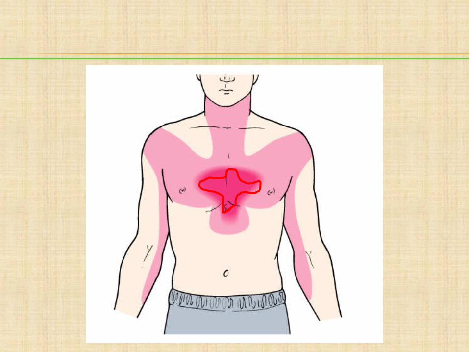
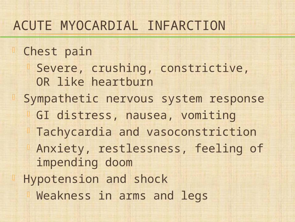
Chest pain Severe, crushing, constrictive, OR like
heartburn Sympathetic nervous system response
GI distress, nausea, vomiting Tachycardia and vasoconstriction Anxiety, restlessness, feeling of
impending doom Hypotension and shock
Weakness in arms and legs
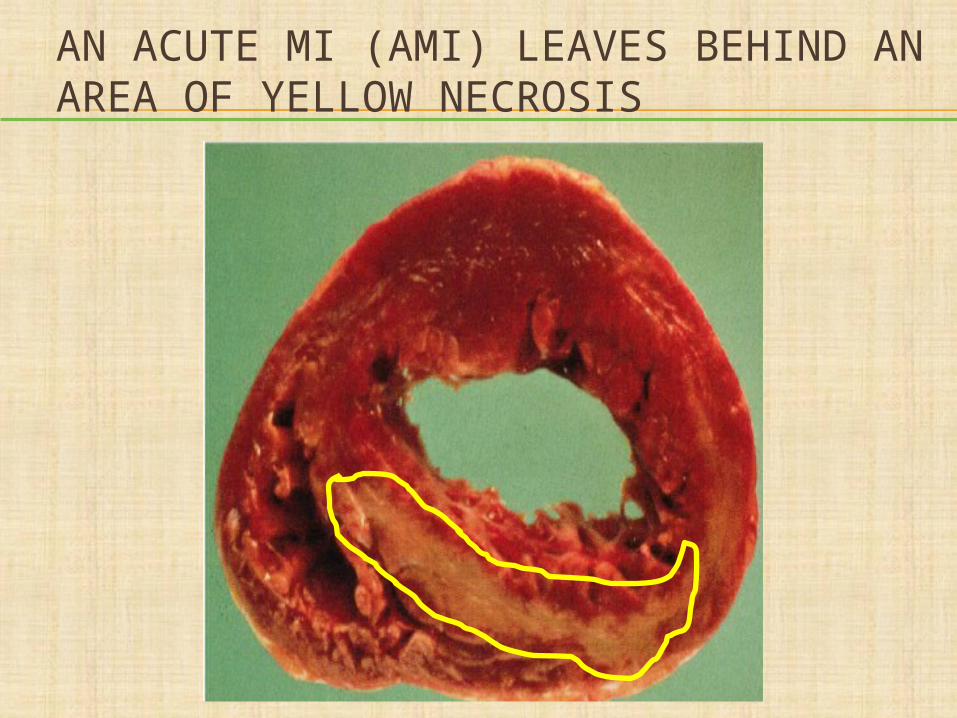
AN ACUTE MI (AMI) LEAVES BEHIND AN AREA OF YELLOW NECROSIS
COMPLICATIONS OF AMI
Heart failure
Cardiogenic shock
Pericarditis
Thromboemboli
Rupture of the heart
Ventricular aneurysms
MALFUNCTIONING HEART MUSCLE Malfunctioning heart muscle can cause
heart failure if: Ventricles are unusually thick so there is
not a normal amount of room for blood inside them (hypertrophic cardiomyopathy)
Ventricles are too stiff to stretch (restrictive cardiomyopathy)
Ventricles are too weak to pump out the blood that is in them (MI, myocarditis, dilated cardiomyopathy)
MYOCARDIAL DISORDERS
Myocarditis Cardiomyopathies
Dilated cardiomyopathies Hypertrophic cardiomyopathies Restrictive cardiomyopathies Peripartum cardiomyopathy
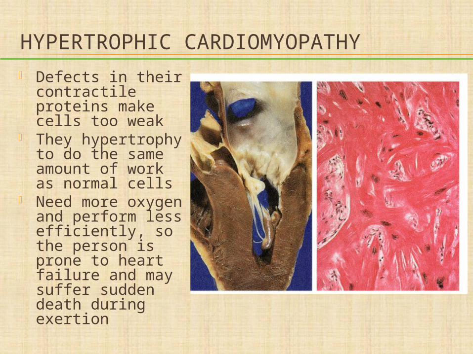
HYPERTROPHIC CARDIOMYOPATHY Defects in their
contractile proteins make cells too weak
They hypertrophy to do the same amount of work as normal cells
Need more oxygen and perform less efficiently, so the person is prone to heart failure and may suffer sudden death during exertion
QUESTION
Which type of cardiomyopathy is characterized by weakened ventricles?
a. Dilated cardiomyopathyb. Hypertrophic cardiomyopathyc. Restrictive cardiomyopathyd. Peripartum cardiomyopathy
ANSWER
a. Dilated cardiomyopathyRationale: In dilated cardiomyopathy,
the ventricles are too weak to pump blood, resulting in a diminished cardiac output (CO). The other types listed are caused by thick ventricles, stiff ventricles, or LV dysfunction in late pregnancy or postpartum, respectively.
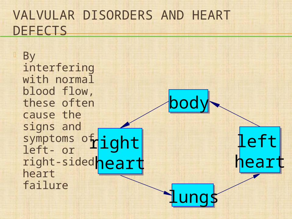
VALVULAR DISORDERS AND HEART DEFECTS
By interfering with normal blood flow, these often cause the signs and symptoms of left- or right-sided heart failure
right heart
lungs
left heart
body
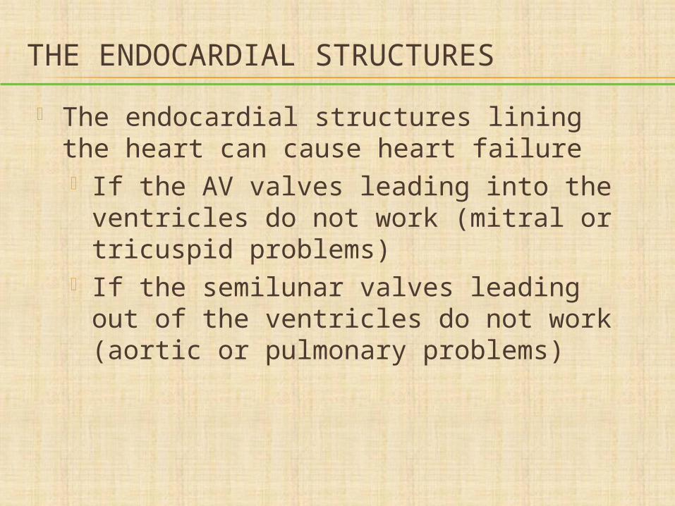
THE ENDOCARDIAL STRUCTURES
The endocardial structures lining the heart can cause heart failure If the AV valves leading into the
ventricles do not work (mitral or tricuspid problems)
If the semilunar valves leading out of the ventricles do not work (aortic or pulmonary problems)
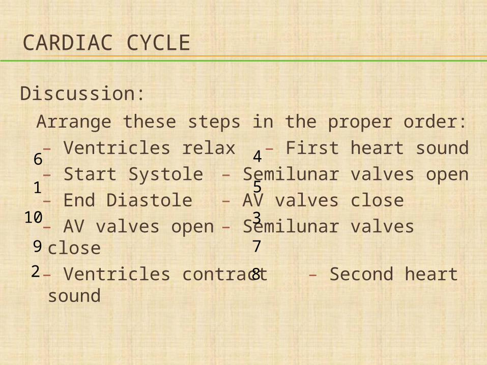
CARDIAC CYCLE
Discussion:Arrange these steps in the proper order:
– Ventricles relax – First heart sound– Start Systole – Semilunar valves open– End Diastole – AV valves close– AV valves open – Semilunar valves close– Ventricles contract – Second heart sound
1
2
3
4
5
6
7
8
9
10
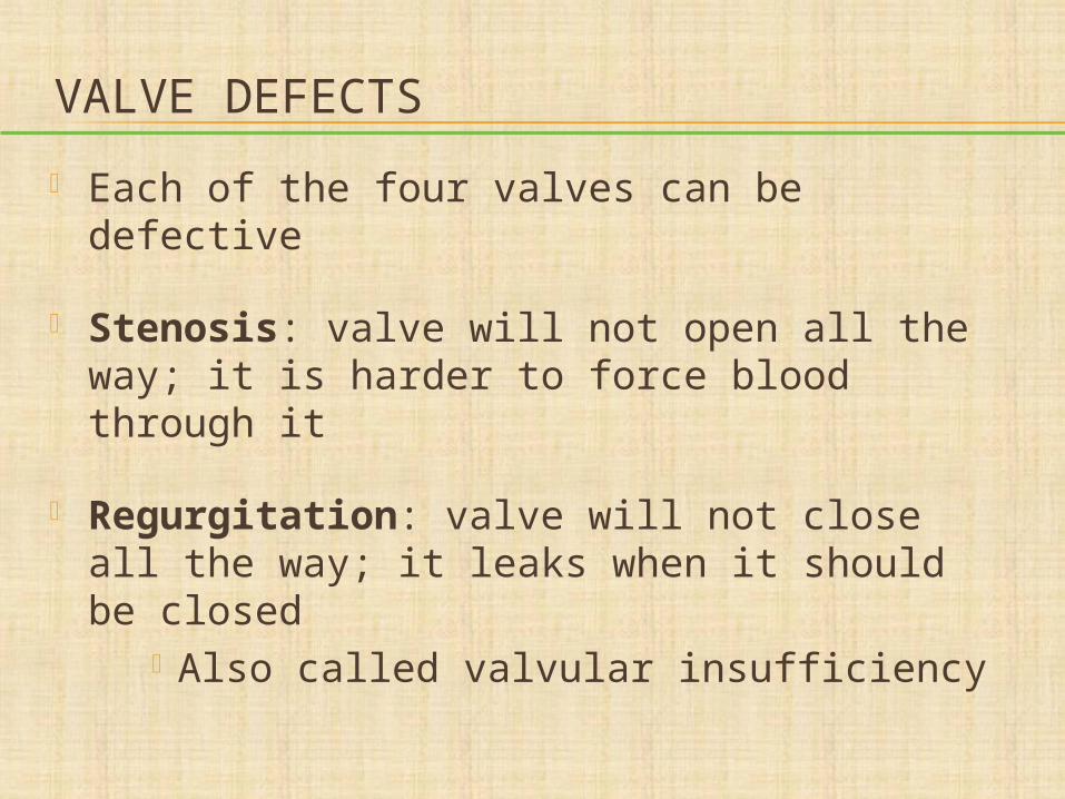
VALVE DEFECTS
Each of the four valves can be defective
Stenosis: valve will not open all the way; it is harder to force blood through it
Regurgitation: valve will not close all the way; it leaks when it should be closed
Also called valvular insufficiency
QUESTION
Tell whether the following statement is true or false.
Mitral valve regurgitation results in a diminished stroke volume.
ANSWER
TrueRationale: If the mitral valve does not
close as it should, a portion of the stroke volume (amount of blood ejected by the ventricle/beat) leaks back into the left atrium, decreasing the amount of blood that is ejected during that beat (SV).
DISCUSSION
Defects in which valves might cause: Severe dependent edema? Paroxysmal nocturnal dyspnea? Congested liver? Distended jugular veins? Productive cough with frothy sputum?
IDENTIFYING DEFECTIVE VALVES
The blood going through the valve makes a noise
These are called heart murmurs You can identify them by:
Where they are—which valve are they near?
How they sound—high- or low-pitched? When they happen—systole or diastole?
WHEN WILL YOU HEAR MURMURS?
If a valve is stenotic, you will hear a murmur of blood shooting through the narrow opening when the valve is open
If a valve is regurgitant, you will hear a murmur of blood leaking back through when the valve should be closed
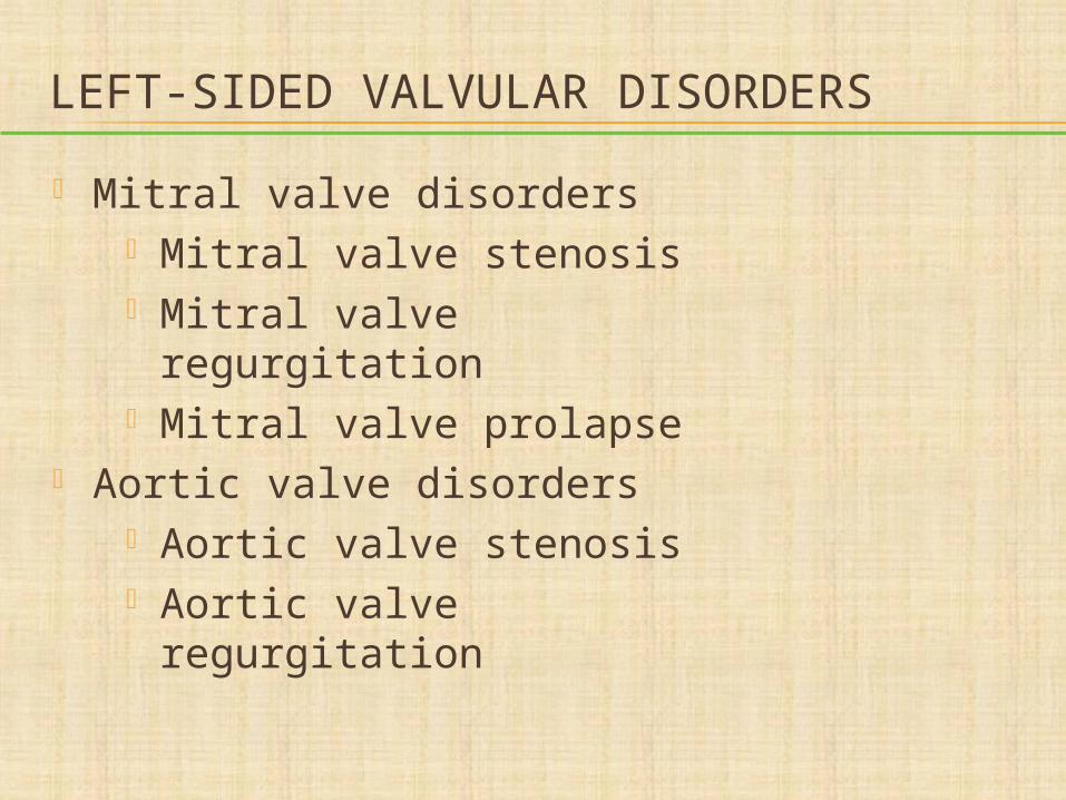
LEFT-SIDED VALVULAR DISORDERS
Mitral valve disorders Mitral valve stenosis Mitral valve regurgitation Mitral valve prolapse
Aortic valve disorders Aortic valve stenosis Aortic valve regurgitation
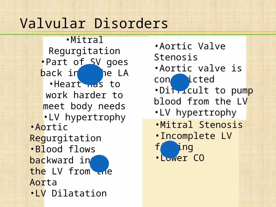
•Aortic Valve Stenosis•Aortic valve is constricted•Difficult to pump blood from the LV•LV hypertrophy
•Mitral Regurgitation•Part of SV goes back
into the LA•Heart has to work
harder to meet body needs
•LV hypertrophy•Mitral Stenosis•Incomplete LV filling•Lower CO
•Aortic Regurgitation•Blood flows backward into the LV from the Aorta•LV Dilatation
Valvular Disorders
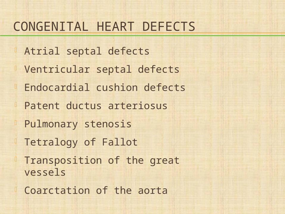
CONGENITAL HEART DEFECTS
Atrial septal defects
Ventricular septal defects
Endocardial cushion defects
Patent ductus arteriosus
Pulmonary stenosis
Tetralogy of Fallot
Transposition of the great vessels
Coarctation of the aorta
SHUNTS
A shunt is an opening or connection that lets blood move from one side of the circulation to the other
Most shunts occur in the heart and move blood either from the left to the right or from the right to the left
Because the left side is stronger, blood is usually pushed from the left to the right side
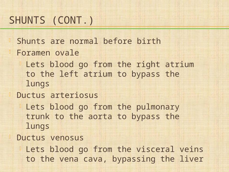
SHUNTS (CONT.)
Shunts are normal before birth Foramen ovale
Lets blood go from the right atrium to the left atrium to bypass the lungs
Ductus arteriosus Lets blood go from the pulmonary trunk
to the aorta to bypass the lungs Ductus venosus
Lets blood go from the visceral veins to the vena cava, bypassing the liver
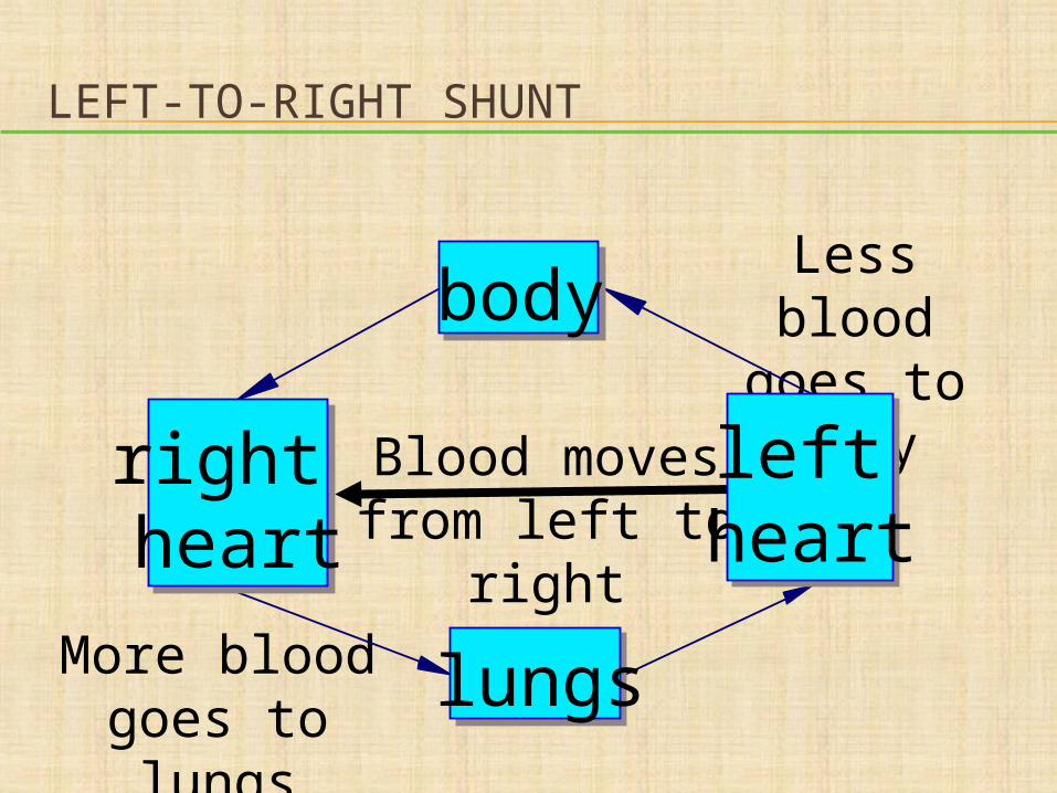
LEFT-TO-RIGHT SHUNT
Less blood goes to body
More blood goes to lungs
Blood moves from left to right
right heart
lungs
left heart
body
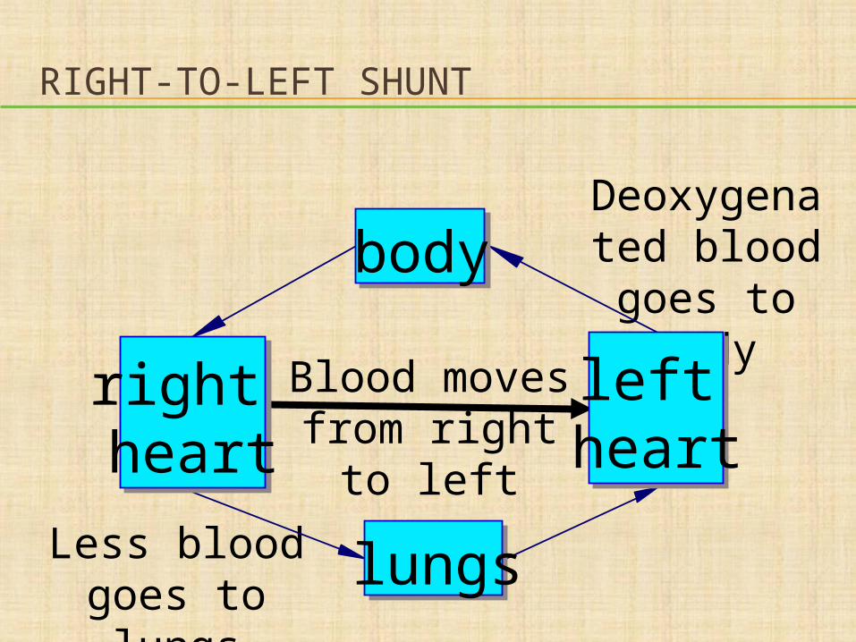
RIGHT-TO-LEFT SHUNT
Deoxygenated blood goes to
body
Less blood goes to lungs
Blood moves from right to left
right heart
lungs
left heart
body
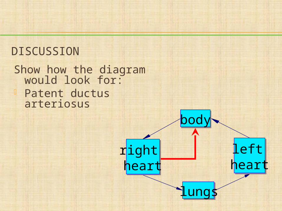
DISCUSSION
Show how the diagram would look for:
Patent ductus arteriosus
right heart
lungs
left heart
body
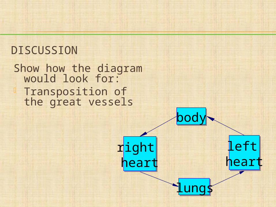
DISCUSSION
Show how the diagram would look for:
Transposition of the great vessels
lungs
body
left heart
right heart
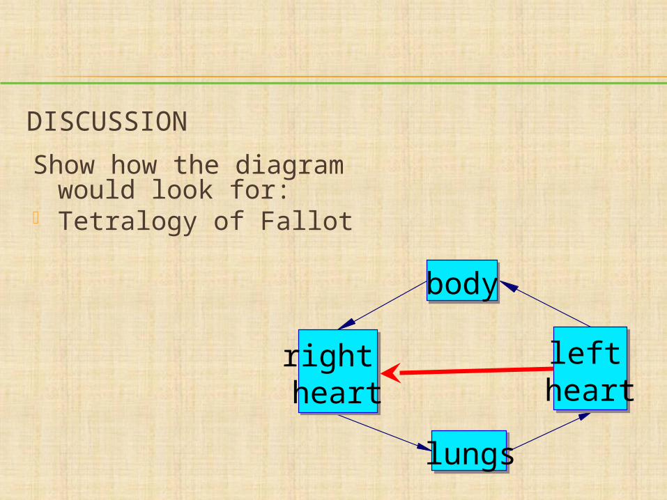
DISCUSSION
Show how the diagram would look for:
Tetralogy of Fallot
right heart
lungs
left heart
body