chapter 18 chromosomes. 18.1 introduction 18.2 condensing viral genomes into their coats 18.3 the...
TRANSCRIPT

Chapter Chapter 1818
ChromosomesChromosomes

18.1 Introduction18.2 Condensing viral genomes into their coats18.3 The bacterial genome is a nucleoid18.4 The bacterial genome is supercoiled18.5 Loops, domains, and scaffolds in eukaryotic DNA18.6 Specific sequence attach DNA to the matrix18.7 The contrast between interphase chromatin and mitotic chromosomes18.8 Lampbrush chromosomes are extended18.9 Polytene chromosomes form bands18.10 Polytene chromosomes expand at sites of gene expression18.11 The eukaryotic chromosome is a segregation device18.12 Centromeres have short DNA sequences in S. cerevisiae18.13 Centromeres may contain repetitious DNA18.14 Telomeres are simple repeats that seal the ends of chromosomes18.15 Telomeres are synthesized by a ribonucleoprotein enzyme

Chromatin describes the condition of the chromosomal material during the interphase (between mitoses) of the cell cycle.Chromosome is a discrete unit of the genome carrying many genes. Each chromosome consists of a very long molecule of duplex DNA and an approximately equal mass of proteins. It is visible as a morphological entity only during cell division.Nucleoid is the compact body that contains the genome in a bacterium.Packing ratio is the ratio of the length of DNA to the unit length of the fiber containing it.
18.1 Introduction

Figure 18.1 The length of nucleic acid is much greater than the dimensions of the surrounding compartment.
18.1 Introduction

Capsid is the external protein coat of a virus particle.
18.2 Condensing viral genomes into their coats
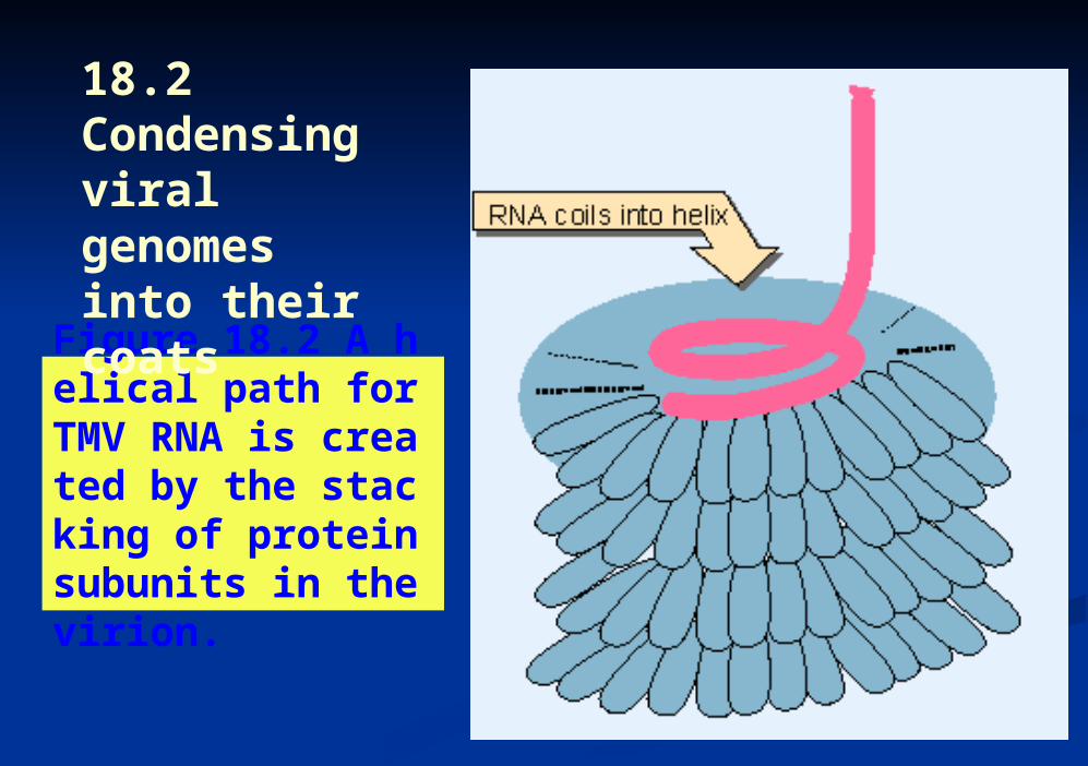
Figure 18.2 A helical path for TMV RNA is created by the stacking of protein subunits in the virion.
18.2 Condensing viral genomes into their coats

Figure 18.3 Maturation of phage lambda passes through several stages. The empty head changes shape and expands when it becomes filled with DNA. The electron micrographs show the particles at the start and end of the maturation pathway. Photographs kindly provided by A. F. Howatson.
18.2 Condensing viral genomes into their coats
Domain of a chromosome may refer either to a discrete structural entity defined as a region within which supercoiling is independent of other domains; or to an extensive region including an expressed gene that has heightened sensitivity to degradation by the enzyme DNAase I.Nucleoid is the compact body that contains the genome in a bacterium.
18.3 The bacterial genome is a nucleoid with many supercoiled loops

Figure 18.4 A thin section shows the bacterial nucleoid as a compact mass in the center of the cell. Photograph kindly provided by Jack Griffith.
18.3 The bacterial genome is a nucleoid with many supercoiled loops

Figure 18.5 The nucleoid spills out of a lysed E. coli cell in the form of loops of a fiber. Photograph kindly provided by Jack Griffith.
18.3 The bacterial genome is a nucleoid with many supercoiled loops

Figure 18.6 The bacterial genome consists of a large number of loops of duplex DNA (in the form of a fiber), each secured at the base to form an independent structural domain.
18.3 The bacterial genome is a nucleoid with many supercoiled loops
MAR (matrix attachment site; also known as SAR for scaffold attachment site) is a region of DNA that attaches to the nuclear matrix.Nuclear matrix is a network of fibers surrounding and penetrating the nucleus.Scaffold of a chromosome is a proteinaceous structure in the shape of a sister chromatid pair, generated when chromosomes are depleted of histones.
18.4 Loops, domains, and scaffolds in eukaryotic DNA
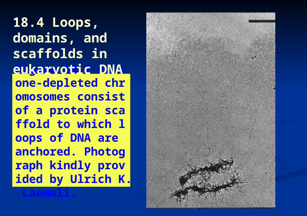
Figure 18.7 Histone-depleted chromosomes consist of a protein scaffold to which loops of DNA are anchored. Photograph kindly provided by Ulrich K. Laemmli.
18.4 Loops, domains, and scaffolds in eukaryotic DNA
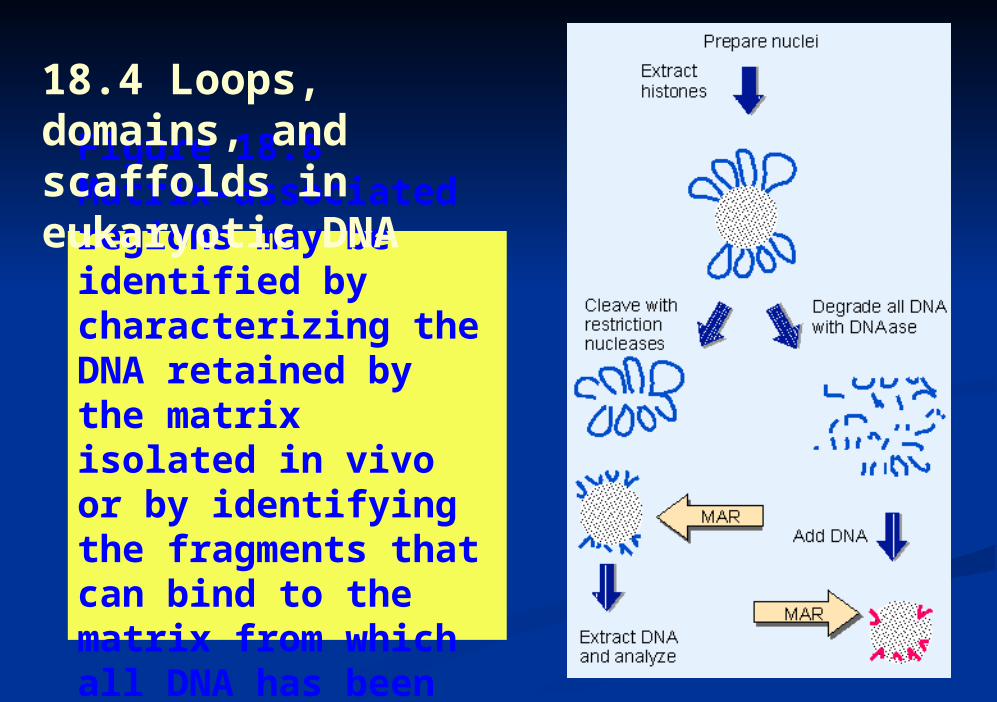
Figure 18.8 Matrix-associated regions may be identified by characterizing the DNA retained by the matrix isolated in vivo or by identifying the fragments that can bind to the matrix from which all DNA has been removed.
18.4 Loops, domains, and scaffolds in eukaryotic DNA

Chromocenter is an aggregate of heterochromatin from different chromosomes.Euchromatin comprises all of the genome in the interphase nucleus except for the heterochromatin.Heterochromatin describes regions of the genome that are permanently in a highly condensed condition are not transcribed, and are late-replicating. May be constitutive or facultative.
18.5 The contrast between interphase chromatin and mitotic chromosomes

Figure 18.9 The sister chromatids of a mitotic pair each consist of a fiber (~30 nm in diameter) compactly folded into the chromosome. Photograph kindly provided by E. J. DuPraw.
18.5 The contrast between interphase chromatin and mitotic chromosomes

Figure 18.10 A thin section through a nucleus stained with Feulgen shows heterochromatin as compact regions clustered near the nucleolus and nuclear membrane. Photograph kindly provided by Edmund Puvion.
18.5 The contrast between interphase chromatin and mitotic chromosomes

Figure 18.11 G-banding generates a characteristic lateral series of bands in each member of the chromosome set. Photograph kindly provided by Lisa Shaffer.
18.5 The contrast between interphase chromatin and mitotic chromosomes

Figure 18.12 The human X chromosome can be divided into distinct regions by its banding pattern. The short arm is p and the long arm is q; each arm is divided into larger regions that are further subdivided. This map shows a low resolution structure; at higher resolution, some bands are further subdivided into smaller bands and interbands, e.g. p21 is divided into p21.1, p21.2, and p21.3.
18.5 The contrast between interphase chromatin and mitotic chromosomes
Chromomeres are densely staining granules visible in chromosomes under certain conditions, especially early in meiosis, when a chromosome may appear to consist of a series of chromomeres.Lampbrush chromosomes are the large meiotic chromosomes found in amphibian oocytes.
18.6 The extended state of lampbrush chromosomes

Figure 18.13 A lampbrush chromosome is a meiotic bivalent in which the two pairs of sister chromatids are held together at chiasmata (indicated by arrows). Photograph kindly provided by Joe Gall.
18.6 The extended state of lampbrush chromosomes

Figure 18.14 A lampbrush chromosome loop is surrounded by a matrix of ribonucleoprotein. Photograph kindly provided by Oscar Miller.
18.6 The extended state of lampbrush chromosomes

Bands of polytene chromosomes are visible as dense regions that contain the majority of DNACytological hybridization means the same as in situ hybridization.Interbands are the relatively dispersed regions of polytene chromosomes that lie between the bands.Polytene chromosomes are generated by successive replications of a chromosome set without separation of the replicas.
18.7 Transcription disrupts the structure of polytene chromosomes

Figure 18.15 The polytene chromosomes of D. melanogaster form an alternating series of bands and interbands. Photograph kindly provi
ded by Jose Bonner.
18.7 Transcription disrupts the structure of polytene chromosomes

Figure 18.16 Individual bands containing particular genes can be identified by in situ hybridization.
18.7 Transcription disrupts the structure of polytene chromosomes

Figure 18.17 A magnified view of bands 87A and 87C shows their hybridization in situ with labeled RNA extracted from heat-shocked cells. Photograph kindly provided by Jose Bonner.
18.7 Transcription disrupts the structure of polytene chromosomes

Figure 18.18 Chromosome IV of the insect C. tentans has three Balbiani rings in the salivary gland. Photograph kindly provided by Bertil Daneholt.
18.7 Transcription disrupts the structure of polytene chromosomes

Acentric fragment of a chromosome (generated by breakage) lacks a centromere and is lost at cell division.Centromere is a constricted region of a chromosome that includes the site of attachment (the kinetochore) to the mitotic or meiotic spindle.Kinetochore is the structural feature of the chromosome to which microtubules of the mitotic spindle attach. Its location determines the centromneric region.MTOC (microtubule organizing center) is a region from which microtubules emanate. The major MTOCs in a mitotic cell are the centrosomes.
18.8 The eukaryotic chromosome as a segregation device
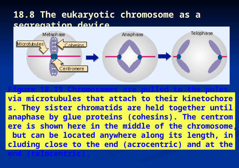
Figure 18.19 Chromosomes are pulled to the poles via microtubules that attach to their kinetochores. They sister chromatids are held together until anaphase by glue proteins (cohesins). The centromere is shown here in the middle of the chromosome, but can be located anywhere along its length, including close to the end (acrocentric) and at the end (telocentric).
18.8 The eukaryotic chromosome as a segregation device

Figure 18.9 The sister chromatids of a mitotic pair each consist of a fiber (~30 nm in diameter) compactly folded into the chromosome. Photograph kindly provided by E. J. DuPraw.
18.8 The eukaryotic chromosome as a segregation device

Figure 18.20 C-banding generates intense staining at the centromeres of all chromosomes. Photograph kindly provided by Lisa Shaffer.
18.8 The eukaryotic chromosome as a segregation device

Figure 18.21 The centromere is identified by a DNA sequence that binds specific proteins. These proteins do not themselves bind to microtubules, but establish the site a which the microtubule-binding proteins in turn bind.
18.8 The eukaryotic chromosome as a segregation device

Figure 18.22 Three conserved regions can be identified by the sequence homologies between yeast CEN elements.
18.8 The eukaryotic chromosome as a segregation device

Telomere is the natural end of a chromosome; the DNA sequence consists of a simple repeating unit with a protruding single-stranded end that may fold into a hairpin.
18.9 Telomeres are simple repeats that seal the ends of chromosomes

Figure 18.24 Telomerase positions itself by base pairing between the RNA template and the protruding single-stranded DNA primer. It adds G and T bases one at a time to the primer, as directed by the template. The cycle starts again when one repeating unit has been added.
18.10 Telomeres are synthesized by a ribonucleoprotein enzyme
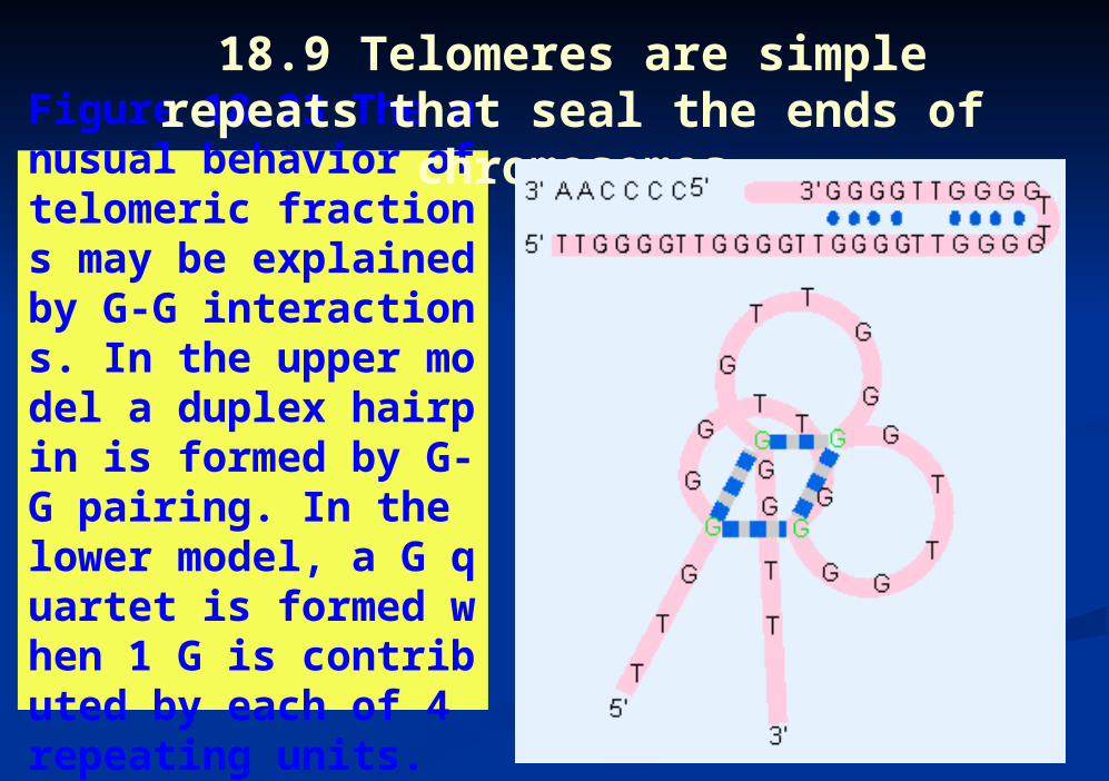
Figure 18.23 The unusual behavior of telomeric fractions may be explained by G-G interactions. In the upper model a duplex hairpin is formed by G-G pairing. In the lower model, a G quartet is formed when 1 G is contributed by each of 4 repeating units.
18.9 Telomeres are simple repeats that seal the ends of chromosomes

Telomerase is the ribonucleoprotein enzyme that creates repeating units of one strand at the telomere, by adding individual bases to the DNA 3 end, as directed by an RNA sequence in the RNA component of the enzyme.
18.10 Telomeres are synthesized by a ribonucleoprotein enzyme

Figure 18.25 A loop forms at the end of chromosomal DNA. Photograph kindly provided by Jack Griffith.
18.10 Telomeres are synthesized by a ribonucleoprotein enzyme

Figure 18.26 The 3’single-stranded end of the telomere (TTAGGG)n displaces the homologous repeats from duplex DNA to form a t-loop. The reaction is catalyzed by TRF2.
18.10 Telomeres are synthesized by a ribonucleoprotein enzyme

The genetic material of all organisms and viruses takes the form of tightly packaged nucleoprotein. Transcriptionally active sequences reside within the euchromatin that comprises the majority of interphase chromatin. Lampbrush chromosomes of amphibians and polytene chromosomes of insects have unusually extended structures, with packing ratios <100. The centromeric region contains the kinetochore, which is responsible for attaching a chromosome to the mitotic spindle. Telomeres make the ends of chromosomes stable.
18.11 Summary