chapter 11: anesthesia - fraser hale - anesthesia.pdf · 2019-01-04 · chapter 11 anesthesia. 74...
TRANSCRIPT

Chapter 11 Anesthesia.
74
Chapter 11: AnesthesiaLet me remind you before we go any further, I am not an anesthesiologist. I anesthetize almost every patient that comes to see me and many of them could be consider dubious anesthetic candidates. However, for information on anesthetic and fluid management, the reader is referred to current texts on veterinary anesthesia. I do have some thought to share, none-the-less.
I want to stress an oft-used caveat. There are no safe anesthetics, only safe anesthetists. What this means is that no agent or drug is inherently safe. They must all be used properly and with respect.
I have been using isoflurane exclusively for maintenance for over fourteen years. In my days as a mobile service, if I traveled to a clinic that was not using isoflurane, I would bring my own vaporizer and hook it up to their machine. This decision was based on two factors. One is the relative safety of isoflurane to the patient compared to other inhaled agents of the day. The other factor has to do with my own safety. When I am doing a dental procedure, I am working within a foot of the patient's mouth for two or three hours at a time. The animal is always fitted with a cuffed endotracheal tube, but there is the possibility of leakage of anesthetic gases. As well, not every clinic has an efficient scavenger unit in use. Inhaling stray isoflurane vapor is much less dangerous to my health than inhaling stray methoxyflurane or halothane vapor.
One danger with isoflurane is complacency. We have all been told how safe it is, but I was informed that the College of Veterinarians of Ontario Complaints Committee had been hearing more cases involving anesthetic death with isoflurane than with other inhaled agents (this goes back many years ago when many clinics were converting to isoflurane). The explanation proposed is that practitioners had been "jumping on the band wagon" and using isoflurane without being sufficiently familiar with its properties.
Some of the properties that make isoflurane so attractive are that animals can be induced rapidly, the plane of anesthesia can be adjusted rapidly and the animals recover rapidly. However, all this rapidity makes it potentially more dangerous. If your patient is too light and you have your technician turn the vaporizer up to 4 % and then he/she leaves the room to get you some suture material, by the time you think about it again, your patient could be dead.
A few years ago, I converted to propafol for virtually all inductions and I love it, but again, if I do not use it properly, there will be trouble.
What's the answer? If you are converting to a new agent from any other agent, read up on the new one first. Become familiar with its physical and pharmacological properties. Use a precision, out-of-circle vaporizer that has been properly calibrated for the new agent. Monitor your anesthetics carefully, especially for the first few months until you become familiar with how animals respond to the new agent. As always, proper pre-operative assessment of your patients is essential as is intra-operative life support (IV fluids, heat, IPPV).
In time, isoflurane and propofol will be passé as they replaced the next generations of anesthetic agents. The above comments will apply regardless of the agent in questions.
When I have students visiting my practice I always like to ask them “What is the dose of induction agent?” They then usually want to know which agent and I tell them it does not matter. The answer is the same regardless of the size of the animal, the degree of sedation or the induction agent used. The answer is “To Effect”.
The “No Anesthetic” Dental Cleaning It is a fact that no matter how careful we are, anesthetic deaths happen. They happen in human medicine where the amount of technology available to prevent them far exceeds the budgets of the average general practice. I have offered some information which might help keep the mortality rate down but there can never be a guarantee.
Owner concerns about anesthetic coupled with our inability to provide a guarantee of safety has prompted some clinics to offer an option known as the Standing Dental, No Anesthetic Dental or Twilight Dental . In this case, the pet is given mild sedation or less and has the calculus chipped off the crowns of the teeth.
This is a practice that must be stopped. I would actually rather see an animal receive no “professional” dental care than have it be the victim of a Standing Dental. I used quotes around “professional” because, in my mind,

Chapter 11 Anesthesia.
75
Standing Dentals are very un-professional, approaching mal-practice.
A few of the problems with a Standing Dental are as follows:
♦ only removes gross calculus from easily assessable areas of the crowns leaving much plaque and calculus behind in inter-dental spaces, lingually, palatally, at the back of the mouth and, most seriously, subgingivally,
♦ can gouge or scratch the enamel, making it rougher which speeds the return of plaque and calculus,
♦ does not allow for polishing to remove plaque,
♦ can cause iatrogenic trauma to oral soft tissue if the animal moves,
♦ can cause pain making the animal head shy which makes instituting a home-care program very difficult,
♦ does not allow operator to do a thorough oral exam so subtle or hidden (subgingival) lesions go undetected and untreated,
♦ does not protect the airways so the animal may aspirate chunks of calculus,
♦ gives the owners a false sense of security thinking their pet has received professional dental care and so many problems go untreated and are allowed to progress.
Since there are no written standards regarding the dental prophylaxis in most jurisdictions, practices can make up their own protocols and
charges. This means that the range in the quality of care offered is vast. It also means that a client might be told three or four contradictory things by three or four different hospitals while phoning around for advice and estimates. This lack of consistency reflects badly on the profession in general and veterinary dentistry in particular.
Those of us who try to do a very complete job and have to charge for the time it takes, are made to look over-priced when compared to those of them who will do half a job at half the price. One problem is that many owners have no notion of what a proper dental prophylaxis involves. They compare apples with oranges and may end up with a lemon.
I am confident that my readers are doing a better than average job so I offer these comments not to convince you, but to help you convince your clients.
To help you avoid being compared to a low priced colleague, here is a suggestion. Do not refer to the job as a "dental". This non-descriptive term over-simplifies and does not indicate what your clients are getting for their money. Instead, call it a "complete oral examination and hygiene procedure" or a "complete dental prophylaxis". Then make sure your client knows what is involved. I will quote the American Veterinary Dental College which states that a dental prophylaxis is:
a procedure to remove all hard and soft substances from the tooth surfaces. The primary objective is always a thorough and complete removal of bacterial plaque, calculus and extrinsic stains. This is accomplished by a procedure known as scaling and polishing. If calculus is present below the gingival margin, subgingival scaling is performed. Dental record keeping, thorough charting and client education are also a necessary portion for the prevention of disease.
Any procedure that does not include all of these components cannot be considered a prophylaxis. Other components which should also be routinely incorporated into a prophy include sulcar lavage and radiology as indicated.
Once you explain to your clients what is involved, they will better understand the absolute need for a general anesthetic. They will also have a better understanding of why the procedure costs what it does. Now, when they are calling around for estimates, they can compare what you charge for a complete procedure to what others
Figure #11.1. There is an 8-mm deep periodontal pocket on the palatal aspect of this canine tooth. This major periodontal defect could not have been found let alone treated if the animal were not under a general anesthetic.

Chapter 11 Anesthesia.
76
charge for a complete procedure. If they choose to have a cheap, incomplete job done elsewhere, at least they will know they are getting what they pay for.
Local Anesthesia In Veterinary Dentistry One of the early veterinary dental textbooks was criticized for devoting considerable space to the administration of dental nerve blocks. Many readers felt this was a waste of paper as dental patients are under general anesthetic anyway and so have no need for nerve blocks. Wrong!
It has long been established that pain is far easier to control preemptively rather than reactively. That is, if you block a nerve prior to traumatizing the tissue ‘down-stream’, there is far less post-operative pain than if you do the surgery and then deal with the pain later. Also the plane of anesthesia required to keep a patient still depends on the level of stimulation the patient perceives. A light plane may be sufficient for minor scaling and polishing, but would not do for a surgical extraction.
In many dental practices, it is routine to use local anesthesia for invasive dental procedures to reduce the plane of general anesthesia required intra-operatively and the amount of pain post-operatively.
I have been using bupivicaine 0.5% with epinephrine (Marcaine E™) to block infra-orbital and mandibular nerves for any procedure likely to cause pain (extractions, root canal, periodontal flap surgery...), and I have been very pleased with the results. The blocks are easy to place, we can maintain patients at a much lighter plane of anesthesia and their recovery is very smooth and comfortable.
There are still a number of questions to answer, such as:
♦ what is the onset and duration in cats and dogs?
♦ in cats and small dogs, is a high volume/diluted bolus better than a low volume/high concentration bolus?
♦ when doing bilateral mandibular blocks in cats, is there the potential to cause laryngeal paralysis? (I have never seen evidence of this.)
♦ how much is enough?
♦ can you block the second maxillary molar with an infra-orbital block?
These questions, and others, will be answered in the coming months/years. For now, I want you to know that local anesthesia has found a home in veterinary dentistry. This is good news, especially for those clients who are nervous about general anesthetic. Every patient still needs a pre-anesthetic assessment and judicious use of anesthetic agents, but now we have one more tool to reduce the risk of anesthetic problems.
Properties and Characteristics of Local Anesthetics Used in Veterinary Dentistry Local anesthetic agents are weak organic bases and their penetration in tissue is impaired in acidic environments, as found in areas of infection or abscessation. Therefore, local nerve blocks are contraindicated in areas of infection and abscessation, as they will not work but may result in inoculation of infection into deeper tissues.
The more basic amide agents (lidocaine, mepivicaine, bupivicaine) have a faster onset of action and give more profound analgesia than the ester agents (procaine, propoxycaine, benzocaine). The amide agents also have longer duration of action due to protein binding and are less likely to result in allergic reaction than the ester agents (in humans at least). Amide agents are metabolized by the liver while ester agents are metabolized in the circulation.
All local anesthetic agents in use today cause vasodilation, which increases systemic absorption and decreases duration of effect. To counter this, a vasoconstrictor such as epinephrine is often added. This decreases rate of systemic absorption (reducing risk of systemic toxicity), increases duration of action and decreases local hemorrhage.
Vasoconstrictors may lead to serious side effects including tachycardia and arrhythmias (epinephrine and halothane are a bad combination for this). Bronchospasm may result due to allergic reaction to sulfites added to vasoconstrictor-containing solutions as antioxidants. Epinephrine can potentiate seizures if injected intra-vascularly.
Duration of effect has been well documented in humans, but is still a matter of conjecture and extrapolation in dogs and cats. The chart shows the duration of pulpal and oral soft-tissue analgesia in humans in minutes.

Chapter 11 Anesthesia.
77
Agent Pulp Soft tissue
Lidocaine with epinephrine
60-90 180-240
Mepivicaine with levonordefrin
45-75 120-240
Etidocaine with epinephrine
45-240 240-540
Bupivicaine with epinephrine
45-240 240-540
Duration of analgesia in dogs and cats with bupivicaine and epinephrine may be as much as 6 to 8 hours as in humans, but more research is needed.
Onset of action for bupivicaine is 3 to 5 minutes.
Landmarks For Local Dental Nerve Blocks In human, canine and feline patients, there are six basic dental nerve blocks, all of which block sensory branches of the trigeminal nerve.
The majority of blocks are placed around nerves as they enter or exit various bony foramina. Typically, a vein and artery lie alongside the nerve. As the bony canals and foramina are small and the vessels are very close to the nerve, there is potential for intravascular injection. Dental syringes (see page 80) typically have a thumb-ring on the end of the plunger to allow for one-handed aspiration during placement of the needle. The syringe should be aspirated in two planes prior to the start of the injection and a few times during the injection to ensure that the needle is extra-vascular.
If the needle is advanced through a narrow foramen (major palatine, for example), there is a risk of causing physical trauma to the nerve with the needle. Therefore, it is recommended to place the tip of the needle just at or barely into the foramen. Once the agent has been deposited, digital pressure for one minute can force the agent to flow deeper into the canal, providing anesthesia distal to the foramen.
Infraorbital Nerve Block The infraorbital canal starts at the rostral floor of the orbit and runs rostrally, dorsal to the fourth premolar and opens on to the surface of the maxilla rostral to the zygomatic arch and dorsal (apical) to the maxillary third premolar tooth at the infraorbital foramen. The canal contains the infraorbital nerve as well as the infraorbital artery and vein.
Placement of the infraorbital block is done intra-orally. The infraorbital foramen is palpated as a depression dorsal to the distal root of the third premolar and the needle inserted to the opening of the canal or just into the canal. Anesthesia will affect the ipsilateral premolar, canine and incisor teeth as well as the bone and soft tissue buccal to the teeth.
If direct pressure is placed over the foramen for one minute after placement of the dose, the agent will flow distally and block the caudal maxillary alveolar nerve to anesthetize the maxillary molars and the associated buccal bone and soft tissue.
Figure #11.2. View of a needle just entering the infraorbital foramen of a dog. This is the most spacious of the canals into which needles may be advanced.
Figure #11.3. The infraorbital canal in most dogs is longer than in cats, allowing the needle to be advanced further before it reaches the suborbital space.

Chapter 11 Anesthesia.
78
Figure #11.4. View of a needle just entering the infraorbital foramen of a cat.
Figure #11.5. The infraorbital canal in a cat is very short and so advancing the needle only a short distance with place the tip below the globe.
Maxillary or Second Division Block The landmark for this block in dogs and cats is the major palatine foramen. It is located on an imaginary line that runs half way between the dental arcade and the palatal mid line, at the level of the middle of the maxillary fourth premolar tooth. This small foramen cannot be palpated through thick palatal mucosa.
Figure #11.6. Needle placed at the major palatine foramen in a dog.
Figure #11.7. A needle placed at the major palatine foramen in a cat.
As this foramen is quite small, even a 30-gauge needle may be too large to safely fit within the lumen. Therefore, the needle should be advanced only until there is a loss of resistance at the opening of the foramen and not advanced further.
The maxillary block provides anesthesia for the entire hemimaxilla and associated soft tissue and so is indicated for procedures of the maxillary molars and the palate.
Mental Nerve Block The middle mental foramen is the largest of the three mental foramina and can often be palpated in medium to large dogs. In dogs, it is located apical to the first or second mandibular premolar at the junction of the ventral and middle thirds to the mandibular body. In cats, the middle mental foramen is located midway between the mandibular canine tooth and the third premolar tooth (cats have no first or second mandibular premolar teeth) at the dorsoventral midpoint of the mandible.
From an intraoral approach, the needle enters the mucosa rostral to the mandibular frenulum and is advanced distoventrally into the opening of the foramen. This block will anesthetize the buccal mucosa and lip rostral to the foramen to the midline. If the needle is advanced deeper through the foramen into the mandibular canal, the canine and incisors will also be anesthetized. However, due to the potential to cause trauma to the nerve and vessels, it is preferable to place the needle tip just inside the foramen and then apply digital pressure to force the agent deeper into the canal.

Chapter 11 Anesthesia.
79
Figure # 11.8. Anesthetic needle placed at the middle mental foramen of a dog.
Figure #11.9. Anesthetic needle placed at the middle mental foramen of a cat.
Inferior Alveolar Nerve Block The inferior alveolar nerve is located within the mandibular canal, ventral to the mandibular tooth roots. Therefore, to block this nerve, it must be accessed before it enters the body of the mandible. The site at which the nerve is accessible is just before it enters the inferior alveolar foramen on the medial side of the ramus of the mandible. This block may be achieved with either an intraoral or an extraoral approach.
The landmark for locating the inferior alveolar foramen is most obvious in dogs. Along the ventral border of the mandible, just cranial to the angular process, there is a concavity. The foramen is dorsal to the middle of this concavity, on the medial side, half way between the ventral and dorsal borders of the mandible.
The extraoral injection starts by soaking the skin over the ventral concavity with alcohol. The needle is passed through the skin to the ventral border of the mandible and then walked off the medial side and advanced about half the width of the mandible. At this stage, the tip of the needle should be adjacent to the opening of the foramen and the anesthetic agent can be deposited.
Cats lack the concavity in the ventral mandibular border. The foramen lies ventral to the midpoint of the zygomatic arch.
Figure #11.11. Anesthetic needle tip placed adjacent to the inferior alveolar foramen on the medial side of the mandible of a cat. There is no concavity in the ventral border of the mandible, as seen in the dog. This foramen can be found ventral to the midpoint of the zygomatic arch.
The intraoral approach to the inferior alveolar foramen is a bit more challenging. In the dog, the needle is directed from the side of the mouth opposite to the one to be blocked. The barrel of the syringe lies over the space between first and second mandibular molars and the needle is directed caudal or medial to the pterygomandibular raphe. The foramen can be palpated below the mucosa in larger patients.
In cats, the needle passes over the mandibular molar on its way to the spot lateral to the pterygomandibular raphe.
In dogs and cats, the needle is advanced until it contacts the bone at the foramen. After the agent is delivered, another dose can be deposited as the
Figure #11.10. Anesthetic needle tip at the inferior alveolar foramen of a dog, on the medial side of the mandible directly dorsal to the middle of the concavity in the ventral border.
Chapter 11 Anesthesia.
80
needle is withdrawn half way to block the lingual nerve. This will give anesthesia to all ipsilateral mandibular teeth and bone as well as all of the soft tissue lingual to the mandible, including half of the tongue.
Figure #11.12. Needle directed at the inferior alveolar foramen in a dog. The needle passes over the space between the mandibular first and second molars on the contralateral side.
Figure #11.13. Anesthetic needle passing over the mandibular molar of a cat on its was to the inferior alveolar foramen.
Infiltration Nerve Block Injection of anesthetic agent in the periapical region of a tooth will provide anesthesia for that tooth, providing the overlying bone is thin enough to allow diffusion of the agent. This would include maxillary teeth and the incisors. Infiltration can also be used to block areas of soft tissue such as the palate adjacent to maxillary teeth prior to extraction.
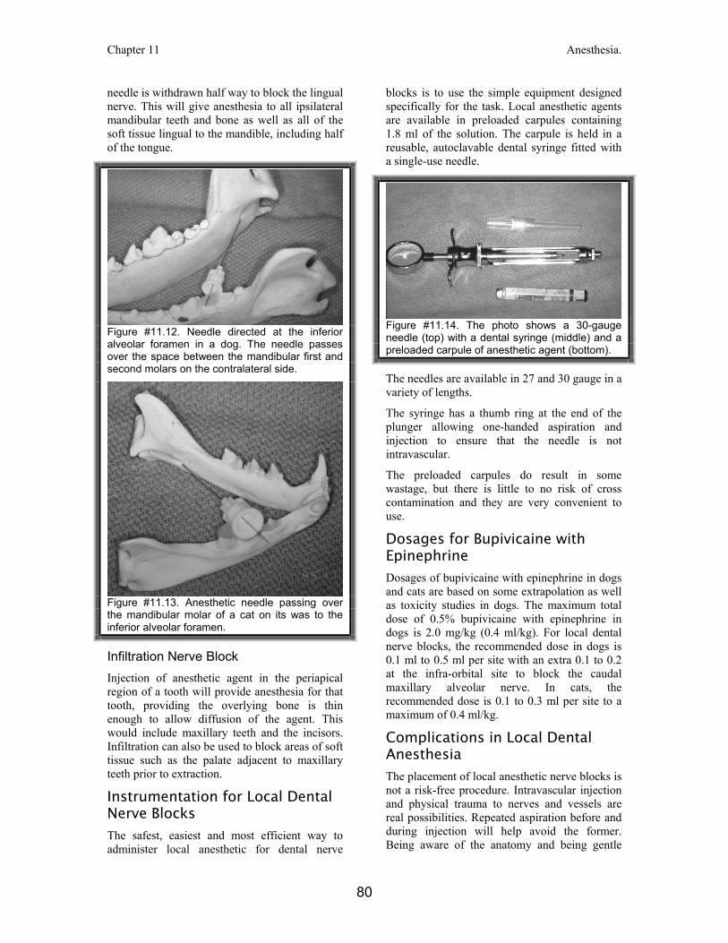
Instrumentation for Local Dental Nerve Blocks The safest, easiest and most efficient way to administer local anesthetic for dental nerve
blocks is to use the simple equipment designed specifically for the task. Local anesthetic agents are available in preloaded carpules containing 1.8 ml of the solution. The carpule is held in a reusable, autoclavable dental syringe fitted with a single-use needle.
Figure #11.14. The photo shows a 30-gauge needle (top) with a dental syringe (middle) and a preloaded carpule of anesthetic agent (bottom).
The needles are available in 27 and 30 gauge in a variety of lengths.
The syringe has a thumb ring at the end of the plunger allowing one-handed aspiration and injection to ensure that the needle is not intravascular.
The preloaded carpules do result in some wastage, but there is little to no risk of cross contamination and they are very convenient to use.
Dosages for Bupivicaine with Epinephrine Dosages of bupivicaine with epinephrine in dogs and cats are based on some extrapolation as well as toxicity studies in dogs. The maximum total dose of 0.5% bupivicaine with epinephrine in dogs is 2.0 mg/kg (0.4 ml/kg). For local dental nerve blocks, the recommended dose in dogs is 0.1 ml to 0.5 ml per site with an extra 0.1 to 0.2 at the infra-orbital site to block the caudal maxillary alveolar nerve. In cats, the recommended dose is 0.1 to 0.3 ml per site to a maximum of 0.4 ml/kg.
Complications in Local Dental Anesthesia The placement of local anesthetic nerve blocks is not a risk-free procedure. Intravascular injection and physical trauma to nerves and vessels are real possibilities. Repeated aspiration before and during injection will help avoid the former. Being aware of the anatomy and being gentle

Chapter 11 Anesthesia.
81
with your technique will help minimize the latter.
I have been using local dental nerve blocks for many years now and overall, have been very happy with the results. Some patients, however, seem distressed on recovery following local nerve blocks. I have assumed that they were reacting to the very strange feeling associated with a dental nerve block. It is also possible that there is hyperesthesia due to a traumatic placement. In either case, some dogs and cats will paw at the anesthetized area upon recovery. Commonly, this behavior stops once the animal has recovered to standing. However, staff should be aware of this potential problem and watch animals carefully during recovery to prevent self-mutilation.
Bupivicaine contains epinephrine as the vasoconstrictor. All vasoconstrictors are contra-indicated in patients with unstable cardiac disease, uncontrolled diabetes or hyperthyroidism, steroid-dependent asthma or pheochromocytoma.
Tricyclic antidepressants such as amytryptyline and beta-blocking agents such as propranolol can potentiate the effects of epinephrine. Halothane sensitizes the heart to epinephrine increasing the risk of arrythmias.
Conclusion Local dental nerve blocks are a great boon to the practice of veterinary dentistry. They are relatively quick and easy to administer and require very little investment in materials and equipment.
Pre-emptive pain management helps animals recover faster and go home happier (which makes the owners happier). With local anesthetic to block sensation, the animal can be maintained at a much lighter plane of general anesthesia which reduces the risk of the procedure.