chapter 1 introduction - gov.uk...chapter 1 first aid 7 dressings, bandages, slings and splints...
TRANSCRIPT

Firs
t ai
d
Introduction
Priorities
General principles of firstaid
General assessment ofthe situation
Dressings, bandages,slings and splints
First aid satchels andboxes
Severe bleeding
Unconscious casualty
Burns and scalds
Suffocation (asphyxia)
Strangulation
Choking
Epileptic fits
Shock
Bleeding
Wounds
Fractures
Dislocations
Head injuries
Chest injuries
Blast injuries
Transportation
CHAPTER 1
5
IntroductionWhen a ship is in port, or near to port where hospital andother expert medical attention are available, the first aidtreatment necessary aboard ship is similar to that practisedashore. At sea, in the absence of these facilities, trained ships’officers are required to give types of treatment beyond thataccepted as normal first aid.
The content of this chapter covers the knowledge of firstaid necessary for the safe and efficient immediate treatmentof casualties before they are transported to the ship’shospital or to a cabin for any necessary definitive treatmentof the type described in Chapter 4.
However, anyone aboard ship may find a casualty andevery seaman should know three basic life-saving actions tobe given immediately while waiting for trained help toarrive. These are:
■ to give artificial respiration by the mouth to nose/mouthmethod;
■ to place an unconscious casualty in the unconsciousposition;
■ to stop severe bleeding.
PrioritiesOn finding a casualty:
■ ensure your own safety;
if necessary, remove the casualty from danger or dangerfrom the casualty (but see the note below on enclosedspaces);
■ give immediate treatment to the casualty who is notbreathing and/or whose heart has stopped, is bleedingseverely or unconscious – others can be treated later;
■ send for help.
If there is more than one unconscious or bleeding casualty:
■ send for help;
■ treat the most serious injury first in the order of:
• not breathing and/or heart stopped;
• unconsciousness.
• serious bleeding;
If the casualty is in an ENCLOSED SPACE:
■ DO NOT enter the enclosed space unless you are a trainedmember of a rescue team acting under instructions;
■ send for help and inform the master.
It must be assumed that the atmosphere in the space ishostile. The rescue team MUST NOT enter unless wearingbreathing apparatus which must also be fitted to the casualtyas soon as possible. The casualty must be removed quickly tothe nearest safe adjacent area outside the enclosed spaceunless his injuries and the likely time of evacuation makessome treatment essential before movement.
WITHDRAWN PUBLICATION

6 THE SHIP CAPTAIN’S MEDICAL GUIDE
General principles of first aid on board shipThe general principles are:
■ make a rapid examination of the patient to assess responsiveness and the extent of theinjury;
■ check breathing, heart and look for serious bleeding;
• if breathing has stopped, give artificial respiration;
• if the heart has stopped, give heart compression and artificial respiration;
• arrest serious bleeding;
■ handle the patient as little and as gently as possible so as to:
• prevent further injuries; and
• prevent further shock;
■ see that the patient is put in the most comfortable position possible and loosen tightclothing so that he can breathe easily;
■ do not remove more clothing than is necessary and, when you do, remove it gently. With aninjured limb, get the sound limb out of the clothing first and then peel the clothes off theinjured limb, which should be supported by another person during the process. If cuttingclothes is indicated to expose the injured part, do so. In removing a boot or shoe remove thelace and, if necessary, cut the upper down towards the toecap; keep onlookers away.
■ always remember that shock can be a great danger to life and one of the main objects offirst aid is to prevent this;
■ you may have to improvise splints, bandages etc. (Figure 1.23);
■ do not give alcohol in any form;
■ do not move the patient until he is fit to be moved. Bleeding should be arrested, fracturesimmobilised and shock treated. See that the necessary personnel and equipment forsmooth and efficient transport are available;
■ never consider anyone to be dead until you and others agree that:
• breathing has stopped;
• no pulse is felt and no sounds are heard when the examiner’s ear is put to the chest;
• the eyes are glazed and pupils are dilated;
• there is a progressive cooling of the body.
(For a further description of the diagnosis of death Chapter 12).
General assessment of the situationOnce it has been established that there is no immediate threat to life there will be time to takestock of the situation. Reassurance and quick and effective attention to injuries andcompassionate treatment of the injured person will alleviate his condition. Remember:
■ a calm and systematic approach should be adopted;
■ give nothing by mouth;
■ protect the casualty from heat or cold, remembering that in the tropics open steel decks canbe very hot;
■ never underestimate and do not treat as minor injuries:
• unconsciousness
• suspected internal bleeding
• stab or puncture wounds
• wounds near joints (see fractures);
• possible fractures
• eye injuries
WITHDRAWN PUBLICATION
Chapter 1 FIRST AID 7
Dressings, bandages, slings and splintsStandard dressing
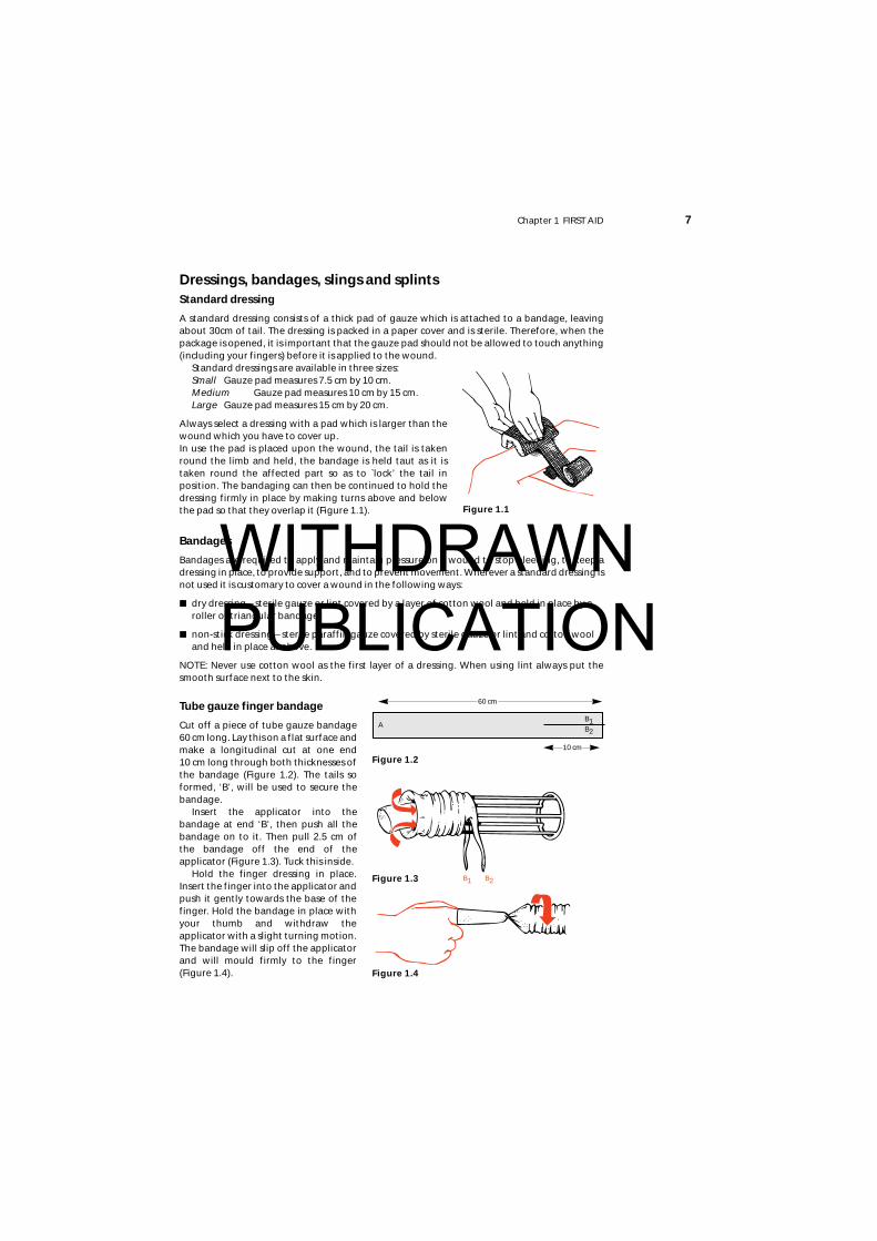
A standard dressing consists of a thick pad of gauze which is attached to a bandage, leavingabout 30cm of tail. The dressing is packed in a paper cover and is sterile. Therefore, when thepackage is opened, it is important that the gauze pad should not be allowed to touch anything(including your fingers) before it is applied to the wound.
Standard dressings are available in three sizes:Small Gauze pad measures 7.5 cm by 10 cm.Medium Gauze pad measures 10 cm by 15 cm.Large Gauze pad measures 15 cm by 20 cm.
Always select a dressing with a pad which is larger than thewound which you have to cover up.In use the pad is placed upon the wound, the tail is takenround the limb and held, the bandage is held taut as it istaken round the affected part so as to `lock’ the tail inposition. The bandaging can then be continued to hold thedressing firmly in place by making turns above and belowthe pad so that they overlap it (Figure 1.1).
Bandages
Bandages are required to apply and maintain pressure on a wound to stop bleeding, to keep adressing in place, to provide support, and to prevent movement. Wherever a standard dressing isnot used it is customary to cover a wound in the following ways:
■ dry dressing – sterile gauze or lint covered by a layer of cotton wool and held in place by aroller or triangular bandage;
■ non-stick dressing – sterile paraffin gauze covered by sterile gauze or lint and cotton wooland held in place as above.
NOTE: Never use cotton wool as the first layer of a dressing. When using lint always put thesmooth surface next to the skin.
Tube gauze finger bandage
Cut off a piece of tube gauze bandage60 cm long. Lay this on a flat surface andmake a longitudinal cut at one end 10 cm long through both thicknesses ofthe bandage (Figure 1.2). The tails soformed, ‘B’, will be used to secure thebandage.
Insert the applicator into thebandage at end ‘B’, then push all thebandage on to it. Then pull 2.5 cm ofthe bandage off the end of theapplicator (Figure 1.3). Tuck this inside.
Hold the finger dressing in place.Insert the finger into the applicator andpush it gently towards the base of thefinger. Hold the bandage in place withyour thumb and withdraw theapplicator with a slight turning motion.The bandage will slip off the applicatorand will mould firmly to the finger(Figure 1.4).
Figure 1.1
A
60 cm
10 cm
B1B2
Figure 1.3 B1 B2
Figure 1.4
Figure 1.2
WITHDRAWN PUBLICATION
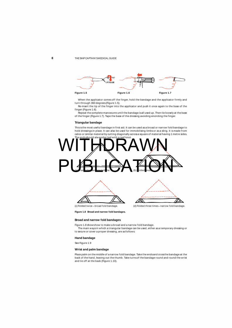
When the applicator comes off the finger, hold the bandage and the applicator firmly andturn through 360 degrees (Figure 1.5).
Re-insert the tip of the finger into the applicator and push it once again to the base of thefinger (Figure 1.6).
Repeat the complete manoeuvre until the bandage is all used up. Then tie loosely at the baseof the finger (Figure 1.7). Tape the base of the dressing avoiding encircling the finger.
Triangular bandage
This is the most useful bandage in first aid. It can be used as a broad or narrow fold bandage tohold dressings in place. It can also be used for immobilising limbs or as a sling. It is made fromcalico or similar material by cutting diagonally across a square of material having 1 metre sides.The ends should always be tied with a reef knot.
Broad and narrow fold bandages
Figure 1.8 shows how to make a broad and a narrow fold bandage.The main ways in which a triangular bandage can be used, either as a temporary dressing or
to secure or cover a proper dressing, are as follows:
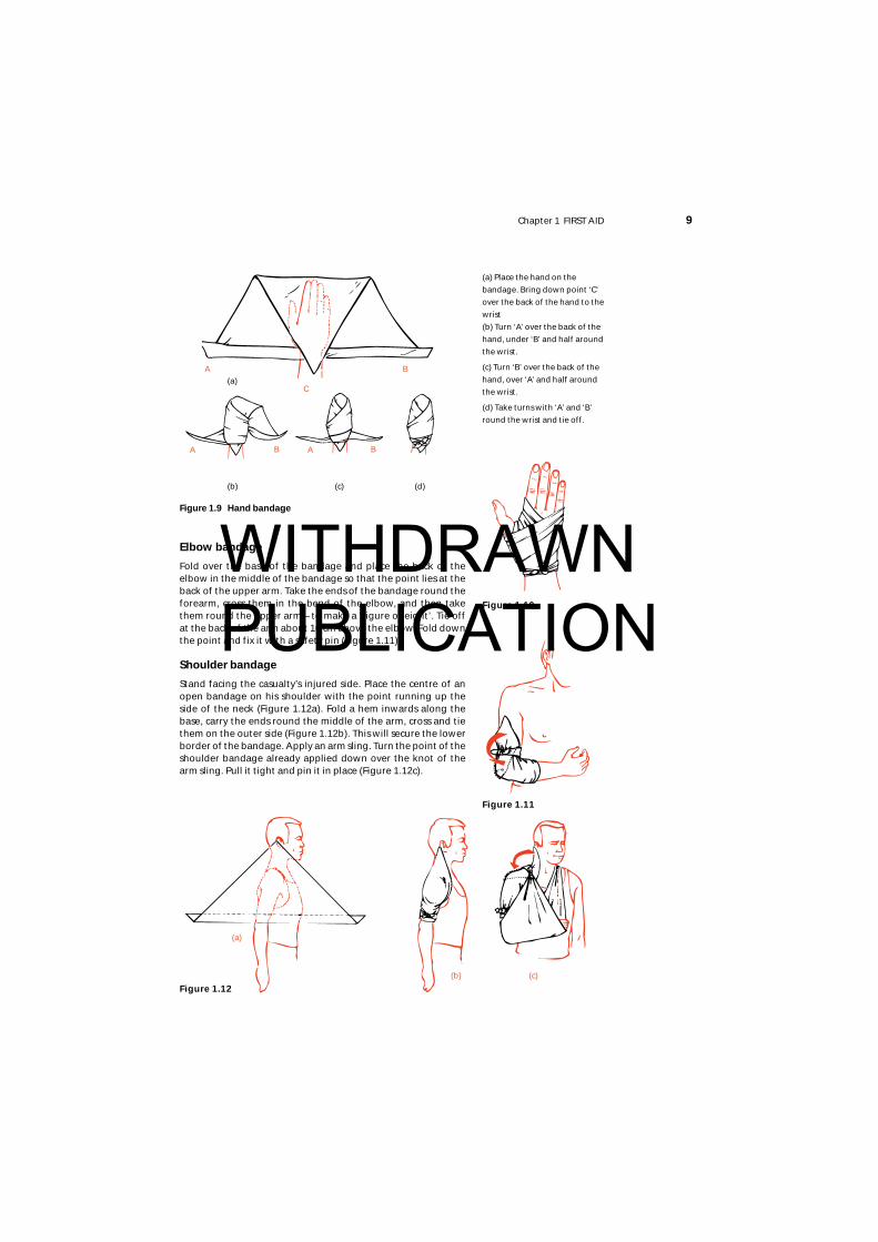
Hand bandage
See Figure 1.9
Wrist and palm bandage
Place palm on the middle of a narrow fold bandage. Take the ends and cross the bandage at theback of the hand, leaving out the thumb. Take turns of the bandage round and round the wristand tie off at the back (Figure 1.10).
8 THE SHIP CAPTAIN’S MEDICAL GUIDE
Figure 1.6Figure 1.5 Figure 1.7
Figure 1.8 Broad and narrow fold bandages.
(a) Triangular bandage laid flat. (b) Folded once.
(d) Folded three times – narrow fold bandage.(c) Folded twice – broad fold bandage.
WITHDRAWN PUBLICATION
Chapter 1 FIRST AID 9
Elbow bandage
Fold over the base of the bandage and place the back of theelbow in the middle of the bandage so that the point lies at theback of the upper arm. Take the ends of the bandage round theforearm, cross them in the bend of the elbow, and then takethem round the upper arm – to make a ‘figure of eight’. Tie offat the back of the arm about 10 cm above the elbow. Fold downthe point and fix it with a safety pin (Figure 1.11).
Shoulder bandage
Stand facing the casualty’s injured side. Place the centre of anopen bandage on his shoulder with the point running up theside of the neck (Figure 1.12a). Fold a hem inwards along thebase, carry the ends round the middle of the arm, cross and tiethem on the outer side (Figure 1.12b). This will secure the lowerborder of the bandage. Apply an arm sling. Turn the point of theshoulder bandage already applied down over the knot of thearm sling. Pull it tight and pin it in place (Figure 1.12c).
Figure 1.10
Figure 1.11
(a) Place the hand on the
bandage. Bring down point ‘C’
over the back of the hand to the
wrist
(b) Turn ‘A’ over the back of the
hand, under ‘B’ and half around
the wrist.
(c) Turn ‘B’ over the back of the
hand, over ‘A’ and half around
the wrist.
(d) Take turns with ‘A’ and ‘B’
round the wrist and tie off.
(a)
(b) (c) (d)
Figure 1.12
A
C
B
A B A B
(a)
(b) (c)
Figure 1.9 Hand bandage
WITHDRAWN PUBLICATION
10 THE SHIP CAPTAIN’S MEDICAL GUIDE
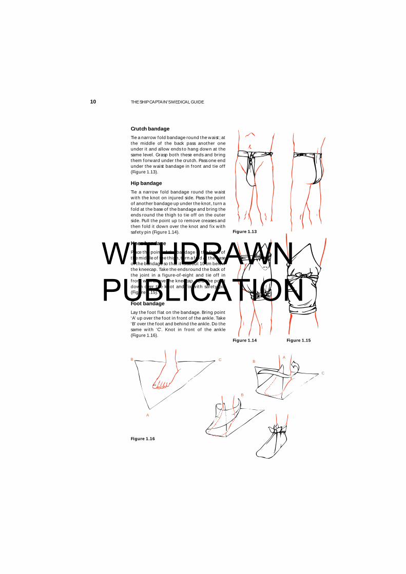
Crutch bandage
Tie a narrow fold bandage round the waist; atthe middle of the back pass another oneunder it and allow ends to hang down at thesame level. Grasp both these ends and bringthem forward under the crutch. Pass one endunder the waist bandage in front and tie off(Figure 1.13).
Hip bandage
Tie a narrow fold bandage round the waistwith the knot on injured side. Pass the pointof another bandage up under the knot, turn afold at the base of the bandage and bring theends round the thigh to tie off on the outerside. Pull the point up to remove creases andthen fold it down over the knot and fix withsafety pin (Figure 1.14).
Knee bandage
Place the point of the bandage in the front ofthe middle of the thigh, turn a fold at the baseof the bandage so that it is about 10 cm belowthe kneecap. Take the ends round the back ofthe joint in a figure-of-eight and tie off infront well above the kneecap. Fold the pointdown over the knot and fix with safety pin(Figure 1.15).
Foot bandage
Lay the foot flat on the bandage. Bring point‘A’ up over the foot in front of the ankle. Take‘B’ over the foot and behind the ankle. Do thesame with ‘C’. Knot in front of the ankle(Figure 1.16).
Figure 1.13
Figure 1.14
Figure 1.16
Figure 1.15
A
CB
B
AB
C
WITHDRAWN PUBLICATION
Chapter 1 FIRST AID 11
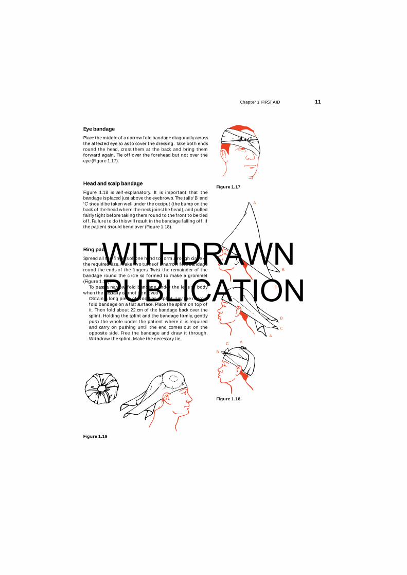
Eye bandage
Place the middle of a narrow fold bandage diagonally acrossthe affected eye so as to cover the dressing. Take both endsround the head, cross them at the back and bring themforward again. Tie off over the forehead but not over theeye (Figure 1.17).
Head and scalp bandage
Figure 1.18 is self-explanatory. It is important that thebandage is placed just above the eyebrows. The tails ‘B’ and‘C’ should be taken well under the occiput (the bump on theback of the head where the neck joins the head), and pulledfairly tight before taking them round to the front to be tiedoff. Failure to do this will result in the bandage falling off, ifthe patient should bend over (Figure 1.18).
Ring pad
Spread all the fingers of one hand to form a rough circle ofthe required size. Make two turns of a narrow fold bandageround the ends of the fingers. Twist the remainder of thebandage round the circle so formed to make a grommet(Figure 1.19).
To pass a narrow-fold bandage under the legs or bodywhen the casualty cannot be moved –
Obtain a long piece of wood or a splint. Lay the narrowfold bandage on a flat surface. Place the splint on top ofit. Then fold about 22 cm of the bandage back over thesplint. Holding the splint and the bandage firmly, gentlypush the whole under the patient where it is requiredand carry on pushing until the end comes out on theopposite side. Free the bandage and draw it through.Withdraw the splint. Make the necessary tie.
Figure 1.19
Figure 1.18
Figure 1.17
A
C
B
A
C
B
AC
B
WITHDRAWN PUBLICATION
Slings
Slings are usually made from triangular bandages, or they can be improvised. The main ways inwhich to make a sling are as follows:
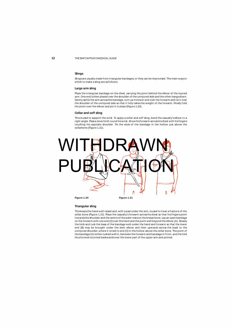
Large arm sling
Place the triangular bandage on the chest, carrying the point behind the elbow of the injuredarm. One end is then placed over the shoulder of the uninjured side and the other hangs down.Gently settle the arm across the bandage, turn up its lower end over the forearm and tie it overthe shoulder of the uninjured side so that it fully takes the weight of the forearm. Finally foldthe point over the elbow and pin it in place (Figure 1.20).
Collar and cuff sling
This is used to support the wrist. To apply a collar and cuff sling, bend the casualty’s elbow to aright angle. Pass a clove hitch round his wrist. Move his forearm across his chest with his fingerstouching his opposite shoulder. Tie the ends of the bandage in the hollow just above thecollarbone (Figure 1.21).
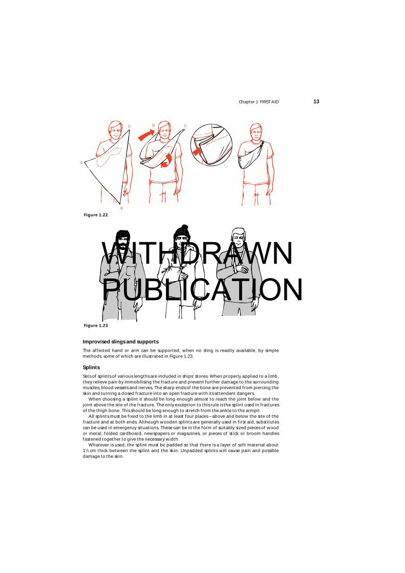
Triangular sling
This keeps the hand well raised and, with a pad under the arm, is used to treat a fracture of thecollar bone (Figure 1.22). Place the casualty’s forearm across his chest so that his fingers pointtowards the shoulder and the centre of the palm rests on the breast bone. Lay an open bandageon the forearm with one end (C) over the hand and the point well beyond the elbow (A). Steadythe limb and tuck the base of the bandage well under the hand and forearm so that the lowerend (B) may be brought under the bent elbow and then upwards across the back to theuninjured shoulder, where it is tied to end (C) in the hollow above the collar bone. The point ofthe bandage (A) is then tucked well in, between the forearm and bandage in front, and the foldthus formed is turned backwards over the lower part of the upper arm and pinned.
12 THE SHIP CAPTAIN’S MEDICAL GUIDE
Figure 1.20 Figure 1.21
WITHDRAWN PUBLICATION
Chapter 1 FIRST AID 13
Improvised slings and supports
The affected hand or arm can be supported, when no sling is readily available, by simplemethods, some of which are illustrated in Figure 1.23.
Splints
Sets of splints of various lengths are included in ships’ stores. When properly applied to a limb,they relieve pain by immobilising the fracture and prevent further damage to the surroundingmuscles, blood vessels and nerves. The sharp ends of the bone are prevented from piercing theskin and turning a closed fracture into an open fracture with its attendant dangers.
When choosing a splint it should be long enough almost to reach the joint below and thejoint above the site of the fracture. The only exception to this rule is the splint used in fracturesof the thigh bone. This should be long enough to stretch from the ankle to the armpit.
All splints must be fixed to the limb in at least four places – above and below the site of thefracture and at both ends. Although wooden splints are generally used in first aid, substitutescan be used in emergency situations. These can be in the form of suitably sized pieces of woodor metal, folded cardboard, newspapers or magazines, or pieces of stick or broom handlesfastened together to give the necessary width.
Whatever is used, the splint must be padded so that there is a layer of soft material about 11/2 cm thick between the splint and the skin. Unpadded splints will cause pain and possibledamage to the skin.
Figure 1.23
Figure 1.22
A
C
B
CB
WITHDRAWN PUBLICATION
14 THE SHIP CAPTAIN’S MEDICAL GUIDE
Inflatable splints are a useful method for temporarily immobilising limb fractures but areunsuitable for fractures which are more than a short distance above the knee or elbow as theycannot provide sufficient immobilisation in these places. The splint is applied to the limb andinflated by mouth. Other methods of inflation can make the splint too tight and thus slowdown or stop the circulation. Inflatable splints can be applied over wound dressings.
The splints are made of clear plastic and any bleeding from a wound can easily be seen.Needless to say, all sharp objects and sharp edges must be kept well clear of the plastic to avoida puncture.
Inflatable splints may be used to transport a patient about the ship or during moving tohospital. They should not be left in place for more than a few hours. Other means ofimmobilising the fracture should be used after that period.
Remember that the sound leg is a very good splint to which an injured leg can be securedpending more elaborate measures, and, similarly, the arms can be immobilised against thetrunk. If the patient is to be moved by Neil Robertson stretcher, no additional splints may benecessary during first aid.
First aid satchels or boxesThese should contain at least the items required by MSN 1726 for the ‘first aid kit’. One shouldbe kept close to the ship’s medical store for swift transfer to the site of an accident. If you havemore than one, the other(s) should be placed away from the medical store so that if the store isdestroyed by fire you have an easily reached first aid kit. These kits should be checkedfrequently and re-stocked as required.
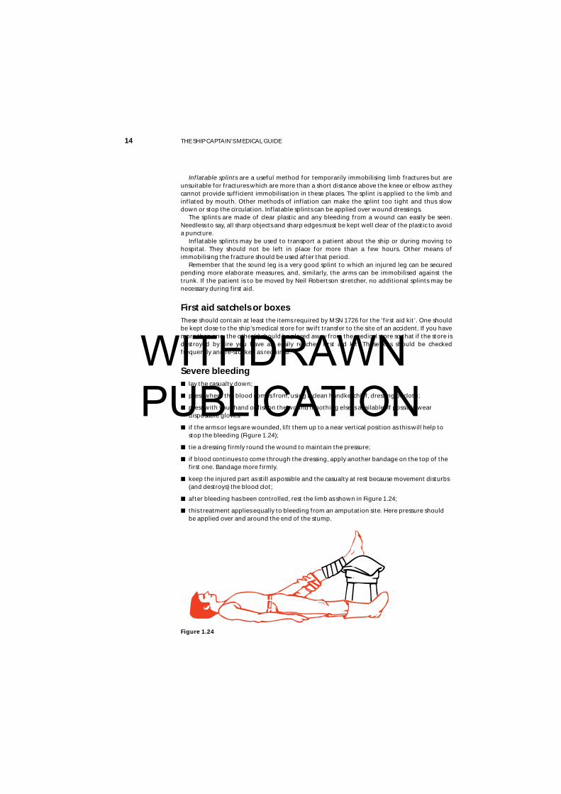
Severe bleeding■ lay the casualty down;
■ press where the blood comes from, using a clean handkerchief, dressing or cloth;
■ press with your hand or fist on the wound if nothing else is available. If possible weardisposable gloves.
■ if the arms or legs are wounded, lift them up to a near vertical position as this will help tostop the bleeding (Figure 1.24);
■ tie a dressing firmly round the wound to maintain the pressure;
■ if blood continues to come through the dressing, apply another bandage on the top of thefirst one. Bandage more firmly.
■ keep the injured part as still as possible and the casualty at rest because movement disturbs(and destroys) the blood clot;
■ after bleeding has been controlled, rest the limb as shown in Figure 1.24;
■ this treatment applies equally to bleeding from an amputation site. Here pressure shouldbe applied over and around the end of the stump.
Figure 1.24
WITHDRAWN PUBLICATION
Chapter 1 FIRST AID 15
Unconscious casualtyThe immediate threat to life may be:
■ breathing obstructed by the tongue falling back andblocking the throat;
■ stopped heart.
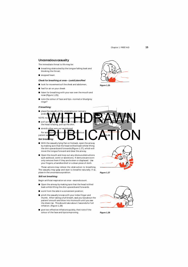
Check for breathing at once – Look/Listen/Feel
■ look for movements of the chest and abdomen;
■ feel for air on your cheek
■ listen for breathing with your ear over the mouth andnose (Figure 1.25);
■ note the colour of face and lips – normal or blue/greytinge?
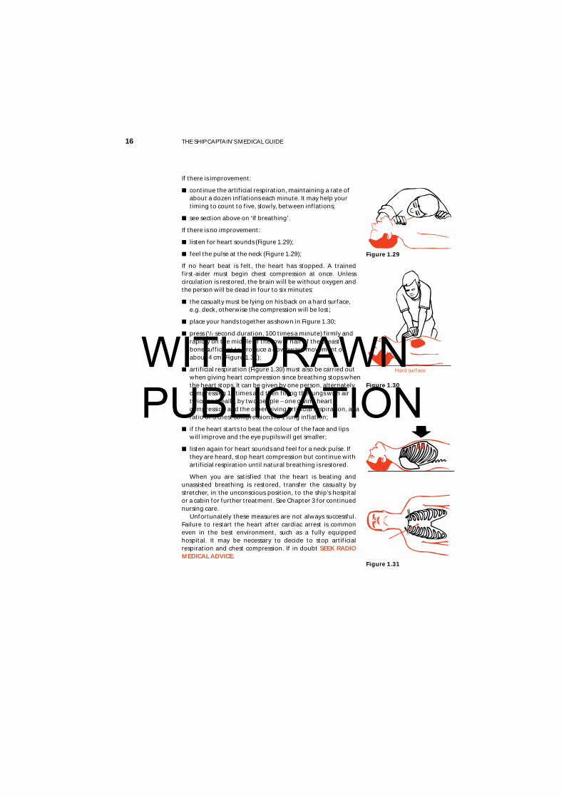
If breathing:
■ place the casualty in the unconscious or recoveryposition (Figure 1.26);
NOTE: no pillows should be used under the head;
■ pull up the leg and the arm on the side to whichthe head is facing, pull up the chin;
■ stretch other arm out as pictured
For subsequent treatment of an unconscious patient see Chapter 3.
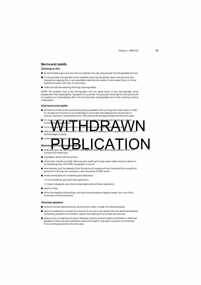
Not breathing:
■ With the casualty lying flat on his back, open the airwayby making sure that the head is tilted back whilst liftingthe chin upwards and forwards (Figure 1.27), which willmove the tongue forward and clear the airway.
■ Open the mouth and mop out any obvious obstructionssuch as blood, vomit or secretions. If dentures are wornonly remove them if they are broken or displaced. Useyour fingers, a handkerchief or a clean piece of cloth.
These actions may relieve the obstruction to breathing.The casualty may gasp and start to breathe naturally. If so,place in the unconscious position.
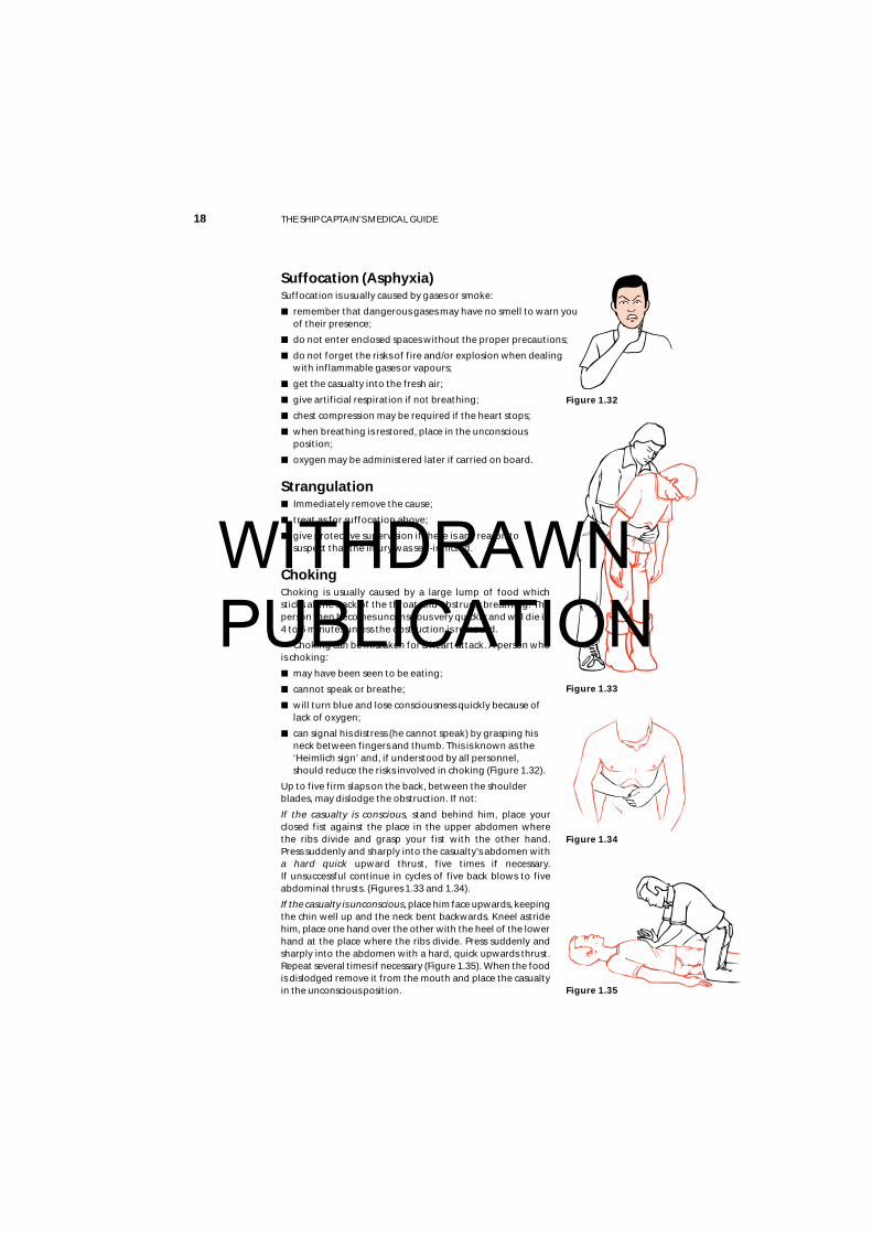
Still not breathing:
Begin artificial respiration at once – seconds count.
■ Open the airway by making sure that the head is tiltedback whilst lifting the chin upwards and forwards.
■ work from the side in a convenient position;
■ pinch the casualty’s nose with your index finger andthumb. After taking a full breath, seal you lips about thepatient’s mouth and blow into his mouth until you seethe chest rise. This should take about 2 seconds for fullinflation. (Figure 1.28)
■ give two effective inflations quickly, then note if thecolour of the face and lips is improving.
Figure 1.25
Figure 1.26
Figure 1.27
Figure 1.28
The unconscious position
WITHDRAWN PUBLICATION
16 THE SHIP CAPTAIN’S MEDICAL GUIDE
If there is improvement:
■ continue the artificial respiration, maintaining a rate ofabout a dozen inflations each minute. It may help yourtiming to count to five, slowly, between inflations;
■ see section above on ‘If breathing’.
If there is no improvement:
■ listen for heart sounds (Figure 1.29);
■ feel the pulse at the neck (Figure 1.29);
If no heart beat is felt, the heart has stopped. A trained first-aider must begin chest compression at once. Unlesscirculation is restored, the brain will be without oxygen andthe person will be dead in four to six minutes:
■ the casualty must be lying on his back on a hard surface,e.g. deck, otherwise the compression will be lost;
■ place your hands together as shown in Figure 1.30;
■ press (1/2 second duration, 100 times a minute) firmly andrapidly on the middle of the lower half of the breastbone sufficient to produce a downward movement ofabout 4 cm (Figure 1.31);
■ artificial respiration (Figure 1.30) must also be carried outwhen giving heart compression since breathing stops whenthe heart stops. It can be given by one person, alternatelycompressing 15 times and then filling the lungs with airtwice or, ideally, by two people – one giving heartcompression and the other giving artificial respiration, at aratio of 5 chest compressions to 1 lung inflation;
■ if the heart starts to beat the colour of the face and lipswill improve and the eye pupils will get smaller;
■ listen again for heart sounds and feel for a neck pulse. Ifthey are heard, stop heart compression but continue withartificial respiration until natural breathing is restored.
When you are satisfied that the heart is beating andunassisted breathing is restored, transfer the casualty bystretcher, in the unconscious position, to the ship’s hospitalor a cabin for further treatment. See Chapter 3 for continuednursing care.
Unfortunately these measures are not always successful.Failure to restart the heart after cardiac arrest is commoneven in the best environment, such as a fully equippedhospital. It may be necessary to decide to stop artificialrespiration and chest compression. If in doubt SEEK RADIOMEDICAL ADVICE.
Figure 1.29
Figure 1.31
Hard surface
Figure 1.30
WITHDRAWN PUBLICATION
Chapter 1 FIRST AID 17
Burns and scaldsClothing on fire
■ by far the best way to put out a fire on a person is to use a dry powder fire extinguisher at once;
■ if a dry powder extinguisher is not available, then lay the person down and smother theflames by wrapping him in any available material (not made of man-made fibre), or throwbuckets of water over him, or use a hose;
■ make sure all smouldering clothing is extinguished.
NOTE: The powder from a fire extinguisher will not cause much, if any, eye damage. Mostpeople shut their eyes tightly if sprayed with powder. Any powder which gets in the eye shouldbe washed out immediately after the fire has been extinguished and while cooling is beingundertaken.
Heat burns and scalds
■ all heat burns should be cooled as quickly as possible with running cold water (sea or fresh)for at least ten minutes, or by immersing in cold water and keeping the injured part inmotion; cooling of extensive burns (>15%) should be avoided as hypothermia will result.
■ if it is not possible to cool the burn on the spot, the casualty should be taken to wherecooling can be carried out;
■ try to remove clothing gently but do not tear off any which adheres to the skin;
■ then cover the burned areas with a dry, non-fluffy, dressing which is larger than the burnsand bandage in place;
■ further treatment as in Chapter 4.
Electrical burns and electrocution
■ make sure you do not become the next casualty when approaching any person who is incontact with electricity:
■ if possible, switch off the current;
■ otherwise, insulate yourself. Remove your watch and rings, wear rubber boots or stand onan insulating mat, thick DRY newspaper or wood;
■ alternatively, pull the casualty from the source of supply with an insulated flex or push himaway with a strong non-conductor, such as a piece of DRY wood;
■ check immediately for breathing and heartbeat:
• if not breathing, give artificial respiration;
• if heart is stopped, give chest compression and artificial respiration;
■ send for help;
■ when the casualty is breathing, cool any burned areas and apply a clean, dry, non-fluffycovering to the burned area.
Chemical splashes
■ remove contaminated clothing. Drench with water to wash the chemical away;
■ carry on washing for at least ten minutes. If you are in any doubt that the chemical has beencompletely cleared from the skin, repeat the washing for a further ten minutes;
■ give priority to washing the eyes if affected, as they are particularly vulnerable to chemicalsplashes. If only one eye is affected, incline the head to that side to prevent the chemicalfrom running across into the other eye.
WITHDRAWN PUBLICATION
18 THE SHIP CAPTAIN’S MEDICAL GUIDE
Suffocation (Asphyxia)Suffocation is usually caused by gases or smoke:
■ remember that dangerous gases may have no smell to warn youof their presence;
■ do not enter enclosed spaces without the proper precautions;
■ do not forget the risks of fire and/or explosion when dealingwith inflammable gases or vapours;
■ get the casualty into the fresh air;
■ give artificial respiration if not breathing;
■ chest compression may be required if the heart stops;
■ when breathing is restored, place in the unconsciousposition;
■ oxygen may be administered later if carried on board.
Strangulation■ Immediately remove the cause;
■ treat as for suffocation above;
■ give protective supervision if there is any reason tosuspect that the injury was self-inflicted.
ChokingChoking is usually caused by a large lump of food whichsticks at the back of the throat and obstructs breathing. Theperson then becomes unconscious very quickly and will die in4 to 6 minutes unless the obstruction is removed.
Choking can be mistaken for a heart attack. A person whois choking:
■ may have been seen to be eating;
■ cannot speak or breathe;
■ will turn blue and lose consciousness quickly because oflack of oxygen;
■ can signal his distress (he cannot speak) by grasping hisneck between fingers and thumb. This is known as the‘Heimlich sign’ and, if understood by all personnel,should reduce the risks involved in choking (Figure 1.32).
Up to five firm slaps on the back, between the shoulderblades, may dislodge the obstruction. If not:
If the casualty is conscious, stand behind him, place yourclosed fist against the place in the upper abdomen where the ribs divide and grasp your fist with the other hand. Press suddenly and sharply into the casualty’s abdomen with a hard quick upward thrust, five times if necessary. If unsuccessful continue in cycles of five back blows to fiveabdominal thrusts. (Figures 1.33 and 1.34).
If the casualty is unconscious, place him face upwards, keepingthe chin well up and the neck bent backwards. Kneel astridehim, place one hand over the other with the heel of the lowerhand at the place where the ribs divide. Press suddenly andsharply into the abdomen with a hard, quick upwards thrust.Repeat several times if necessary (Figure 1.35). When the foodis dislodged remove it from the mouth and place the casualtyin the unconscious position.
Figure 1.32
Figure 1.33
Figure 1.34
Figure 1.35
WITHDRAWN PUBLICATION

Chapter 1 FIRST AID 19
Epileptic fits – convulsionsThe fit may vary from a momentary loss of consciousness (petit mal) in which the patient maysway but does not actually fall, to a major attack (grand mal) as follows: the patient suddenlyloses consciousness and falls to the ground, possibly with a cry; he remains rigid for someseconds, during which he stops breathing and the face becomes flushed; the convulsion thenstarts with irregular, jerky movements of the limbs, rolling of the eyes, gnashing of the teeth,with perhaps some frothing at the mouth. He may lose control and pass urine or faeces. After avariable time, but usually in a few minutes, the convulsion ceases and he falls into what appearsto be a deep sleep.
Treatment
■ prevent the patient from hurting himself in the convulsive stage;
■ never restrain him forcibly, as this may cause injury, but remove hard objects and surroundhim by pillows, clothing or other soft material;
■ after the fit is over, check for injuries. Assuming the patient is uninjured, let him sleep it off.He may be rather confused and dazed when he comes round. Reassure him and do notleave him until you are sure he is aware of his surroundings and knows what he is doing.
In the event of the patient having several fits, one after the other, it may be necessary to givehim an anti-epileptic drug such as Diazepam. SEEK URGENT RADIO MEDICAL ADVICE.
Shock and circulatory collapseShock occurs when the body’s circulatory system is unable to distribute oxygen enriched bloodto all parts of the body. If untreated, the body’s vital organs (brain, heart, lungs, kidneys) canfail, leading to collapse, unconsciousness and eventually death.
Causes
The commonest cause is loss of body fluid from the circulation. It can result, either from externalor internal bleeding, (e.g. as occurs in fractures of the thigh), the formation of large blisters andthe weeping of fluid from large burns and from damaged blood vessels in crush injuries. Shockcan also be found in severe heart attacks, and in certain diseases characterised by excessivevomiting and diarrhoea.
The first-aider should always be on the look-out for this condition as it can develop evenwhile the casualty is under close observation and it may be missed. Fear, pain and exposure tocold make shock worse.
Symptoms and signs
The patient:
■ will usually lie still, taking little notice of his surroundings
■ will complain of feeling faint, cold and thirsty. He may shiver;
■ his lips and the edges of the ears may be blue;
■ his skin will be pale, cold and clammy;
■ his pulse will be rapid and weak;
■ his respiration will be rapid and shallow and, as shock deepens, he will give frequent sighs;
■ he may start to vomit;
■ if untreated, he may lapse into unconsciousness and later die.
WITHDRAWN PUBLICATION

20 THE SHIP CAPTAIN’S MEDICAL GUIDE
TreatmentThe primary aim is to treat whatever condition is causing the shock;
■ lay the patient flat and, if injuries permit, elevate the feet and legs so that blood flows tothe heart and brain (see note below on exceptions to this rule);
■ do not move him unless in a position of danger;
■ stop any blood loss. Cover burns and scalds. Immobilise fractures.
■ loosen any tight clothing which restricts breathing movement;
■ keep warm but do not overheat.
■ deal with any pain. Morphine may be given as necessary
■ give small sips of water if there is no suspicion of abdominal injury but NEVER give fluids toan unconscious casualty. A badly burned or scalded person may require much more fluid;
■ move to a place of safety as gently as possible. Rough handling will increase the pain andthe shock.
Exceptions to the lay flat rule:■ if there is an injury to the face, mouth or jaw with a lot of bleeding, place in the unconscious
position with the head turned with the damaged side underneath and, if possible, with ahead-down tilt. This will prevent blood running down into the throat and lungs;
■ if there is a penetrating wound of the chest, or if breathing is difficult, prop up to assistbreathing;
■ if unconsciousness occurs, put into the unconscious position with as little disturbance aspossible to the injured part.
Bleeding
External bleedingBleeding from small blood vessels occurs when there is a minor cut or abrasion of the skin. Bloodoozes from the wound; it usually stops by itself or when a dressing is applied. It is generally ofno consequence.
In large and deep wounds, the blood wells up in a steady stream. The volume of blood lossdepends on the number of blood vessels damaged and, although it may appear alarming, it isnot usually dangerous, unless allowed to continue.
When large arteries are damaged, bright red blood will spurt from the wound in time withthe heart beats. This bleeding is usually profuse and the patient’s life will be endangered. This isa rare situation.
In all cases of external bleeding, follow the three cardinal rules:
■ lay the patient down;
■ lift up the affected part if possible;
■ press firmly where the blood comes from. Use a dressing or a clean cloth or handkerchiefbut, if none should be available, use the bare hand or fingers. When possible disposablegloves should be worn to protect yourself.
This procedure will stop the flow of blood.
When bleeding has been controlled, apply a standard dressing to the wound and bandagefirmly and widely in position. There may be a slight staining of blood through the dressing,which is of little consequence, but if blood soaks quickly through the pad it is a sign that thebleeding has not been properly controlled. If this happens, do not disturb the dressing, but putanother standard dressing on top and bandage more firmly. This will usually stop the bleeding.Very occasionally, a third dressing may be required.
Do not disturb the dressings until you are prepared to undertake definitive treatment. Thebleeding stops because of the formation of a clot. If you remove the dressing, the clot will breakand bleeding will start again.
WITHDRAWN PUBLICATION
Chapter 1 FIRST AID 21
Special types of external bleeding
From an open fracture
The bleeding comes mainly from around the break and not fromthe bone.
■ do not attempt to elevate the part, this will cause further painand damage;
■ apply a dressing, sterile if possible, padding around thewound. Firm bandaging will apply the necessary pressure tothe tissue around the exposed bone ends.
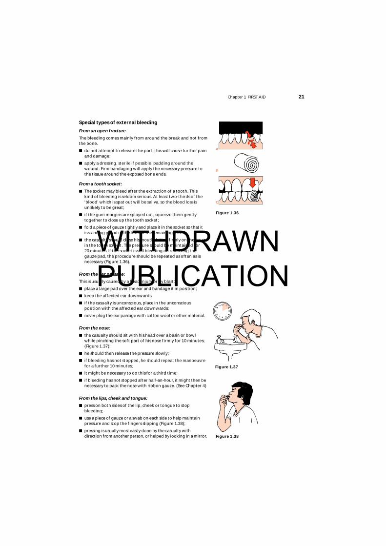
From a tooth socket:
■ The socket may bleed after the extraction of a tooth. This kind of bleeding is seldom serious. At least two-thirds of the‘blood’ which is spat out will be saliva, so the blood loss isunlikely to be great;
■ if the gum margins are splayed out, squeeze them gentlytogether to close up the tooth socket;
■ fold a piece of gauze tightly and place it in the socket so that it is standing proud of the level of the remaining teeth;
■ the casualty should close his mouth, biting firmly on the gauzein the tooth socket. The pressure should be maintained for 20 minutes. If the socket is still bleeding on removing thegauze pad, the procedure should be repeated as often as isnecessary (Figure 1.36).
From the ear passage:
This is usually caused by a head injury or by blast:
■ place a large pad over the ear and bandage it in position;
■ keep the affected ear downwards;
■ if the casualty is unconscious, place in the unconscious position with the affected ear downwards;
■ never plug the ear passage with cotton wool or other material.
From the nose:
■ the casualty should sit with his head over a basin or bowl while pinching the soft part of his nose firmly for 10 minutes;(Figure 1.37);
■ he should then release the pressure slowly;
■ if bleeding has not stopped, he should repeat the manoeuvrefor a further 10 minutes;
■ it might be necessary to do this for a third time;
■ if bleeding has not stopped after half-an-hour, it might then benecessary to pack the nose with ribbon gauze. (See Chapter 4)
From the lips, cheek and tongue:
■ press on both sides of the lip, cheek or tongue to stopbleeding;
■ use a piece of gauze or a swab on each side to help maintain pressure and stop the fingers slipping (Figure 1.38);
■ pressing is usually most easily done by the casualty withdirection from another person, or helped by looking in a mirror.
Figure 1.36
Figure 1.37
Figure 1.38
A
C
B
WITHDRAWN PUBLICATION
22 THE SHIP CAPTAIN’S MEDICAL GUIDE
Internal bleeding
Internal bleeding may be caused by injury, disease, or by the action of certain poisons. Anysevere injury to the body will cause bleeding of varying degree. Bleeding may be limited to thesoft tissues, such as muscles, but when a bone breaks there is always bleeding at the fracturesite. Minor injury will affect only the superficial tissues and the bleeding may be limited to smallamounts which will appear as bruising. Greater force will result, in addition to bruising, in theformation of a collection of blood within the deeper tissues (a haematoma). This causes painfulswelling of the affected part and may be difficult to distinguish from a fracture. Whatever thenature of such injuries, the blood loss very rarely endangers life.
In contrast, bleeding from injury to internal organs is always very serious and may quicklyendanger life. Such bleeding is always concealed and its presence has to be deduced from thehistory of the injury, a rising pulse rate and the signs and symptoms of shock which occur rapidly.The abdominal organs are poorly protected by the abdominal wall and they are particularlyliable to injury by direct or crushing forces. These internal injuries require expert treatmenturgently and every effort must be made to deliver the casualty to medical care. Always getRADIO MEDICAL ADVICE. There is little that can be done aboard because a blood transfusionmay be needed.
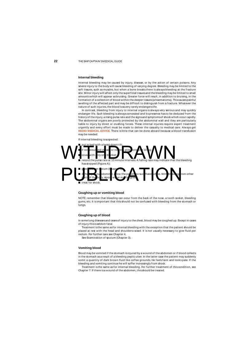
If internal bleeding is suspected:
■ put in bed with a head-down tilt;
■ if conscious and in pain or restless, give morphine 10 mg;
■ cover with only one blanket;
■ record the pulse rate at 10 minute intervals. A falling rate may indicate that the bleedinghas stopped (Figure A);
■ give fluid per rectum (Chapter 3);
■ if the injury is abdominal, allow the patient to suck flakes of ice. With bleeding from otherparts of the body, sips of water may be given;
■ treat for shock.
Coughing up or vomiting blood
NOTE: remember that bleeding can occur from the back of the nose, a tooth socket, bleedinggums, etc. It is important that this should not be confused with bleeding from the stomach orlungs.
Coughing up of blood
In some lung diseases and cases of injury to the chest, blood may be coughed up. Except in casesof injury this is seldom fatal.
Treatment is the same as for internal bleeding with the exception that the patient should beplaced at rest with the head and shoulders raised. It is not usually necessary to give fluid perrectum. For further care see Chapter 4.
See Examination of sputum (Chapter 3).
Vomiting blood
Blood may be vomited if the stomach is injured by a wound of the abdomen or if blood collectsin the stomach as a result of a bleeding peptic ulcer. In the latter case the patient may suddenlyvomit a quantity of dark brown fluid like coffee grounds. He feels faint and looks pale. If thebleeding and vomiting continue he will suffer increasingly from shock.
Treatment is the same as for internal bleeding. For further treatment of this condition, seeChapter 7. If there is a wound of the abdomen, this should be treated.
WITHDRAWN PUBLICATION

Chapter 1 FIRST AID 23
WoundsA wound at any site in the body poses three problems:
■ control of bleeding;
■ prevention of shock
■ prevention of infection
There are some simple rules:
■ never wash the wound – except in cases of an animal bite
■ never try to remove pieces of metal or glass from a wound unless they are superficial andcan be easily lifted out. If pieces can be removed, do it by grasping the material with sterilegauze or use sterile forceps, if available;
■ do not pour antiseptic into a wound;
■ as soon as possible, cover the wound with a suitable dressing.
Bullet or metal fragment wounds
In this type of injury, look for and treat any exit wound. This is usually larger than the entrywound. Remember that there may be underlying bone fractures and that the bullet or metalfragment may have been deflected from the bone to cause serious internal damage, the onlysigns of which may be increasing shock.
Figure A Haemorrhage – the falling temperature and the rising pulse rate
WITHDRAWN PUBLICATION
24 THE SHIP CAPTAIN’S MEDICAL GUIDE
Chest woundsA superficial chest wound should be treated as for any wound elsewhere but a penetratingwound (a sucking wound) of the chest must be sealed immediately, otherwise air is drawn intothe chest cavity and the lungs cannot inflate as the vacuum inside the chest is destroyed.A useful dressing for a sucking wound can be made from a paraffin gauze dressing. Place theparaffin gauze over the wound, smooth the foil on to the chest wall and seal three edges onlywith zinc oxide adhesive plaster. In emergency, a suitable dressing may be improvised frompetroleum jelly, gauze and kitchen foil or polythene or, alternatively, a wet dressing may beused to provide an airtight seal. If nothing else is available, use the casualty’s own bloodstainedclothing to plug the wound temporarily. The aim is to prevent air entering the chest but toallow it to escape if necessary.
The usual rules about stopping bleeding by pressing where the blood comes from also apply.Start a pulse chart soon to check on possible internal bleeding in all chest injuries. Therespiratory rate should also be recorded. See also sections on chest injuries.
Conscious casualties should be placed in the half-sitting-up position because breathing iseasier in this position.NOTE: DO NOT GIVE MORPHINE to a patient with this type of wound, even if he is sufferingfrom a lot of pain, as the morphine will increase the breathing difficulties.Get RADIO MEDICAL ADVICE.
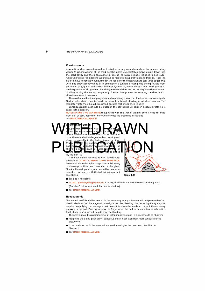
Abdominal woundsA superficial abdominal wound will require thesame treatment as any wound, but for more seriouswounds, if the abdominal contents do not protrude,cover the wound with a large standard dressing andplace the casualty in the half-sitting-up position(Figure 1.39). In this position the wound will notgape open. As the abdominal muscles are slack, theabdominal contents will not bulge through. If thewound runs more or less vertically, it may be best tolay the man flat.
If the abdominal contents do protrude throughthe wound, DO NOT ATTEMPT TO PUT THEM BACK.Cover with a loosely applied large standard dressingor dressings until further treatment can be given.Shock will develop quickly and should be treated asdescribed previously, with the following importantexceptions:
■ prop up if necessary;
■ DO NOT give anything by mouth. If thirsty, the lips should be moistened; nothing more.
(See also Crush wounds and Stab wounds below).
■ Get RADIO MEDICAL ADVICE.
Head woundsThe wound itself should be treated in the same way as any other wound. Scalp wounds oftenbleed briskly. A firm bandage will usually arrest the bleeding, but some ingenuity may berequired in applying the bandage so as to keep it firmly on the head and transmit the necessarypressure to the pad. Firm pressure by the fingers over the pad for a few minutes before it isfinally fixed in position will help to stop the bleeding.
The possibility of brain damage is of greater importance and two rules should be observed:
■ morphine should be given only if conscious and in much pain from more serious injurieselsewhere;
■ if unconscious, put in the unconscious position and give the treatment described in Chapter 4.
■ Get RADIO MEDICAL ADVICE.
Figure 1.39
WITHDRAWN PUBLICATION
Chapter 1 FIRST AID 25
Face and jaw wounds
There may be danger of suffocation as a result of blood running into the throat. Lay flat in theunconscious position (Figure 1.26) with the more damaged side underneath. If the casualty is tobe removed by stretcher, see that he remains in that position. With severe wounds there may beloss of the power of speech. Give reassurance; speech will probably return to normal whenhealing has taken place.
Palm of the hand wounds
A deep wound of the palm of the hand may cut the large artery in this area. If this occurs:
■ stop the bleeding by pressing where the blood comes from;
■ cover the wound with a sterile gauze dressing and ask the patient to grasp firmly on arolled-up 7.5 cm bandage;
■ a hand bandage, firmly applied, will hold the dressing in place and will maintain thepressure necessary to control the bleeding.
Crush injuries
Limbs
After a crush injury, at first there may be very little to see. However, considerable damage mayhave been done to the muscles and other soft tissues and gross swelling may take place later.Shock, which may be very severe, may also develop.
■ treat any wound;
■ the affected limb should be immobilised and supported in its most comfortable position;
■ treat shock as described but:
• do not give large amounts of fluid at once as the casualty will vomit;
• give frequent small amounts of water only.
■ GET RADIO MEDICAL ADVICE.
Chest
Crushing of the chest may stop breathing and then artificial respiration will be required.
If ribs have been fractured, treat as described under fractures.
See also section on chest injuries.
Abdomen
Severe crushing of the abdomen may cause rupture of the internal organs and/or internalbleeding. If you suspect that this has occurred, Get RADIO MEDICAL ADVICE. See general adviceon abdominal wounds at beginning of this section and stab wounds below.
Stab wounds
Stab wounds are especially dangerous because the underlying structures will have beenpenetrated and infection will have been carried into the deep tissues.
Chest:
■ if the lung has been penetrated, it will collapse giving rise to breathlessness and coughingof bright red frothy blood;
■ a sucking wound can be created;
■ the heart can be damaged.
■ Get RADIO MEDICAL ADVICE
■ see also section on chest injuries.
WITHDRAWN PUBLICATION
26 THE SHIP CAPTAIN’S MEDICAL GUIDE
Abdomen
Depending on the position of the wound (see Anatomy Diagrams, Annex II), an organ may bepierced, giving rise to peritonitis and internal bleeding. See general advice at beginning of thissection. Get RADIO MEDICAL ADVICE.
Limbs
Muscles, nerves and blood vessels may be cut. Bleeding, both internal and external, will occur.Whatever the site of the stab wound, the immediate treatment is the same:
■ stop external bleeding by pressure
■ prevent further infection by applying suitable dressings
■ treat shock if necessary.
FracturesA fracture is a broken bone. The bone may be broken into two or more pieces with separationof the fragments or it may have one or more fissured cracks without any separation.
Most fractures are caused by direct force, but force may be transmitted through the body tocause injury indirectly elsewhere. Two classical examples are: a fall on the outstretched hand,causing a fracture of the collar bone; and a fall from a height on to the heels, causing a fractureof the base of the skull.
A much less common type is a stress fracture. The bone becomes weakened in a waycomparable to metal fatigue. Sudden, strong muscular effort may snap the bone.
In simple terms, a fracture may be open to infection or closed to infection.
A closed fracture
There is no communication between the fracture and the surface of the body.
An open fractureThere is communication between a skin wound and the fracture. Open fractures are alwaysserious because germs may enter through the wound to cause infection of the broken bone andthe surrounding tissues.
NOTE: A skin wound may be present but, unless it is deep enough to reach the broken bone,the fracture is still closed. Open or closed fractures are sometimes complicated by damage toimportant structures such as the brain, lung, blood vessels or nerves.
Principles of treatment
It is not possible to set fractures on board ship. Indeed, many fractures may not require settingand unskilled attempts might prejudice healing. First aid measures should ensure adequateimmobilisation. Wherever a fracture case has to be kept on board for more than two or threedays, the joints above and below the fracture site should be gently put through a full range ofmovements, morning and night.
Lasting damage may result if a joint surface is involved in the fracture and in all cases wherethis is suspected, RADIO MEDICAL ADVICE must be sought.
Antibiotic treatment must always be given as soon as an open fracture is diagnosed orsuspected.
Examination
The following signs and symptoms will indicate that the bone is probably broken:
■ a heavy blow or other force has been applied to the body or limbs. The casualty or othersmay have heard the bone break;
■ intense pain, especially on pressure or movement at the site;
■ swelling. The site may be swollen and/or bruised. This may be due to internal bleeding;
■ loss of use. The casualty may be unable or unwilling to use the injured part because of the
WITHDRAWN PUBLICATION

Chapter 1 FIRST AID 27
pain. He may also experience severe pain if an attempt, even very gently, is made to helphim make the movement. Watch his face for signs of pain. Occasionally, if the broken endsof a bone are impacted together, the person may be able to use the part but usually onlywith a fair amount of pain;
■ distortion. Compare good and bad limbs or sides of the body to see if the part is swollen,bent, twisted or shortened;
■ irregularity. The irregular edges of a broken bone can sometimes be seen in an openfracture. They may be seen or felt under the skin in a closed fracture;
■ unnatural movement and grating of bone ends. Neither of these symptoms should besought deliberately. A limb may feel limp and wobbly and grating may be felt when tryingto apply support to the limb. In either of these situations, the bone is certainly broken.
General treatment
■ bleeding should be treated as described;
■ rest the affected part by immobilisation. This prevents further damage, relieves pain andstops further bleeding;
■ all fractures or suspected fractures must be immobilised before making any attempt tomove the casualty. This can be done using wooden, improvised or inflatable splints, or byfixing a limb to the body, or – in the case of the legs – by lashing one to the other.
Immobilise a limb in the position in which it isfound, if it is comfortable. If it does becomenecessary to move an injured limb, because ofpoor circulation or for any other reason, firstapply traction by pulling the limb gently andfirmly away from the body before attempting tomove it (Figure 1.40).
Keep pulling until it has been securelyimmobilised and then release the traction veryslowly. Sudden release can cause pain.
Circulation of the blood in a fractured limb.Check that the circulation to the limb is intact. Todo this, press on the nail of the thumb or of thebig toe. When circulation is normal the nailbecomes white when pressed and pink whenreleased. Continue checking until you aresatisfied that all is well. Danger signs are:
■ blueness or whiteness of fingers and toes;
■ coldness of the parts below the fracture;
■ loss of feeling below the injury. Test for this by touching lightly on fingers and toes andasking the casualty if he can feel anything;
■ absence of pulse.
If there is any doubt at all about the circulation, loosen all tight and limb-encircling dressingsat once and straighten out the limb, remembering to use traction when doing so. Checkcirculation again. If the limb does not become pink and warm and you cannot detect a pulse,then medical help is urgently necessary if amputation is to be avoided. Get RADIO MEDICALADVICE.
■ remember that fractures can cause severe internal bleeding ;
■ always look for and treat for shock;
■ morphine may be necessary to control pain.
Figure 1.40
WITHDRAWN PUBLICATION

28 THE SHIP CAPTAIN’S MEDICAL GUIDE
Collar bone, shoulder blade and shoulder
Fractures in these areas are often the result either of a fall onthe outstretched hand or a fall on to the shoulder. Directviolence to the parts is a less common cause of thesefractures.
Place loose padding about the size of a fist into thearmpit. Support the arm using a triangular sling (Figure 1.41).Then tie the arm to the body, using a narrow fold bandage.Keep the casualty sitting up as he will probably be mostcomfortable in this position.
Upper arm
Upper arm fractures are usually caused by direct violence.Bind the upper arm to the body, using a broad fold
bandage. Bend the elbow gently and apply a collar and cuffsling (Figure 1.42). Keep the casualty sitting up so that theweight of the arm can supply traction to the lower fragment.
Alternatively, upper arm fractures may be splinted. Bendthe elbow gently. Use three well padded splints. Place onebehind the upper arm, one in front and the third from the tipof the shoulder to the elbow. Bandage the splints securely inplace. Support the arm with a collar and cuff sling (see alsoFigure 1.21).
Elbow
Fractures in this area can be especially dangerous because ofdamage to blood vessels and nerves around the elbow.Check circulation and feeling in the fingers. If the finger tipsare white or blue and feeling is absent or altered, the elbowmust be straightened at once. Tell the casualty to lie down.Be gentle. Apply traction on the hand and forearm. Bring thearm and forearm slowly and carefully to the casualty’s side.Now place plenty of loose padding between the arm and thebody and also around the arm. Then bind the forearm to thebody by encircling ties. Check the circulation again when youhave made the encircling ties. If the circulation is poor, theties should be loosely secured until the casualty has to bemoved (Figure 1.43).
Figure 1.42
Figure 1.43
Figure 1.41
WITHDRAWN PUBLICATION
Chapter 1 FIRST AID 29
Forearm and wrist
Fractures in this area commonly result from a fall on the outstretched hand. Bend the elbow untilthe forearm is across the body. Then apply an arm sling (Figure 1.20). Remove any finger rings.
Later, apply two well padded splints to the back and front of the forearm and secure firmly,using narrow fold bandages. Support the arm with a broad arm sling. For fractures of the wristbones, put a broad, well padded splint on the front of the forearm and the palm of the hand.Put plenty of padding on the back of the forearm and hand and secure. Use a broad arm slingfor support.
Hand and fingers
Fractures of the hand bones (metacarpals) and the finger bones are a common result ofshipboard accidents and expert treatment may be many days away. As fixation in a straightsplint is only permissible for a short time, the treatment described in the following paragraphsshould be undertaken if the casualty has to be kept on board. Always remove ringsimmediately.
The hand bones (metacarpals):
■ apply a crepe bandage around the hand and wrist firmly enough to support the injuredpart but not so tight as to prevent movement of the wrist and finger joints;
■ check that circulation to the fingers is present;
■ elevate the hand by placing the arm in a triangular sling to reduce the swelling;
■ encourage the casualty to move the wrist and all the finger joints frequently.
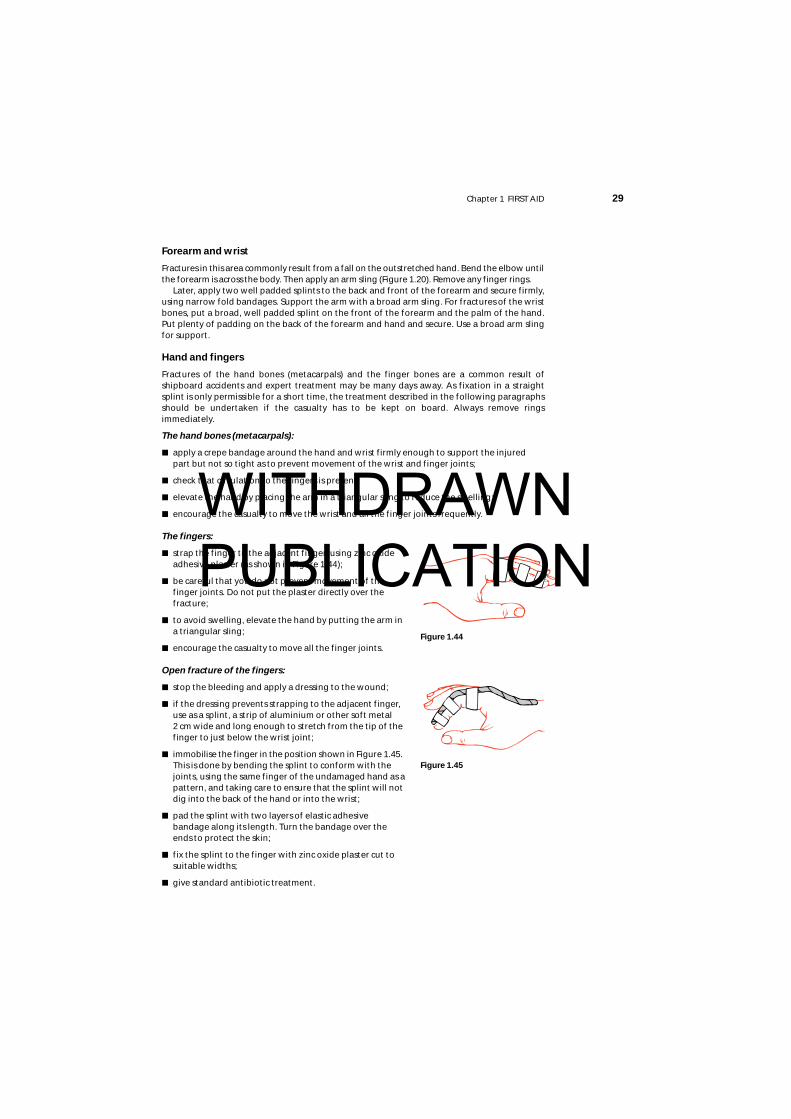
The fingers:
■ strap the finger to the adjacent finger, using zinc oxideadhesive plaster (as shown in Figure 1.44);
■ be careful that you do not prevent movement of thefinger joints. Do not put the plaster directly over thefracture;
■ to avoid swelling, elevate the hand by putting the arm ina triangular sling;
■ encourage the casualty to move all the finger joints.
Open fracture of the fingers:
■ stop the bleeding and apply a dressing to the wound;
■ if the dressing prevents strapping to the adjacent finger,use as a splint, a strip of aluminium or other soft metal 2 cm wide and long enough to stretch from the tip of thefinger to just below the wrist joint;
■ immobilise the finger in the position shown in Figure 1.45.This is done by bending the splint to conform with thejoints, using the same finger of the undamaged hand as apattern, and taking care to ensure that the splint will notdig into the back of the hand or into the wrist;
■ pad the splint with two layers of elastic adhesivebandage along its length. Turn the bandage over theends to protect the skin;
■ fix the splint to the finger with zinc oxide plaster cut tosuitable widths;
■ give standard antibiotic treatment.
Figure 1.44
Figure 1.45
WITHDRAWN PUBLICATION
30 THE SHIP CAPTAIN’S MEDICAL GUIDE
Crush injuries to the hand
Severe crushing injuries to the hands may cause multiple open or closed fractures of themetacarpal or finger bones. Other wounds are likely to be present.
■ stop the bleeding and apply dressings;
■ pain will be severe. Give analgesics (Morphine if necessary);
■ if hospital treatment is not available quickly, read the section on definitive treatment ofwounds and treat accordingly.
Hip to knee
A broken thigh bone is a potentially serious injury. It causes significant internal bleeding intothe muscles of the thigh and, with the associated pain, shock very quickly develops. If it iscombined with other serious injuries, the blood loss may be so great as to require bloodreplacement. Get RADIO MEDICAL ADVICE.
■ a break of the neck of the thigh bone causes shortening of the injured leg and the casualtywill lie with the whole lower limb and foot flopped outwards. There will be severe pain inthe region of the hip;
■ fractures of the shaft of the thigh bone exhibit the usual signs and symptoms of a fracture.Severe pain is a normal feature.
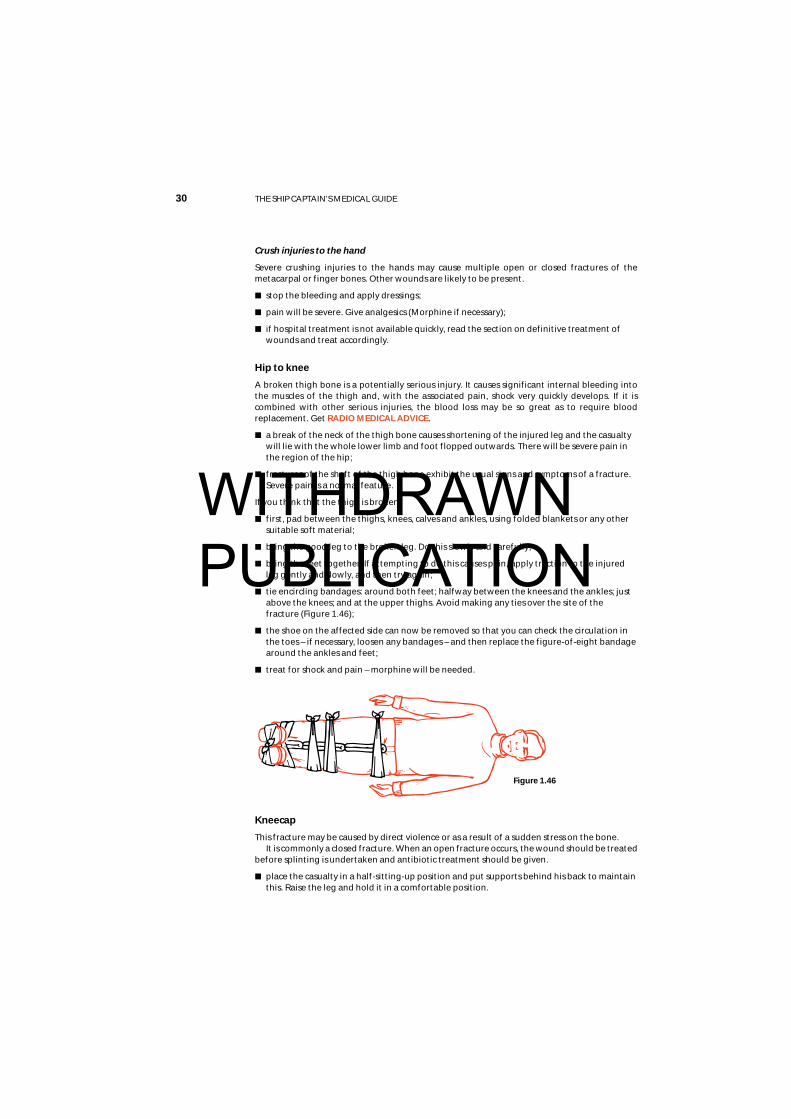
If you think that the thigh is broken:
■ first, pad between the thighs, knees, calves and ankles, using folded blankets or any othersuitable soft material;
■ bring the good leg to the broken leg. Do this slowly and carefully;
■ bring the feet together. If attempting to do this causes pain, apply traction to the injuredleg gently and slowly, and then try again;
■ tie encircling bandages: around both feet; halfway between the knees and the ankles; justabove the knees; and at the upper thighs. Avoid making any ties over the site of thefracture (Figure 1.46);
■ the shoe on the affected side can now be removed so that you can check the circulation inthe toes – if necessary, loosen any bandages – and then replace the figure-of-eight bandagearound the ankles and feet;
■ treat for shock and pain – morphine will be needed.
Kneecap
This fracture may be caused by direct violence or as a result of a sudden stress on the bone.It is commonly a closed fracture. When an open fracture occurs, the wound should be treated
before splinting is undertaken and antibiotic treatment should be given.
■ place the casualty in a half-sitting-up position and put supports behind his back to maintainthis. Raise the leg and hold it in a comfortable position.
Figure 1.46
WITHDRAWN PUBLICATION
Chapter 1 FIRST AID 31
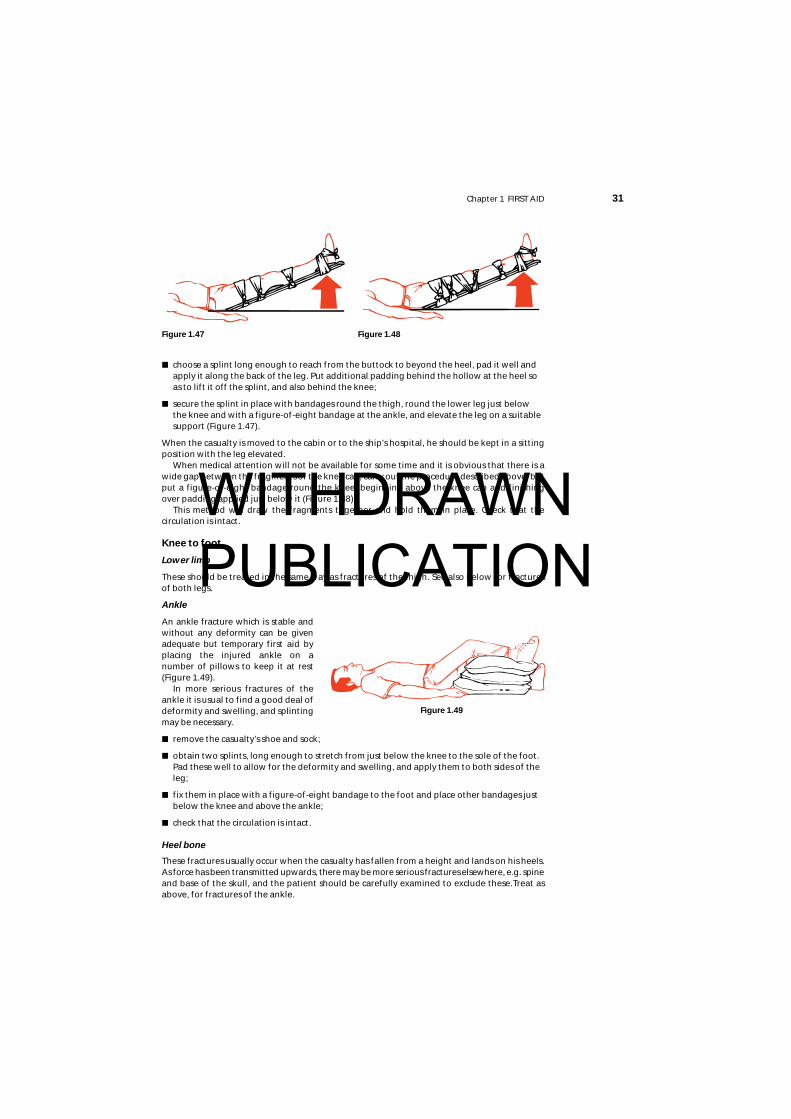
■ choose a splint long enough to reach from the buttock to beyond the heel, pad it well andapply it along the back of the leg. Put additional padding behind the hollow at the heel soas to lift it off the splint, and also behind the knee;
■ secure the splint in place with bandages round the thigh, round the lower leg just belowthe knee and with a figure-of-eight bandage at the ankle, and elevate the leg on a suitablesupport (Figure 1.47).
When the casualty is moved to the cabin or to the ship’s hospital, he should be kept in a sittingposition with the leg elevated.
When medical attention will not be available for some time and it is obvious that there is awide gap between the fragments of the knee cap, carry out the procedure described above, butput a figure-of-eight bandage round the knee, beginning above the knee cap and finishingover padding applied just below it (Figure 1.48).
This method will draw the fragments together and hold them in place. Check that thecirculation is intact.
Knee to foot
Lower limb
These should be treated in the same way as fractures of the thigh. See also below for fracturesof both legs.
Ankle
An ankle fracture which is stable andwithout any deformity can be givenadequate but temporary first aid byplacing the injured ankle on anumber of pillows to keep it at rest(Figure 1.49).
In more serious fractures of theankle it is usual to find a good deal ofdeformity and swelling, and splintingmay be necessary.
■ remove the casualty’s shoe and sock;
■ obtain two splints, long enough to stretch from just below the knee to the sole of the foot.Pad these well to allow for the deformity and swelling, and apply them to both sides of theleg;
■ fix them in place with a figure-of-eight bandage to the foot and place other bandages justbelow the knee and above the ankle;
■ check that the circulation is intact.
Heel bone
These fractures usually occur when the casualty has fallen from a height and lands on his heels.As force has been transmitted upwards, there may be more serious fractures elsewhere, e.g. spineand base of the skull, and the patient should be carefully examined to exclude these.Treat asabove, for fractures of the ankle.
Figure 1.47 Figure 1.48
Figure 1.49
WITHDRAWN PUBLICATION
32 THE SHIP CAPTAIN’S MEDICAL GUIDE
Bones of the foot
Severe injuries are usually the result of heavy weights beingdropped on to unprotected feet or of crushing. Fractures of thetoes may occur when they are stubbed against some hard object.
■ remove the boot or shoe and the socks carefully;
■ treat any wound.
■ keep the foot elevated and use pillows to keep it in acomfortable position.
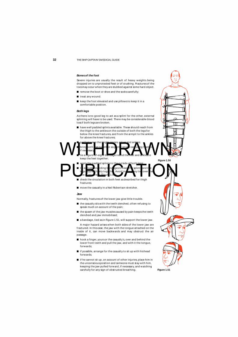
Both legs
As there is no good leg to act as a splint for the other, externalsplinting will have to be used. There may be considerable bloodloss if both legs are broken.
■ have well padded splints available. These should reach fromthe thigh to the ankles on the outside of both the legs forbelow the knee fractures, and from the armpit to the anklesfor above the knee fractures;
■ pad between the thighs, knees, calves and ankles;
■ bring both feet together as gently as you can, using tractionif necessary.
■ tie a figure-of-eight bandage round the feet and ankles tokeep the feet together;
■ apply the padded splints to the outside of both legs;
■ tie enough encircling bandages to keep the splints and thelegs secured firmly together. Avoid making any ties over thesite of any break (Figure 1.50);
■ check the circulation in both feet as described for thighfractures;
■ move the casualty in a Neil Robertson stretcher.
Jaw
Normally, fractures of the lower jaw give little trouble.
■ the casualty sits with the teeth clenched, often refusing tospeak much on account of the pain;
■ the spasm of the jaw muscles caused by pain keeps the teethclenched and jaw immobilised;
■ a bandage, tied as in Figure 1.51, will support the lower jaw.
A major hazard arises when both sides of the lower jaw arefractured. In this case, the jaw with the tongue attached on theinside of it, can move backwards and may obstruct the airpassage.
■ hook a finger, yours or the casualty’s, over and behind thelower front teeth and pull the jaw, and with it the tongue,forwards;
■ if possible, arrange for the casualty to sit up with his headforwards;
■ if he cannot sit up, on account of other injuries, place him inthe unconscious position and someone must stay with him,keeping the jaw pulled forward, if necessary, and watchingcarefully for any sign of obstructed breathing.
Figure 1.50
Figure 1.51
WITHDRAWN PUBLICATION
Chapter 1 FIRST AID 33
Spine
Always suspect a fracture of the spine if a person has fallen a distance of over two metres.Check carefully how the injury happened. Ask if there is pain in the back. Most people withfractures of the spine have pain but a very few DO NOT. If in doubt, treat the injury as afractured spine.A FRACTURED SPINE IS POTENTIALLY A VERY SERIOUS INJURY. IF YOU SUSPECT AFRACTURED SPINE, TELL THE CASUALTY TO LIE STILL AND DO NOT ALLOW ANYONE TOMOVE HIM UNTIL FIRST AID TREATMENT HAS BEEN COMPLETED.
Any careless movement of a casualty with a fractured spine could damage or sever the spinalcord, resulting in permanent paralysis and loss of feeling in the legs, and double incontinencefor life. He can, however, be safely rolled over onto one side or the other because, if this is donevery gently and carefully, there is very little movement of the spine.
First, establish whether the spinal cord has been damaged. To do this:
■ ask the casualty if he can feel any tingling of the feet or legs. Tingling usually means thatthere is some pressure on the spinal cord;
■ ask him to move his toes. If he is unable to do this, then paralysis is present and indicatessevere damage to the spinal cord;
■ run your fingers lightly over the skin of the lower legs and feet. Absence of sensationindicates severe damage to the spinal cord.
If any of these are found, get RADIO MEDICAL ADVICE.
■ next, place padding between the legs;
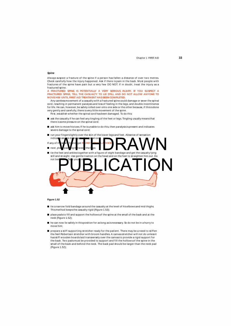
■ tie the feet and ankles together with a figure-of-eight bandage and get the casualty lyingstill and straight. Use gentle traction on the head and on the feet to straighten him out. Donot bend him. Take your time;
■ tie a narrow fold bandage around the casualty at the level of his elbows and mid thighs.This method keeps the casualty rigid (Figure 1.53);
■ place pads to fill and support the hollows of the spine at the small of the back and at theneck (Figure 1.52);
■ he can now lie safely in this position for as long as is necessary. So do not be in a hurry tomove him;
■ prepare a stiff supporting stretcher ready for the patient. There may be a need to stiffenthe Neil Robertson stretcher with broom handles. A canvas stretcher will not do unless ithas stiff wooden boards laid transversely over the canvas to provide a rigid support forthe back. Two pads must be provided to support and fill the hollows of the spine in thesmall of the back and behind the neck. The back pad should be larger than the neck pad(Figure 1.52);
Figure 1.52
WITHDRAWN PUBLICATION
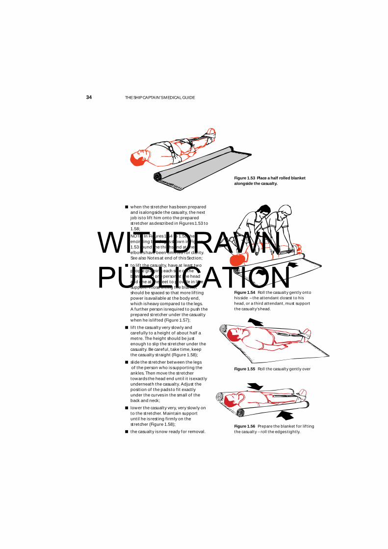
■ when the stretcher has been preparedand is alongside the casualty, the nextjob is to lift him onto the preparedstretcher as described in Figures 1.53 to1.58;
NOTE: In Figures 1.54 to 1.58 theencircling bandages shown in Figure1.53 round the thighs and at theelbows have been omitted for clarity.See also Notes at end of this Section;
■ to lift the casualty, have at least twopeople grasping each side of theblanket and one person at the head and one at the feet to provide in linesupport. Those lifting the blanketshould be spaced so that more liftingpower is available at the body end,which is heavy compared to the legs. A further person is required to push theprepared stretcher under the casualtywhen he is lifted (Figure 1.57);
■ lift the casualty very slowly andcarefully to a height of about half ametre. The height should be justenough to slip the stretcher under thecasualty. Be careful, take time, keepthe casualty straight (Figure 1.58);
■ slide the stretcher between the legsof the person who is supporting theankles. Then move the stretchertowards the head end until it is exactlyunderneath the casualty. Adjust theposition of the pads to fit exactlyunder the curves in the small of theback and neck;
■ lower the casualty very, very slowly onto the stretcher. Maintain supportuntil he is resting firmly on thestretcher (Figure 1.58);
■ the casualty is now ready for removal.
34 THE SHIP CAPTAIN’S MEDICAL GUIDE
Figure 1.56 Prepare the blanket for liftingthe casualty – roll the edges tightly.
Figure 1.55 Roll the casualty gently over
Figure 1.53 Place a half rolled blanketalongside the casualty.
Figure 1.54 Roll the casualty gently ontohis side – the attendant closest to hishead, or a third attendant, must supportthe casualty’s head.
WITHDRAWN PUBLICATION

Chapter 1 FIRST AID 35
When the casualty has been very carefully transported to amattress on the deck, or other very firm bed, where he mayremain undisturbed flat on his back, the mostimportant single point is to keep him as still aspossible. He must continue to besupported with pillows, etc., as describedlater in the text. Every care and attention,and encouragement must be given to helphim to remain still, whether or not any paralysis ispresent. Bags filled with sand should be placed asnecessary to prevent the body or limbs rolling. A urinebottle should be constantly available, and a catheter shouldbe used to relieve him if necessary. He should pass any faeceson to cotton wool or other material: he must not be lifted onto a bed pan. His back should be treated, so far as possible, toprevent sores. He must be put ashore at the very earliestpossible moment. Get RADIO MEDICAL ADVICE.
NOTES:(1) As there are a number of peoplehelping and since it is important to takegreat care in handling the casualty, itmay be helpful to have a person readout the particular instruction beforeeach operation is carried out.(2) At least seven people are required tocarry out this manoeuvre. In ships withsmall crews, there may be insufficientnumbers of men available. In this case,do not attempt to move the casualty butcarry out the instructions given above onimmobilising him and padding the naturalcurves of the spine. The casualty shouldthen be kept warm, his pain should be treated(see section on analgesics and, if he is on the deck, he should beprotected from the elements with suitable waterproof coverings.
Neck
Injuries to the neck are often compression fractures of thevertebrae due, for example, to a person standing upsuddenly and bumping his head violently, or by somethingfalling on his head. Falls from a height can also produce neckinjuries. Treatment is similar to that described above forfractures of the spine, because the neck is the upper part ofthe spine.
■ the casualty should be laid flat, if not already in thisposition, and should be kept still and straight;
■ a semi-rigid neck collar should then be applied gently tostop movement of the neck while an assistant steadiesthe head. An improvised neck collar can be made quiteeasily from a newspaper. Fold the newspaper so that thewidth is about 10 cm at the front. Fold the bottom edgeover to produce a slightly narrower back. Then fold thisaround the neck with the top edge under the chin andthe bottom edge over the top of the collar bones;
■ tie a bandage, scarf or a necktie over the newspaper tohold it in place. This will keep the neck still (Figure 1.59).
Figure 1.58
Figure 1.59
Figure 1.57
WITHDRAWN PUBLICATION
36 THE SHIP CAPTAIN’S MEDICAL GUIDE
Chest
See Fractured ribs.
Pelvis
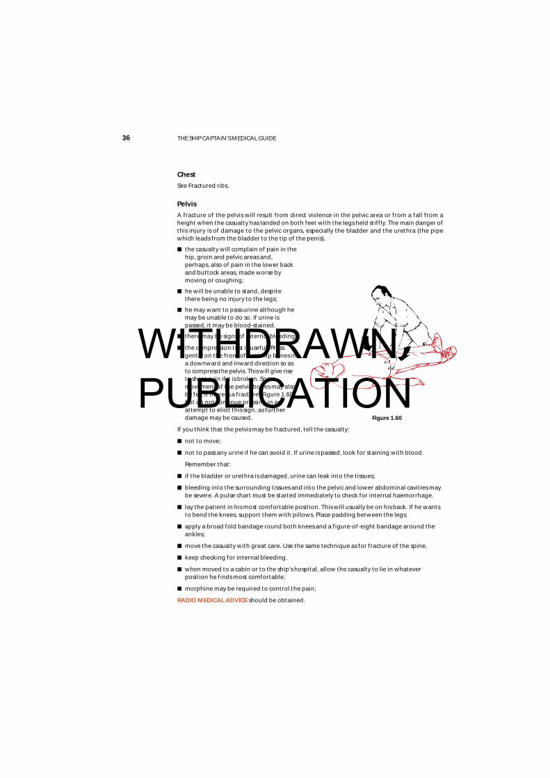
A fracture of the pelvis will result from direct violence in the pelvic area or from a fall from aheight when the casualty has landed on both feet with the legs held stiffly. The main danger ofthis injury is of damage to the pelvic organs, especially the bladder and the urethra (the pipewhich leads from the bladder to the tip of the penis).
■ the casualty will complain of pain in thehip, groin and pelvic areas and,perhaps, also of pain in the lower backand buttock areas, made worse bymoving or coughing;
■ he will be unable to stand, despitethere being no injury to the legs;
■ he may want to pass urine although hemay be unable to do so. If urine ispassed, it may be blood-stained.
■ there may be signs of internal bleeding;
■ the compression test is useful. Pressgently on the front of both hip bones ina downward and inward direction so asto compress the pelvis. This will give riseto sharp pain if it is broken. Somemovement of the pelvic bones may alsobe felt if there is a fracture (Figure 1.60),but do not continue pressing in anattempt to elicit this sign, as furtherdamage may be caused.
If you think that the pelvis may be fractured, tell the casualty:
■ not to move;
■ not to pass any urine if he can avoid it. If urine is passed, look for staining with blood.
Remember that:
■ if the bladder or urethra is damaged, urine can leak into the tissues;
■ bleeding into the surrounding tissues and into the pelvic and lower abdominal cavities maybe severe. A pulse chart must be started immediately to check for internal haemorrhage.
■ lay the patient in his most comfortable position. This will usually be on his back. If he wantsto bend the knees, support them with pillows. Place padding between the legs;
■ apply a broad fold bandage round both knees and a figure-of-eight bandage around theankles;
■ move the casualty with great care. Use the same technique as for fracture of the spine.
■ keep checking for internal bleeding.
■ when moved to a cabin or to the ship’s hospital, allow the casualty to lie in whateverposition he finds most comfortable;
■ morphine may be required to control the pain;
RADIO MEDICAL ADVICE should be obtained.
Figure 1.60
WITHDRAWN PUBLICATION
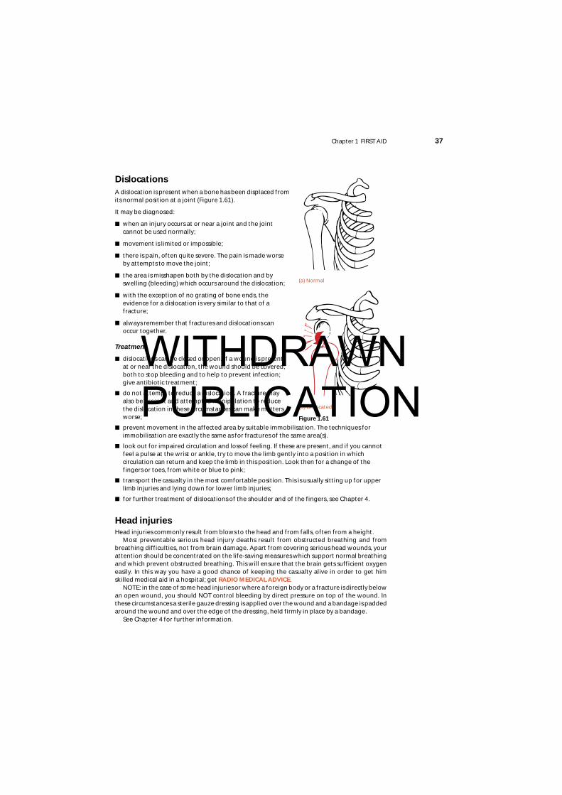
DislocationsA dislocation is present when a bone has been displaced fromits normal position at a joint (Figure 1.61).
It may be diagnosed:
■ when an injury occurs at or near a joint and the jointcannot be used normally;
■ movement is limited or impossible;
■ there is pain, often quite severe. The pain is made worseby attempts to move the joint;
■ the area is misshapen both by the dislocation and byswelling (bleeding) which occurs around the dislocation;
■ with the exception of no grating of bone ends, theevidence for a dislocation is very similar to that of afracture;
■ always remember that fractures and dislocations canoccur together.
Treatment
■ dislocations can be closed or open. If a wound is present,at or near the dislocation, the wound should be covered,both to stop bleeding and to help to prevent infection;give antibiotic treatment;
■ do not attempt to reduce a dislocation. A fracture mayalso be present and attempted manipulation to reducethe dislocation in these circumstances can make mattersworse;
■ prevent movement in the affected area by suitable immobilisation. The techniques forimmobilisation are exactly the same as for fractures of the same area(s).
■ look out for impaired circulation and loss of feeling. If these are present, and if you cannotfeel a pulse at the wrist or ankle, try to move the limb gently into a position in whichcirculation can return and keep the limb in this position. Look then for a change of thefingers or toes, from white or blue to pink;
■ transport the casualty in the most comfortable position. This is usually sitting up for upperlimb injuries and lying down for lower limb injuries;
■ for further treatment of dislocations of the shoulder and of the fingers, see Chapter 4.
Head injuriesHead injuries commonly result from blows to the head and from falls, often from a height.
Most preventable serious head injury deaths result from obstructed breathing and frombreathing difficulties, not from brain damage. Apart from covering serious head wounds, yourattention should be concentrated on the life-saving measures which support normal breathingand which prevent obstructed breathing. This will ensure that the brain gets sufficient oxygeneasily. In this way you have a good chance of keeping the casualty alive in order to get himskilled medical aid in a hospital; get RADIO MEDICAL ADVICE.
NOTE: in the case of some head injuries or where a foreign body or a fracture is directly belowan open wound, you should NOT control bleeding by direct pressure on top of the wound. Inthese circumstances a sterile gauze dressing is applied over the wound and a bandage is paddedaround the wound and over the edge of the dressing, held firmly in place by a bandage.
See Chapter 4 for further information.
Chapter 1 FIRST AID 37
Figure 1.61
(b) Dislocated
(a) Normal
WITHDRAWN PUBLICATION
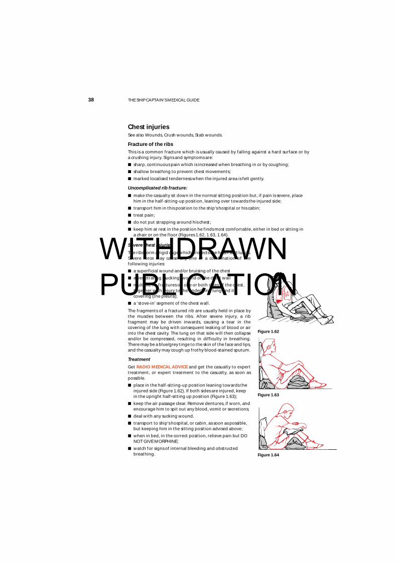
Chest injuriesSee also Wounds, Crush wounds, Stab wounds.
Fracture of the ribsThis is a common fracture which is usually caused by falling against a hard surface or by a crushing injury. Signs and symptoms are:
■ sharp, continuous pain which is increased when breathing in or by coughing;
■ shallow breathing to prevent chest movements;
■ marked localised tenderness when the injured area is felt gently.
Uncomplicated rib fracture:
■ make the casualty sit down in the normal sitting position but, if pain is severe, placehim in the half-sitting-up position, leaning over towards the injured side;
■ transport him in this position to the ship’s hospital or his cabin;
■ treat pain;
■ do not put strapping around his chest;
■ keep him at rest in the position he finds most comfortable, either in bed or sitting in a chair or on the floor (Figures 1.62, 1.63, 1.64).
Severe chest injuries
The ribs form a rigid cage which protects the heart and lungs.Severe force may cause any one or a combination of thefollowing injuries:
■ a superficial wound and/or bruising of the chest
■ a penetrating (sucking) wound of the chest wall
■ multiple rib fractures on one or both sides of the chest,together with injury to the underlying lung and itscovering (the pleura);
■ a ‘stove-in’ segment of the chest wall.
The fragments of a fractured rib are usually held in place bythe muscles between the ribs. After severe injury, a ribfragment may be driven inwards, causing a tear in thecovering of the lung with consequent leaking of blood or airinto the chest cavity. The lung on that side will then collapseand/or be compressed, resulting in difficulty in breathing.There may be a blue/grey tinge to the skin of the face and lips,and the casualty may cough up frothy blood-stained sputum.
Treatment
Get RADIO MEDICAL ADVICE and get the casualty to experttreatment, or expert treatment to the casualty, as soon aspossible.
■ place in the half-sitting-up position leaning towards theinjured side (Figure 1.62). If both sides are injured, keepin the upright half-sitting up position (Figure 1.63);
■ keep the air passage clear. Remove dentures, if worn, andencourage him to spit out any blood, vomit or secretions;
■ deal with any sucking wound.
■ transport to ship’s hospital, or cabin, as soon as possible,but keeping him in the sitting position advised above;
■ when in bed, in the correct position, relieve pain but DONOT GIVE MORPHINE;
■ watch for signs of internal bleeding and obstructedbreathing.
38 THE SHIP CAPTAIN’S MEDICAL GUIDE
Figure 1.64
Figure 1.63
Figure 1.62
WITHDRAWN PUBLICATION
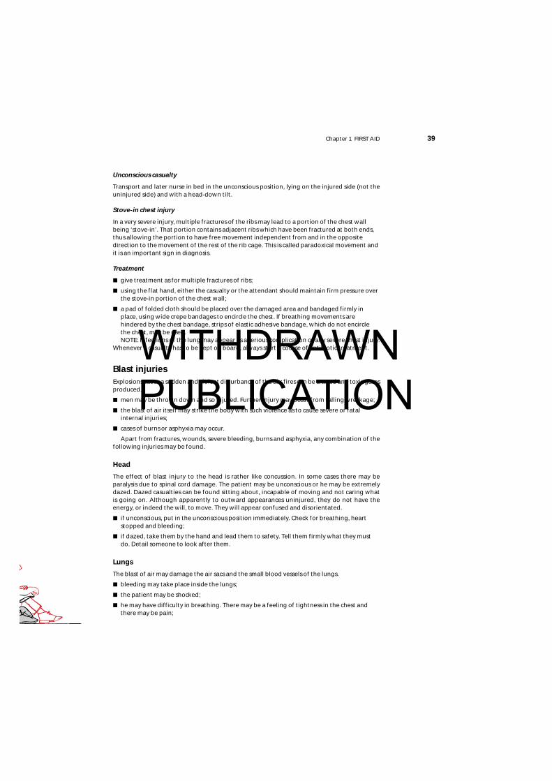
Chapter 1 FIRST AID 39
Unconscious casualty
Transport and later nurse in bed in the unconscious position, lying on the injured side (not theuninjured side) and with a head-down tilt.
Stove-in chest injury
In a very severe injury, multiple fractures of the ribs may lead to a portion of the chest wallbeing ‘stove-in’. That portion contains adjacent ribs which have been fractured at both ends,thus allowing the portion to have free movement independent from and in the oppositedirection to the movement of the rest of the rib cage. This is called paradoxical movement andit is an important sign in diagnosis.
Treatment
■ give treatment as for multiple fractures of ribs;
■ using the flat hand, either the casualty or the attendant should maintain firm pressure overthe stove-in portion of the chest wall;
■ a pad of folded cloth should be placed over the damaged area and bandaged firmly inplace, using wide crepe bandages to encircle the chest. If breathing movements arehindered by the chest bandage, strips of elastic adhesive bandage, which do not encirclethe chest, may be used.NOTE: Infections of the lung may appear as a serious complication of any severe chest injury.
Whenever a casualty has to be kept on board, always start a course of antibiotic treatment.
Blast injuriesExplosions cause a sudden and violent disturbance of the air; fires can be started and toxic gasesproduced.
■ men may be thrown down and so injured. Further injury may occur from falling wreckage;
■ the blast of air itself may strike the body with such violence as to cause severe or fatalinternal injuries;
■ cases of burns or asphyxia may occur.
Apart from fractures, wounds, severe bleeding, burns and asphyxia, any combination of thefollowing injuries may be found.
Head
The effect of blast injury to the head is rather like concussion. In some cases there may beparalysis due to spinal cord damage. The patient may be unconscious or he may be extremelydazed. Dazed casualties can be found sitting about, incapable of moving and not caring what is going on. Although apparently to outward appearances uninjured, they do not have theenergy, or indeed the will, to move. They will appear confused and disorientated.
■ if unconscious, put in the unconscious position immediately. Check for breathing, heartstopped and bleeding;
■ if dazed, take them by the hand and lead them to safety. Tell them firmly what they mustdo. Detail someone to look after them.
Lungs
The blast of air may damage the air sacs and the small blood vessels of the lungs.
■ bleeding may take place inside the lungs;
■ the patient may be shocked;
■ he may have difficulty in breathing. There may be a feeling of tightness in the chest andthere may be pain;
WITHDRAWN PUBLICATION
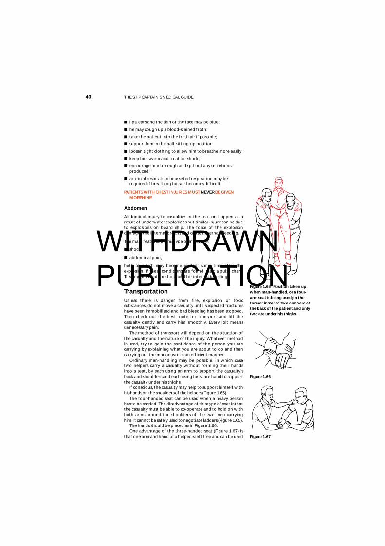
■ lips, ears and the skin of the face may be blue;
■ he may cough up a blood-stained froth;
■ take the patient into the fresh air if possible;
■ support him in the half-sitting-up position
■ loosen tight clothing to allow him to breathe more easily;
■ keep him warm and treat for shock;
■ encourage him to cough and spit out any secretionsproduced;
■ artificial respiration or assisted respiration may berequired if breathing fails or becomes difficult.
PATIENTS WITH CHEST INJURIES MUST NEVER BE GIVENMORPHINE
Abdomen
Abdominal injury to casualties in the sea can happen as aresult of underwater explosions but similar injury can be dueto explosions on board ship. The force of the explosiondamages the internal organs and causes internal bleeding.
The main features of this type of injury are:
■ shock;
■ abdominal pain;
both of which may become evident some time after theexplosion. If these conditions are found, start a pulse chart.Treatment is that for shock and for internal bleeding.
TransportationUnless there is danger from fire, explosion or toxicsubstances, do not move a casualty until suspected fractureshave been immobilised and bad bleeding has been stopped.Then check out the best route for transport and lift thecasualty gently and carry him smoothly. Every jolt meansunnecessary pain.
The method of transport will depend on the situation ofthe casualty and the nature of the injury. Whatever methodis used, try to gain the confidence of the person you arecarrying by explaining what you are about to do and thencarrying out the manoeuvre in an efficient manner.
Ordinary man-handling may be possible, in which casetwo helpers carry a casualty without forming their handsinto a seat, by each using an arm to support the casualty’sback and shoulders and each using his spare hand to supportthe casualty under his thighs.
If conscious, the casualty may help to support himself withhis hands on the shoulders of the helpers (Figure 1.65).
The four-handed seat can be used when a heavy personhas to be carried. The disadvantage of this type of seat is thatthe casualty must be able to co-operate and to hold on withboth arms around the shoulders of the two men carryinghim. It cannot be safely used to negotiate ladders (Figure 1.65).
The hands should be placed as in Figure 1.66.One advantage of the three-handed seat (Figure 1.67) is
that one arm and hand of a helper is left free and can be used
40 THE SHIP CAPTAIN’S MEDICAL GUIDE
Figure 1.66
Figure 1.65 Position taken upwhen man-handled, or a four-arm seat is being used; in theformer instance two arms are atthe back of the patient and onlytwo are under his thighs.
Figure 1.67
WITHDRAWN PUBLICATION
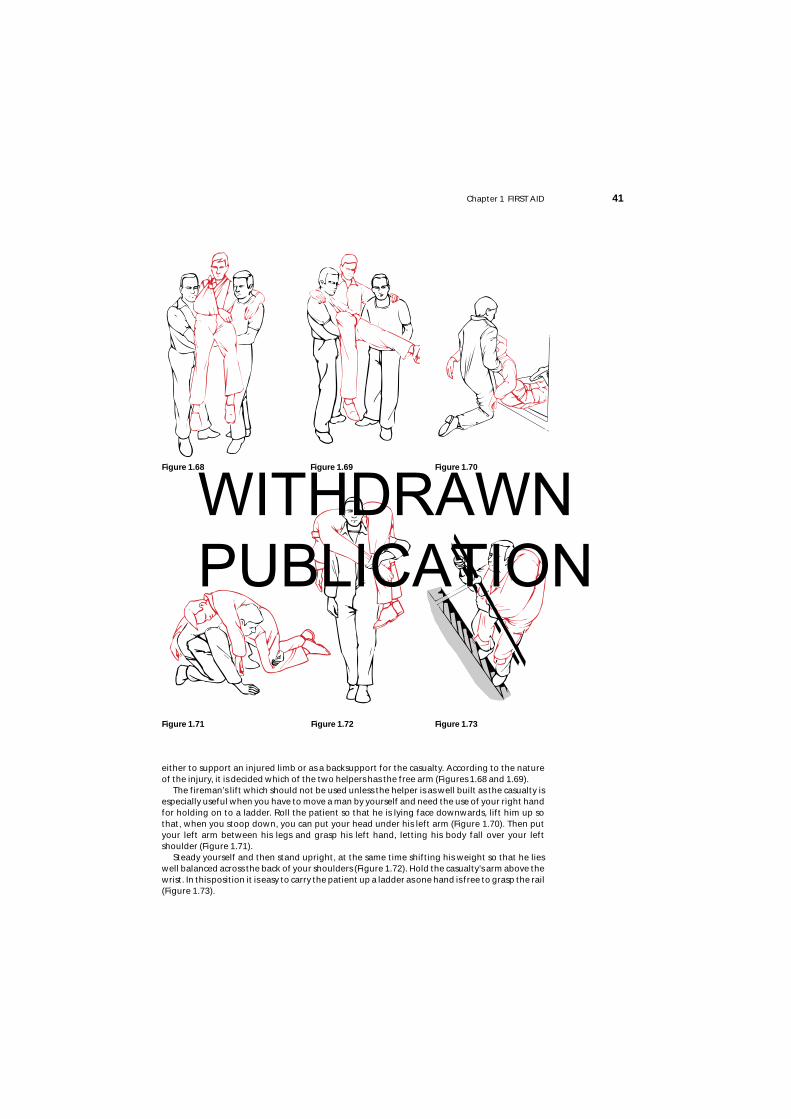
either to support an injured limb or as a backsupport for the casualty. According to the natureof the injury, it is decided which of the two helpers has the free arm (Figures 1.68 and 1.69).
The fireman’s lift which should not be used unless the helper is as well built as the casualty isespecially useful when you have to move a man by yourself and need the use of your right handfor holding on to a ladder. Roll the patient so that he is lying face downwards, lift him up sothat, when you stoop down, you can put your head under his left arm (Figure 1.70). Then putyour left arm between his legs and grasp his left hand, letting his body fall over your leftshoulder (Figure 1.71).
Steady yourself and then stand upright, at the same time shifting his weight so that he lieswell balanced across the back of your shoulders (Figure 1.72). Hold the casualty’s arm above thewrist. In this position it is easy to carry the patient up a ladder as one hand is free to grasp the rail(Figure 1.73).
Chapter 1 FIRST AID 41
Figure 1.69
Figure 1.73Figure 1.71 Figure 1.72
Figure 1.70Figure 1.68
WITHDRAWN PUBLICATION
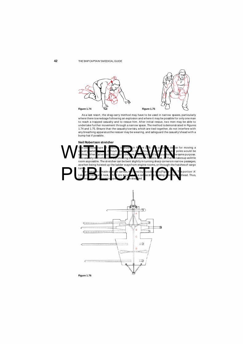
As a last resort, the drag-carry method may have to be used in narrow spaces, particularlywhere there is wreckage following an explosion and where it may be possible for only one manto reach a trapped casualty and to rescue him. After initial rescue, two men may be able toundertake further movement through a narrow space. The method is demonstrated in Figures1.74 and 1.75. Ensure that the casualty’s wrists, which are tied together, do not interfere withany breathing apparatus the rescuer may be wearing, and safeguard the casualty’s head with abump hat if possible.
Neil Robertson stretcher
This particular type of stretcher is shown in Figure 1.76. It is a simple device for moving acasualty safely from a difficult place where the ordinary stretcher with stiff poles would beuseless. Other patterns of rescue stretcher are available but all aim to achieve the same purpose.The casualty is enveloped in a protecting but somewhat flexible case, so that he takes up as littleroom as possible. The stretcher can be bent slightly in turning sharp corners in narrow passages,as when being hoisted up the ladder ways from engine-rooms, or through the hatches of cargotanks.
The stretcher is made of stout canvas, stiffened by wooden slats (Figure 1.76). The portion ‘A’takes the head and neck, which are steadied by a canvas strap passing over the forehead. Thus,the head of an unconscious patient can be steadied.
42 THE SHIP CAPTAIN’S MEDICAL GUIDE
Figure 1.76
Figure 1.74 Figure 1.75
A
B
C
D
WITHDRAWN PUBLICATION
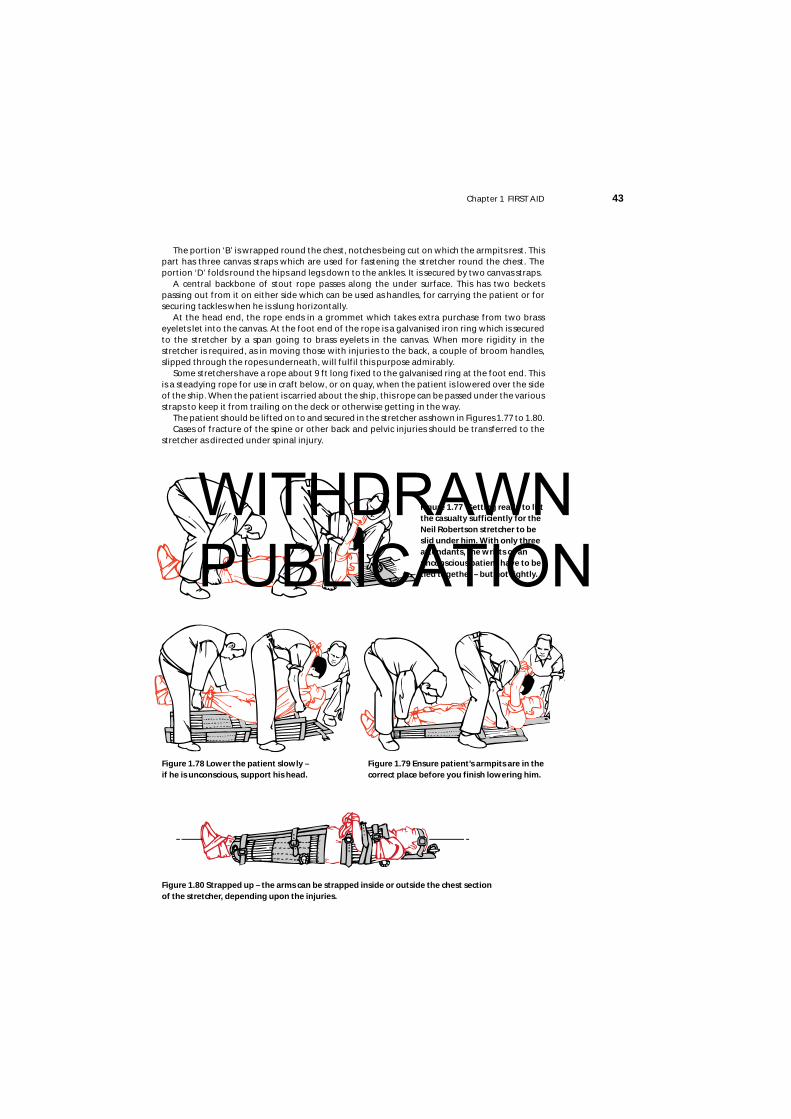
Chapter 1 FIRST AID 43
The portion ‘B’ is wrapped round the chest, notches being cut on which the armpits rest. Thispart has three canvas straps which are used for fastening the stretcher round the chest. Theportion ‘D’ folds round the hips and legs down to the ankles. It is secured by two canvas straps.
A central backbone of stout rope passes along the under surface. This has two becketspassing out from it on either side which can be used as handles, for carrying the patient or forsecuring tackles when he is slung horizontally.
At the head end, the rope ends in a grommet which takes extra purchase from two brasseyelets let into the canvas. At the foot end of the rope is a galvanised iron ring which is securedto the stretcher by a span going to brass eyelets in the canvas. When more rigidity in thestretcher is required, as in moving those with injuries to the back, a couple of broom handles,slipped through the ropes underneath, will fulfil this purpose admirably.
Some stretchers have a rope about 9 ft long fixed to the galvanised ring at the foot end. Thisis a steadying rope for use in craft below, or on quay, when the patient is lowered over the sideof the ship. When the patient is carried about the ship, this rope can be passed under the variousstraps to keep it from trailing on the deck or otherwise getting in the way.
The patient should be lifted on to and secured in the stretcher as shown in Figures 1.77 to 1.80.Cases of fracture of the spine or other back and pelvic injuries should be transferred to the
stretcher as directed under spinal injury.
Figure 1.77 Getting ready to liftthe casualty sufficiently for theNeil Robertson stretcher to beslid under him. With only threeattendants, the wrists of anunconscious patient have to betied together – but not tightly.
Figure 1.80 Strapped up – the arms can be strapped inside or outside the chest section of the stretcher, depending upon the injuries.
Figure 1.78 Lower the patient slowly – if he is unconscious, support his head.
Figure 1.79 Ensure patient’s armpits are in thecorrect place before you finish lowering him.
WITHDRAWN PUBLICATION
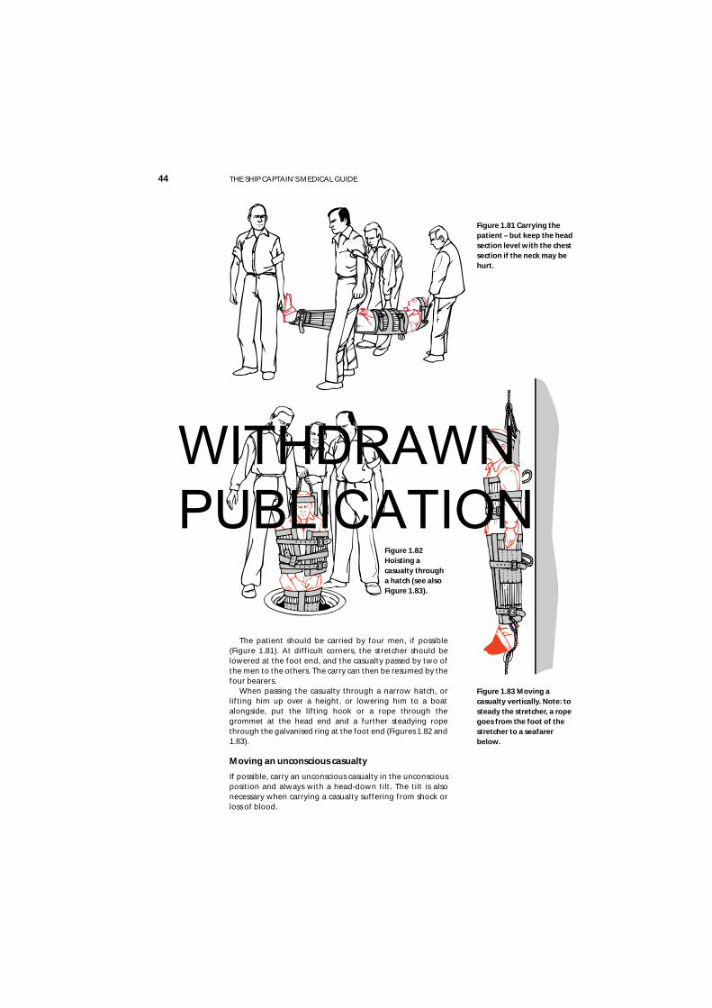
44 THE SHIP CAPTAIN’S MEDICAL GUIDE
Figure 1.82Hoisting acasualty througha hatch (see also Figure 1.83).
Figure 1.81 Carrying thepatient – but keep the headsection level with the chestsection if the neck may behurt.
The patient should be carried by four men, if possible(Figure 1.81). At difficult corners, the stretcher should belowered at the foot end, and the casualty passed by two ofthe men to the others. The carry can then be resumed by thefour bearers.
When passing the casualty through a narrow hatch, orlifting him up over a height, or lowering him to a boatalongside, put the lifting hook or a rope through thegrommet at the head end and a further steadying ropethrough the galvanised ring at the foot end (Figures 1.82 and1.83).
Moving an unconscious casualty
If possible, carry an unconscious casualty in the unconsciousposition and always with a head-down tilt. The tilt is alsonecessary when carrying a casualty suffering from shock orloss of blood.
Figure 1.83 Moving acasualty vertically. Note: tosteady the stretcher, a ropegoes from the foot of thestretcher to a seafarerbelow.
WITHDRAWN PUBLICATION