channel gene cacophony disrupts locomotion in drosophila larvae · research article deletion of a...
TRANSCRIPT
RESEARCH ARTICLE
Deletion of a specific exon in the voltage-gated calciumchannel gene cacophony disrupts locomotion in DrosophilalarvaeKayly M. Lembke1,2, Alexander D. Law2, Jasmine Ahrar2 and David B. Morton2,*
ABSTRACTTAR DNA-binding protein 43 (TDP-43) is an RNA-binding protein thatregulates transcription, translation and alternative splicing of mRNA.We have shown previously that null mutations of the Drosophilaortholog, Tar DNA-binding homolog (tbph), causes severelocomotion defects in larvae that are mediated by a reduction in theexpression of a type II voltage-gated calcium channel, cacophony(cac). We also showed that TDP-43 regulates the inclusion ofalternatively spliced exons of cacophony; tbph mutants showedsignificantly increased expression of cacophony isoforms lackingexon 7, a particularly notable finding as only one out of the 15predicted isoforms lacks exon 7. To investigate the function of exon 7,we generated Drosophila mutant lines with a deletion that eliminatesexon 7. This deletion phenocopies many defects in tbph mutants: areduction in cacophony protein (Dmca1A) expression, locomotiondefects in male and female third instar larvae, disrupted larval motoroutput, and also reduced activity levels in adult male flies. All thesedefects were rescued by expression of cacophony transcriptscontaining exon 7. By contrast, expression of a cacophony cDNAlacking exon 7 resulted in reduced cacophony protein levels andfailed to rescue larval locomotion.
KEY WORDS: ALS, Model organism, Motor program, TDP-43,Dmca1A, Neurodegeneration
INTRODUCTIONThe amyotrophic lateral sclerosis (ALS)- and frontotemporal lobardegeneration (FTLD)-associated protein TARDNA-binding protein43 (TDP-43) is an RNA- and DNA-binding protein that regulatesthe expression and splicing of thousands of gene transcripts(Sephton et al., 2011; Hazelett et al., 2012). We have shownpreviously that loss of the Drosophila TDP-43 ortholog, tbph,caused a decrease in the expression of the primary type II voltage-gated channel, cacophony (Chang et al., 2014). Although tbphmutants showed a 50% reduction in cacophony protein (Dmca1A)expression, there was no decrease in total cacophony transcriptlevels (Chang et al., 2014). Of the cacophony transcripts expressed,there was a significant enrichment for transcripts lacking exon 7,which codes for a portion of the C-terminal cytoplasmic domain of
the protein, suggesting that tbph functions to regulate the inclusionof specific cacophony exons (Chang et al., 2014). Of the 15 reportedcacophony isoforms, only one, cac-RM, lacks exon 7, suggestingthat this exon is of functional importance to the channel (Fig. 1A).
There is evidence that functional specificity of channels can beconferred by the differential expression of discreet channel isoforms(Lipscombe et al., 2013). The cacophony gene encodes the α-subunit of the Drosophila type II voltage-gated calcium channel,which is homologous to the vertebrate type II, N-type voltage-gatedcalcium channel (CaV2.2) (Kawasaki et al., 2002). The CaV2.2channel has been implicated in synaptogenesis, regulation of geneexpression, and neurotransmission at the neuromuscular junction(Brosenitsch and Katz, 2001; Kawasaki et al., 2004). The C-terminus of the gene coordinates channel inactivation, modulationby G-proteins, modulation by calmodulin (CAM; as there is a CAM-binding site), and protein–protein interactions that regulate activityand/or target the channel to specific cellular compartments (Grayet al., 2007). Inclusion and exclusion of specific exons of thechannel are tissue specific and confer functional specificity. Forexample, in mice, exon 37a is expressed preferentially in the dorsalroot ganglia, where it functions to increase the sensitivity of theN-type channel to the voltage-independent form of G-proteinmodulation (Gray et al., 2007). Therefore, natural variants of Cav2.2can regulate the global activity of the channel within the context ofthe cellular milieu. Examples of disease-causing mutations thataltered splicing of voltage-gated calcium channels include FTLD,Parkinsonism linked to chromosome 17, ALS, spinocerebellarataxia 8, myotonic dystrophy and Timothy syndrome (Splawskiet al., 2004; Cooper et al., 2009).
We have shown that loss of tbph caused disrupted larvallocomotion, loss of motor burst pattern and coordination, and ledto altered splicing and reduced levels of cacophony protein(Hazelett et al., 2012; Chang et al., 2014; Lembke et al., 2017).All of these effects were rescued by genetically restoring levels ofcacophony. To further investigate the relationship between splicingof cacophony and motor burst rhythmicity and coordination, wespecifically deleted the exon that appears to be the primary target oftbph, exon 7, using the CRISPR/Cas9 (clustered regularlyinterspaced short palindromic repeats/CRISPR associated gene 9)genome editing system (Gratz et al., 2014).
We found that targeted deletion of exon 7 reduced cacophonyprotein expression and caused locomotion defects in third instarlarvae, similar to those reported in tbph mutants (Hazelett et al.,2012; Chang et al., 2014), but did not change the total expression ofcacophony transcripts. Despite the fact that cacophony is theprimary voltage-gated calcium channel at the neuromuscularjunction (NMJ) and necessary for full evoked neurotransmission(Kawasaki et al., 2002, 2004), we found no consistent reduction inevoked release. Deletion of exon 7 also caused defects in larvalReceived 22 August 2018; Accepted 29 October 2018
1Program in Molecular and Cellular Biosciences, Department of Physiology andPharmacology, Oregon Health & Science University, Portland, OR 97239, USA.2Department of Integrative Biosciences, Oregon Health & Science University,Portland, OR 97239, USA.
*Author for correspondence ([email protected])
A.D.L., 0000-0002-0954-7147; D.B.M., 0000-0002-1320-653X
1
© 2019. Published by The Company of Biologists Ltd | Journal of Experimental Biology (2019) 222, jeb191106. doi:10.1242/jeb.191106
Journal
ofEx
perim
entalB
iology
locomotion and unpatterned, arrhythmic motor bursts similar tothose of tbph mutants. These results also demonstrate a novelapproach to assessing the function of specific exons in alternativelyspliced genes: by precisely deleting specific exons, the contributionof these splice variants in physiology and development can beassessed at the whole-animal level.
MATERIALS AND METHODSFly stocksAll Drosophila stocks were reared at 25°C using standard procedures(Greenspan, 2004). The following fly strains were obtained fromthe Bloomington Drosophila Stock Center (http://flystocks.bio.
indiana.edu): D42-GAL4 motor neuron driver (w[*];P{w[+mW.hs]=GawB}D42), OK6-GAL4 motor neuron driver(P{w[+mW.hs]=GawB}OK6), UAS-cacophony-EGFP (w[*];P{w[+mC]=UAS-cac1-EGFP}786C), genomic duplication coveringthe cacophony gene (w[1118]; Dp(1;3)DC131, PBac{y[+mDint2]w[+mC]=DC131}VK00033). The duplication line carries a77,150 bp genomic fragment on the third chromosome, whichincludes the cacophony locus and an additional 23,342 bp offlanking sequence. The cacexon7Δ mutants were crossed with w1118
at least four times to minimize any off-target effects caused byCRISPR/Cas9 (Lee et al., 2016) and to reduce differences in thegenetic background between lines.
Exon 7
cac-RM
A
B
D
E
FC
Contro
l
cac
cac
bp400
EF handCAM binding
Exon 7N
C
Ala Leu His Leu Leu Asp
mRNA
300200
100
Exon 6 Exon 8
p1
6
6
6
7 8
8
6 8
DsRed
6 7 8
6 8
7 8
p2
Cas9pHD-DsRed-attP
Permanentexon removal
cacexon7Δ
cace
xon7Δ
cacexon7Δ
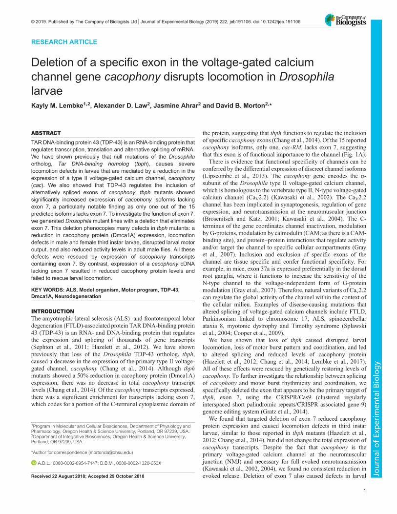
Fig. 1. Generation of cacophony exon 7 deletion lines. (A) Predicted alternatively spliced cacophony transcripts from FlyBase, showing the position of exon 7and that a single predicted isoform, cac-RM, lacks exon 7. (B) Schematic diagram showing the process of using CRISPR/Cas9 to delete exon 7 of cacophony.Cas9 was targeted to cut the genomic DNA on either side of exon 7, resulting in two double-stranded breaks, which were repaired via homologousrecombination with a donor template containing DsRed, which was then incorporated into the genome. Splicing between exon 6 and 8 excludes the DsRedcassette, yielding cacexon7Δ mRNA. (C) Schematic diagram of the predicted cacophony protein, showing the location of exon 7 near the C-terminus.(D) Location of the primers in exon 6 and 8 used for RT-PCR to confirm the deletion of exon 7. (E) Representative DNA gel of the RT-PCR products from adultheads, showing a major 300 bp band that includes exon 7 and a minor 100 bp band lacking exon 7 from control animals. By contrast, RT-PCR from thecacexon7Δ line only yielded the 100 bp band. (F) Sequencing chromatogram resulting from the sequencing of the 100 bp band from the cacexon7Δ line showedthat exon 6 splices directly to exon 8, excluding exon 7.
2
RESEARCH ARTICLE Journal of Experimental Biology (2019) 222, jeb191106. doi:10.1242/jeb.191106
Journal
ofEx
perim
entalB
iology

Generation of the cacexon7Δ mutantsA schematic diagram of the strategy used to generate flies in whichexon 7 of cacophony was deleted is shown in Fig. 1. Fly embryosexpressing the endonuclease Cas9 under the control of vas regulatorysequences (w[1118]; PBac{y[+mDint2]=vas-Cas9}VK00037/CyO,P{w[+mC]=Tb[1]}Cpr[CyO-A]) were injected by BestGene (ChinoHills, CA, USA) with two plasmids. One plasmid contained a donortemplate and was injected at a concentration of 500 ng µl−1. Thedonor template contained two regions, each of about 1 kb, ofcacophony sequence from either side of exon 7, which were separatedby sequence coding for a red fluorescent protein (DsRed) under thecontrol of an eye-specific promoter. The other plasmid contained twoguide RNAs and was injected at a concentration of 200 ng µl−1. Theguide RNAs target Cas9 to sites on either side of exon 7 – 382 bpupstream and 562 bp downstream – resulting in two double-strandedbreaks. This break was then repaired by homologous recombinationusing the donor template.The donor template was generated by using PCR to amplify two
homology arms that immediately flank exon 7 of cacophony andincorporate them into the pHD-DsRed-attP vector (Gratz et al.,2014). Since both homology arms did not perfectly flank exon 7,this incorporation resulted in a loss of 41 bp of intron on the 5′ endof exon 7 and a loss of 27 bp on the 3′ end of exon 7. About 1 kb ofcacophony sequence was amplified from the P[acman] BACCH321-64N05 library (Venken et al., 2009) for both the 5′ andthe 3′ homology arms (using primers 5′ homology arm S and R andprimers 3′ homology arm S and R, Table 1, respectively). The 5′fragment was incorporated into the AarI site of the 5′ multiplecloning site (MCS) in the pHD-DsRed-attP vector and the 3′fragment was incorporated into the SapI site of 3′ MCS in the pHD-DsRed-attP vector. Protospacer adjacent motif (PAM) sequences thatwould be used as targets by the guide RNAs were then removed fromthe homology arms using primers [5′ and 3′ PAM site-directedmutagenesis (SDM); Table 1] designed through the NEBaseChangerweb tool. The mutated homology armswere then generated followingthe instructions of the Q5 site-directedmutagenesis kit (New EnglandBioLabs, MA, USA) and confirmed by sequencing.The guide RNA (gRNA) sequences were chosen using the
Drosophila RNAi Screening Center CRISPR2 web tool (http://www.flyrnai.org/crispr2/). The two sequences selected were located
in the introns between exons 7 and 8 and between exons 6 and 7 ofcacophony. These two gRNAs (see Table 1, 5′ gRNA and 3′ gRNA)were cloned into two sibling vectors, pBFv-U6.2 and pBFv-U6.2B,respectively (Kondo and Ueda, 2013). Finally, these twoindependent gRNA cassettes were fused into a single plasmid forincreased injection efficiency (Kondo and Ueda, 2013).
Generation of the UAS-cacophony-EGFP and UAS-cacexon7Δ-EGFP fliesFly embryos with phiC31 (ϕC31) integration sites (y[1] w[67c23];P{y[+t7.7]=CaryP}attP2) were injected by BestGene with one oftwo transformation plasmids. One plasmid contained a cacophonycDNA (cac1) fused to the EGFP coding sequence as describedpreviously (Kawasaki et al., 2002), while the other was constructedwith the same cac EGFP-fused coding sequence with a deletion toexclude exon 7 of cacophony. To generate cacophony sequencewithout exon 7, DNAwas synthesized (Biomatik, Wilmington, DE,USA) at a length of 1515 bp using sequence from FlyBase as areference with exon 7 removed. The synthesized sequencecontained the restriction sites AgeI on the 5′ end and KpnI on the3′ end, which were also present in cac1. The synthesized sequencewas cut out of its cloning vector using AgeI and KpnI and shuttledinto cac1. The final constructs were incorporated into thetransformation vector pUASTattB (Bischof et al., 2007) andsequenced to confirm the lack of exon 7. The transformationconstructs were prepared for injection using ZymoPURE PlasmidMini Prep Kit (Zymo Research, Irvine, CA, USA) and injected at aconcentration of 500 ng µl−1.
Genotyping of the cacexon7Δ mutantsTwo sets of primers were designed to span a region that includedthe DsRed sequence at the 5′ end of cacophony and the 5′homology arm (primers 5′ GT S and 5′ GT R; Table 1) and theregion spanning DsRed at the 3′ end of cacophony and 3′homology arm (primers 3′ GT S and 3′ GT R; Table 1). PCR wasthen performed using both sets of primers and Q5 high-fidelitypolymerase (New England BioLabs). Both fragments were thenTA-cloned for sequencing to confirm the expected sequence ofthe cacophony genomic region in which exon 7 was replaced bythe DsRed cassette.
Table 1. Sequences of the primers used in this study (5′–3′)5′ homology arm S AAAAGCTCTTCATATATTAAGGATGTCGGGGCACA5′ homology arm R AAAAGCTCTTCAGATTTCCCCAAACCCCGTAA3′ homology arm S AAAACACCTGCAAAATCGCTGCCTGCATATCTGTGGTAGT5′ homology arm R AAAACACCTGCAAAACTACGTCCAACAAGCTCAATCAAAA5′ gRNA AAACTACATACGAGATGTGATTAC3′ gRNA CTTCGTAATCACATCTCGTATGTA5′ PAM SDM S TGTGTGTGCCAATTGCAAGCCGATC5′ PAM SDM R GAAAACAAAGGGACTTGGAC3′ PAM SDM S CTCGTATGTATAATGCTGACATGTACAG3′ PAM SDM R ATGTGATTATAATATAGGTGGATG5′ GT S ACGGCTCCTTCATCTACAAGG5′ GT R CGATTCGACCATCTGGAAGT3′ GT S ACATTGCCTGCATATCTGTGG3′ GT R AAGCGCATGAACTCCTTGATcacEx6-8 S GCCAGTGTTGGGTAACTATGCcacEx6-8 R TGTTGTAGAGATGGTCAAGGAGACqRT-cac S ATAGCGTTCGAGAGGTCGTGqRT-cac R AGAGCAAGGCGAAGCTGAGTqRT-EF2b S GCGTTCACCCTCAAGCAGTTCTqRT-EF2b R AGCGTTTGTTGTCAGCCTCTTTCT
All primers were designed using Primer3 and modified using protocols found at flycrispr.molbio.wisc.edu as needed. S, sense; R, reverse; gRNA, guide RNA;PAM, protospacer adjacent motif; SDM, site-directed mutagenesis; GT, genotype.
3
RESEARCH ARTICLE Journal of Experimental Biology (2019) 222, jeb191106. doi:10.1242/jeb.191106
Journal
ofEx
perim
entalB
iology

Real-time reverse transcription (RT)-PCRTotal RNA was extracted from either adult heads or larval CNSusing Trizol (Life Technologies, Carlsbad, CA, USA) and cDNAsynthesis was performed using SuperScript III (Life Technologies).To confirm that splicing took place as expected in the absence ofexon 7, sequence from exon 6 to exon 8 was amplified with Q5using primers cacEx6-8 S and cacEx6-8 R (see Table 1) and TA-cloned for sequencing. Real-time RT-PCR was used to determinethe relative expression levels of cacophony in control flies and in thecacexon7Δ flies. Primer pairs against exon 23 and exon 24 ofcacophony, and against EF2b as an internal control, were used for real-time RT-PCR reactions (see Table 1). Exons 23 and 24 were chosenbecause they are conserved in all predicted transcripts on FlyBase.Real-time RT-PCR was performed in a StepOne thermocycler (LifeTechnologies) using Power SYBR Green PCR Master Mix (AppliedBiosystems, Warrington, UK). The PCR reaction conditions were 95°C for 10 min, followed by 40 cycles of 15 s at 95°C, 30 s at 63°C and40 s at 72°C.Delta Ct (ΔCt) values were calculated as the mean value of
cacophony Ct minus the mean value of EF2b Ct. For larval CNSsamples, a single technical replicate and 3 biological replicates wereused and, for adult heads, 3 technical replicates and 4 biologicalreplicates were processed. For each sample, the ΔCt for eachbiological replicate was calculated and normalized to the values ofcontrols for relative expression levels.
ImmunoblottingThe following antisera and antibodies (dilution; catalog number;source) were used for immunoblotting: rabbit anti-cacophony[1:4000; as previously described (Chang et al., 2014)], mouseanti-GAPDH (1:333; SC-365062; Santa Cruz, Dallas, TX, USA),chicken anti-GFP (1:2500; GFP-1020; Aves Labs, Inc., Tigard, OR,USA), mouse anti-rabbit H+L (1:2500; 211-005-109), goat anti-mouse H+L (1:2500; 115-005-146), goat anti-mouse peroxidaseconjugate (1:5000; 115-035-003), rabbit anti-goat peroxidase conjugate(1:5000; 305-035-045) and goat anti-chicken peroxidase conjugate(1:10,000; 103-035-155) all from Jackson ImmunoResearch, WestGrove, PA, USA.Adult fly heads were dissected on a cold plate, homogenized in
LDS loading buffer (Life Technologies) containing proteaseinhibitor (Roche Molecular Systems, Pleasanton, CA, USA) andallowed to incubate at room temperature for 45 min. TCEP was usedto reduce samples at 75°C for 10 min, followed by centrifugation at10,000 g for 15 min. The supernatant was kept, denatured at 75°Cfor an additional 10 min and proteins were fractionated using SDS-PAGE. Following transfer to a PVDF (polyvinylidene difluoride)membrane (Life Technologies), the membranes were blocked with0.1% gelatin in PBST (PBS containing 0.05% Tween-20). Blotswere then incubated in antisera against cacophony, GFP or GAPDH,followed by incubation with mouse anti-rabbit H+L or goat anti-mouse H+L antisera, and finally HRP-conjugated goat anti-mouse,HRP-conjugated rabbit anti-goat antisera or HRP-conjugated goatanti-chicken antisera as appropriate. The proteins were then detectedwith enhanced chemiluminescence DuoLuX (Vector laboratories,Inc., Burlingame, CA, USA). The relative cacophony levels werenormalized to GAPDH levels using Fiji (Schindelin et al., 2012).
Longevity and behavioral assaysThe cacexon7Δ mutants were outcrossed to control flies (describedabove) at least 4 times to minimize the effects of different geneticbackgrounds and potential CRISPRoff-target effects. The longevityof each linewas assessed by collecting virgin males and females and
placing them in separate vials of 10–15 animals in each vial. Vialswere inspected for dead flies every other day and the remaining fliesplaced in new vials every 7 days.
To assess the climbing ability of adult flies, we carried outnegative geotaxis assays on adult male flies as previously described(Gargano et al., 2005). Groups of 15 flies were placed in vials andthe distance that individual flies climbed up the wall of a vial in 4 swas measured each week. Adult locomotory activity was assessedfor 1-week-old individual male flies using an activity monitor(TriKinetics, Waltham, MA, USA) as previously described(Vanderwerf et al., 2015). Adult activity was logged as the totalactivity over the first 24 h the flies were in the recorder.
Larval locomotion was determined as previously described(Lembke et al., 2017). Third instar larvae were rinsed in PBS andplaced on 2% agarose plates at room temperature. The crawlingpaths of the larvae were recorded for 5 min using a moticam 1000connected to a PC and using theMIPlus07 software (Motic Images).The distance traveled in each video was traced and quantified usingImageJ software (http://imagej.nih.gov/ij/). The number of fullposterior–anterior peristaltic waveforms was also recorded fromthese videos for each genotype.
Electrophysiological methodsIntracellular recordings were made from larval body wall muscle 6in abdominal segment 3. Recordings were performed using glassmicroelectrodes as previously described (Lembke et al., 2017) andwere performed at room temperature in extracellular HL3 salinecontaining (in mM): 70 NaCl, 5 KCl, 20 MgCl2, 10 NaHCO3, 115sucrose, 5 trehalose, 5 HEPES and 1.0 CaCl2. Membrane potentialswere recorded using an Axoclamp-2A amplifier (MolecularDevices) connected to a PC (Dell) and only recordings withresting membrane potential at or more negative than −55 mV wereused. Excitatory junctional potentials (EJPs) were evoked byinjection of current into severed axons, at 0.5 Hz, via a suctionelectrode and an A310 Accupulser (World Precision Instruments)through an isolation transformer. Miniature end plate potentials(mEPPs) were recorded over 3 min and analyzed using MiniAnalysis 6.0.0.7 (Synaptosoft). The average single EJP amplitudeof each recording was taken from 30 EPSPs, whose amplitudes weremeasured using Clampfit 10.2 software (Molecular Devices, AxonInstruments).
Motor activity recordings were made from peripheral nervesprojecting from the second and seventh neuromeres of the intactCNS in third instar larvae as previously reported (Lembke et al.,2017). Nerves were suctioned en passant with a glass suctionelectrode in HL saline containing the following (in mmol ml–1): 70NaCl, 5 KCl, 4 MgCl2, 10 NaHCO3, 115 sucrose, 5 trehalose, 5HEPES and 1.8 CaCl2. Preparations were acutely incubated with 30μmol ml–1 pilocarpine to stimulate fictive crawling and activate themotor program (Johnston and Levine, 1996). Recordings were doneusing an A-M Systems Differential AC Amplifier, digitized at10 kHz, and stored with the Digidata 1440A digitizer as above.Recordings were made over 10 min and bandpass filtered (100 Hzto 10 kHz) using Clampex software (Molecular Devices).Autocorrelations and cross-correlations were computed for eachtrace as previously described (Lembke et al., 2017).
Experimental design and statistical analysisGraphPad Prism 6 software was used to generate the graphs in thiswork. Data sets were analyzed for significance using one-way ortwo-way ANOVA followed by Dunnett’s multiple comparisoncorrection two-tailed t-test or Mantel–Cox log ranked test as stated
4
RESEARCH ARTICLE Journal of Experimental Biology (2019) 222, jeb191106. doi:10.1242/jeb.191106
Journal
ofEx
perim
entalB
iology

in each figure legend. A 95% confidence interval was used todetermine significance (P<0.05). See figure legends for samplesizes, P-values, and associated F- and t-values.
RESULTSDeletion of exon 7 caused a decrease in cacophony proteinexpression but not total transcript levelsOur previous data showed that loss of tbph resulted in defectivelarval locomotion and reduction in the inclusion of exon 7 incacophony (Hazelett et al., 2012; Chang et al., 2014). Furthermore,a causal relationship between cacophony and tbph-dependent larvallocomotion was demonstrated by rescuing larval locomotion withincreased expression of cacophony (Chang et al., 2014; Lembkeet al., 2017). The cacophony cDNA used in these studies includedexon 7, raising the question of whether larval locomotion requiredthe specific inclusion of exon 7 or whether simply increased levelsof cacophony were sufficient. To begin to address this question weused the CRISPR/Cas9 genome editing system to create a deletioncovering exon 7 of cacophony (Fig. 1). Two plasmids wereconstructed, one that contained sequences complementary to thegenomic sequence to direct Cas9-mediated cleavage of the genomeon either side of exon 7 and the other containing complementarysequences on either side of exon 7, to direct homologous mediatedrecombination after exon 7 had been removed (Fig. 1B). Thissecond plasmid also contained a cassette for eye-specific expressionof DsRed flanked by splice acceptor and donor sequences (Gratzet al., 2014). Exon 7 codes for a 66-residue sequence at theC-terminal tail of cacophony (Fig. 1C). Drosophila embryos thatexpressed Cas9 in germline cells (Gratz et al., 2014) were injectedwith these two plasmids and 21 independent lines expressing DsRedwere isolated. Two of these lines were homozygous viable, whereasthe remaining 19 lines were homozygous lethal. We crossed the 19lethal lines to a line containing a duplication that covers the genomicregion of cacophony (DC131) and found that it rescued viability,indicating that the lethality locus was close to, or within, thecacophony locus. We then tested to see whether the lethality couldbe rescued by expressing a cacophony cDNA using the pan-neuronal Appl-GAL4 driver. We failed to observe any rescue oflethality, suggesting that the lethality was not due to loss ofcacophony expression, but possibly due to off-target CRISPReffects, located nearby cacophony. For all the results described inthis article, both of the homozygous viable lines gave similar resultsto each other and the results reported are for one of these lines. RT-PCR using primers that hybridized to exon 6 and 8 (Fig. 1D) usingRNA from control animals generated a major 306 bp band expectedfor a product that included exon 7 and a minor band of 105 bpexpected for a product that lacked exon 7. The mutant line, namedcacexon7Δ, generated only this 105 bp product (Fig. 1E). When wesequenced this PCR product, the results showed that correct splicingbetween exon 6 and 8 had occurred, with a resulting sequence thatwas in frame and predicted to generate a full-length protein(Fig. 1F).To determine whether the deletion had an impact on the overall
transcription of cacophony or stability of the transcript, we usedreal-time RT-PCR with a primer pair spanning the intron betweenexon 24 and exon 23 (common to all cacophony isoforms)(Table 1). Using heads from adult males or females or wholelarvae as a source of RNA showed that there was no difference in thelevels of cacophony transcript between control or cacexon7Δ flies(Fig. 2A). We then examined the levels of cacophony protein, usingimmunoblots and an antibody specific to cacophony (Chang et al.,2014). Most of the different predicted isoforms of cacophony
generate proteins with predicted sizes ranging from 212.1 to212.6 kDa with cac-PM, the isoform lacking exon 7, having apredicted size of 204.9 kDa. These differences in size are notdiscernable on immunoblots and, as predicted, a single band with anapparent size of about 205 kDa was detected on immunoblots(Chang et al., 2014). Immunoblots of either adult heads or larvalCNS from the cacexon7Δ line also showed a single band of this size(Fig. 2B). In addition, these blots showed that there was a reduction
Adult m
ales
Adult f
emale
sLa
rvae
0
0.5
1.0
1.5
0
0.5
1.0
1.5
Rel
ativ
e ex
pres
sion
Rel
ativ
e ex
pres
sion
Controlcacexon7Δ
Contro
l
cace
xon7Δ
Contro
l
cace
xon7Δ
*** ***
A
B
Adult
Larva
e
cacGAPDH
cacGAPDH
Fig. 2. Quantification of cacophony transcripts and protein. (A) Real-timeRT-PCR showed no significant difference in the levels of cacophony transcriptbetween controls and the cacexon7Δ line for male or female heads or larvalCNS. For each sample, primers against exons conserved across all predictedcacophony transcripts and against the loading control, EF2b, were used. Therelative expression levels were calculated relative to EF2b and normalized tothe relative expression in control flies. Mean and s.e.m. from four independentsamples for adult heads and three independent samples for larval CNS areshown. Two-way ANOVA showed no significant differences between controland mutant lines (F2,16=1.62; P=0.23; P>0.05). (B) The levels of cacophonyprotein are reduced in cacexon7Δ mutants in both larvae and adults. Proteinsfrom heads of adults or whole larvae were separated by PAGE, transferred toPVDF membrane and probed with cacophony protein antisera (Chang et al.,2014) and GAPDH as a loading control. The intensity of the cacophonyimmunoreactive band was quantified, divided by the intensity of itscorresponding GAPDH band and normalized to the ratio obtained for thecontrol sample. Mean and s.e.m. from four independent samples for adultheads and three independent samples for larvae are shown. Two-way ANOVAfollowed by Dunnett’s multiple comparison test showed a significant reductionof cacophony protein levels in the cacexon7Δ mutant in both adults and larvaecompared with controls (F1,12=42.7; ***P<0.001).
5
RESEARCH ARTICLE Journal of Experimental Biology (2019) 222, jeb191106. doi:10.1242/jeb.191106
Journal
ofEx
perim
entalB
iology
in the intensity of the band in the cacexon7Δ line compared withcontrols (Fig. 2B). We quantified the intensity of the bands,revealing a significant reduction in the level of cacophony proteindetected in the mutant lines using either adult heads or larval CNS(Fig. 2B). After characterizing the molecular characteristics of themutant line, we examined their phenotypes.
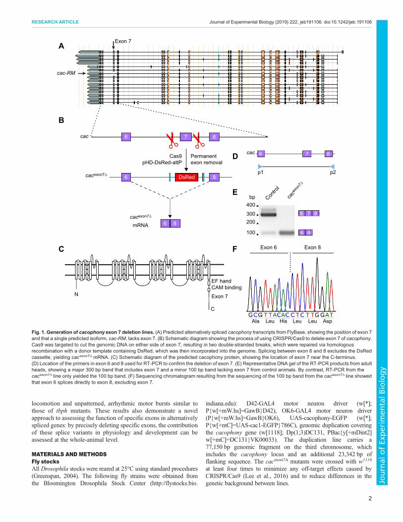
Deletion of exon 7 caused reduced locomotory activity inadultsTo determine whether there were any deleterious effects of thisreduced level of cacophony, we assessed the longevity of cacexon7Δ
flies. These data, shown in Fig. 3A and B, revealed that there was aslight, but statistically significant, reduction in the median lifespanin both male and female flies.We also assessed the general activity levels of the cacexon7Δ flies
in a bioactivity recorder, which showed that male cacexon7Δ flies hadreduced levels of activity compared with controls. To confirm thatthis defect was due to changes in cacophony, we used a duplicationline (DpDC131) that contained a duplication of a portion of the Xchromosome that spans cacophony and includes the predicted
promotor region. When we combined this duplication with the exon7 deletion, the activity of the flies was restored to normal (Fig. 3C).We then used the pan-neuronal Appl-GAL4 driver and a UAS-cacophony construct that contained exon 7 (Kawasaki et al., 2004)to determine whether neuronal expression of exon 7 (+) cacophonywas required for normal adult activity. The results shown in Fig. 3Cshowed that this was sufficient to restore the levels of activity in thecacexon7Δ flies. To assess the climbing ability of adult flies, weperformed negative geotaxis assays by measuring the distance theyclimbed in 4 s. The results of this assay are shown in Fig. 3D:cacexon7Δ flies performed as well as control flies and showed similarlevels of reduction in performance with age. In addition to assessingthe adult phenotypes, we also examined phenotypes in larvae.
Deletion of exon 7 caused locomotion defects in third instarlarvaeWe had previously shown that tbph mutants show cacophony-dependent defective larval locomotion (Chang et al., 2014; Lembkeet al., 2017). We therefore tested whether the cacexon7Δ mutantsshowed similar defects in larval locomotion. Total distance crawled
A
1 2 3 4 5 6 70
1
2
3
4
Age (weeks)
Dis
tanc
e cl
imbe
d (c
m)
Negative geotaxisC D
0 20 40 60 80 1000
20
40
60
80
100
Longevity – Males
0 20 40 60 80 100Time (days)
Per
cent
sur
viva
l
Controlcacexon7Δ
Controlcacexon7Δ
Contro
l
cace
xon7Δ
cace
xon7Δ ;D
pDC13
1
0
20
40
60
80
100
Longevity – Females
Appl, c
acex
on7Δ ; c
ac0
500
1000
1500
Adult activity
Act
ivity
(cou
nts
24 h
–1)
**
**
B
Fig. 3. Adult phenotypes of the cacexon7Δmutant. Survival plots of male (A) and female (B) adult flies, showing a slightly reduced median lifespan in both maleand female flies: males: cacexon7Δ, 54 days (N=38), compared with controls, 70 days (N=39) (Mantel–Cox log ranked test, P=0.02); females: cacexon7Δ, 52 days(N=34), compared with controls, 70 days (N=40) (Mantel–Cox log ranked test, P=0.0002). (C) Activity levels of individual adult male flies. The level of activityover 24 h of the cacexon7Δ mutant (N=36) was significantly reduced compared with control flies (N=57). This reduced activity was restored with the presenceof a genomic duplication (DpDC131) (N=31) or by expressing cacophony in all neurons using a pan-neuronal GAL4 driver (Appl) (N=21). The data were analyzedusing one-way ANOVA followed by Dunnett’s multiple comparison test. (F3,144=12.1; **P<0.01). (D) Negative geotaxis assay showed no significant differencein the performance of the cacexon7Δ mutant (N=9) compared to controls (N=8) (two-way ANOVA followed by Dunnett’s multiple comparison test; P>0.05).
6
RESEARCH ARTICLE Journal of Experimental Biology (2019) 222, jeb191106. doi:10.1242/jeb.191106
Journal
ofEx
perim
entalB
iology

wasmeasured in third instar larvae and bothmale and female mutantlarvae showed decreased crawling distance (Fig. 4A). To confirmthat this defect was due to changes in cacophony, we again used theDpDC131 duplication line. When we combined this duplication withthe cacexon7Δ mutants, the distance crawled by the larvae wasrestored to normal (Fig. 4B). We then used a variety of cell-specificGAL4 drivers and a UAS-cacophony construct that contained exon7 (Kawasaki et al., 2004) to determinewhich neurons required exon7 (+) cacophony for normal larval locomotion. Broad motor neuronGAL4 drivers (D42 and OK6) were sufficient to fully rescue thetotal distance crawled by larvae (Fig. 4B). We also used acholinergic driver (Cha-GAL4) to drive expression of cacophonyin sensory neurons and interneurons, which failed to rescue thecrawling defect (Fig. 4B). Our previous studies (Lembke et al.,2017) identified a GAL4 driver line (R75C03) that expressed intwo pairs of neurons in the brain that, when used to expresscacophony in tbphmutants, was capable of fully rescuing the larvalcrawling defects. When we used this line to drive cacophony in thecacexon7Δ mutant larvae, we also measured a significant increase incrawling distance, although it did not fully rescue the defect(Fig. 4B).
Larval crawling is a highly stereotyped behavior consisting ofposterior to anterior peristaltic waves that travel the length of thelarva (Fox et al., 2006; Inada et al., 2011). To analyze in more detailthe locomotion defect of the deletion lines, we measured thefrequency of the peristaltic waves. As expected, the cacexon7Δ
mutant larvae showed a significant reduction in the frequency ofperistaltic waves, which was rescued with the DpDC131 duplicationand expression of cacophony using the D42 motor neuron driver(Fig. 4C). These defects in larval locomotion suggested defects atthe NMJ, which we also examined.
Synaptic neurotransmission at the NMJDrosophila cacophony is necessary for evoked neurotransmission atthe NMJ (Kawasaki et al., 2004; Lee et al., 2014) and the reducedlevels of cacophony protein in the exon 7 deletion lines suggestedthat there could also be defects in neurotransmission at the NMJ. Toexamine whether there were any defects in the cacexon7Δ mutants,we recorded evoked and spontaneous transmission at the larval NMJof body wall muscle 6 (Fig. 5). Surprisingly, when we measuredthe EJP amplitudes of the cacexon7Δ mutants, we found that therewas no change in the EJP amplitude when compared to controls(Fig. 5A,B).
We had previously shown that reduction in endogenouscacophony protein at the larval NMJ caused reduced frequenciesof spontaneous neurotransmitter release (Lembke et al., 2017), andwe also examined spontaneous neurotransmission by measuring theamplitude and frequency of mEPPs (Fig. 5C–E). The mEPPamplitude was unchanged in cacexon7Δ mutants, but the frequencywas significantly reduced (Fig. 5D,E). We then examined the effectof incorporating the DpDC131 duplication and also of expressingcacophony cDNA in all motor neurons, and found that neither theduplication nor expressing cacophony in all motor neuronsincreased the frequency of mEPPs, suggesting that the reductionin mEPP frequency was not due to the deletion of exon 7 fromcacophony (Fig. 5E). The absence of major defects in synaptictransmission suggested defects in the motor output of the nervoussystem.
Motor pattern outputThe cacexon7Δ mutant animals showed a reduction in the frequencyof peristaltic waves (Fig. 4C). This phenotype suggested that there
0
0.2
0.4
0.6
0.8
1.0
Freq
uenc
y of
per
ista
ltic
wav
es (H
z)
****
********
Contro
l
Contro
l
0
5
10
15
20
Dis
tanc
e cr
awle
d (c
m)
Dis
tanc
e cr
awle
d (c
m)
MalesFemales
*****
Contro
l
cace
xon7Δ ;D
42>c
ac
cace
xon7Δ ;O
K6>ca
c
cace
xon7Δ ;C
ha>c
ac
cace
xon7Δ ;D
42>c
ac
cace
xon7Δ ;R
75CO3>
cac
0
5
10
15
20
*******
****
****
**
A
C
Bca
cexo
n7Δ
cace
xon7Δ
cace
xon7Δ
cace
xon7Δ ;D
pDC13
1
cace
xon7Δ ;D
pDC13
1
Fig. 4. Larval locomotion is disrupted in the cacexon7Δ mutant. Third instarlarvae were allowed to crawl across plain agar plates and the total distancetraveled in 5 min was measured. (A) The total distance crawled in 5 min wassignificantly reduced in the cacexon7Δ mutant in both male (N=53 for controlsand N=38 for mutants) and female (N=8 for controls and N=10 for mutants)larvae (two-way ANOVA followed by Sidak’s multiple comparisons test,F2105=31.0; *P<0.05, ****P<0.0001). (B) Restoration of crawling in male larvaeby expression of cacophony in motor neurons. The distance crawled by thecacexon7Δ mutants (N=38) was significantly reduced compared with controls(N=53) and was significantly increased by the presence of a genomicduplication (DpDC131; N=32) or by expressing cacophony in motor neuronsusing either the D42 (N=32) or OK6 (N=20) GAL4 drivers. No significantincrease was seen with a cholinergic driver (Cha-GAL4; N=17) and asignificant rescue was seen using the R75CO3 driver (N=19). One-wayANOVA followed by Dunnett’s multiple comparisons test; F6,204=15.3;**P<0.01, ***P<0.001, ****P<0.0001. (C) The frequency of peristaltic waves inmale larvae was significantly reduced in cacexon7Δ mutants (N=9) comparedwith controls (N=19) and was significantly increased by the presence of agenomic duplication (DpDC131) (N=10) or by expressing cacophony in motorneurons using the D42 GAL4 driver (N=10). One-way ANOVA followed byDunnett’s multiple comparisons test; F3,44=13.9; ****P<0.0001.
7
RESEARCH ARTICLE Journal of Experimental Biology (2019) 222, jeb191106. doi:10.1242/jeb.191106
Journal
ofEx
perim
entalB
iology

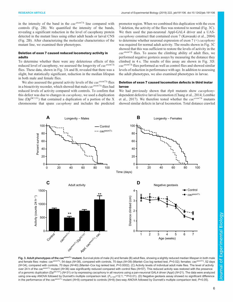
was also defective motor output from the CNS (Fox et al., 2006;Lembke et al., 2017), and we therefore monitored the motor outputfrom the CNS in semi-intact larvae.
Focal extracellular recordings were made en passant from intactperipheral nerves projecting to muscle 6/7 in abdominal segment 2(A2) and abdominal segment 7 (A7), as previously described(Lembke et al., 2017) (Fig. 6A). In the presence of pilocarpine, thecacexon7Δ deletion line show a significant reduction in the frequencyof non-random motor bursts (Fig. 6A,B), which was rescued bydriving cacophony in motor neurons using the OK6-GAL4 driver(Fig. 6B), suggesting that deletion of exon 7 significantly affectsmotor burst frequency.
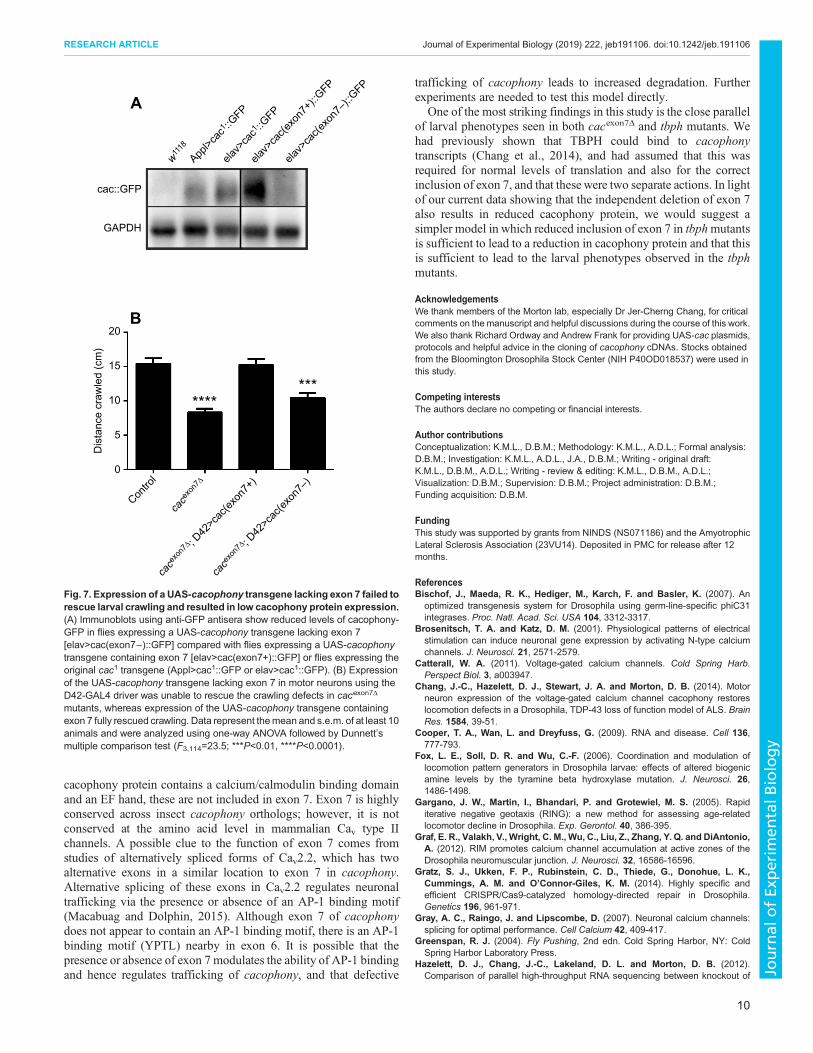
Exon 7 is required for full protein expression and restorationof larval locomotionDeletion of exon 7 caused reduced cacophony protein levels anddefective larval locomotion. To determine whether these twophenotypes are causally related, we generated two new UAS-cacophony lines. One was generated using the same cacophonycDNA used to make the original UAS-cac1 line (Kawasaki et al.,2002) and the other was constructed using the same cDNA asstarting material but engineered to delete exon 7. Both constructswere generated in plasmids containing attB sites for site-specificintegration into the genome using the phiC3 system to ensureequivalent expression levels (Bischof et al., 2007). We first crossedboth lines with a pan-neuronal GAL4 driver and immunoblottedadult heads with an anti-GFP antibody to determine whetherequivalent levels of cacophony-EGFP protein were present(Fig. 7A). Both the UAS-cac (exon 7+) line and the originalUAS-cac1 line resulted in a robust GFP signal, whereas there was anotably lower intensity signal from the UAS-cac (exon 7−) line.
To test whether the presence of exon 7 in the cDNA had anyeffect on larval crawling, we drove its expression in motor neuronsusing the D42-GAL4motor neuron driver. The larval locomotion ofthe cacexon7Δ mutant was rescued by cac(exon7+), whereas drivingcac(exon7−) expression with D42-GAL4 failed to rescue crawling,suggesting that normal larval movement depended on theexpression of exon 7.
DISCUSSIONDrosophila cacophony is a member of the type II voltage-gatedcalcium channel family, member of which act to regulate manyneuronal processes, including synaptic neurotransmission at theNMJ, synaptogenesis and synaptic homeostasis, as well asregulation of gene expression (Catterall, 2011). Here, we describethe generation of a new cacophony mutant fly line that has a singleexon deletion, which eliminates exon 7 in the coding region of theprotein. The cacophony gene has multiple splice variants and, inwild-type animals, exon 7 is included in all but one predicted splicevariant. RT-PCR showed that, in wild-type larvae, approximately50% of the total level of cacophonymRNA contains exon 7 (Changet al., 2014), whereas, in adult heads fromwild-type animals, almostall of the cacophony transcripts include exon 7 (Fig. 1E). Thiswould have suggested that any phenotypes resulting from the loss ofexon 7 would be more severe in adults compared with larvae.Surprisingly, however, we did not detect any dramatic phenotypesin adults: their survival and longevity were similar to controlanimals and negative geotaxis was unaffected. The only significanteffect was that the total level of locomotory activity was reduced in acacophony-dependent manner. Interestingly, larval locomotion wasalso reduced in cacexon7Δ animals, which could be rescued by theexpression of cacophony in motor neurons.
Our rationale for developing these mutants was our finding thatnull mutations in tbph, the Drosophila ortholog of the ALS- andFTLD-associated gene, TARDBP, caused a reduction in the
5 m
V
50 ms
0
0
0
1
2
3
4
5
****
0.2
0.4
0.6
0.8
1.0
1.2
5
10
15
20
25
A B
C D
E
0.5m
V
200 ms
mE
PP
frequ
ency
(Hz)
mE
PP
ampl
itude
(mV
)E
JP a
mpl
itude
(mV
)Con
trol
cace
xon7Δ
Contro
l
cace
xon7Δ
Contro
l
cace
xon7Δ
cace
xon7Δ ; D
pDC13
1
cace
xon7Δ ; D
42>c
ac
Fig. 5. Synaptic physiology at the larval NMJ in the cacexon7Δ mutant. (A)Representative examples of excitatory junctional potentials (EJPs) fromcontrol (top) and cacexon7Δ mutant (bottom) larvae. (B) EJP amplitude wasunchanged in cacexon7Δ mutants (N=11) compared to controls (N=17) (two-tailed t-test, t=0.04 d.f.=26; P>0.05). (C) Representative example of miniatureend plate potentials (mEPPs) in control (top) and cacexon7Δ mutant (bottom)larvae. (D) The mEPP amplitude was unchanged in the cacexon7Δ mutant(N=11) compared to controls (N=17) (two-tailed t-test, t=0.04 d.f.=26; P>0.05).(E) The mEPP frequency was significantly reduced in the cacexon7Δ mutant(N=11) compared with controls (N=17) and was not rescued by either theDpDC131 duplication (N=5) or by expressing cacophony in motor neurons usingthe D42 GAL4 driver (N=9) (one-way ANOVA followed by Dunnett’s multiplecomparisons test, F3,38=11.2; ****P<0.0001). All experiments were carried outin 1 mmol l−1 calcium using male larvae.
8
RESEARCH ARTICLE Journal of Experimental Biology (2019) 222, jeb191106. doi:10.1242/jeb.191106
Journal
ofEx
perim
entalB
iology
incorporation of exon 7 in cacophony transcripts and a correspondingcacophony-dependent reduction in larval locomotion (Chang et al.,2014). In addition to both tbph and cacexon7Δ mutants showingdefective larval locomotion, in both mutants there is also a reductionin the levels of cacophony protein, but no change in the total levels ofcacophony transcript (Chang et al., 2014; Fig. 2).Given the similarity of the cacophony-dependent effects in both
tbph and cacexon7Δ mutants, we decided to investigate the causes ofthe defects in larval locomotion in cacexon7Δmutants in more detail.We first examined synaptic transmission and, despite there being areduced level of cacophony protein, there was no reduction inevoked neurotransmitter release. This was unexpected as anotherhypomorphic cacophony allele, cacTS2, led to a dramatic reductionin EJP amplitude (Kawasaki et al., 2002; Macabuag and Dolphin,2015). However, it is consistent with our previous report thatdecreased endogenous cacophony protein expression in tbphmutants did not display reduced evoked neurotransmission(Lembke et al., 2017). This suggests that there is sufficientcacophony protein present at the NMJ for normal evokedtransmission. We also found that the frequency of spontaneousmEPPs was significantly reduced in cacexon7Δ mutants, althoughthis could not be restored by the expression of cacophony in motorneurons. This was particularly noteworthy as tbph mutants alsoexhibited a cacophony-dependent reduction in the frequency ofmEPPs and, also, no effect on EJP amplitude was observed(Lembke et al., 2017). Although there are no reports that there is areduction in mEPP frequency in the cacTS2 allele, studies on genesthat lead to a reduction in cacophony protein at active zones showedno effect on mEPP frequency, although they also found a reductionin EJP amplitude (Kittel et al., 2006; Graf et al., 2012).Nevertheless, we have previously shown that pharmacologicalblockage of cacophony protein also led to reduced frequency ofmEPPs (Lembke et al., 2017). These results seem to suggest thatthere is not a simple relationship between the levels of cacophony
protein in the pre-synaptic terminal and evoked and spontaneoustransmitter release.
Similarly, both tbph and cacexon7Δ mutants exhibited a disruptedlarval motor program. We previously reported that tbph mutantlarvae displayed cacophony-dependent unpatterned motor burstsand loss of coordination between body wall segments (Lembkeet al., 2017). In the current study we also showed that cacexon7Δ
mutants produced disrupted motor output (Fig. 6B), which could berestored by the motor neuron expression of cacophony. However,the phenotype did not appear to be as severe as in the tbph mutantsas we still detected patterned output in the cacexon7Δ mutants(Fig. 6A).
To better understand the relationship between the presence ofexon 7, cacophony protein levels and defects in larval locomotion,we generated two additional UAS constructs: one containing thesame cacophony cDNA used for all previous rescues and the othercontaining an identical cDNA, with the exception that the 201 bpcoding for exon 7 was deleted. Both constructs were targeted to thesame insertion positions in the genome to ensure comparable levelsof expression. Immunoblot analysis showed that, despite similarexpression levels, cacophony protein levels were dramaticallyreduced in flies expressing the exon7− cDNA (Fig. 7A). Notsurprisingly, when expressed in motor neurons in cacexon7Δ
mutants, the cac(exon7+) cDNA fully rescued the larvallocomotion defects, whereas cac(exon7−) cDNA failed to restorelarval locomotion. These data suggest that the loss of exon 7 directlyleads to reduced levels of protein, and that the reduced levels ofcacophony protein in cacexon7Δ mutants is responsible for thephenotypes observed.
A significant unanswered question is the specific molecular and/or cellular function of exon 7. The data presented here clearly showthat loss of exon 7 leads to reduced levels of cacophony protein anddefective behavior. Exon 7 codes for a 66-residue portion of thecytoplasmic C-terminus of the channel. Although the C-terminus of
Control
cacexon7Δ
A2
A7
A2
A7
12 s
200 μV
A
Contro
l
cace
xon7
∆
cace
xon7
∆ ; OK6>
cac
0
5
10
15
Freq
uenc
y of
bur
sts
(cyc
les
min
–1)
**
Controlcacexon7Δ
B
Fig. 6. The motor output of cacexon7Δ mutants was disrupted. (A) Representative examples of the motor output shows that control larvae exhibit regular,patterned motor bursts that progress from abdominal segment 7 (A7) and abdominal segment 2 (A2). By contrast, recordings from the cacexon7Δ mutants showirregular and poorly defined bursts. (B) Frequency of motor bursts. An autocorrelation analysis was performed on each recording to test whether the burstingshowed non-randomness. The cacexon7Δ mutants showed non-random bursting at a frequency significantly reduced compared with control larvae, whichwas rescued by expressing cacophony in motor neurons using the OK6 GAL4 driver (one-way ANOVA followed by Dunnett’s multiple comparison test;F2,18=12.8; **P<0.01). All recordings were done in HL3.1 saline solution containing 1.8 mmol l−1 calcium and 30 μmol l–1 pilocarpine. Recordings were takenfrom A2 and A7 motor nerves. Data represent the mean and s.e.m. of 7 animals for each genotype.
9
RESEARCH ARTICLE Journal of Experimental Biology (2019) 222, jeb191106. doi:10.1242/jeb.191106
Journal
ofEx
perim
entalB
iology
cacophony protein contains a calcium/calmodulin binding domainand an EF hand, these are not included in exon 7. Exon 7 is highlyconserved across insect cacophony orthologs; however, it is notconserved at the amino acid level in mammalian Cav type IIchannels. A possible clue to the function of exon 7 comes fromstudies of alternatively spliced forms of Cav2.2, which has twoalternative exons in a similar location to exon 7 in cacophony.Alternative splicing of these exons in Cav2.2 regulates neuronaltrafficking via the presence or absence of an AP-1 binding motif(Macabuag and Dolphin, 2015). Although exon 7 of cacophonydoes not appear to contain an AP-1 binding motif, there is an AP-1binding motif (YPTL) nearby in exon 6. It is possible that thepresence or absence of exon 7 modulates the ability of AP-1 bindingand hence regulates trafficking of cacophony, and that defective
trafficking of cacophony leads to increased degradation. Furtherexperiments are needed to test this model directly.
One of the most striking findings in this study is the close parallelof larval phenotypes seen in both cacexon7Δ and tbph mutants. Wehad previously shown that TBPH could bind to cacophonytranscripts (Chang et al., 2014), and had assumed that this wasrequired for normal levels of translation and also for the correctinclusion of exon 7, and that thesewere two separate actions. In lightof our current data showing that the independent deletion of exon 7also results in reduced cacophony protein, we would suggest asimpler model in which reduced inclusion of exon 7 in tbphmutantsis sufficient to lead to a reduction in cacophony protein and that thisis sufficient to lead to the larval phenotypes observed in the tbphmutants.
AcknowledgementsWe thank members of the Morton lab, especially Dr Jer-Cherng Chang, for criticalcomments on the manuscript and helpful discussions during the course of this work.We also thank Richard Ordway and Andrew Frank for providing UAS-cac plasmids,protocols and helpful advice in the cloning of cacophony cDNAs. Stocks obtainedfrom the Bloomington Drosophila Stock Center (NIH P40OD018537) were used inthis study.
Competing interestsThe authors declare no competing or financial interests.
Author contributionsConceptualization: K.M.L., D.B.M.; Methodology: K.M.L., A.D.L.; Formal analysis:D.B.M.; Investigation: K.M.L., A.D.L., J.A., D.B.M.; Writing - original draft:K.M.L., D.B.M., A.D.L.; Writing - review & editing: K.M.L., D.B.M., A.D.L.;Visualization: D.B.M.; Supervision: D.B.M.; Project administration: D.B.M.;Funding acquisition: D.B.M.
FundingThis study was supported by grants from NINDS (NS071186) and the AmyotrophicLateral Sclerosis Association (23VU14). Deposited in PMC for release after 12months.
ReferencesBischof, J., Maeda, R. K., Hediger, M., Karch, F. and Basler, K. (2007). An
optimized transgenesis system for Drosophila using germ-line-specific phiC31integrases. Proc. Natl. Acad. Sci. USA 104, 3312-3317.
Brosenitsch, T. A. and Katz, D. M. (2001). Physiological patterns of electricalstimulation can induce neuronal gene expression by activating N-type calciumchannels. J. Neurosci. 21, 2571-2579.
Catterall, W. A. (2011). Voltage-gated calcium channels. Cold Spring Harb.Perspect Biol. 3, a003947.
Chang, J.-C., Hazelett, D. J., Stewart, J. A. and Morton, D. B. (2014). Motorneuron expression of the voltage-gated calcium channel cacophony restoreslocomotion defects in a Drosophila, TDP-43 loss of function model of ALS. BrainRes. 1584, 39-51.
Cooper, T. A., Wan, L. and Dreyfuss, G. (2009). RNA and disease. Cell 136,777-793.
Fox, L. E., Soll, D. R. and Wu, C.-F. (2006). Coordination and modulation oflocomotion pattern generators in Drosophila larvae: effects of altered biogenicamine levels by the tyramine beta hydroxylase mutation. J. Neurosci. 26,1486-1498.
Gargano, J. W., Martin, I., Bhandari, P. and Grotewiel, M. S. (2005). Rapiditerative negative geotaxis (RING): a new method for assessing age-relatedlocomotor decline in Drosophila. Exp. Gerontol. 40, 386-395.
Graf, E. R., Valakh, V., Wright, C. M., Wu, C., Liu, Z., Zhang, Y. Q. and DiAntonio,A. (2012). RIM promotes calcium channel accumulation at active zones of theDrosophila neuromuscular junction. J. Neurosci. 32, 16586-16596.
Gratz, S. J., Ukken, F. P., Rubinstein, C. D., Thiede, G., Donohue, L. K.,Cummings, A. M. and O’Connor-Giles, K. M. (2014). Highly specific andefficient CRISPR/Cas9-catalyzed homology-directed repair in Drosophila.Genetics 196, 961-971.
Gray, A. C., Raingo, J. and Lipscombe, D. (2007). Neuronal calcium channels:splicing for optimal performance. Cell Calcium 42, 409-417.
Greenspan, R. J. (2004). Fly Pushing, 2nd edn. Cold Spring Harbor, NY: ColdSpring Harbor Laboratory Press.
Hazelett, D. J., Chang, J.-C., Lakeland, D. L. and Morton, D. B. (2012).Comparison of parallel high-throughput RNA sequencing between knockout of
A
B
w11
18
Appl>c
ac1 ::G
FP
elav>
cac1 ::G
FP
elav>
cac(e
xon7
–)::G
FP
elav>
cac(e
xon7
+)::G
FP
cac::GFP
GAPDH
Dis
tanc
e cr
awle
d (c
m)
*******
0
Contro
l
cace
xon7
∆
cace
xon7
∆ ; D42
>cac
(exon
7+)
cace
xon7
∆ ; D42
>cac
(exon
7–)
5
10
15
20
Fig. 7. Expression of a UAS-cacophony transgene lacking exon 7 failed torescue larval crawling and resulted in low cacophony protein expression.(A) Immunoblots using anti-GFP antisera show reduced levels of cacophony-GFP in flies expressing a UAS-cacophony transgene lacking exon 7[elav>cac(exon7−)::GFP] compared with flies expressing a UAS-cacophonytransgene containing exon 7 [elav>cac(exon7+)::GFP] or flies expressing theoriginal cac1 transgene (Appl>cac1::GFP or elav>cac1::GFP). (B) Expressionof the UAS-cacophony transgene lacking exon 7 in motor neurons using theD42-GAL4 driver was unable to rescue the crawling defects in cacexon7Δ
mutants, whereas expression of the UAS-cacophony transgene containingexon 7 fully rescued crawling. Data represent themean and s.e.m. of at least 10animals and were analyzed using one-way ANOVA followed by Dunnett’smultiple comparison test (F3,114=23.5; ***P<0.01, ****P<0.0001).
10
RESEARCH ARTICLE Journal of Experimental Biology (2019) 222, jeb191106. doi:10.1242/jeb.191106
Journal
ofEx
perim
entalB
iology

TDP-43 and its overexpression reveals primarily nonreciprocal andnonoverlapping gene expression changes in the central nervous system ofDrosophila. G3 2, 789-802.
Inada, K., Kohsaka, H., Takasu, E., Matsunaga, T. and Nose, A. (2011). Opticaldissection of neural circuits responsible for Drosophila larval locomotion withhalorhodopsin. PLoS ONE 6, e29019.
Johnston, R. M. and Levine, R. B. (1996). Crawling motor patterns induced bypilocarpine in isolated larval nerve cords of Manduca sexta. J. Neurophysiol. 76,3178-3195.
Kawasaki, F., Collins, S. C. and Ordway, R. W. (2002). Synaptic calcium-channelfunction in Drosophila: analysis and transformation rescue of temperature-sensitive paralytic and lethal mutations of cacophony. J. Neurosci. 22, 5856-5864.
Kawasaki, F., Zou, B., Xu, X. and Ordway, R.W. (2004). Active zone localization ofpresynaptic calcium channels encoded by the cacophony locus of Drosophila.J. Neurosci. 24, 282-285.
Kittel, R. J., Wichmann, C., Rasse, T. M., Fouquet, W., Schmidt, M., Schmid, A.,Wagh, D. A., Pawlu, C., Kellner, R. R., Willig, K. I. et al. (2006). Bruchpilotpromotes active zone assembly, Ca2+ channel clustering, and vesicle release.Science 312, 1051-1054.
Kondo, S. and Ueda, R. (2013). Highly improved gene targeting by germline-specific Cas9 expression in Drosophila. Genetics 195, 715-721.
Lee, J., Ueda, A. and Wu, C.-F. (2014). Distinct roles of Drosophila cacophony andDmca1D Ca 2+ channels in synaptic homeostasis: Genetic interactions withslowpoke Ca 2+ -activated BK channels in presynaptic excitability andpostsynaptic response. Dev. Neurobiol. 74, 1-15.
Lee, C. M., Cradick, T. J., Fine, E. J. and Bao, G. (2016). Nuclease target siteselection for maximizing on-target activity and minimizing off-target effects ingenome editing. Mol. Ther 24, 475-487.
Lembke, K.M., Scudder, C. andMorton, D. B. (2017). Restoration of motor defectscaused by loss of Drosophila TDP-43 by expression of the voltage-gated calciumchannel, Cacophony, in central neurons. J. Neurosci. 37, 9486-9497.
Lipscombe, D., Andrade, A. andAllen, S. E. (2013). Alternative splicing: functionaldiversity among voltage-gated calcium channels and behavioral consequences.Biochim. Biophys. Acta. 1828, 1522-1529.
Macabuag, N. and Dolphin, A. C. (2015). Alternative splicing in Ca(V)2.2 regulatesneuronal trafficking via adaptor protein complex-1 adaptor protein motifs. J.Neurosci. 35, 14636-14652.
Schindelin, J., Arganda-Carreras, I., Frise, E., Kaynig, V., Longair, M., Pietzsch,T., Preibisch, S., Rueden, C., Saalfeld, S., Schmid, B. et al. (2012). Fiji: anopen-source platform for biological-image analysis. Nat. Methods 9, 676-682.
Sephton, C. F., Cenik, C., Kucukural, A., Dammer, E. B., Cenik, B., Han, Y.,Dewey, C. M., Roth, F. P., Herz, J., Peng, J. et al. (2011). Identification ofneuronal RNA targets of TDP-43-containing ribonucleoprotein complexes. J. Biol.Chem 286, 1204-1215.
Splawski, I., Timothy, K. W., Sharpe, L. M., Decher, N., Kumar, P., Bloise, R.,Napolitano, C., Schwartz, P. J., Joseph, R. M., Condouris, K. et al. (2004).Ca(V)1.2 calcium channel dysfunction causes a multisystem disorder includingarrhythmia and autism. Cell 119, 19-31.
Vanderwerf, S. M., Buck, D. C., Wilmarth, P. A., David, L. L., Sears, L. M.,Morton, D. B. and Neve, K. A. (2015). Role for Rab10 in methamphetamine-induced behavior. PloS ONE 10, e0136167.
Venken, K. J. T., Carlson, J.W., Schulze, K. L., Pan, H., He, Y., Spokony, R.,Wan,K. H., Koriabine, M., de Jong, P. J.,White, K. P. et al. (2009). Versatile P[acman]BAC libraries for transgenesis studies in Drosophila melanogaster. Nat. Methods6, 431-434.
11
RESEARCH ARTICLE Journal of Experimental Biology (2019) 222, jeb191106. doi:10.1242/jeb.191106
Journal
ofEx
perim
entalB
iology