changes in the components of the diffusing
TRANSCRIPT

Thorax (1963), 18, 275
Changes in the components of the diffusingcapacity in pulmonary sarcoidosis
NEIL A. J. HAMER'
From the Medical Unit, King's College Hospital, London, S.E.5
Previous studies in pulmonary sarcoidosis havedemonstrated that impairment of the diffusingcapacity is the most frequent abnormality ofpulmonary function in this condition (Marshall,Smellie, Baylis, Hoyle, and Bates, 1958). The workof Roughton and Forster (1957) showed that thediffusing capacity could be divided into twocomponents: one involves the transfer of oxygenthrough the alveolar-capillary membrane, and theother its combination with haemoglobin in the redcells of the lung capillaries (Fig. 1). The presentstudy was undertaken to determine the effect ofpulmonary sarcoidosis on these two factors.Carbon monoxide may be used to determine the
diffusing capacity as it behaves in the same wayALVEOLAR-CAPILLARYMEMBRANE
ALVEOLUS 1, CAPILLARY
RESISTANCE I IN THE REDCELLeTO DIFFUSION ,I r DL
I IN THE MEMBRANE: -------/ DM
FIG. 1. Diagrammatic representation of diffusion in thelung. Oxygen or carbon monoxide diffuse across thealveolar-capillary membrane and combine with haemo-globin in the pulmonary capillary red cells. The com-petition between the two gases for the available haemo-globin can be used to separate the membrane and thered cell components of the diffusing capacity.
1 Present address: The Institute of Cardiology, 35 Wimpole Street,London, W. I
as oxygen. Roughton and Forster (1957) showedthat oxygen and carbon monoxide compete forthe available haemoglobin in the pulmonarycapillaries, so that an increase in oxygen tensionreduces the uptake of carbon monoxide. As thiseffect is confined to the red cell it can be usedto separate the two parts of the diffusion process.Diffusion of carbon monoxide through thealveolar-capillary membrane is unaffected byvariations in oxygen tension.The alveolar-capillary membrane and the
pulmonary capillary red cells may be regarded asforming two consecutive resistances to the transferof gas from the alveoli to the blood stream. Thesum of these two components forms the overallresistance to diffusion, which is the reciprocal of
the diffusing capacity ( Only the red cell
component is affected by variations in oxygentension.By analogy with the diffusing capacity (DL), the
membrane resistance may be regarded as thereciprocal of the diffusing capacity of the alveolar-capillary membrane (DM), and the red cellresistance as the reciprocal of the capacity of thered cell to take up carbon monoxide (OVc), whereVc is the pulmonary capillary volume and 0 isthe rate of combination of carbon monoxide withblood. Values for 0 have been determined byRoughton and Forster (1957), so the Vc can becalculated.At the oxygen tension obtained breathing air
the two components are of approximately equalimportance in producing a resistance to gastransfer in normal subjects. The range of valuesfor DM and Vc have been established in apreliminary study of 25 normal subjects (Hamer,1962). A tendency for the DM to decrease withage was demonstrated. There was considerablevariation in the values obtained for Vc, probablydue in part to the effects of the respiratorymanceuvres during measurement.
275

Neil A. J. Hamer
TABLE IBODY SIZE AND LUNG V'OLUMES IN 30 SUBJECTS WITH PULMONARY SARCOIDOSISSex Age Body Size
Height Weight B.S.A.(in.) (lb.) (m.2)
P.C.J.P.L.N.B.S.C.W.H.H.G.S.P.J.I.S.P.W.K.C.A.D.E.G.T.M.B.O.E.S.V.S.H.B.F.M.J.D.W.K.T.N.K.S.R.B.D.B.V.F.E.C.R.M.R.R.G.W.R.B.2B.0.2C.W.2
MMFFMMMMMMFMFMFFFMMMFMFFFFFFMMFFM
273629302839352837335621543630302343464051525939353743413358403128
687167646870687071696371666759666665687167636464616064656963646068
13716611213614018017213718418713013514815510613890164153155148138164132120140135110156120148110145
1741941-581-661-742 001 921 772 002 011631771X761 811X401 701421 821 831 891 771 651 801-641 511 601 651521 861 561 72145177
Number of Tests
Air Oxygen
23422223
2222222222
2
322222I2
12
2
232222
3
222222
2222
22
22
Total LungVolume(I. B.T.P.S.)Mean of All
Single-breathMeasurements
5.756 164 804 526 465-385.794-006-225 683 074.943 514.533 483.533-224.745 184.373 805s093-624 153-593 603 043s584-563 404 003s586 62
Residual Volume(I. B.T.P.S.)
Mean of All Closed-Single-breath circuitMeasurements Method
1X931-681-791-901321-251 481651.901-431261-781 791371531191351 252-291-391 351 801 091-761 240o901.051 291 411.511 531 261 54
1o00
2 071 63
1 390 85
1 461*111 62
0 61
1-281 47
METHODS
Thirty patients with pulmonary infiltration due tosarcoidosis were studied. Details of age, sex, and bodysize are given in Table I. These subjects had beenunder observation in a special clinic for varyingperiods up to 16 years with regular radiologicalexamination, and their clinical course was welldocumented. In seven cases pulmonary infiltrationappeared while the patients were under observation,so the duration of the lung changes was accuratelyknown. The remaining patients had pulmonaryparenchymal changes on their chest radiograph whenfirst seen. The diagnosis was confirmed histologicallyby lymph node, conjunctival or hepatic biopsy in 20patients.An attempt was made to study patients at all stages
of the disease, so the cases cannot be regarded as
representative of the relative incidence of varioustypes of pulmonary sarcoidosis. An analysis of thesymptoms and physical signs was not consideredworth while in this study, as changes were usuallyminor except in the few severely affected patients.The radiographs were reviewed without knowledge ofthe results of the diffusion measurements, and thecurrent film was classified as normal or as showingpulmonary infiltration, with or without pulmonaryfibrosis (Figs. 2 and 3). The criteria for pulmonaryfibrosis were those proposed by Smellie and Hoyle(1960), i.e., linear streaking and local condensation,
especially around elevated hilar shadows, tending tospread horizontally into the lower parts of the upperzones of both lungs. It is recognized that diffusefibrosis may be present histologically without thesefindings which indicate more coarse localized changes.In view of the changes in radiographic appearancethat can be produced by differences in technique, noestimate of the severity of infiltration or fibrosis wasthought justified. The known duration of each typeof radiographic appearance was noted.
Eight patients were receiving steroid therapy at thetime of study, and six others had received treatmentof this type in the past. In addition three subjectswere studied before and during steroid treatment.Seven patients were cigarette smokers and two (P. J.and V. S.) were thought to have chronic bronchitis inaddition to pulmonary sarcoidosis. One patient (P. C.)had had Myco. tuberculosis in the sputum and hadreceived suitable chemotherapy several yearspreviously. In another patient (J. S.) a spontaneouspneumothorax had required pleurodesis in the past.
In most subjects the studies were performed fromthe out-patient department without special preparation.Diffusing capacity for carbon monoxide was measuredat two oxygen tensions, using the technique describedby Ogilvie, Forster, Blakemore, and Morton (1957)with some modifications. After a full but unforcedexpiration a maximal breath of gas containingapproximately 0-2% carbon monoxide and 150%helium was held for about 10 seconds. Two gas
276

FIG. 3
FIGS. 2 and 3. Typical radiological appearances of pulmonary infiltration insarcoidosis. Fig. 2 (A.D.) shows uncomplicated infiltration, but in Fig. 3(R.M.) there are also changes due to pulmonary fibrosis.
I Ki. 2

Neil A. J. Hamer
mixtures were used, one containing only oxygen inaddition to the carbon monoxide and helium(referred to as the 'oxygen' mixture) and the other200o oxygen with the remainder nitrogen (referredto as the 'air' mixture). A single three-way tap was
used to reduce the dead space of the apparatus.Rubber bags were applied to the outlet of the tapusing tapered air-tight connectors and were thenevacuated by a suction line. A sample was collectedafter the expiration of about 1 litre to flush out thedead space. Sampling time was kept as short aspossible, and in general approximately 1 litre ofexpirate was collected in about half a second.
Part of each sample was transferred to a mercurytonometer for the subsequent measurement of theoxygen and carbon dioxide contents by the Scholandermethod. The remainder of the sample was then drawnthrough a carbon dioxide absorber to the gasanalysers at approximately 05 litre/minute. The gasto be inspired was drawn through a humidifier to theanalysers before each estimation. Approximately 0 5litre of gas was needed for accurate measurement ofcarbon monoxide and helium. The gas passed first toa Cambridge catharometer set to read helium concen-
trations in moist air. The catharometer readings were
corrected for variations in oxygen content on thebasis of a reading of 1-90° helium with 100°o oxygen,assuming a linear increase in the reading between 21 00
and 1000/ oxygen in the gas carrying the helium.After passing through a water absorber (magnesiumperchlorate) the gas passed through a carbonmonoxide analyser' (type SCLA). This instrument was
calibrated before each study with a standard gas
mixture supplied by the makers. One scale, extendingfrom 0-03 to 0-25 carbon monoxide, was used formeasurement of both the inspired and expired gas,
and the linearity of the response was confirmed bycomparison with the catharometer readings, usingserial dilutions of mixtures of carbon monoxide andhelium. All measurements were made as soon as theinstruments had reached a steady reading after flowhad stopped. The carbon monoxide and helium con-
tents of the expired sample were corrected for thecarbon dioxide absorbed.The duration of breath holding and the volume of
gas inspired were obtained from the spirometer chart.Breath holding was measured from the beginning ofinspiration to the beginning of sample collection, andthe value corrected as suggested by Jones and Meade(1961), i.e., half the sampling time was added andthree-tenths of the inspiratory time subtracted, toallow for variations in the timing of inspiration andexpiration. The lung volume during breath holdingwas calculated from the dilution of the inspiredhelium as suggested by McGrath and Thomson (1959),after subtraction of an estimated value for the deadspace. The measurements thus correspond to thosedenoted VA' and DL' by McGrath and Thomson. Innormal subjects the residual volume calculated in thisway differs little from that obtained by the closed-' Inf-a Red Development Co.
circuit method (McGrath and Thomson, 1959;Hamer, 1962). and errors due to variations in theextent of the preliminary expiration are reduced as aseparate residual volume is calculated each time. In11 subjects the residual volume was also measuredby the closed-circuit method for comparison.From one to four measurements of diffusing
capacity were made at each oxygen tension (Table IIand II A). The measurements using the oxygen mix-ture were performed first to minimize the correctionnecessary from the progressive increase in circulatingcarboxyhaemoglobin. The pulmonary capillarv carbonmonoxide concentration was estimated bv equilibra-tion before and after each study, either by rebreathingin a six-litre closed circuit with a carbon dioxideabsorber after four minutes' preliminary washout with1000" oxygen (Siosteen and Sjostrand, 1951), or froman expired alveolar sample after a maximal breathhad been held for two minutes following hyperventi-lation with 1000O oxyg-n (Forster, Roughton, Cander,Briscoe, and Kreuzer. 1957). Values for each experi-ment were obtained b\ interpolation and corrected tothe appropriate oxygen tension by direct proportion.The estimated pulmonary capillary carbon monoxideconcentration was subtracted from both the initial andfinal alveolar carbon monoxide concentrations beforecalculation of the diffusing capacity. A small furthercorrection was made to the initial concentration forthe carbon monoxide in the residual volumes beforeeach experiment.The rate of uptake of carbon monoxide by the
blood (Ii) was calculated for each experiment from
the relation =0 33+00057 P02, where Po. is the0
oxygen tension in the pulmonar\ capillary red cell,obtained from Fig. 1 of Roughton and Forster (1957)for X oo, i.e., on the assumption that there is noadditional increase in the resistance to gas transfer atthe red cell surface. Recent work shows no evidenceof such a resistance in vSivo (Thews and Niesel, 1959;Yahr and Kreuzer, 1960). In five subjects examinedwhile in hospital for review. f was corrected forvariations in the systemic venous haematocrit. Thevalues ranged from 10-6 to 14 2 g. Hb per 100 ml.The appropriate oxygen tension for the calculation of0 was obtained as suggested by McNeill, Rankin, andForster (1958) by adding 5 mm. Hg to the oxygentension of the expired sample to give an estimate ofthe mean alveolar oxygen tension, and subtractingthe alveolar-capillary oxygen difference estimated asthe oxygen consumption divided by the diffusingcapacity. Oxygen uptake was taken from tables,assuming a metabolic rate 200 above basal, and thediffusing capacity for oxygen was assumed to be 1.23times that for carbon monoxide (Krogh, 1915). Thephysical process of diffusion, from which this ratiois derived, is only one of the factors contributing tothe diffusing capacity of the lung, but the errorproduced by this assumption is relatively small.
Pairs of measurements of the diffusing capacity(DL) and the rate of uptake of carbon monoxide by
278
Changes in the components of the diffusing capacity in pulmonary sarcoidosis 279
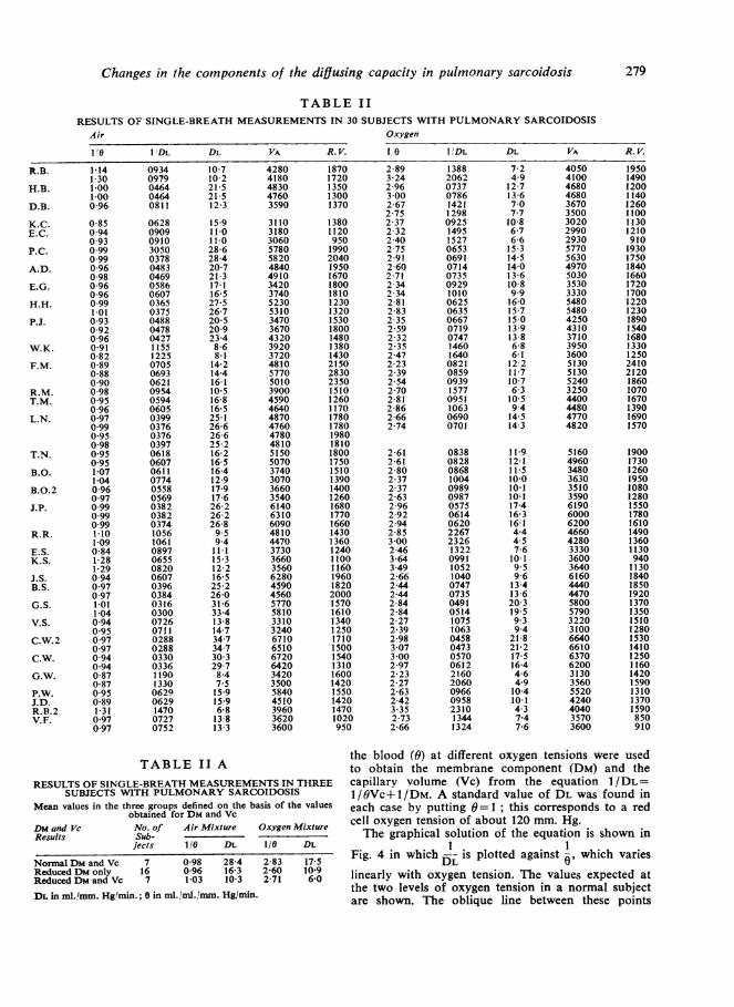
TABLE IIRESULTS OF SINGLE-BREATH MEASUREMENTS IN 30 SUBJECTS WITH PULMONARY SARCOIDOSIS
Air Oxygen
10 I1DL DL VA R. V. 10( I/DL DL VA R.V.
R.B. 1-14 0934 10-7 4280 1870 2-89 1388 7-2 4050 19501-30 0979 10-2 4180 1720 3-24 2062 4-9 4100 1490
H.B. 1-00 0464 21-5 4830 1350 2-96 0737 12-7 4680 12001-00 0464 21-5 4760 1300 3-00 0786 13-6 4680 1140
D.B. 0-96 0811 12-3 3590 1370 2-67 1421 7-0 3670 12602-75 1298 7-7 3500 1100
K.C. 0-85 0628 15-9 3110 1380 2-37 0925 10-8 3020 1130E.C. 0-94 0909 11-0 3180 1120 2-32 1495 6-7 2990 1210
0-93 0910 11-0 3060 950 2-40 1527 6-6 2930 910P.C. 0-99 3050 28-6 5780 1990 2-75 0653 15-3 5770 1930
0-99 0378 28-4 5820 2040 2-91 0691 14-5 5630 1750A.D. 0-96 0483 20-7 4840 1950 2-60 0714 14-0 4970 1840
0-98 0469 21-3 4910 1670 2-71 0735 13-6 5030 1660E.G. 0-96 0586 17-1 3420 1800 2-34 0929 10-8 3530 1720
0-96 0607 16-5 3740 1810 2-34 1010 9-9 3330 1700H.H. 0-99 0365 27-5 5230 1230 2-81 0625 16-0 5480 1220
1-01 0375 26-7 5310 1320 2-83 0635 15-7 5480 1230P.J. 0-93 0488 20-5 3470 1530 2-35 0667 15-0 4250 1890
0-92 0478 20-9 3670 1800 2-59 0719 13-9 4310 15400-96 0427 23-4 4320 1480 2-32 0747 13-8 3710 1680
W.K. 0-91 1155 8-6 3920 1380 2-35 1460 6-8 3950 13300-82 1225 8-1 3720 1430 2-47 1640 6-1 3600 1250
F.M. 0-89 0705 14-2 4810 2150 2-23 0821 12-2 5130 24100-88 0693 14-4 5770 2830 2-39 0859 11-7 5130 21200-90 0621 16-1 5010 2350 2-54 0939 10-7 5240 1860
R.M. 0-98 0954 10-5 3900 1510 2-70 1577 6-3 3250 1070T.M. 0-95 0594 16-8 4590 1260 2-81 0951 10-5 4400 1670
0-96 0605 16-5 4640 1170 2-86 1063 9-4 4480 1390L.N. 0-97 0399 25-1 4870 1780 2-66 0690 14-5 4770 1690
0-99 0376 26-6 4760 1780 2-74 0701 14-3 4820 15700-95 0376 26-6 4780 19800-98 0397 25-2 4810 1810
T.N. 0-95 0618 16-2 5150 1800 2-61 0838 11-9 5160 19000-95 0607 16-5 5070 1750 2-61 0828 12-1 4960 1730
B.O. 1-07 0611 16-4 3740 1510 2-80 0868 11-5 3480 12601-04 0774 12-9 3070 1390 2-37 1004 10-0 3630 1950
B.O.2 0-96 0558 17-9 3660 1400 2-37 0989 10.1 3510 10800-97 0569 17-6 3540 1260 2-63 0987 10-1 3590 1280
J.P. 0-99 0382 26-2 6140 1680 2-96 0575 17-4 6190 15500-99 0382 26-2 6310 1770 2-92 0614 16-3 6000 17800-99 0374 26-8 6090 1660 2-94 0620 16-1 6200 1610
R.R. 1-10 1056 9-5 4810 1430 2-85 2267 4-4 4660 14901-09 1061 9-4 4470 1360 3-00 2326 4-5 4280 1360
E.S. 0-84 0897 11-1 3730 1240 2-46 1322 7-6 3330 1130K.S. 1-28 0655 15-3 3660 1100 3-64 0991 10-1 3600 940
1-29 0820 12-2 3560 1160 3-49 1052 9-5 3640 1130J.S. 0-94 0607 16-5 6280 1960 2-66 1040 9-6 6160 1840B.S. 0-97 0396 25-2 4590 1820 2-44 0747 13-4 4440 1850
0-97 0384 26-0 4560 2000 2-44 0735 13-6 4470 1920G.S. 1-01 0316 31-6 5770 1570 2-84 0491 20-3 5800 1370
1-04 0300 33-4 5810 1610 2-84 0514 19-5 5790 1350V.S. 0-94 0726 13-8 3310 1340 2-27 1075 9-3 3220 1510
0-95 0711 14-7 3240 1250 2-39 1063 9-4 3100 1280C.W.2 0-97 0288 34-7 6710 1710 2-98 0458 21-8 6640 1530
0-97 0288 34-7 6510 1500 3-07 0473 21-2 6610 1410C.W. 0-94 0330 30-3 6720 1540 3-00 0570 17-5 6370 1250
0-94 0336 29-7 6420 1310 2-97 0612 16-4 6200 1160G.W. 0-87 1190 8-4 3420 1600 2-23 2160 4-6 3130 1420
0-87 1330 7-5 3500 1420 2-27 2060 4-9 3560 1590P.W. 0-95 0629 15-9 5840 1550 2-63 0966 10-4 5520 1310J.D. 0-89 0629 15-9 4510 1420 2-42 0958 10-1 4240 1370R.B.2 1-31 1470 6-8 3960 1470 3-35 2310 4-3 4040 1590V.F. 0-97 0727 13-8 3620 1020 2-73 1344 7-4 3570 850
0-97 0752 13-3 3600 950 2-66 1324 7-6 3600 910
TABLE11 A ~~~~the blood (9) at different oxygen tensions were usedTABLEII A ~~to obtain the membrane component (DM) and the
RESULTS OF SINGLE-BREATH MEASUREMENTS IN THREE capillary volume (Vc) from the equation 1/1DL=SUBJECTS WITH PULMONARY SARCOIDOSIS I//9Vc+ 1/Dm. A standard value of DL was found in
Mean values in the three groups defined on the basis of the values each case by putting 9= 1 ;this corresponds to a redobtained for Dm and Vc cell oxygen tension of about 120 mm. Hg.
Dm and Vc No. of Air Mixture Oxygen Mixture Tegahclslto fteeuto ssoniResults Sub- Tegahclslto fteeuto ssoni
jects 110 DL 1/0 DL Fi.4i1hc ispotdaan 1t wihvreNormnalDmand Vc 7 0-98 28-4 2-83 17-5 Fi.4iwhc --splte agnt , hchvrs
Reduced Dm only 16 0-96 16-3 2-60 10-9 linearly with oxygen tension. The values expected atReduceDmadVc7 1-03 0-3 2-1 6-0
the two levels of oxygen tension in a normal subjectDL in ml. Immf. Hglmin.; 0 in ml. noil/mmn. Hg/min. are shown. The oblique line between these points

Neil A. J. Hamer
evident correlation between the lung volumes and. --= +-! the severity of the changes judged on otherDL Du OVc
grounds. In 11 patients residual volume was alsomeasured by helium dilution in a closed circuitfor 7 minutes ; the results were similar to thoseobtained during breath holding (Table I), but theclosed-circuit measurements tended to be smaller,the average difference being 92 ml. (1,322
)5- ~ SLOPE Vc 80 compared to 1,414 ml.).DLAIR = 30 The results of the diffusion studies (Table IV)
INTERCEPT Dm = 50 fell into three groups; seven subjects showednormal values, 16 had impairment of DM with a
o I _ ,_ ,_,_ normal Vc, and in seven cases both components2 3 (DM and Vc) were reduced. The mean values in
6 200 400 P02 each group are shown in Table V. In Fig. 5 these
Calculation of DM and Vc in a normal subject. average findings are shown graphically, 1 /DL
shows the effect of variations in oxygen tension onthe diffusing capacity for carbon monoxide. The slopeof the line gives the Vc, a steeper slope indicating a
smaller volume, and the intercept, at -=0, gives the
DM, which is unaffected by variations in oxygentension. The diffusing capacities (DL and DM)are expressed throughout in ml./mm. Hg/min. andcapillary volume (Vc) in millilitres. The DL, DM, andVc were regarded as abnormal if more than threestandard errors from the regression equation for agein normal subjects (Table III), using the less skew
reciprocal values, I, ' and I No correctionDL Dm Vc-
was attempted for variation in body size.
TABLE IIINORMAL VALUES IN 25 SUBJECTS (HAMER, 1962)
EXPRESSED AS REGRESSION EQUATION FOR AGE! C 100 m S.E.
0-0250DLIDM 0 0122
0-0129
0-0282
0-0221
0 0060
y = mx + c; x = age in completed years
Lung volumes were expressed as percentages of thevalues predicted from the regression equations forresidual volume and vital capacity given in Table Xof Needham, Rogan, and McDonald (1954), adding6% to bring the values to B.T.P.S.
RESULTS
The total lung volume estimated from the dilutionof helium during the breath-holding manceuvre wason the average 77% (53 to 93 %), and thecorresponding residual volume 79% (56 to 122%)of the value predicted (Table IV). There was no
FIG. 5. A verage findings in the three groups of patients
with sarcoidosis. DM is given by the intercept at 0, DL
by the value at - 1, and Vc by the slope of the line in
each gr'oulp.
being plotted against 1/0 as in Fig. 4. The firstgroup shows a normal slope and intercept, thesecond group shows elevation of the interceptwithout any change in the slope of the line, andthe third group shows a similar intercept but theline slopes more steeply as Vc is reduced.
Several types of radiographic change were foundin each of the groups defined on the basis of thediffusion studies (Table VI). In five patientsradiographic changes had been present for severalyears but had resolved a few years before theinvestigation. Two of these patients had normaldiffusion, and the other three had impairment ofDM only.Pulmonary infiltration was present at the time
of the study in 13 patients. Three of these haddeveloped infiltration while under observation a
DL
OiK
0 1<O
00
DL
0-
20-
4080-
FIG. 4.
I
280
I

Changes in the components of the diffusing capacity in pulmonary sarcoidosis 281
TABLE IVRESULTS OF DIFFUSION AND LUNG VOLUME MEASUREMENTS WITH DETAILS OF RADIOGRAPHIC CHANGES
AND STEROID THERAPY IN 30 PATIENTS WITH PULMONARY SARCOIDOSISInitials Diffusion Measurements Lung Volwnes Duration of Radiographic Changes Duration of Steroid Therapy
(as % predicted) (years) (years)DL DM Vc
Total Residual Infiltration Subsequent Additional Current Previous CeasedClearing Fibrosis
Normal values:Clear radiograph at time ofstudy
P.C. 27-4 51 60 83 94 2-9 3-2J.P. 26-0 32 83 85 73 1-7 2-0
Pulmonary infiltrationL.N. 25-9 48 56 93 115 2-9*B.S. 25-6 62 38 93 122 0-8*C.W. 29-3 47 79 93 64 1-3* 2-5 0-8
Localfibrosis (one upper lobe)H.H. 27-0 44 70 79 59 8-9 3-3 2-8 3-2G.S. 32-8 51 93 89 78 5.8* 2-0Mean 27-7 49 68
Impaired DM only:Clear radiograph at time ofstudy
P.J. 21-1 33 60 53 72 0 9 4-6J.S. 16-5 26 41 89 90 2-9* 1-5 1-6P.W. 15-9 23 50 86 81 18 9.1
Pulmonary infiltrationK.C. 21-6 15 51 69 70 5-8 2-6 2-8A.D. 20-8 30 68 64 80 1-5* 1.1 0-6E.G. 16-5 30 37 73 100 4-5T.M. 15-3 25 38 70 70 3-3 3-0B.O. 14-6 19 63 81 98 2-8 2-4 0-3E.S. 19-8 36 49 80 60 3-2V.S. 13-7 21 39 63 90 9 0
Localfibrosis (one upper lobe)H.B. 21-5 32 68 77 57 13-4 4-4 0 5 2-9 0-8F.M. 14-6 18 75 79 97 15-4 3-3
Generalized fibrosisJ.D. 15-3 23 47 60 56 6-6 3-1W.K. 8-4 10 43 77 77 8-6 5-5 8-6T.N. 16 3 20 75 92 84 6-3 4-1 2-9 0 3K.S. 14 2 17 80 80 60 8-5 5-5 5-3Mean 16-6 24 55
Impaired DM and Vc:Pulmonary infiltration
RB. 11-6 22 25 87 107 14-0D.B. 12-1 20 12 80 78 8-2* 0-3 7-3V.F. 13-3 25 29 83 56 5-0 1 2 2-0 3 0
GeneralizedfibrosisE.C. 10-7 20 24 65 62 7-3 1-8R.M. 10-5 17 28 74 78 7-6 4-6R.R. 10-1 31 15 66 67 8-8 3-5 3-3G.W. 7-5 14 16 60 61 12-4 4-4 4-8Mean 10-8 21 21
Second studies after steroid therapy:R.B. 7-5 11 24 84 93 1 5B.O. 17-5 31 41 81 80 1-0C.W. 34-5 49 116 96 78 0-1
* Onset under observation
TABLE V TABLE VIMEAN VALUES OF DL, DM, AND Vc IN EACH GROUP RELATION BETWEEN RADIOGRAPHIC AND DIFFUSIONNVc CHANGES IN 30 PATIENTS WITH PULMONARY
Number DL DM VC SARCOIDOSISNormal series Normal Low DM Low DM(Hamer, 1962) 25 29-4 52 75 Only and VcSarcoid:normal values 7 27-7 49 68Sarcoid:Clardogah 2 3low DM only 16 16 6 24 55 Infiltration 3 7 3Sarcoid: Local fibrosis 2 2low Dm and Vc 7 10-8 21 21 Bilateral fibrosis 4 4
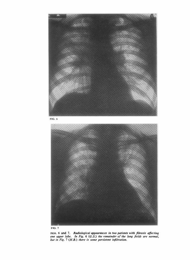
FIG. 6
FIG. 7
FIGS. 6 and 7. Radiological appearances in two patients with fibrosis affectingone upper lobe. In Fig. 6 (G.S.) the remainder of the lung fields are normal,but in Fig. 7 (H.B.) there is some persistent infiltration.

Changes in the components of the diffusing capacity in pulmonary sarcoidosis
short time previously and had normal diffusion.Seven with moderately long-standing infiltrationhad reduction in DM only, and three with a longhistory of infiltration had low values for bothcomponents (DM and Vc).
Local fibrosis affecting one upper lobe was foundin four patients with long-standing radiologicalchanges. There was complete clearing elsewherein two subjects who showed normal diffusion(Fig. 6), but some infiltration persisted in the othertwo who had impairment of DM only (Fig. 7).Bilateral fibrosis was evident in the radiograph ineight cases, all with a long history. Four of thesepatients had a reduced DM only and four had lowvalues for both components (DM and Vc).
In the patients with pulmonary infiltration butno fibrosis there is a trend to more severe changesin diffusion the longer the history of radiologicalchanges in the lungs. When the diffusing capacityof the alveolar-capillary membrane for the wholegroup is compared with the known duration ofradiographic changes (Fig. 8) there is a tendency
TABLE VIIEFFECT OF STEROID THERAPY IN THREE PATIENTS WITH
PULMONARY SARCOIDOSISDuration of Duration Diffusion Changes Lung VolumeRadiographic of (%) ChangesChanges Steroid -- ()before Therapy DL DM VcTherapy (years) T. L. C. R.V.(years)
R.B. 14 0B.O. 2-8C.W. 1 3Mean 6 0
151*00*10 9
-35 -50+20 +63+18 +4+1 +6
-4-35+47+3
-4+3+3+1
-13-18+17-5
radiological evidence of fibrosis. Each type ofdiffusion abnormality was represented (Table VII).There was no definite change in radiological ordiffusion findings as a result of steroid therapyin these three patients. One patient showeddeterioration in DM despite steroid therapy,whereas the other two improved in some para-meters. The mean changes were negligible.
DISCUSSION
00 0
0 0
0 0
O 0
000o0
x
0
O * 0
0
O NORMAL Vc* REDUCED VcX LOCAL FIBROSIS
x
0
0
KNOWN DURATION OF RADIOGRAPHIC CHANGES IN YEARS
FIG. 8. Relation between DM and the known duration ofradiographic changes.
for a rapid fall in the first few years and a moregradual reduction in more long-standing disease.'The patients with local fibrosis show less impair-ment in the membrane component than expectedfrom the duration of the disease. Reduction incapillary volume is confined to patients withknown radiographic changes for more than fiveyears, and nearly half the patients with disease ofthis duration are affected in this way.The effects of steroid therapy were studied in
three patients with pulmonary infiltration but no
1 The correlation between DM and the known duration of radio-graphic changes is statistically significant (r=0-47, t=3-4. P<O0O1).
x
FUNCTIONAL CHANGES IN THE LUNGS The classi-fication of the functional abnormalities in pul-monary sarcoidosis as ventilatory restriction,sometimes complicated by a diffusion defect,or as predominantly ventilatory obstruction(McClement, Renzetti, Himmelstein, andCournand, 1953) has been largely confirmed bysubsequent studies (Stone, Schwartz, Feltman, andLovelock, 1953; Marshall et a{., 1958; Svanborg,1961). Early work indicated that restrictivechanges, with reduced lung volumes and a tendencyto hyperventilation but no evidence of airwayobstruction, were common in pulmonary sarcoid-osis (Bruce and Wassen, 1940; Leitner, 1946;Baldwin, Cournand, and Richards, 1949; Small,1951; Shulman, Schoenrich, and Harvey, 1952;Williams, 1953; Gray and Gray, 1957; Morrison,Fulton, and Hickam, 1957), and this has beenconfirmed in more recent studies. The correspond-ing changes in the mechanical properties of thelung are usually small (Marshall and DuBois,1956; Marshall et al., 1958; Svanborg, 1961).The frequency with which abnormalities of
diffusion have been recognized in these patientshas varied with the techniques available. Riley,Riley, and Hill (1952) reported three cases ofpulmonary sarcoidosis with impaired diffusion foroxygen, and similar cases were reported byWilliams (1953) and by Austrian, McClement,Renzetti, Donald, Riley, and Cournand (1951).Five of the nine patients with restrictive changesreported by McClement and his colleagues (1953)showed impairment of oxygen diffusion. Stone
DM A 0
50-
40-
30-
20 -
I 0 -
283
Neil A. J. Hamer
et al. (1953) found similar evidence of reduceddiffusion in five of 12 such patients.The introduction of the more convenient carbon
monoxide method for the measurement of diffusionled to more frequent reports of diffusion changesin sarcoidosis (Bates, 1958). Marshall et al. (1958),in their study of 21 patients with pulmonarysarcoidosis, found that a reduction of diffusingcapacity was the principal disturbance. Low valueswere found in 13 of these subjects. Svanborg(1961) studied 26 patients and found impaireddiffusion in 19.
Obstructive changes, with an increase in residualvolume and an abnormal distribution of ventila-tion, were reported by Coates and Comroe (1951).Later studies have shown this type of abnormalityin a minority of cases (McClement et al., 1953;Stone et al., 1953 ; Marshall et al., 1958 ; Svanborg,1961), usually without any increase in residualvolume. These ventilatory abnormalities arethought to indicate bronchial constriction withoutalveolar destruction or bronchiolar collapse, asthere is little evidence of functional changes of thetypo found in chronic obstructive emphysema. Inthe present study lung volumes were in generalsomewhat less than predicted, and there was noevidence of a pathological increase in residualvolume in any case. The detailed studies ofMarshall and his colleagues (1958) and of Svanborg(1961) suggest that restrictive changes are relativelymild and obstructive changes uncommon inpulmonary sarcoidosis, and that impairment ofdiffusion is the predominant abnormality.
There has been little previous work on thecomponents of the diffusing capacity in patientswith pulmonary sarcoidosis. McNeill et al. (1958)studied two patients with diffuse pulmonaryfibrosis due to sarcoidosis and found themembrane component (DM) to be reduced to agreater extent than the capillary volume (Vc).Johnson, Lawson, and Wilcox (1961) report insummary the findings in nine patients withpulmonary sarcoidosis. Reduction of DM was themajor change, the Vc being relatively little affected,even in two cases of 'cor pulmonale'. The changesreported in these studies are similar to those foundin the present work.
Bates, Varvis, Donevan, and Christie (1960)studied the components of the diffusing capacityin two patients with pulmonary sarcoidosis, usinga steady-state method on exercise instead of thesingle-breath technique. The main change foundwas a low Vc with a normal DM in one subject.This pattern of impairment was not encounteredin the present study.
CORRELATION OF RADIOLOGICAL APPEARANCES WITHCHANGES IN DIFFUSING CAPACITY The absence ofany correspondence between the changes indiffusion and the radiological status at the timeof the study suggests that the diffusion findingsshould be considered in relation to the precedingchanges in the lungs. The natural history andprognosis of patients with pulmonary sarcoidosishave been analysed by Hoyle (1961). He classified125 patients into three main groups which togetherform a spectrum of the disease (Fig. 9). Group I
v)wz
IU-J
U
000
incrOD
DEATH
,m 19010
II 400/0
41/1
05,0,
O 5 10~~~~~l 15
z0
z
YEARS
FIG. 9. Classification of sarcoidosis proposed by Hoyle(1961). The diagram correlates the radiographic changeswith the duration of the disease.
consisted of 41 % of the patients, in whompulmonary infiltration was transient, the chestradiograph becoming normal within about twoyears. Group II formed 40% of the total and wasmade up of patients in whom pulmonary infiltra-tion persisted but did not increase progressively,so that eventual fibrosis was not severelydestructive. The remaining 19% of subjects wereclassed as group III; they showed progressiveinfiltration followed by severe radiological fibrosisleading to dyspnoea and heart failure.The five patients studied here who showed
resolution of previous pulmonary infiltration fallinto group I of this classification. In three of thesepatients an abnormality of DM persists in spite ofradiological clearing. A similar persistence ofimpaired DL has been reported by Marshall et al.(1958). In one patient in the present series theimprovement coincided with steroid treatment, soradiological clearing following such therapy cannot
284

Changes in the components of the diffusing capacity in pulmonary sarcoidosis
be taken to indicate a corresponding resolution ofthe pathological changes in the lungs.Three patients with pulmonary infiltration of
short duration had normal diffusion. In thepatients with pulmonary infiltration for more thana few years there was always a reduction in DM,and in some of those with changes for more thanfive years the Vc was also reduced. It seems likelythat this is the course followed by the patientsin group II of Hoyle's (1961) classification. Atendency to more severe changes in overalldiffusing capacity (DL) when radiographicabnormalities have been present for more than afew years is reported by Marshall et al. (1958)and by Stone et al. (1953).The absence of changes in diffusion with
pulmonary infiltration of relatively short durationsuggests that the pathological changes responsiblefor the radiographic shadows may differ duringthe course of the disease. Though deposits ofsarcoid tissue might be expected to increase thethickness of the alveolar-capillary membrane, theydo not seem to interfere significantly with gastransfer at first. In fact, the scattered sarcoidfollicles probably leave large areas of thediffusion pathway unimpaired (Longcope andFreiman, 1952). The reduction in DM found incases with pulmonary infiltration for more thantwo or three years might then be attributed to acritical increase in the sarcoid deposits interferingwith gas transfer. However, there is no correspond-ing increase in the density of the radiographicchanges.The reduction in DM after several years of
pulmonary infiltration may be attributable to finefibrotic changes not evident in the radiograph butreducing the area available for diffusion. Theprogressive impairment of the membrane compo-nent over the years may be explained in this way.An alternative possibility is that the DM isdetermined only in part by the physical processof diffusion in the alveolar-capillary membraneand is in the main an index of uneven distributionof ventilation and perfusion in the lung (Asmussenand Nielsen, 1960; Staub, 1961; Chinard, Enns,and Nolan, 1961). The absence of changes earlyin the course of pulmonary infiltration would notbe unexpected in these circumstances, and the laterdeterioration might be attributed to the distortingeffects of fine fibrosis.
Reduction of the Vc appears to be confined topatients with long-standing disease. It was notfound in the absence of a reduction in DM, andmay indicate a more severe degree of diffusefibrosis, not necessarily evident radiologically.
However, these subjects show no greater changein DM than those with a normal Vc, suggestingthat a different mechanism is involved. Apredominantly perivascular distribution of thesarcoid follicles (Longcope and Freiman, 1952)may be responsible. Marshall et al. (1958) suggestthat the impairment of diffusion in pulmonarysarcoidosis is mainly due to perivascular lesions,as the mechanical properties of the lungs arerelatively little affected. The reduced Vc found inone patient by Bates et al. (1960) is further supportfor this hypothesis.The four patients with considerable radiological
resolution but persistent local fibrosis showrelatively little change in diffusion considering thelong history of lung disease. Two have in factnormal diffusion measurements. These findingsindicate that local fibrosis has little effect ondiffusion, the relatively unaffected lung tending topredominate. Both Marshall and his colleagues(1958) and Svanborg (1961) report normal valuesfor overall diffusing capacity (DL) in a few patientswith only minor signs of fibrosis.The patients with radiological evidence of
pulmonary fibrosis have serious long-standingdisease and correspond to group III of Hoyle's(1961) classification. The changes in diffusion,however, are similar to those found in patientswith a long history of pulmonary infiltrationwithout fibrosis. Although all the patients withradiological fibrosis were known to have hadradiographic changes for more than five years andhad an impaired DM, only half showed reductionin Vc. The changes in the radiographs interpretedas pulmonary fibrosis are mainly confined to theupper lobes, and there is often apparent over-distension of the lower lobes. Relatively normalfunction in these regions may account for the lackof correlation between the radiological evidenceof fibrosis and the reduction of Vc. Svanborg(1961) found evidence of an increased gravitationalshift of blood to the legs in many patients withpulmonary sarcoidosis, which might be expectedto reduce the Vc and DL; however, measurementof DL, sitting and supine, in three cases is reportedas showing little change.As the Vc was not reduced without serious
impairment of DM in the present study, the DLgave a good indication of the severity of thedisease. Nevertheless, a study of the componentsof the diffusing capacity can give helpful additionalinformation at times. In two subjects (P.J.and H.B.) a significant reduction in DM wasaccompanied by an unusually large Vc, sothat the DL was normal. Similarly, measurement
285
Neil A. J. Hamer
of DL cannot detect the stage at which the Vcbegins to fall. This may be a critical point in theprogress of the disease as it suggests that thepulmonary vascular bed is becoming seriouslydamaged.
EFFECTS OF STEROID THERAPY Studies before andduring steroid therapy in three subjects withpulmonary infiltration but no radiological fibrosisshowed little improvement in the diffusion changesin spite of some clearing of the radiographicshadows in the two cases with shorter histories.Another patient (J.S.) developed a normal chestfilm on steroid therapy, but an abnormality ofdiffusion persisted. In the remaining seven patientsstudied during steroid therapy pulmonary infiltra-tion had not cleared, and there was radiologicalevidence of fibrosis in all but two; serious impair-ment of diffusion was found in these patients.An increase in lung volumes has been reported
frequently following steroid therapy in pulmonarysarcoidosis (Small, 1951 ; Riley et al., 1952;Shulman et al., 1952; Stone et al., 1953; Rudberg-Roos and Roos, 1958), as in spontaneous resolution(Bruce and Wassen, 1940), but Riley et al. (1952)found no increase in diffusing capacity for oxygenafter A.C.T.H. therapy in three patients, andMcClement et al. (1953) found improvement inonly one of four cases treated with cortisone.Smellie, Apthorp, and Marshall (1961) studied theeffect of steroid therapy in six patients withsarcoidosis and abnormal pulmonary function, andfound some improvement in lung volume anddiffusing capacity, but the values did not reachnormal levels.The results in these few cases suggest that though
pulmonary infiltration of short duration clearsduring steroid therapy much of the effect ondiffusion persists. This is consistent with thesuggestion that the impairment of diffusion is duelo fine fibrotic changes which would be expectedto remain after resolution of the exudative com-ponents of the sarcoid granulomata on steroidtherapy. Biopsies have shown a change from agranulomatous to a fibrotic lesion during treatmentwith steroids (McClement et al., 1953), tending tosupport this view.
CONCLUSION
The changes in the diffusion process found inpatients with pulmonary sarcoidosis are probablydue to diffuse fibrosis. Although individual patientswere not studied throughout the course of thedisease, an overall picture of the changes in
diffusion can be built up from the separate casereports. No abnormality of diffusion is found withpulmoniary infiltration of short duration, but afterone to three years the membrane component (DM)becomes impaired. This component (DM) fallsprogressively during the subsequent three to fiveyears if pulmonary infiltration persists, but thereis little further deterioration after this time. Inhalf the patients with pulmonary infiltration formore than five years the pulmonary capillaryvolume (Vc) becomes reduced.Although these changes are interpreted as
evidence of fibrosis there is little correlation withthe conventional radiological signs of fibrosis, andsubjects with radiographic changes confined to oneupper lobe show only minor changes in diffusion.The radiological appearances probably indicatelocal effects whereas the diffusion findings aredetermined by diffuse changes throughout thelungs.
It seems likely that diffusion measurements willprove to be complementary to radiologicalexamination in assessing the progress andprognosis of patients with pulmonary sarcoidosis,and that measurement of the two components ofdiffusion will be helpful in defining the changesin the lungs more precisely than is possible fromthe overall diffusing capacity.
I am grateful to Dr. Clifford Hoyle for hisencouragement and advice in carrying out this work,and for permission to study patients under his care.
REFERENCES
Asmussen, E., and Nielsen, M. (1960). Alveolo-arterial gas exchangeat rest and during work at different 02 tensions. Acta physiol.wcand., 50, 153.
Austrian, R., McClement, J. H., Renzetti, A. D., Donald, K. W.,Riley, R. L., and Cournand, A. (1951). Clinical and physiologicfeatures of some types of pulmonary diseases with impairment ofalveolar-capillary diffusion: the syndrome of " alveolar-capillaryblock." Amer. J. Med., 11, 667.
Baldwin, E. deF., Cournand, A., and Richards, D. W. (1949). Pul-monary insufficiency 1I. A study of thirty-nine cases of pulmonaryfibrosis. Medicine (Baltimore), 28, 1.
Bates, D. V. (1958). The measurement of the pulmonary diffusingcapacity in the presence of lung disease. J. clin. Invest., 37, 591.- Varvis, C. J., Donevan, R. E., and Christie, R. V. (1960).
Variations in the pulmonary capillary blood volume and mem-brane diffusion component in health and disease. Ibid., 39, 1401.
Bruce, T., and Wassen, E. (1940). Clinical observations on the courseand prognosis of lymphogranulomatosis benigna Schaumann,particularly in regard to the pulmonary lesions. Acta med. scand.,104, 63.
Chinard, F. P., Enns, T.. and Nolan, M. F. (1961). Diffusion andsolubility factors in pulmonary inert gas exchanges. J. appi.Physiol., 16, 831.
Coates, E. O., and Comroe, J. H. (1951). Pulmonary function studiesin sarcoidosis. J. clin. Invest., 30, 848.
Forster, R. E., Roughton, F. . W., Cander, L., Brscoe, W. A., andKreuzer, F. (1957). Apparent pulmonary diffusing capacity forCO at varying alveolar O0 tensions. J. appl. Physiol., 11, 277.
Gray, F. D., and Gray, F. G. (1957). Pulmonary sarcoidosis: aphysiopathologic analysis. J. chron. Dis., 6, 572.
Hamer, N. A. J. (1962). The effect of age on the components of thepulmonary diffusing capacity. Clin. Sci., 23, 85.
Hoyle, C. (1961). Prognosis of pulmonary sarcoidosis. Lancet, 2, 611.
286

Changes in the components of the diffusing capacity in pulmonary sarcoidosis
Johnson, R. L., Lawson, W. H., andWilcox,W C. N. (1961). Alveolarcapillary block in sarcoidosis. Clin. Res., 9, 196.
Jones, R. S., and Meade, F. (1961). A theoretical and experimentalanalysis of anomalies in the estimation of pulmonary diffusingcapacity by the single breath method. Quart. J. exp. Physiol.,46, 131.
Krogh, M. (1915). The diffusion of gases through the lungs of man.J. Physiol. (Lond.), 49, 271.
Leitner, St. J. (1946). Elektrokardiographische und spirometrischeUntersuchungen bei der epitheloidzelligen Granulomatose(Morbus Besnier-Boeck-Schaumann). Cardiologia (Basel), 10, 379.
Longcope, W. T., and Freiman, D. G. (1952). A study of sarcoidosisbased on a combined investigation of 160 cases including 30autopsies from the Johns Hopkins Hospital and MassachusettsGeneral Hospital. Medicine (Baltimore), 31, 1.
McClement, J. H., Renzetti, A. D., Himmelstein, A., and Cournand,A. (1953). Cardiopulmonary function in the pulmonary form ofBoeck's sarcoid and its modification by cortisone therapy. Amer.Rev. Tuberc., 67, 154.
McGrath, M. W., and Thomson, M. L. (1959). The effect of age, bodysize and lung volume change on alveolar-capillary permeabilityand diffusing capacity in man. J. Physiol. Lond., 146, 572.
McNeill, R. S., Rankin, J., and Forster, R. E. (1958). The diffusingcapacity of the pulmonary membrane and the pulmonarycapillary blood volume in cardiopulmonary disease. Clin. Sci.,17, 465.
Marshall, R., and DuBois, A. B. (1956). The viscous resistance of lungtissue in patients with pulmonary disease. Ibid., 15, 473.- Smellie, H., Baylis, J. H., Hoyle, C., and Bates, D. V. (1958).
Pulmonary function in sarcoidosis. Thorax, 13, 48.Morrison, C., Fulton, J., and Hickam, J. B. (1957). Ventilatory
function in pulmonary sarcoidosis. Clin. Res. Proc., 5, 119.Needham, C. D., Rogan, M. C., and McDonald, I. (1954). Normal
standards for lung volumes, intrapulmonary gas-mixing, andmaximum breathing capacity. Thorax, 9, 313.
Ogilvie, C. M., Forster, R. E., Blakemore, W. S., and Morton, J. W.(1957). A standardized breath holding technique for the clinicalmeasurement of the diffusing capacity of the lung for carbonmonoxide. J. clin. Invest., 36, 1.
Riley, R. L., Riley, M. C., and Hill, H. McD. (1952). Diffuse pul-monary sarcoidosis: diffusing capacity during exercise and otherlung function studies in relation to ACTH therapy. Bull. JohnsHopk. Hosp., 91, 345.
Roughton, F. J. W., and Forster, R. E. (1957). Relative importanceof diffusion and chemical reaction rates in determining rate ofexchange of gases in the human lung, with special reference totrue diffusing capacity of pulmonary membrane and volume ofblood in the lung capillaries. J. appl. Physiol., 11, 290.
Rudberg-Roos, I., and Roos, B. E. (1958). Pulmonary function insarcoidosis before and after ACTH and cortisone therapy. Actatubere. scand., 35, 49.
Shulman, L. E., Schoenrich, E. H., and Harvey, A. McG. (1952). Theeffects of adrenocorticotropic hormone (ACTH) and cortisoneon sarcoidosis. Bull. Johns Hopk. Hosp., 91, 371.
Siosteen, S. M., and Sjostrand, T. (1951). A method for the deter-mination of low concentrations of CO in the blood and therelation between the CO-concentration in the blood and that inthe alveolar air. Acta physiol. scand., 22, 129.
Small, M. J. (1951). Favorable response of sarcoidosis to cortisonetreatment. J. Amer. med. Ass., 147, 932.
Smellie, H., Apthorp, G. H., and Marshall, R. (1961). The effect ofcorticosteroid treatment on pulmonary function in sarcoidosis.Thorax, 16, 87.and Hoyle, C. (1960). The natural history of pulmonary sar-coidosis. Quart. J. Med., n.s. 29, 539.
Staub, N. C. (1961). Is there ever a measurable alveolar-arterial 02grtdient due to diffusion ? Physiologist, 4, 115.
Stone, D. J., Schwartz, A., Feltman, J. A., and Lovelock, F. J. (1953).Pulmonary function in sarcoidosis: results with cortisonetherapy. Amer. J. Med., 15, 468.
Svanborg, N. (1961). Studies on the cardiopulmonary function insarcoidosis. Acta med. scand., 170, suppl. 366, pp. 7, 34, and 75.
Thews, G., and Niesel, W. (1959). Zur Theorie der Sauerstoffdiffusionim Erythrocyten. Pflugers Arch. ges. Physiol., 268, 318.
Williams, M. H. (1953). Pulmonary function in Boeck's sarcoid. J.clin. Invest., 32, 909.
Yahr, W. Z., and Kreuzer, F. (1960). Influence of red cell membraneon 02 diffusion. Fed. Proc., 19, 382.
287