cervical venous reflux in dynamic brain scintigraphy - journal of
TRANSCRIPT
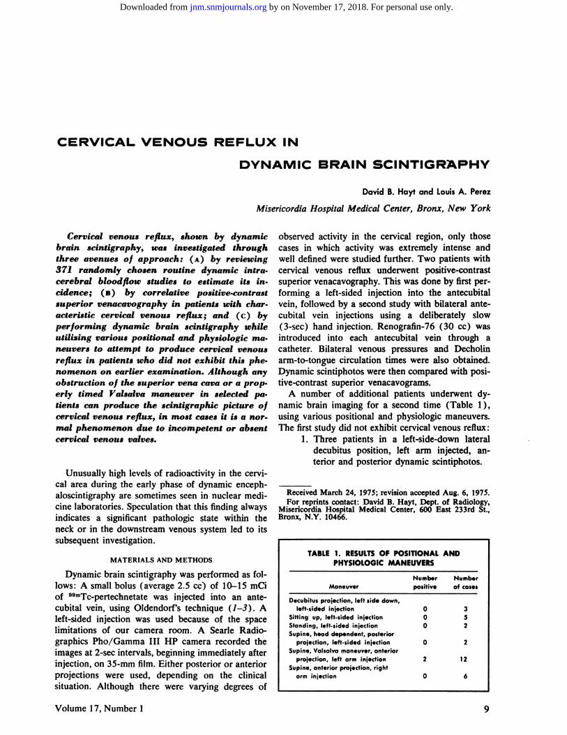
NumberNumberManeuverpositiveof cases
Misericordia Hospital Medical Center, Bronx, New York
Cervical venous re/lux, shown by dynamicbrain scintigraphy, was investigated throughthree avenues of approach: (A) by reviewing371 randomly chosen routine dynamic intra
cerebral blood/low studies to estimate its incidence; (B) by correlative positive-contrastsuperior venacavography in patients with characteristic cervical venous reflux; and (c) by
performing dynamic brain scintigraphy whileutilizing various positional and physiologic maneuvers to attempt to produce cervical venousreflux in patients who did not exhibit this phe.nomenon on earlier examination. Although anyobstruction of the superior vena cava or a properly timed Valsalva maneuver in selected patients can produce the scintigraphic picture ofcervical venous reflux, in most cases it is a nor
mal phenomenon due to incompetent or absentcervical venous valves.
Unusually high levels of radioactivity in the cervical area during the early phase of dynamic encephaloscintigraphy are sometimes seen in nuclear medicine laboratories. Speculation that this finding alwaysindicates a significant pathologic state within theneck or in the downstream venous system led to itssubsequent investigation.
MATERIALS AND METHODS
Dynamic brain scintigraphy was performed as follows: A small bolus (average 2.5 cc) of 10—15mCiof o9mTc@pertechnetate was injected into an antecubital vein, using Oldendorf's technique (1—3). Aleft-sided injection was used because of the spacelimitations of our camera room. A Searle Radiographics Pho/Gamma III HP camera recorded theimages at 2-sec intervals, beginning immediately afterinjection, on 35-mm film. Either posterior or anteriorprojections were used, depending on the clinicalsituation. Although there were varying degrees of
observed activity in the cervical region, only thosecases in which activity was extremely intense andwell defined were studied further. Two patients withcervical venous reflux underwent positive-contrastsuperior venacavography. This was done by first performing a left-sided injection into the antecubitalvein, followed by a second study with bilateral antecubital vein injections using a deliberately slow(3-sec) hand injection. Renografin-76 (30 cc) wasintroduced into each antecubital vein through acatheter. Bilateral venous pressures and Decholinarm-to-tongue circulation times were also obtained.Dynamic scintiphotos were then compared with positive-contrast superior venacavograms.
A number of additional patients underwent dynamic brain imaging for a second time (Table 1),using various positional and physiologic maneuvers.The first study did not exhibit cervical venous reflux:
1. Three patients in a left-side-down lateraldecubitus position, left arm injected, anterior and posterior dynamic scintiphotos.
Received March 24, 1975; revision accepted Aug. 6, 1975.For reprints contact: David B. Hayt, Dept. of Radiology,
Misericordia Hospital Medical Center, 600 East 233rd St.,Bronx, N.Y. 10466.
TABLE 1. RESULTSOF POSITIONAL ANDPHYSIOLOGIC MANEUVERS
Decubitusprojection, left side down,left-sided injection
Sitting up, left-sided injectionStanding, left-sided injectionSupine, head dependent, posterior
projection, left-sided injectionSupine, Valsalva maneuver, anterior
proiection, left arm injectionSupine, anterior projection,right
arm injection
0 30 50 2
0 2
2 12
0 6
Volume 17, Number 1 9
CERVICAL VENOUS REFLUX IN
DYNAMIC BRAIN SCINTIGRAPHY
David B. Hayt and LouisA. Perez
by on November 17, 2018. For personal use only. jnm.snmjournals.org Downloaded from
@—
@ I@
SEC 18 SEC@\
-@-‘
21SEC,t@.
@
@-.
42SEC 45SEC!,
q'._*
@‘
.@
-@-
485EC
r@
HAYT AND PEREZ
2. Five patients in sitting position, left arminjected, anterior dynamic scintiphotos.
3. Two patients in a standing position, left arminjected, anterior dynamic scintiphotos.
4. Two patients supine, head dependent, leftarm injected, posterior dynamic scintiphotos.
5. Twelve patients supine performing a Valsalva maneuver, prior to and during left arminjection, anterior scintiphotos.
6. Six patients supine, right arm injected, antenor scintiphotos.
RESULTS
Of the 37 1 randomly chosen cases, 14 were posilive for early intense well-defined radioactivity withinthe neck, an incidence of 3.5% . Of these, ten showedonly left-sided cervical activity. Analysis of the current diagnosis, sex, anatomic position during thestudy, and cardiovascular status of the 14 positivecases showed no common pattern that would aid inexplaining the phenomenon. The median age was70 years (excluding a 1-year-old infant).
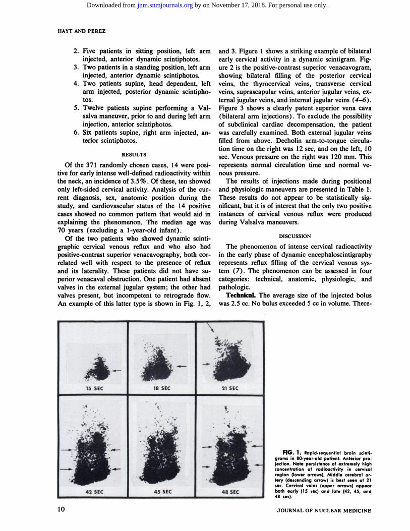
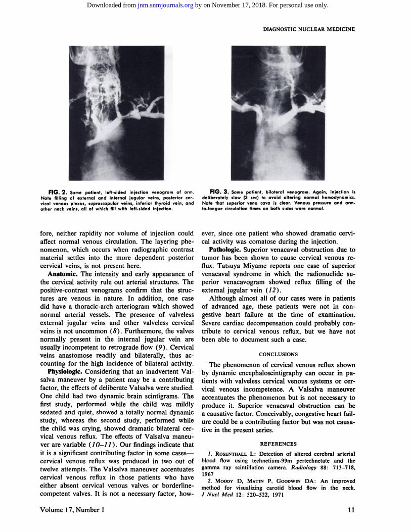
Of the two patients who showed dynamic scintigraphic cervical venous reflux and who also hadpositive-contrast superior venacavography, both correlated well with respect to the presence of refluxand its laterality. These patients did not have superior venacaval obstruction. One patient had absentvalves in the external jugular system; the other hadvalves present, but incompetent to retrograde flow.An example of this latter type is shown in Fig. 1, 2,
and 3. Figure 1 shows a striking example of bilateralearly cervical activity in a dynamic scintigram. Figure 2 is the positive-contrast superior venacavogram,showing bilateral ifihing of the posterior cervicalveins, the thyrocervical veins, transverse cervicalveins, suprascapular veins, anterior jugular veins, cxternal jugular veins, and internal jugular veins (4—6).Figure 3 shows a clearly patent superior vena cava(bilateral arm injections) . To exclude the possibilityof subclinical cardiac decompensation, the patientwas carefully examined. Both external jugular veinsifiled from above. Decholin arm-to-tongue circulation time on the right was 12 5cc, and on the left, 10sec. Venous pressure on the right was 120 mm. Thisrepresents normal circulation time and normal yenous pressure.
The results of injections made during positionaland physiologic maneuvers are presented in Table 1.These results do not appear to be statistically significant,but it is of interest that the only two positiveinstances of cervical venous reflux were producedduring Valsalva maneuvers.
DISCUSSION
The phenomenon of intense cervical radioactivityin the early phase of dynamic encephaloscintigraphyrepresents reflux filling of the cervical venous systern (7). The phenomenon can be assessed in fourcategories : technical, anatomic, physiologic, andpathologic.
Technical.The averagesizeof the injectedboluswas 2.5 cc. No bolus exceeded 5 cc in volume. There
FIG. 1. Rapid-sequentialbrain scintigrams in 80-year-old patient. Anterior projection. Note persistenceof extremely highconcentration of radioactivity in cervicalregion (lower arrows). Middle cerebral ortery (descending arrow) s best seen at 21sec. Cervical veins (upper arrows) appearboth early (15 sec)and late (42, 45, and48 sec)
10 JOURNAL OF NUCLEAR MEDICINE
by on November 17, 2018. For personal use only. jnm.snmjournals.org Downloaded from
DIAGNOSTICNUCLEARMEDICINE
4@,@ @.
FIG. 2. Somepatient, left-sidediniectionvenogramof arm.Note filling of external and internal jugular veins, posterior cervical venous plexus, suprascapular veins, inferior thyroid vein, andother neck veins, all of which fill with left-sided injection.
fore, neither rapidity nor volume of injection couldaffect normal venous circulation. The layering phenomenon, which occurs when radiographic contrastmaterial settles into the more dependent posteriorcervical veins, is not present here.
Anatomic. The intensity and early appearance ofthe cervical activity rule out arterial structures. Thepositive-contrast venograms confirm that the struc
tures are venous in nature. In addition, one casedid have a thoracic-arch arteriogram which showednormal arterial vessels. The presence of valvelessexternal jugular veins and other valveless cervicalveins is not uncommon (8) . Furthermore, the valvesnormally present in the internal jugular vein areusually incompetent to retrograde flow (9) . Cervicalveins anastomose readily and bilaterally, thus accounting for the high incidence of bilateral activity.
Physiologic. Considering that an inadvertent Valsalva maneuver by a patient may be a contributingfactor, the effects of deliberate Valsalva were studied.One child had two dynamic brain scintigrams. Thefirst study, performed while the child was mildlysedated and quiet, showed a totally normal dynamicstudy, whereas the second study, performed whilethe child was crying, showed dramatic bilateral cervical venous reflux. The effects of Valsalva maneuver are variable (1 0—11 ) . Our findings indicate thatit is a significant contributing factor in some casescervical venous reflux was produced in two out oftwelve attempts. The Valsalva maneuver accentuatescervical venous reflux in those patients who haveeither absent cervical venous valves or borderlinecompetent valves. It is not a necessary factor, how
FIG. 3. Samepatient,bilateralvenogram.Again, injectionisdeliberately slow (3 sec) to avoid altering normal hemodynamics.Note that superior vena cava is clear. Venous pressure and armto-tongue circulationtimes on both sides were normal.
ever, since one patient who showed dramatic cervical activity was comatose during the injection.
Pathologic. Superior venacaval obstruction due totumor has been shown to cause cervical venous re
flux. Tatsuya Miyame reports one case of superiorvenacaval syndrome in which the radionuclide superior venacavogram showed reflux filling of the
external jugular vein (12).Although almost all of our cases were in patients
of advanced age, these patients were not in congestive heart failure at the time of examination.Severe cardiac decompensation could probably contribute to cervical venous reflux, but we have notbeen able to document such a case.
CONCLUSIONS
The phenomenon of cervical venous reflux shownby dynamic encephaloscintigraphy can occur in patients with valveless cervical venous systems or cervical venous incompetence. A Valsalva maneuveraccentuates the phenomenon but is not necessary toproduce it. Superior venacaval obstruction can bea causative factor. Conceivably, congestive heart failure could be a contributing factor but was not causative in the present series.
REFERENCES
1. ROSENThALLL: Detection of altered cerebral arterialblood flow using technetium-99m pertechnetate and thegamma ray scintillation camera. Radiology 88: 713—718,1967
2. MOODY D, MAnN P, GOODWIN DA : An improvedmethod for visualizing carotid blood flow in the neck.I Nuci Med 12: 520—522,1971
Volume 17, Number 1 11
by on November 17, 2018. For personal use only. jnm.snmjournals.org Downloaded from
HAYT AND PEREZ
3. OLDENDORF WH, KimNo M, Smt@uzu S: Evaluationof a simple technique for abrupt intravenous injection ofradioisotope. I Nuci Med 6: 205—209,1965
4. SHIMiw-rPM, DOPPMANJK, POWELLD, et al: Demonstration of parathyroid adenomas by retrograde thyroidvenography. Radiology 103 : 63—67,1972
5. GRANTJCB: An Atlas of Anatomy, 3rd ed, Baltimore,Williams & Wilkins, 1951, pp 463—465,511—512
6. THERONJ, DJINDJIAM R: Cervicovertebral phiebography using catheterization: A preliminary report. Radiology108:325—331,1973
7. FRIEDMANBH, LOVEGROVEFTA, WAGNERHN : An
unusual variant in cerebral circulation studies. I Nuci Med15:363—364,1974
8. BRASHJC: Cunningham's Texibook of Anatomy, 9thed, London, Oxford University Press, 1951, pp 1335—1339
9. Goss CM : Anatomy of the Human Body. Philadelphia, Lea & Febiger, 1956, pp 720—725
10. HAYT DB: Upright inferior venacavography. Radiology86:865—870,1966
11. WATSONDD, NELSONJP, GOTrLIEB 5: Rapid bolusinjection of radioisotopes. Radiology 104: 347—352,1973
12. TATSUYAM : Interpretation of ‘@mTcsuperior venacavagram and results of studies in 92 patients. Radiology108:339—352,1973
Thefollowingtitleswill appear in the
JOURNALOF NUCLEARMEDICINETECHNOLOGY
Volume 3, Number 4 (December@ 975)
TechnologistNews Effectsof TechnicianTrainingLevelsin theNuclear Medicine Department
Bylawsof the TechnologistsSection Wanda M. Hibbardof the Society of Nuclear Medicine
Current Topicsin Clinical Nuclear Medicine:Letter from the Editor A Selected Bibliography
1.DavidWells C.J.Klobukowskiand0. 0. Frey
So You Are a RegisteredNuclear Medicine NMT GadgetryTechnologist! Inexpensive and Effective Radiation Protection
DonaldC.Wharton StructuresBonnie J. Baggenstossand Mary E. Maxwell
Safe and AccurateDeterminationofGenerator Eluate Volume
AnthonyR.Benedetto NMTBookshelf
Measurement of Radiation Exposure Resultingfrom NMT Audiovisual ReviewsUsingan Automated,Solvent-Extraction-Type,
TechnetiumGeneratorV. 1.GelezunasandK.P.Lyons What'sNew?
Radiochemical Purity and Stability of Commercial CalendarSPMTc@StannousDTPAKitsUsinga New
ChromatographyTechniquePatriciaA. Cooperand A. Michael Zimmer Placement
Subscriptions to the JOURNAL OF NUCLEAR MEDICINE TECHNOLOGY are available. Please contactSubscription Department, Society of Nuclear Medicine, 475 Park Avenue South, New York, NY. 10016for 1976 subscription rates.
12 JOURNAL OF NUCLEAR MEDICINE
by on November 17, 2018. For personal use only. jnm.snmjournals.org Downloaded from
1976;17:9-12.J Nucl Med. David B. Hayt and Louis A. Perez Cervical Venous Reflux in Dynamic Brain Scintigraphy
http://jnm.snmjournals.org/content/17/1/9This article and updated information are available at:
http://jnm.snmjournals.org/site/subscriptions/online.xhtml
Information about subscriptions to JNM can be found at:
http://jnm.snmjournals.org/site/misc/permission.xhtmlInformation about reproducing figures, tables, or other portions of this article can be found online at:
(Print ISSN: 0161-5505, Online ISSN: 2159-662X)1850 Samuel Morse Drive, Reston, VA 20190.SNMMI | Society of Nuclear Medicine and Molecular Imaging
is published monthly.The Journal of Nuclear Medicine
© Copyright 1976 SNMMI; all rights reserved.
by on November 17, 2018. For personal use only. jnm.snmjournals.org Downloaded from