cerebrospinal elaphostrongylosis in dairy goats in northern norway
TRANSCRIPT
J. Vet. Med. B 38, 755-763 (1991) 0 1991 Paul Parey Scientific Publishers, Berlin and Hamburg ISSN 0931 - 1793
State Veterinary Laboratory for Northern N o w a y , Harstad, Norway
Cerebrospinal Elaphostrongylosis in Dairy Goats in Northern Norway
K. HANDELAND and 0. SPARBOE"
Address of authors: Dr. K. HANDELAND, State Veterinary Laboratory for Northern Norway, P.O. Box 652, N-9401 Harstad, Norway
With 8 figures and one table
(Received for publication July 18, 1990)
S u m m a r y Ten carcasses and three vertebral columns from north Norwegian dairy goats, which had been
killed due to clinical signs of severe neurologic disease, were received for necropsy. Pathological examination revealed nematodes and nematode ova in the central nervous system
(CNS) of nine goats. Worms found by gross examination were identified as Elaphostrongylus rangiferi MITSKEVICH, 1960. Focal traumatic encephalomyelomalacia, apparently caused by migrating worms, perivascular cuffing, eosinophilic leptomeningitis and perineural infiltrations and granulomas, could be demonstrated in CNS sections from all 13 animals examined.
Clinical signs reported were initial pruritus followed by motor weakness, lameness, paresis, reduced vision, circling, abnormal head position, bulging eyes and scoliosis.
The disease occurred from September to January in regions with a considerable migrant reindeer population. It was concluded that the reported outbreaks of neurologic disease represented seasonal occurrence of cerebrospinal elaphostrongylosis caused by Elaphostrongylus rang$&, the elaphostron- gyloid nematode of reindeer (Rangifer tarandus tarandus).
In t roduc t ion Nematodes of the genus Elaphostrongylus (Protostrongylidae : Metastrongyloidea)
are parasites of the skeletal muscles and central nervous system (CNS) of Eurasian cervids. The parasites have also been introduced to N e w Zealand (MASON et al., 1976) and Canada (LANKESTER and NORTHCOTT, 1979). Four species have been described: E. cervi CAME- RON, 1931; E.panticola LUBIMOV, 1945; E. rangiferi MITSKEVICH, 1960; and E. alces STEEN et al., 1989. However, the taxonomy of the genus remains to be clarified.
The life-cycle of Ehphostrongylus spp. has been essentially ascertained. First-stage larvae passed in faeces of infected cervidae after a prepatent period of three to four months, penetrate in to different gastropods, in which they undergo a temperature-dependent development to become infective third-stage larvae. Final hosts are infected by accidental ingestion of infected gastropods during grazing. Eggs are deposited by mature female mainly directly into the venous blood and are carried to the lungs as emboli. Here the first- stage larvae hatch, migrate u p the respiratory tract, are swallowed and finally eliminated in faeces (PROSL and KUTZER, 1980).
'i Present address: Food Control Authority, P.O. Box 477, N-9401 Harstad, Norway.
U.S.Copyiight Clearance Ccnter Code Statement: 0931 - 1793/91/3810-0755$02.50/0
756 HANDELAND and SPARBOE
To the best of our knowledge, elaphostrongylosis has so far not been reported as occurring naturally in non-cervids, i. e. in species regarded as abnormal hosts.
The present paper describes pathological findings, clinical signs and herd histories of 13 dairy goats from Troms county in northern Norway, which were naturally infected with Eluphostrongylus rungiferi, the elaphostrongyloid nematode of reindeer (Rungifer turundus turundus).
Material and Methods Dairy goat farming in Troms county
Approximately % of a total number of about 65,000 Norwegian dairy goats are kept in Tronls county, northern Norway. Average herd size is 65-70, and kidding takes place from January to March. During winter the goats are kept in barns and fed silage, hay and concentrates (ANON., 1988).
The time on pasture is limited to a period from June to late September o r early October. Extensive, uncultivated outlying fields constitute the main grazing areas. During parts of the year herds of semi-domesticated reindeer and a few moose may be present on the pastures grazed by many goat herds. Sheep and cattle may also graze in the same areas during the summer.
Animals The material examined comprised 10 carcasses and three spinal columns from dairy goats,
received for necropsy during the period November to January of three different indoor seasons. The animals, which originated from seven different herds in Troms county, had all been killed after showing signs of severe neurologic disorders. They were of the Norwegian Dairy Goat breed and represented all age classes. Half of the goats were pregnant whereas the others had recently kidded.
The number of animals necropsied from each herd and the date of necropsy are given in Table 1 .
Herd histories and clinical signs From reports accompanying the carcasses, and through telephone interviews of all owners in
October 1989, information was obtained about herd size, total number of goats affected, temporal distribution, clinical signs, and course of the disease. The owners were also questioned about the occurrence of cervids in the pasture areas.
Pathology Necropsy was performed on all carcasses, using standard procedures. The skull was cloven by a sagittal cut. The two brain halves, the whole spinal cord including
cauda equina, 2-3 pairs of the lumbosacral (all animals) and the cervicothoracic ( 3 ) spinal nerve roots, were exposed and examined for the presence of nematodes.
The brain, spinal cord, and samples of the lungs (all carcasses), myocardium (5), skeletal muscles ( 3 ) , liver (5) and kidneys (5) were fixed in a 10% buffered solution of formalin. After fixation, specirncns of tissues were processed routinely, embedded in paraffin, sectioned at 3 to 5pm, and stained with hernatoxylin and eosin (HE) for histologic examination.
Transverse sections of the cerebral hemispheres (frequently both frontal, middle and occipital areas), pons, cerebellum and medulla oblongata were examined from all carcasses.
Cross sections were prepared from the cervical (2-7 sections), thoracic (2-13), lumbar (1-5) and sacral (1 -2) regions of the cords. In addition, longitudinal sections of cauda equina, lumbosacral and cervicothoracic spinal nerve roots, and in one case also the thoracic spinal cord, were studied.
Bacteriology Samples of medulla oblongata from all carcasses were seeded on calf blood agar plates which
were then incubated aerobically at 37°C for 24-48 hours.
Results Herd histories und clinical signs
The total number of goats at the end of the grazing season (herd size), and the number
The neurologic disease characterized by severe locomotor defects occurred during the of animals affected in each herd, are given in Table 1.
period September to January, with a peak incidence in November and December.
Cerebrospinal Elaphostrongylosis in Dairy Goats 75 7
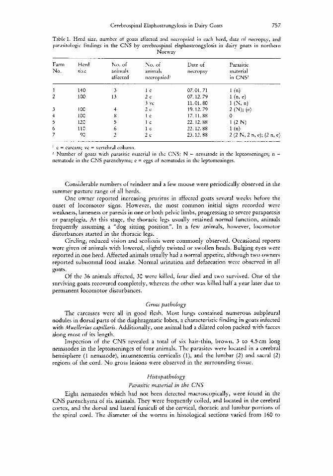
Table 1. Herd size, number of goats affected and necropsied in each herd, date of necropsy, and parasitologic findings in the C N S by cerebrospinal elaphostrongylosis in dairy goats in northern
Norway
Farm Herd No. of No. of Date of Parasitic No. size animals animals necropsy material
affected necropsied’ in CNSZ
1 140 3 l c 2 100 13 2 c
3 100 4 2 c 4 100 8 l c 5 120 5 l c 6 110 6 l c 7 90 2 2 c
3 vc
07.01.71 07.12.79 11.01.80 19.12.79 17.11.88 22.12.88 22.12.88 23.12.88
I c = carcass; vc = vertebral column. 2 Number of goats with parasitic material in the CNS; N = nematode in the leptomeninges; n = nematode in the CNS parenchyma; e = eggs of nematodes in the leptomeninges.
Considerable numbers of reindeer and a few moose were periodically observed in the summer pasture range of all herds.
One owner reported increasing pruritus in affected goats several weeks before the onset of locomotor signs. However, the most common initial signs recorded were weakness, lameness or paresis in one or both pelvic limbs, progressing to severe paraparesis or paraplegia. At this stage, the thoracic legs usually retained normal function, animals frequently assuming a “dog sitting position”. In a few animals, however, locomotor disturbances started in the thoracic legs.
Circling, reduced vision and scoliosis were commonly observed. Occasional reports were given of animals with lowered, slightly twisted or swollen heads. Bulging eyes were reported in one herd. Affected animals usually had a normal appetite, although two owners reported subnormal food intake. Normal urination and defaecation were observed in all goats.
Of the 36 animals affected, 30 were killed, four died and two survived. One of the surviving goats recovered completely, whereas the other was killed half a year later due to permanent locomotor disturbances.
Gross pathology The carcasses were all in good flesh. Most lungs contained numerous subpleural
nodules in dorsal parts of the diaphragmatic lobes, a characteristic finding in goats infected with Muellerius capillaris. Additionally, one animal had a dilated colon packed with faeces along most of its length.
Inspection of the C N S revealed a total of six hair-thin, brown, 3 to 4.5cm long nematodes in the leptomeninges of four animals. The parasites were located in a cerebral hemisphere (1 nematode), intumescentia cervicalis (I), and the lumbar (2) and sacral ( 2 ) regions of the cord. N o gross lesions were observed in the surrounding tissue.
Histopathology Parasitic material in the CNS
Eight nematodes which had not been detected macroscopically, were found in the CNS parenchyma of six animals. They were frequently coiled, and located in the cerebral cortex, and the dorsal and lateral funiculi of the cervical, thoracic and lumbar portions of the spinal cord. The diameter of the worms in histological sections varied from 160 to

75 8 HANDELAND and SPARBOE
Fig. 1. Cross sections of a coiled, female Eluphostrongylrrs rurrgqerz in the subarachnoid space of the sacral cord. HE ~ 1 0 0
Fig. 2. Cluster of nematode ova in the leptomeninges of cerebrum. HE ~ 4 0 0 Fig. 3 . Elongated malacic focus in the cerebral cortex with macrophages and perivascular cuffings. HE
X l O O Pig. 4. Cross sections of a coiled nematode in the crebral cortex. Perivascular cuffings. Meningeal
infiltration in sulcus cerebri. HE x40
Cerebrospinal Elaphostrongylosis in Dairy Goats 759
250 pm. The intestine and genital organs were usually clearly visible in their pseudocoelum (Fig. 1).
A few nematode ova were found in four goats, occurring in the leptomeninges of cerebrum (2 animals), cerebellum and cervical spinal cord (Fig. 2).
Parasitic findings in the CNS by gross and histopathologic examination are sum- marized in Table 1.
Brain lesions Areas showing local or diffuse infiltration of mononuclear cells, and frequently also
eosinophils, could be demonstrated in the leptomeninges of all 10 brains examined. The infiltrations were mild to severe and most prominent in the depths of the sulci. A few small granulomas were also identified, one of which contained a medium-sized, necrotic artery.
Malacic foci containing macrophages were randomly distributed in the parenchyma of 8 brains. Surrounding tissues contained scattered, swollen axons and necrotic neurons, together with light to pronounced perivascular accumulation of mononuclear cells and frequently also of eosinophils (Fig. 3). Glial scarring was also observed, whereas haemor- rhages were rare. Light to moderate perivascular cuffings were the only lesions found in the parenchyma of the remaining two animals.
A probably moribund or recently dead nematode found in the cerebral cortex in one goat, was surrounded by compressed and disrupted tissue. Debris and proliferating macrophages were evident near the worm (Fig. 4).
Spinal cord lesions In dura mater, light to moderate infiltrations of mononuclear cells and scattered
eosinophils were observed, especially near blood vessels and in the sheaths of spinal nerve roots. The infiltrations were usually focal and were most prominent in the caudal parts of the cord.
The leptomeninges showed slight to marked accumulation of mononuclear cells and various numbers of eosinophils. The infiltrations were local or diffuse and frequently extended into the dorsal sulcus and ventral fissure. Occasionally, small granulomas were also observed.
Throughout the white matter, malacic foci with microcavitations were randomly distributed in both dorsal and ventrolateral funiculi. The number of foci on a single slide varied from 1 to 15. They were either round, oval, o r irregular in shape, and had disorganized margins which usually contained varying numbers of macrophages prolifer- ating into the microcavitations. Swollen axons, or small cavitations resulting from axon disintegration, were dominant features in lesions and adjacent tissue (Fig. 5 ) . Fragmenta- tion of swollen, necrotic axons and myelin sheaths was evident in longitudinal sections of the cord (Fig.6). Glial scarring was observed in some sites, whereas haemorrhages were rare.
Light to marked perivascular accumulations of mononuclear cells and frequently eosinophils, were commonly seen near malacic foci. Scattered cuffings and swollen axons were also observed seemingly far distant from malacic lesions.
Five of seven worms found in white matter of the cord were surrounded by small zones of compressed and disrupted tissue showing no inflammatory reactions. Large parasitic granulomas had been formed around the remaining two nematodes. These granulomas contained several cross sections of a necrotic worm and numerous leukocytes, glial cells and scattered epithelioid and foreign body giant cells (Fig. 7).
Malacic foci similar to those described in white matter, were also observed in the grey matter of the cord. However, compared to white matter, lesions were few in number.
Other fairly common findings in the grey matter of’the cord, were a slight though distinct gliosis especially involving the dorsal horns, and perivascular cuffings.
760 HANDELAND and SPARROE
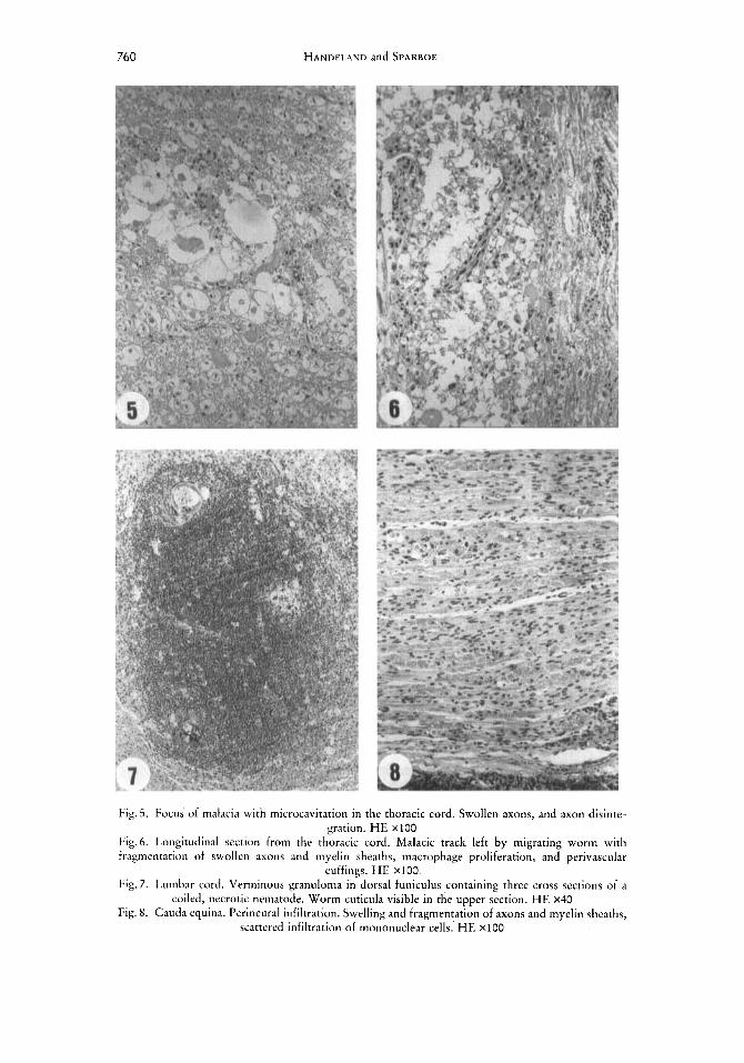
Fig. 5. Focus of malacia with microcavitation in the thoracic cord. Swollen axons, and axon disinte- gration. H E ~ 1 0 0
Fig.6. Longitudinal section from the thoracic cord. Malacic track left by migrating worm with fragmentation of swollen axons and myelin sheaths, macrophage proliferation, and perivascular
cuffings. H E ~ 1 0 0 Fig. 7. Lumbar cord. Verminous granuloma in dorsal funiculus containing three cross sections of a
coiled, necrotic nematode. Worm cuticula visible in the upper section. H E x40 Fig. 8. Cauda equina. Perineural infiltration. Swelling and fragmentation of axons and myelin sheaths,
scattered infiltration of mononuclear cells.' H E ~ 1 0 0

Cerebrospinal Elaphostrongylosis in Dairy Goats 76 1
Cauda equina and spinal nerve root lesions Epi- and perineural infiltrations and granulomas mainly composed of plasma cells,
eosinophils and lymphocytes were found in longitudinal sections of the cauda equina and the extradural roots of the lumbosacral spinal nerves in all animals examined (Fig. 8). That there were remnants of parasitic material within the granulomas could not be demonstrated with complete certainty.
Longitudinal sections of extradural roots of the cervicothoracic spinal nerves revealed slight perineural infiltrations in one of three goats examined.
Focal swelling and fragmentation of axons and myelin sheaths, together with scattered macrophage infiltrations, were seen in nerve fascicles of cauda equina and spinal nerve roots in most animals (Fig. 8).
Other tissues A verminous pneumonia with the presence of adults, larvae and ova of Muellerius
capillaris (tentatively identified) was diagnosed in 8 of 10 lungs examined. In the kidneys, liver and cardiac musculature of five goats examined, minor foci with proliferation of fibrocytes, and accumulation of mononuclear cells and scattered granulocytes, were regularly observed.
Results of bacteriological examination were negative.
Discussion This paper is assumed to be the first report of naturally occurring cerebrospinal
elaphostrongylosis in an abnormal host species, i. e. the goat. The diagnosis was based upon the identification of Elaphostrongylus rangiferi in the
leptomeninges, and of elaphostrongyloids in the CNS parenchyma of eight animals. However, histopathologic lesions such as focal encephalomyelomalacia, eosinophilic lep- tomeningitis and perineural infiltrations, were found in the CNS of all animals examined.
The clinical signs were characterized by motor weakness, lameness and paresis, usually progressing to severe paraparesis. These signs, most probably the clinical manifes- tation of lesions caused by mature or close to mature worms migrating through and lacerating the CNS parenchyma, were observed in all animals reported to be affected. Furthermore, the disease in all herds occurred exclusively during the autumn and early winter of one single indoor season, after the goats had been at pasture in areas frequented by considerable numbers of reindeer, a species commonly found to be infected with Elaphostrongylus rangiferi in northern Norway (GRDHOLT, 1969; BAKKEN and SPARBOE, 1973; KUMMENEJ~, 1974; HALVORSEN et al., 1980).
This leads to the conclusion that the reported outbreaks of neurological disease represented the seasonal occurrence of cerebrospinal elaphostrongylosis caused by Elaphostrongylus rangiferi, the elaphostrongyloid nematode of reindeer (Rangifer tarandus tarandus).
The seasonal occurrence, clinical signs and histopathologic lesions of the neuro- logical disease in our material, were in accordance with reports of cerebrospinal nematodiasis in abnormal host species in North America and Asia caused by Parelapho- strongylus tenuis (KENNEDY et al., 1952; ANDERSON, 1965, 1971; CARPENTER et a]., 1973; TRAINER, 1973; MAYHEW et al., 1976; KISTNER et al., 1977) and Setaria digitata (INNES et al., 1952).
The minor, chronic inflammatory foci, which were regularly found in the liver, kidneys, and cardiac musculature of goats in the present study, probably represented reactive changes in response to larvae spread via the blood stream in the early stage of infection. These findings indicate that ingested, infective larvae of E. rangiferi after penetrating the wall of the digestive tract, travel to the lungs via the venous route, and from there to the organs through the arterial circulation. The C N S probably represents a niche for the neurothropic parasite in which an essential stage of development takes place.
762 HANDELAND and SPARBOE
The presence of nematode ova in the CNS of goats in our material indicates that E. rungifevi is able to mature and reproduce in this animal species. However, due to the difficulty in differentiating first stage larvae of E. vungiferi from Muellerius cupilluris in faeces (BAKKEN and SPARBOE, 1973), the latter parasite infecting the majority of the carcasses examined, the performance of infection experiments will be required to ascertain whether or not the nematode is able to complete its life cycle in goats.
Acknowledgement The authors are grateful to Dr H . NORBERG, Dr G. STUVE, Professor 0. HELLE, Professor
K. NORDSTOCA, Dr C . HACEN and especially Dr T. SLETTBAKK for valuable discussions on the manuscript. Furthermore, we thank Dr J.RACE for linguistic advice, and D r A.SKORPING for assistance with the photomicrographs.
References ANIIERSON, R. C., 1965: An examination of wild moose exhibiting neurologic signs in Ontario.
Can. J. Zool. 43, 635-639. ANDERSON, R. C., 1971 : Neurologic disease in reindeer (Rungifer turundus terruenovue) introduced
into Ontario. Can. J. Zool. 49, 159-166. ANONYMOUS, 1988: Annual Report. Norwegian board of animal production recordings. Otto Falch
A/S, Oslo, 173 pp. BAKKEN, G., and 0. SPAKBOE, 1973: Elaphostrongylose hos rein (Elaphostrongylosis in reindeer).
Nord. Vet.-Med. 25, 203-210. CAMERON, T. W.M., 1931: On two new species of nematodes from the Scottish red deer. J.
Helminthol. 9, 213-216. CAKPENTER, J. W., H. E. JORDAN, and B. C. WARD, 1973: Neurologic disease in wapiti naturally
infected with meningeal worms. J. Wildl. Dis. 9, 149-153. CRUHOLT, L., 1969: Eluphostrongylus rungiferi og dens betydning i praktisk reinkjnttkontroll
(Eluphostrongylus rungiferi and its significance for judgement in reindeer meat inspection). Medlemsbl. Norske Vet.-Foren. 21, 382-388.
HALVORSEN, O., J. ANDERSEN, A. SKORPING, and G. LORENTZEN, 1980: Infection in reindeer with the nematode Eluphostrongylus rungiferi MITSKEVICH in relation to climate and distribution of intermediate hosts. In: REIMERS, E., E. GAARE, and S. SKJENNEBERC (eds.), Proc. 2nd Int. Reindeer/Caribou Symp. DVF, Trondeim, 449-455.
HELLE, O., 1990: Personal communication. INNES, J . R. M., C. SHOHO, and C. P. PILLAI, 1952: Epizootic Cerebro-Spinal Nematodiasis: Focal
Encephalomyelomalacia in Animals in the Orient Caused by Immature Seturzu digitutu. Brit. vet. J . 108, 71-88.
KENNEDY, P. C., J. H . WHITLOCK, and S. J. ROBERTS, 1952: Neurofilariosis, a paralytic disease of sheep: 1. Introduction, symptomatology, and pathology. Cornell Vet. 42, 118- 124.
KISTNER, T. P., G. R. JOHNSEN, and G. A. RILLINC, 1977: Naturally occurring neurologic disease in fallow deer infected with meningeal worm. J. Wildl. Dis. 13, 55-58.
KUMMENEJE, K., 1974: Encephalomyelitis and neuritis in acute cerebrospinal nematodiasis in reindeer calves. Nord. Vet.-Med. 26, 456-458.
LANKEWER, M. W., and T. H. NOKTHCO~T, 1979: Eluphostrongylus cervi CAMERON, 1931 (Nematoda: Metastrongyloidea) in caribou (Rungifer tarandus caribou) of Newfoundland. Can.
LuiIrMov, M. P., 1945: Sbornik nauchoissledovatel’skikh rabot (New worm diseases of the brain o f deer with unossified antlers). 1.aboratory pantovogo olenvodstva Ministerstva sovkhozov SSR 1,
MASON, P.C., N.K. KrDueY, R.J. SUTHEKLAND, D.M. RUTHERFORD, and A.G. GKEEN, 1976: Eluphostrongylus cervi in red deer. N . Z. Vet. J. 24, 22-23.
MAYHEW, 1. G., A. DELAHUNTA, J. R. GEORGI, and D. G. ASPROS, 1976: Naturally occurring cere- brospinal parelaphostrongylosis in goats. Cornell Vet. 66, 56-72.
MITSKE:VTCH, V. Y., 1960: Eluphostrongylus rnngiferi - novyi vid gel’minta olenya (Eluphostrongylus rungiferi - a new species of helminth of reindeer). Parazity Zhivotnykh i Prirodnaya Ocliagovast’Bolezcni Trudy In-ta 2001. A N KazSSR 12, 115-119.
J. Zool. 57, 1386-1392.
225-232.

Cerebrospinal Elaphostrongylosis in Dairy Goats 763
PROSL, H., und E. KUTZER, 1980: Zur Biologie und Bekarnpfung von Eluphostrongylus cerwi. Z. Jagdwiss. 26, 198-207.
S T E ~ N , M., A. G. CHAI~AUD, and C. REHBINDER, 1989: Species of the genus Eluphostrongylus parasite of Swedish cervidae. A description of E. ulces n. sp. Ann. Parasitol. Hum. Comp. 64, 134-142.
TRAINER, D. O., 1973: Caribou mortality due to the meningeal worm (Pureluphostrongylus tenuzs). J. Wildl. Dis. 9, 376-378.