the intracraniovertebral volumes, the cerebrospinal fluid ... · the intracraniovertebral volumes,...
TRANSCRIPT

The Intracraniovertebral Volumes, the Cerebrospinal Fluid Flow and the Cerebrospinal Fluid Pressure, Their Homeostasis and Its Physical Regulation Darko D. Lavrencic, M.D. Slovenia, European Union www.med-lavrencic.si/raziskava.htm www.med-lavrencic.si/correspondence.htm, www.med-lavrencic.si/related.shtml Preface. After publication of the presented hypothesis some predictions were verified independently by other authors: (1) Monro-Kellie "four compartments" doctrine, (2) relation between cerebrospinal fluid (CSF) formation and CSF removal in physiological phase as presented with illustrative curves, (3) hypovolemia during intracranial hypotension syndrome, (4) increased CSF proteins in decreased CSF flow and (5) influence of neuro-vegetative system on CSF pressure. The predictions not yet verified: (1) turning points B-low and B-high that represent physiological borders, (2) pathophysiological self-sustaining phases of low and high CSF pressure with corresponding minimal or maximal CSF volume (maximal dural sac collapse or distension) and no CSF transport, (3) compensated and de-compensated conditions. None of the predictions were disproved yet. There are three types of CSF hydrodynamic processes according to their duration in time: (1) rapid changes (in second(s), e.g., hydrodynamic changes caused by arterial pulsations in craniospinal space, etc), (2) homeostatic changes (in minutes to hours, e.g., as described in this article) and (3) long-term changes and adaptations, e.g., chronic hydrocephalus, etc. This article is not discussing (1) and (3) processes. The purpose of this presentation on the INTERNET is to promote further discussions about unverified predictions and to encourage clinical research and experimenting in this direction Summary. Physiological and pathophysiological processes in the intracraniovertebral space are specific because of its rigid and constant volume (Monro-Kellie doctrine). The hypothesis presents how the homeostasis of the intracraniovertebral compartments’ volumes, cerebrospinal fluid (CSF) flow and CSF pressure is physically regulated. The hypothesis takes into account the quantitative and qualitative relations regulating CSF formation and CSF removal on which the homeostasis is based. Introduction. Monro found in 1783, during studying brain blood circulation, that the processes in intracranial space are specific due to the constant volume of the skull. None of the contained masses can be compressed and one of them can be increased, only if another one is decreased. This occurs when the ossification of the cranial sutures is so strong that it resists any increase of intracranial pressure. Monro-Kellie doctrine includes 3 compartments’ volumes: (1) brain and medulla spinalis with meninges, (2) cerebrospinal blood vessels with blood, (3) CSF space. Dural sac is elastic. It can be distended or collapsed. The spinal extradural space, which contains soft tissues and veins plexuses adapts to the changes of dural sac. The author of this text regards this spinal extradural space and intracranial venous sinuses as compartment IV. By the addition of compartment IV, Monro-Kellie doctrine becomes re-established in wider sense. This hypothesis suggests a possible mechanism, which complies with all demands by Millen and Woollam: “Under normal conditions, the composition, volume and pressure of cerebrospinal fluid remain relatively constant. This homeostasis depends upon maintenance of a balance between the production and absorption of the fluid and upon its free circulation between the sites of its formation and removal. Any disturbance of this balance or interference with circulation will result in alternations in the pressure of the fluid and perhaps in its volume and composition”. The author could not find anywhere in literature the above-mentioned general principles in their integral application. The author has thus collected partial ideas, reports of experiments and facts and confronted those with his analysis of various physiological and pathophysiological situations. After this he proceeded to the analysis of various theoretically possible relations to make a synthesis, which would be a logical and a dynamic one and be based on physical regulation of homeostasis. In author’s opinion the experimenting, if performed in this direction, would resolve questions, thus supporting or disproving the presented hypothesis.
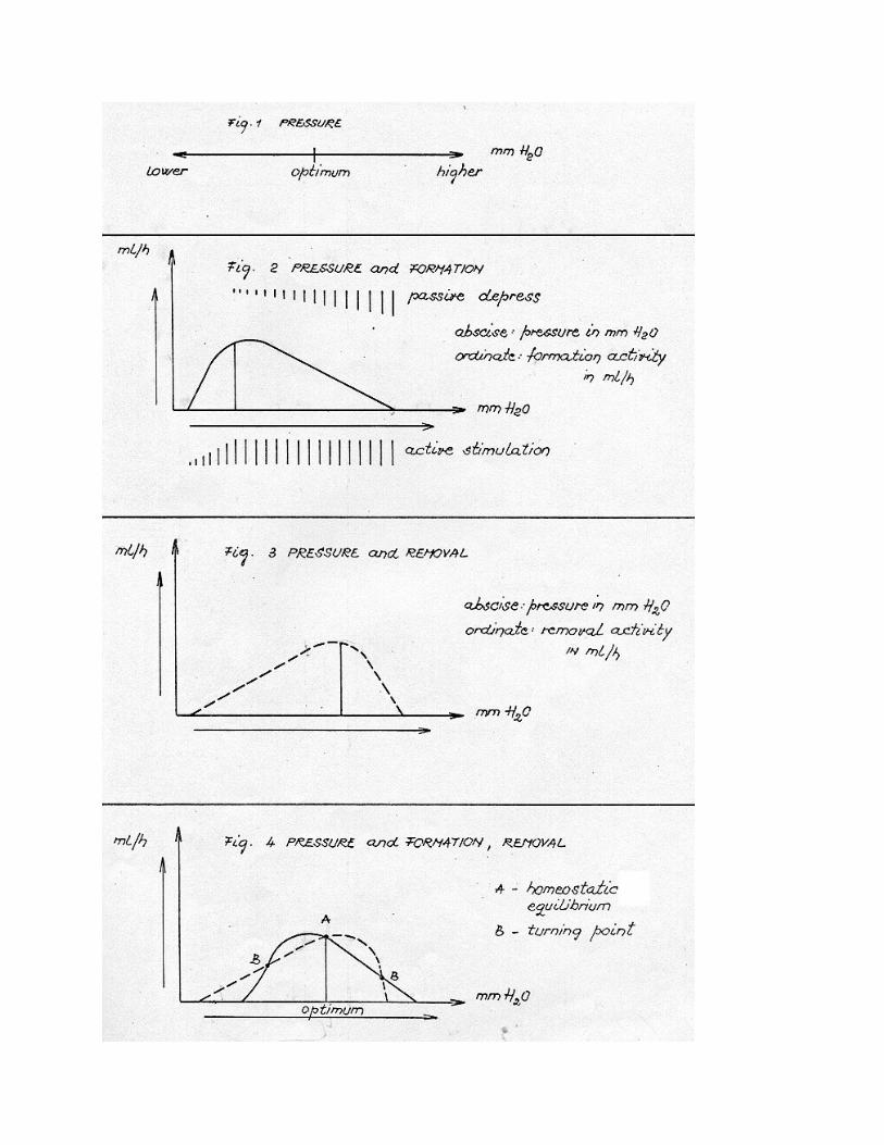
In the following text CSF formation, CSF removal, CSF flow and CSF pressure will be referred to as formation, removal, flow and pressure. Compartments I, II, III and IV are presented in the graphics as volumes I, II, III and IV. The hypothesis will be presented in the following order: (1) Pressure (2) Formation and pressure (3) Removal and pressure (4) Formation, removal and pressure (5) Formation, removal, compartment III, flow and pressure (6) Mechanical adaptations (7) Primal changes in compartment III (8) Primal changes in compartment I (9) Primal changes in compartment II (10) Primal changes in compartment IV (11) Primal changes in flow (12) Primal changes in formation (13) Primal changes in removal (14) Verification (15) Appendix 1) PRESSURE The pressure is the dynamic result of interaction between all four compartments. The pressure transmission to compartment IV is specific due to elastic dural sac, which can resist and maintain a certain pressure difference. The pressure is defined by the homeostasis of the compartments. The disturbance in compartments always occurs primarily and the disturbance in pressure always secondarily. The pressure and other relations are not presented as absolute, but relative relations (Fig. 1). 2) FORMATION AND PRESSURE The formation mainly takes place in plexus chorioideus. The transudation as ‘formation’ will not be discussed here. It is believed that the formation is an active process. Woollam and Millen reopened the question whether the glomus plexus chorioideus is sensitive to changes in pressure and whether it can regulate the formation. The idea of Bowsher and Kappers was that pressure, when higher than normal, can passively depress formation in plexus chorioideus. The analysis of clinical cases by the author supports the above ideas. At certain pressure the glomus plexus chorioideus starts to stimulate the formation (Fig. 2). The formation is increasing and at certain pressure reaches its maximal activity. During further increasing of pressure the glomus stimulation remains maximal and the passive depression is increasing. The formation has therefore two phases: the first one is the increasing phase, due to glomus stimulation and the second one is the decreasing phase, as a result of glomus stimulation and passive depression. At certain pressure the formation stops. 3) REMOVAL AND PRESSURE Experiments show that removal is a passive process through arachnoidal villi. It occurs mainly in cranial venous sinuses and less in spinal extradural veins plexuses. ‘The re-absorption’ through plexus chorioideus will not be discussed here. The removal depends on hydrostatic pressure and oncotic pressure difference between CSF and venous blood in compartment IV. It also depends on quality of arachnoidal villi (Fig. 3). At certain pressure the removal starts. Increasing of removal follows the increasing of pressure. Removal increases to the maximum and then decreases, until it stops at its highest pressure. 4) FORMATION, REMOVAL AND PRESSURE This is the nucleus of the hypothesis and was in the center of author’s attention during building a theory (Fig. 4). The presented relation between formation, removal and pressure enables the automatic
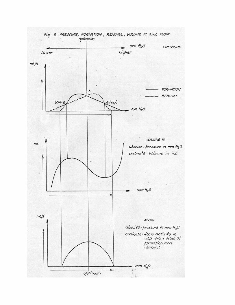
regulation of the entire homeostasis; it identifies its physiological borders and explains the transition to pathophysiological conditions. Formation and removal reach maximal activity at different pressure respectively, the formation at lower and removal at higher. Three times they are balanced. Point A represents homeostatic equilibrium. As we shall see, this equilibrium is automatically regulated and re-established during physiological disturbances. It is the homeostatic equilibrium that defines the optimal pressure. Turning points B-low and B-high represent the physiological borders. No mechanism supports this balance; it is only a temporary and unstable condition, after which the pathophysiological processes begin to take place. 5) FORMATION, REMOVAL, COMPARTMENT III, FLOW AND PRESSURE Relation between those is represented in Fig. 5. Homeostatic equilibrium defines the optimal pressure and consequently the optimal volume of compartment III. If there is increase of pressure towards turning point B-high, the removal will be relatively greater than formation and compartment III will decrease. If the pressure is increased over turning point B-high, the formation will be relatively greater than removal and compartment III will be progressively increasing for as long as formation lasts. The final result is the maximal compartment III. If there is decrease of pressure toward turning point B-low, the formation will be relatively greater than removal and compartment III will increase. If the pressure is decreased over turning point B-low, the removal will be relatively greater than formation and the compartment III will be progressively decreasing for as long as the removal lasts. The final result is the minimal compartment III. Homeostatic equilibrium also defines the optimal flow i.e. maximal flow. Pressure, higher or lower than optimal, will respectively decrease flow. Flow is being defined by formation or by removal; whichever is lower at a certain pressure. If either of them stops, flow also stops, while pure CSF output or pure CSF input remains. Input (inflow) or output (outflow) stops after both formation and removal stop. It can be assumed also that, the slower is the flow, the higher are concentrations of the protein in CSF. 6) MECHANICAL ADAPTATIONS During the rapid changes in the compartments’ volumes, there is no time for any physiological mechanism to interfere. No masses or compartments can be compressed; therefore, the compartment IV with its vein plexuses must carry out rapid mechanical adaptations over elastic dural sac. The blood from compartment IV can be driven out of intracraniovertebral space or it can be retained. 7) PRIMAL CHANGES IN COMPARTMENT III (CSF space) Compartment III is the most responsible for the compartments’ volume homeostasis. Therefore, it will be discussed first. If primal changes occur in compartment III it must re-establish its own optimal volume; and must actively adapt if the primal changes occur in other compartments. Primal increase in compartment III increases pressure towards turning point B-high. In this phase homeostatic mechanism decreases compartment III and consequently the pressure also until the homeostatic equilibrium becomes re-established (Fig. 5), (clinical picture: intrathecal application of solution in CSF space). Primal decrease in compartment III decreases pressure towards turning point B-low. In this phase homeostatic mechanism increases compartment III and consequently the pressure until homeostatic equilibrium becomes re-established (Fig. 5), (clinical picture: CSF taken by lumbar puncture). Primal increase in compartment III which increases the pressure over turning point B-high causes the compartment III and the pressure to increase progressively to the maximal compartment III (Fig. 5), (clinical picture: exacerbation of hydrocephalus after PEG). Primal decrease in compartment III which decreases the pressure over turning point B-low causes the compartment III and the pressure to decrease progressively to the minimal compartment III (Fig. 5), (clinical picture: aliquorrhea after lumbar puncture).
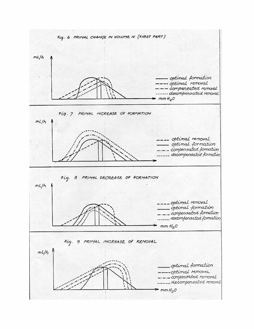
8) PRIMAL CHANGES IN COMPARTMENT I (brain, medulla spinalis and meninges) Under normal conditions the volume of compartment I is perfectly constant. Primal increase in compartment I increases pressure towards turning point B-high. In this phase, the compartment III becomes actively adapted, i.e. decreased (Fig. 5). This process lasts until a new relation is established between compartment I and III at optimal pressure (clinical picture: tumor cerebri). If primal change in compartment I is reversible, the active adaptation of compartment III is reversible as well. Primal decrease in compartment I decreases pressure towards turning point B-low. In this phase compartment III is actively adapted, i.e. increased (Fig. 5). This lasts until a new relation is established at optimal pressure, between compartment I and compartment III (clinical picture: atrophia cerebri with consequential hydrocephalus ex vacuo). If primal change is reversible, the active adaptation of compartment III is reversible, too. If primal change in compartment I increase pressure over turning point B-high, compartment III as a result progressively increases (Fig. 5). If primal changes in compartment I decreases pressure over turning point B-low, compartment III as a result progressively decreases (Fig. 5). 9) PRIMAL CHANGE IN COMPARTMENT II (brain’s blood vessels with blood) The supply of oxygen to the brain must be very constant at any blood and intracranial pressure. Lamina muscularis of brain blood vessels is weaker than elsewhere. The supply with oxygen, if disturbed, becomes regular due to blood vessel auto regulation, namely the speed of blood flow through the brain, rather than vasoconstriction or vasodilatation. In this way the compartment II remains fairly constant in any physiological condition. Supply with oxygen being of the foremost importance for the brain, the compartment II cannot be used as active or passive principle for regulation compartments’ volume homeostasis. Primal changes in compartment II are regulated in the same way as primal changes in compartment I. 10) PRIMAL CHANGES IN COMPARTMENT IV (cranial venous sinuses and spinal extradural space) Compartment IV is separated from other compartments by elastic dural sac; that, and the presence of arachnoidal villi, is causing its particular behavior. Compartment IV is divided in two parts: the first part is the cranial venous sinuses where removal mainly occurs. The changes in compartment’s volume are relatively unimportant, while the changes in venous blood pressure are very important, because of their direct influence on removal curve. Second part is the spinal extradural space. The removal occurs here in minor degree, thus inversely, the changes in blood veins pressure are the less important, while the changes in compartment’s volume are the more important. The fact is that the whole compartment IV cannot be increased immediately, since the other three compartments cannot become compressed. The mechanical adaptation is thus not possible. The influence on the whole compartment IV will merely rapidly increase the pressure, whereas the relations in compartments’ volumes will remain undisturbed. Only the increased venous blood pressure in the first part of compartment IV will be discussed here. Other possibilities are more complex. Primal increase of venous blood pressure in the first part of compartment IV will selectively depress removal curve (Fig. 6). There are two conditions. Compensated condition involves increased compartment III, increased pressure and decreased flow. Decompensation involves maximal compartment III, maximal pressure and no flow (clinical picture: hydrocephalus communicans, due to thrombosis of dural sinuses).
11) PRIMAL CHANGE IN FLOW (CSF circulation) Various blocks represent the primal changes. Block divides compartment III in two independent parts. Each of them depends on their own respective formation and removal. Compartments’ volumes homeostasis is broken. If foramina Luschka and foramen Magendie are blocked, first part (ventricles) will, due to remaining formation and absent removal, reach its maximal volume at maximal pressure and zero flow. Second part (subarachnoid space) will, due to remaining removal and formation, reach the minimal volume and zero flow. 12) PRIMAL CHANGE IN FORMATION Primal increase in formation (Fig. 7): Compensated condition involves increased compartment III at increased pressure and increased flow. Decompensation involves maximal compartment III at maximal pressure and zero flow. Primal decrease in formation (Fig. 8): Compensated condition involves decreased compartment III at decreased pressure and decreased flow. Decompensation involves minimal compartment III at minimal pressure and zero flow. 13) PRIMAL CHANGE IN REMOVAL Primal increase in removal (Fig. 9): Compensated condition involves decreased compartment III at decreased pressure and increased flow. Decompensation involves minimal compartment III at minimal pressure and zero flow. Primal decrease in removal (Fig. 6): Compensated condition involves increased compartment III at increased pressure and decreased flow (clinical picture: arachnitis). Decompensation involves maximal compartment III at maximal pressure and zero flow (clinical picture: meningeal hydrops). 14) VERIFICATION The present hypothesis will be rather difficult to verify, because of the technical factors and complex and dynamic character of homeostasis. The working hypothesis will include two types of experiments, according to: Direct method: (1) to determine absolute relation between formation and pressure, (2) to determine absolute relation between removal and pressure. Indirect method: (1) to determine physiological borders of homeostasis, (2) to provoke pathophysiological conditions, (3) to determine absolute relation between flow and pressure. 15) APPENDIX Speculation in connection with glomus plexus chorioideus would be interesting, namely that it could be regulated by the neuro-vegetative system and would be activated at lower or higher pressure. This could result in CSF hypertension or CSF hypotension within physiological homeostatic borders with the corresponding changes in compartment III, flow and physiological borders of homeostasis. CSF hypertension involves: increased pressure, increased compartment III, increased flow and narrower physiological borders, CSF hypotension involves: decreased pressure, decreased compartment III, decreased flow and wider physiological borders. Submitted: September 1, 1969 Retyped: November 12, 2001 Original paper with references: Lavrencic D. The Intracraniovertebral Volumes, the Cerebrospinal Fluid Flow and the Cerebrospinal Fluid Pressure, Their Homeostasis and Its Physical Regulation. Ljubljana: Darko Lavrencic; 1970.