cerebral hemodynamics and investigations of cerebral blood ... · wojciech rudziński 1, maciej...
TRANSCRIPT
29
Nuclear Medicine Review 2007 Vol. 10, No. 1, pp. 29–42
Copyright © 2007 Via MedicaISSN 1506–9680
www.nmr.viamedica.pl
Review
Wojciech Rudziński1, Maciej Swiat2, Maciej Tomaszewski1,Jaroslaw Krejza1, 3
1Department of Radiology, Division of Neuroradiology of the Universityof Pennsylvania, United States2Department of Neurology, Aging, Degenerative and CerebrovascularDiseases, Medical University of Silesia, Katowice, Poland3Department of Nuclear Medicine, Medical University of Gdansk, Poland
[Received 20 III 2007; Accepted 7 IV 2007]
Abstract
To maintain adequate cerebral blood flow despite frequent chang-es in systemic arterial blood pressure and to constantly adjust bloodsupply to the current metabolic demand dictated by neuronal elec-trical activity, brain developed a myriad of mechanisms. These aredesigned to protect central nervous system from fatal consequenc-es of hypoxia and energy deficit and are collectively called “cere-bral autoregulation”. Despite years of research mechanisms respon-sible for regulation of CBF functioning under physiologic and patho-logic conditions are still not clear. When these mechanisms aredamaged or exhausted, patients life is in danger, as even slight,negligible under normal conditions, systemic hemodynamic distur-bances might lead to cerebral infarct. Even perfect imaging of theirreversible brain damage with MR for the particular patient is toolate action. Thus, detection of cerebral blood flow disturbances andimpaired autoregulation, which are known to be associated withhigh risk of stroke, are extremely important in clinical practice. Se-veral methods have been developed to quantify this process andthus evaluate risk of cerebral ischemia and guide therapeutic pro-cess. This review focuses on current knowledge on physiology ofregulation of cerebral blood flow, mechanisms responsible for braindamage resulted from cerebral ischemia and reviews noninvasivediagnostic tests to assess cerebral autoregulation.Key words: brain, cerebral circulation, cerebrovascularreactivity, autoregulation, hemodynamics
Cerebral hemodynamics and investigationsof cerebral blood flow regulation
Correspondence to: Jaroslaw KrejzaDepartment of Radiology of the University of PennsylvaniaDivision of Neuroradiology, Science Building ste 3703600 Market street, Philadelphia, PA 19104, USAe-mail: [email protected]
Cerebral blood flow regulation
Brain tissue constantly maintains an extremely high metabolicrate. Cerebral oxygen consumption (ª 3.5 ml/100g tissue/min)[1] accounts for about 20% resting total body oxygen consump-tion. The metabolic demand must be matched by high blood flowsupply, which on average exceeds 50 ml/100g tissue/min, ac-counting for 15–20% of the total cardiac output [1, 2]. As the brainalmost exclusively uses glucose for its energy metabolism and ingeneral does not store energy, continuous blood supply, main-tained within a narrow range, is absolutely required for brain func-tion (90%) and cell viability (10%) [3]. Cerebral blood flow (CBF) isdriven by cerebral perfusion pressure (CPP), represented by thedifference between mean arterial blood pressure (ABP) and in-tracranial pressure (ICP), working in concert with cerebrovascularimpedance. The components which make up cerebrovascularimpedance include:— cerebrovascular resistance (CVR), which is inversely propor-
tional to the forth power of the vessel radius when laminar flowoccurs and the flow is in steady state;
— the internal fluid resistance, which depends on viscoelasticproperties of arterial walls, viscosity of the blood and flow ve-locity;
— the blood inductance, which is dependent on its rheostaticproperties and momentum;
— the vascular compliance, which is related to the elasticity ofthe vessel wall [4, 5].Alteration of any of the components, which make up impe-
dance in a non steady-state system, such as occurs in the cere-brovascular system, can significantly impact the blood flow. Mi-crovascular constriction, however, most significantly increases theimpedance [6]. The ability of brain microvasculature to maintaincerebral blood flow relatively constant despite wide variations inCPP is called “cerebral autoregulation” (CA). In the normal stateCA maintains relatively constant CBF within the range of meanABP from about 60 to 150 mm Hg [7]. It should be mentioned,however, that upper and lower limits of CA are not fixed and canbe shifted up or down by endogenous as well as exogenous fac-tors. Sympathetic nervous system activity and increased levels ofAngiotensin II, for example, shift upper and lower limits of CA uptowards higher pressures, while chronic use of antihypertensivemedications have opposite effects [8, 9]. Patients with untreatedhypertension have limits of regulation set on higher level in com-
30
Nuclear Medicine Review 2007, Vol. 10, No. 1
www.nmr.viamedica.pl
Review
parison to healthy people [8]. This is important because in thesepatients overzealous antihypertensive treatment may lead to dan-gerous reduction of CBF at relatively high ABP. The mechanismsof CA are complex and not fully understood. It is surmised fromthe results of numerous experimental studies that CA is maintainedby three different control pathways: vasogenic, metabolic andneurogenic.
The vasogenic mechanism is based on the intrinsic ability ofcerebral vessels to respond to changes in shear stress and trans-luminal pressure [10]. Increase in transluminal pressure activatesvascular smooth muscle cells leading to decrease in the diameterof arteries [11, 12]. On the other hand, contraction of cerebralvessels increases vascular wall shear stress [13]. This triggersendothelial cells to release factors, that relax vascular smoothmuscles and dilates vessels [12]. These counteracting mecha-nisms assure optimal adjustment of the vessel diameter to CPPat any time.
The exact mechanisms of vascular contraction in response toincrease in transluminal pressure remain unclear. It appears, how-ever, that mechanical dilation of smooth muscle cells leads acti-vation of phospholipase C (PLC) [14]. Increased PLC activity leadsto elevated diacylglycerol levels, which activates protein kinase C,which is known to activate nonselective cation channels, depolar-izing the smooth muscle membrane potential and enhancing cal-cium entry through voltage-dependent calcium channels. In-creased concentration of free calcium ions activates the myosinlight chain kinase, which phosphorylates myosin light chains andcauses muscle contraction [15]. It is also possible that stretch ofmuscle cells directly activates calcium channels and leads to in-creased calcium concentration without activation of PLC- depen-dent pathway [16]. In contrary to intraluminal pressure-inducedcontraction, which seems to be dependent only on smooth mus-cle cells, shear stress-induced vasodilatation requires interactionof endothelial cells and vascular muscles. Synthesis of nitric oxi-de (NO) is necessary for this phenomenon to occur [17]. Shearstress increases activity of endothelial nitric synthase (eNOS) andcauses release of NO, which diffuses to adjacent smooth musclecells and induces vasodilatation through activation of potassiumchannels in those cells [18, 19]. Shear stress may also directlyincrease expression of eNOS as a shear stress-response elementhas been found in promoter region of gene for eNOS [20]. It is notclear, how significant a role the vasogenic mechanism plays inmaintaining and regulation CBF and whether it depends some-how on caliber of the involved cerebral arteries.
In metabolic regulation, arterial resistance is modified by wasteproducts of energy metabolism (CO2), partial pressure of O2, andrelease of specific vasoactive substances such as adenosine andpotassium ions from neurons in response to insufficient bloodsupply. The most important metabolic factor is tension of CO2 inperiarteriolar space, although CVR is not directly affected by theCO2 tension. It is the accompanying shift in periarteriolar pH, whichregulates diameter of cerebral vessels [21]. Hypercapnia and theresulting decrease in extracellular pH causes cerebral vasodilata-tion and increase in CBF, while hypocapnia leads to cerebral vas-oconstriction and CBF decrease. Mechanisms responsible forregulation of CVR by CO2 tension and accompanying pH chan-ges are not clear. Hydrogen ions may directly activate potassiumchannels in smooth muscle cells leading to its hyperpolarization
or they may induce release of vasodilatory prostaglandins, ade-nosine or NO from neurons, glia or vessels [22–26]. Hypoxia, po-tassium ions and adenosine also lead to hyperpolarization ofsmooth muscle cells and consequently dilation of cerebral ves-sels. It appears that hypoxia-induced vasodilatation is mediatedby activation of potassium channels, while increased concentra-tion of potassium ions in extracellular fluid activates electrogenicNa/K pumps and smooth muscle inward rectifier potassium chan-nels [27, 28]. Adenosine acts on cerebral vessels through its re-ceptors located in arterial smooth muscle membrane [29]. Acti-vation of these receptors leads to opening of calcium-dependentand ATP-dependent potassium channels [30, 31].
While there is a consensus that vasogenic and metabolicmechanisms play critical role in regulation of cerebrovascular tone,the importance of neurogenic regulation in the control of CBF isstill a matter of debate. The cerebral vessels are innervated byextrinsic and intrinsic systems of nerve fibers. The “extrinsic” sys-tem refers to nerve fibers originating in ganglia belonging to sym-pathetic, parasympathetic and sensory ganglia, while nerves orig-inating within the brain represent an "intrinsic" system [32]. Activa-tion of sympathetic vascular nerves leads mainly to release ofnorepinephrine and neuropeptide Y [33]. Sympathetic stimulationconstricts large cerebral arteries. However, CBF does not de-crease, because the constriction of large cerebral vessels is im-mediately compensated for by dilation of resistance arterioles [34].The role of sympathetic innervation seems to be related to pro-tecting the brain against ABP increases through the mechanismof sympathetic activation shifting the upper and lower limits of CAtowards higher pressures. Parasympathetic vascular nerve fibersrelease vasoactive intestinal polypeptide, acetylcholine and NO[35, 36]. Activation of these nerves causes cerebrovascular dila-tion, the physiological significance of which is still not clear [37].Fibers originating in sensory ganglia contain clacitonin gene-re-lated peptide, substance P, neurokinin A and pituitary adenylatecyclase-activating polypeptide [38–40]. These fibers cause dila-tation of cerebral vessels and their physiological role appears tobe counteracting action of cerebral vasoconstrictors [41]. Activa-tion of these fibers may contribute to increase in CBF occurring inpathologic conditions such as meningitis and may play a role inpathogenesis of migraine [42, 43].
Vascular fibers belonging to the intrinsic system originate indifferent parts of brain such as nucleus basalis, locus coeruleusand raphe nucleus [44–46]. These fibers may modulate vasculartone directly or through stimulation of perivascular interneuronsand glial cells [47, 48]. Activity of the intrinsic system may de-crease or increase local blood flow depending on the rostrocau-dal level of activation of their cells of origin within the brain. Thephysiological significance of this system is not yet completely elu-cidated [45, 46].
For academic purposes, CBF regulation is parceled into se-veral major mechanisms, though the division is somehow artifi-cial. The mechanisms influence and modify each other at any giv-en time and the overall cerebrovascular tone is the product ofinterplay of many processes. It is clear that some of these mech-anisms share common biochemical pathway at the level of vas-cular muscle cells. For instance vascular dilation caused by para-sympathetic activation, shear stress and hypercapnia, at least inpart, are mediated by the same factor — NO [49].
31www.nmr.viamedica.pl
ReviewWojciech Rudziński et al. Cerebral hemodynamics and investigations of CBF regulation
Neurovascular coupling in humans
The brain's information-processing capacity is limited by theamount of oxygen and energy available. When neurons are ac-tive, blood flow in that local brain region increases to meet theenhanced local metabolic demand [50]. The tight coupling be-tween neuronal activity and blood flow is known as the “neurovas-cular coupling”. This phenomenon seems to be dependent onboth astrocytes and perivascular neurons.
Increased local neural activity leads to release of glutamateacting on astrocytic metabotropic glutamate receptors [51]. Acti-vation of glutamatate astrocytic receptors leads to increased syn-thesis of arachidonic acid, which is metabolized to vasodilatoryprostanoids like Prostalgandin E2 (PGE2) and epoxyeicosotrienoicacid (EET) [52, 53]. PGE2 and EET diffuse to vascular musclecells and dilate vessels increasing local blood flow [52, 53]. How-ever, during this process significant amount of arachidonic reachesvascular muscles as well. Smooth muscle cells contain enzymes(CYP4A), which generate powerful vasoconstrictor 20-hydroxye-icosotetraenoic acid (20-HETE) counteracting PGE2 and EET— induced vasodilatation [48, 54]. The question arises, what shiftsthis balance towards vasodilatation? Glutamate released into syn-aptic space activates postsynaptic NMDA-type glutamate recep-tors located on perivascular neurons [55, 56]. This leads to acti-vation of neuronal NO synthase and production of NO, whichreaches smooth muscle cells. NO not only exerts vasodilatory ef-fects but also binds to the heme moiety of CYP4A enzyme andinactivates it, thereby preventing further synthesis of 20-HETE fromarachidonic acid [57, 58]. Thus activation of both nNOS positiveneurons and astrocytes seems to be necessary for proper func-tioning of neurovascular coupling. It is likely that astrocyte-derivedvasodilatory prostanoids are direct mediators while neuronal NOacts as a modulator of this process.
Respiratory and cardiovascular factorsand cerebral hemodynamics
Both oxygen and CO2 tension have powerful effects on car-diovascular system through the peripheral and central chemore-ceptors. Peripheral chemoreceptors are located in the aorta andinternal carotid artery and respond to both hypoxemia and hyper-capnia. The peripheral chemoreflex causes hyperventilation, tran-siently activates sympathetic traffic to peripheral blood vessels,and increases vagal activity to the heart [59]. Activation of periph-eral chemoreceptors does not change CBF [60]. Central chemore-ceptors are located in ventrolateral medulla and detect pH changesof the interstitial fluid [61]. Activation of the central chemoreceptorreflex causes hyperventilation and increased sympathetic stimu-lation to vasculature. Changes in sympathetic tone have a limitedeffect on CBF at normal PaCO2 levels [62]. However, the sympa-thetic nervous system seems to attenuate the CO2-induced in-crease in CBF. This phenomenon may indicate a moderate directeffect of the sympathetic nervous system on the cerebral vascula-ture [62]. Sympathetic stimulation can have more important ef-fects on segmental vascular resistance and cerebral microvascu-lar pressure in pathologic conditions. Generalized increases inthe sympathetic discharge, causing substantial increases in ABP,can prevent concomitant increases in CBF by acting on both small
resistance and large vessels [34, 63].Arterial baroreceptor reflex, which plays an integral role in re-
gulating peripheral vascular tone and heart rate in response tochanges in ABP, seems to have no impact on the CBF. Neitherinterruption nor stimulation of baroreceptor nerves affects CBF orCVR at ABP within the range of CA [64, 65]. However, cerebralvasoconstriction occurs in healthy humans during graded reduc-tions in central blood volume. The magnitude of this response issmall compared with changes in systemic vascular resistance [66].It seems that this degree of cerebral vasoconstriction is not byitself sufficient to cause syncope during orthostatic stress, butcan exacerbate the decrease in CBF associated with hypoten-sion if hemodynamic instability develops.
Cerebral ischemia and investigationsof cerebral blood flow regulation
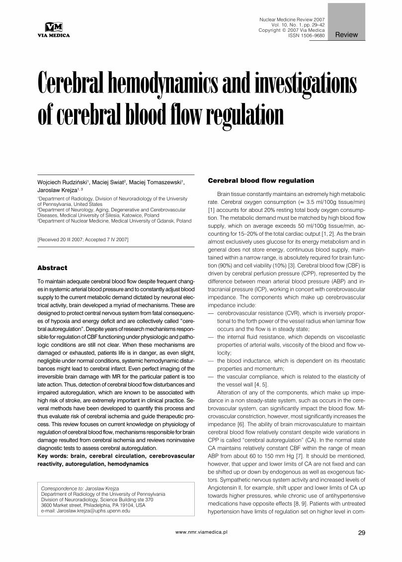
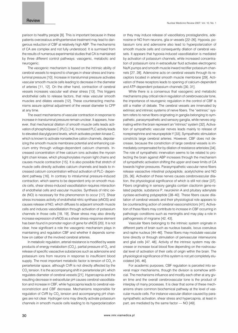
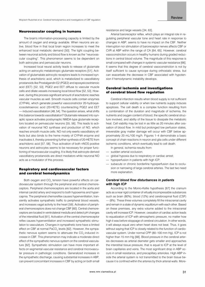
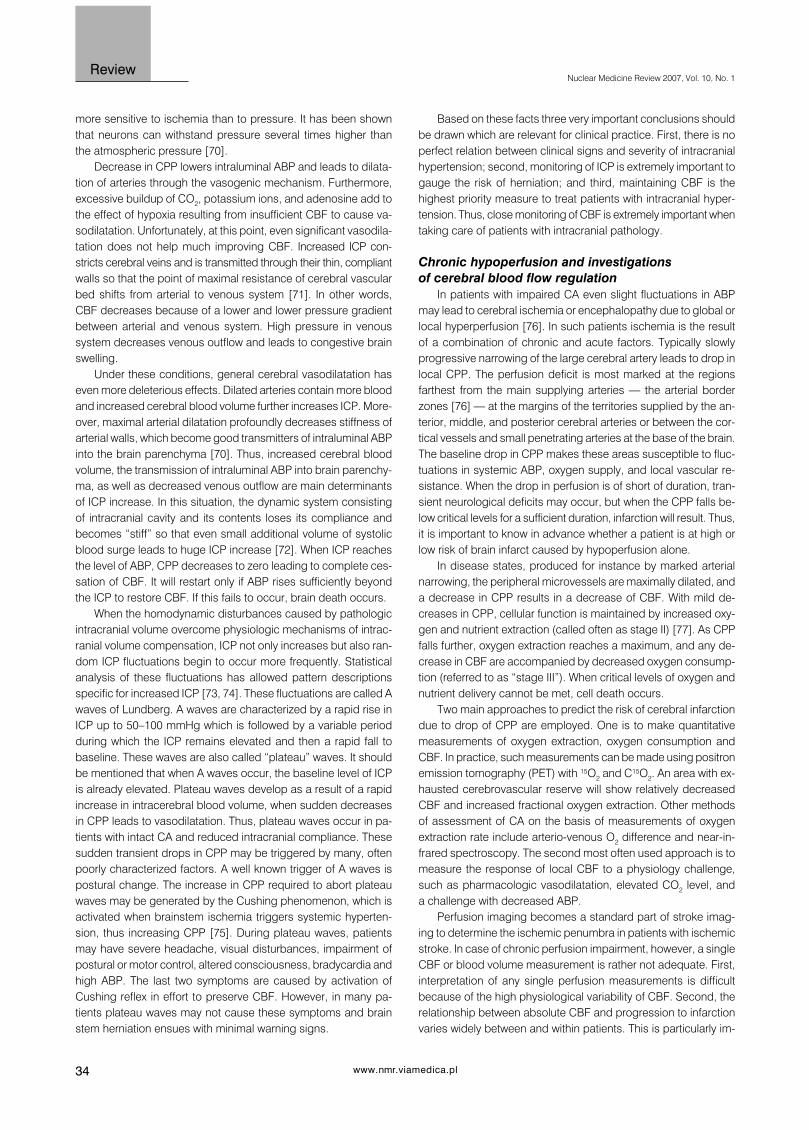
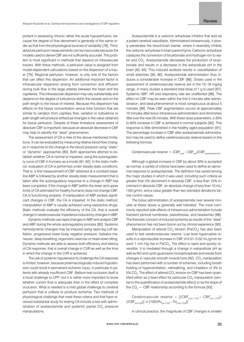
Cerebral infarction results when blood supply is not sufficientto support cellular viability or when low nutrients supply inducesapoptosis. The cell death is a complex function resulting froma combination of the duration and magnitude of ischemia, thenutrients and oxygen content of blood, the specific cerebral struc-ture involved, and ability of the tissue to dissipate the metabolicheat. Cell viability may be lost in as little as 20 minutes after ces-sation of blood flow. In mild but prolonged low perfusion states,irreversible gray matter damage will occur with CBF below ap-proximately 20 mL/100 mg/h. Figures 1–4 demonstrate a basicconcept of chain reactions in neuron and glia cells under differentischemic conditions, which eventually lead to cell death.
In general, ischemia results from:— sudden arterial occlusion;— global hypoxia due to respiratory or cardiac arrest;— hypoperfusion in patients with high ICP;— subacute or chronic borderline hypoperfusion due to occlu-
sion or narrowing of large cerebral arteries. The last two needmore explanation.
Cerebral blood flow disturbances in patientswith high ICP
According to the Monro-Kellie hypothesis [67] the craniumacts as a near rigid container of virtually incompressible substancessuch as brain (80%), blood (12%) and cerebrospinal fluid (CSF)— (8%). These three volumes completely fill the intracranial cavityand remain in a state of dynamic equilibrium with each other. Basedon these premises, any extra volume added to the intracranialcavity will increase ICP. However, cessation of cardiac action leadsto equalization of ICP with atmospheric pressure, no matter howhigh it was before stoppage of cerebral circulation. In other words,it will always equal zero when heart does not beat. Thus, it goeswithout saying that ICP is closely related to the function of cardio-vascular system. Under normal CPP (80–100 mm Hg), ICP is nothigher than 15 mm Hg [68]. Blood pressure in the cerebral arter-ies decreases as arterial diameter gets smaller and approachesthe interstitial tissue pressure, that is equal to ICP at the level ofbrain capillaries and veins. The most significant drop in ABP oc-curs in small resistance, and precapillary arterioles. High ABP in-side the arterial system is not transmitted to the brain tissue be-cause it is confined within the arteries by thick arterial walls. More-

32
Nuclear Medicine Review 2007, Vol. 10, No. 1
www.nmr.viamedica.pl
Review
over, during every cardiac systole, some CSF and venous bloodis pushed out of the cranium to accommodate “cerebral” portionof stroke volume. Thus, under normal conditions, cranial cavityexhibits some compliance and there is only slight rise in ICP dueto distention of cerebral arteries during cardiac systole.
As mentioned before, any extra space occupying by intracra-nial lesion is at the cost of the volume of other intracranial compo-nents in order to maintain the ICP at normal levels. Thus, during
growth of tumors or the development of hematoma, CSF is ab-sorbed and venous blood is pushed out of the cerebral veins toaccommodate the volume of the abnormal lesion inside the skull.When these compensatory mechanisms are exhausted, ICP in-creases logarithmically as pathologic volume further increases [69].As ICP increases, the CPP and consequently CBF begin to de-crease. From then on, the highest priority is maintaining CBF, evenat the price of further increase in ICP, as neural tissue is much
Figure 1. Physiological mechanisms of ion equilibrium in the neuron. The energy of in-flowing sodium ions is utilized for expelling the ions of calcium.Sodium is then removed by the Na\K pump. Negative potential of the cell interior helps keep the NMDA-coupled calcium channel locked with the ions ofmagnesium and inhibits the voltage-dependent calcium channels. nNOS — neuronal nitric oxide synthase, NMDA — glutamate (N-methyl-D-spartate)receptor, ATP — adenosinotriphosphate.
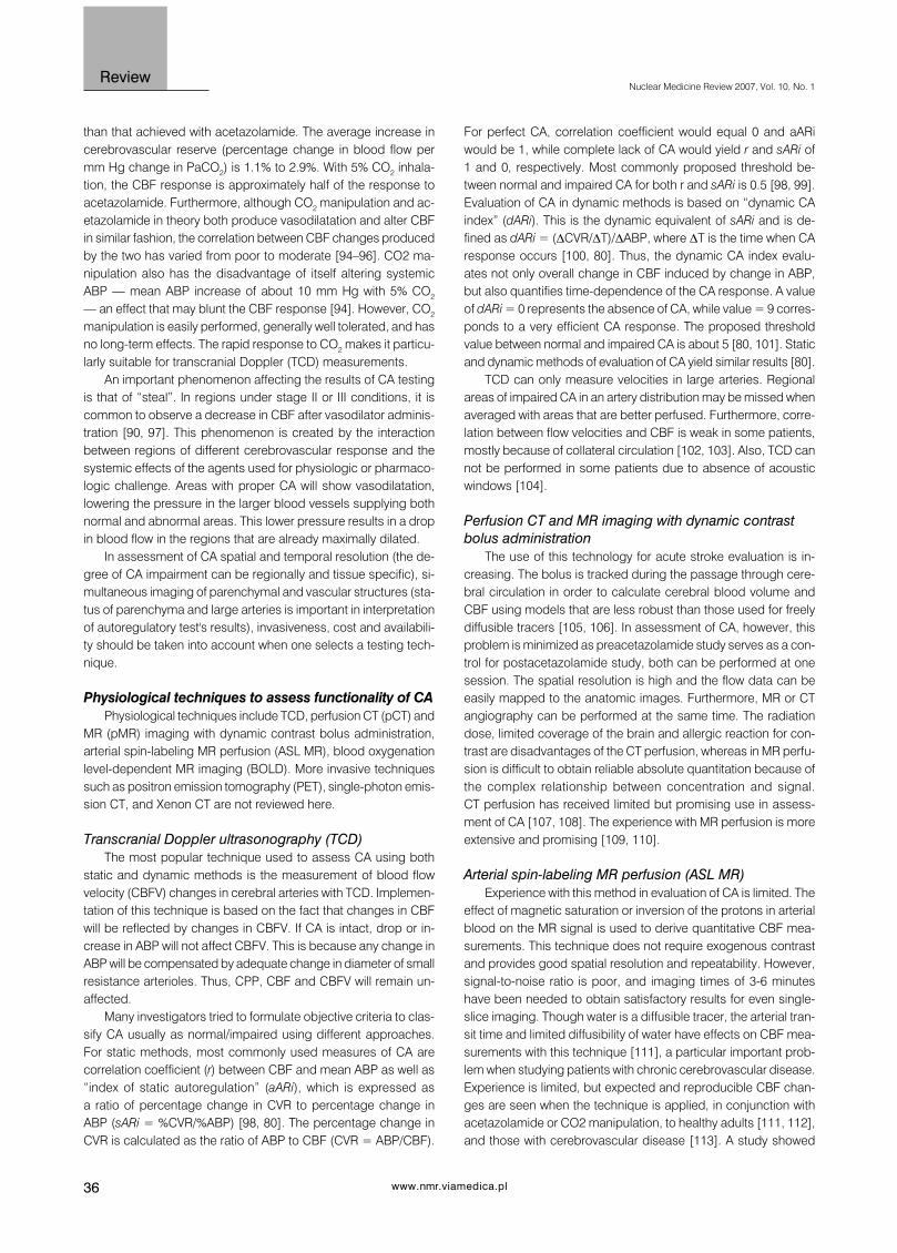
Figure 2. Ischemia and consequent energy deficit cause depolarization of the cell membrane, reverses the action of the sodium-calcium exchanger andallow influx of sodium and calcium ions into the cell. Depolarization opens the voltage-dependent calcium channels and releases magnesium-dependentblock of NMDA-coupled calcium channel. The latter is made patent by glutamate, thus leading to increased synthesis of nitric oxide. Excessive concentra-tion of nitric oxide and calcium leads to cell death.

33www.nmr.viamedica.pl
ReviewWojciech Rudziński et al. Cerebral hemodynamics and investigations of CBF regulation
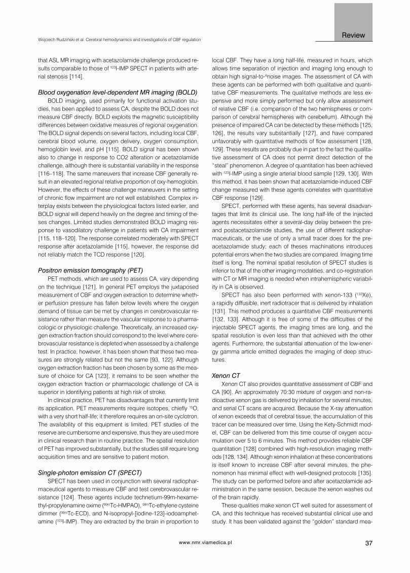
Figure 3. Biochemical mechanism of excitotoxicity. Energy deficit attenuates re-uptake of glutamate in the synaptic cleft. Increased content of this mediatorcauses overstimulation of NMDA receptor, excessive synthesis of nitric oxide and influx of calcium into the cell. This leads to inactivation of mitochondrialenzymes, free radicals production and activation of Phospholipase A2. The phospholipase releases arachidonic acid which again attenuates re-uptake ofglutamate, thus producing the vicious circle of excitotoxicity.
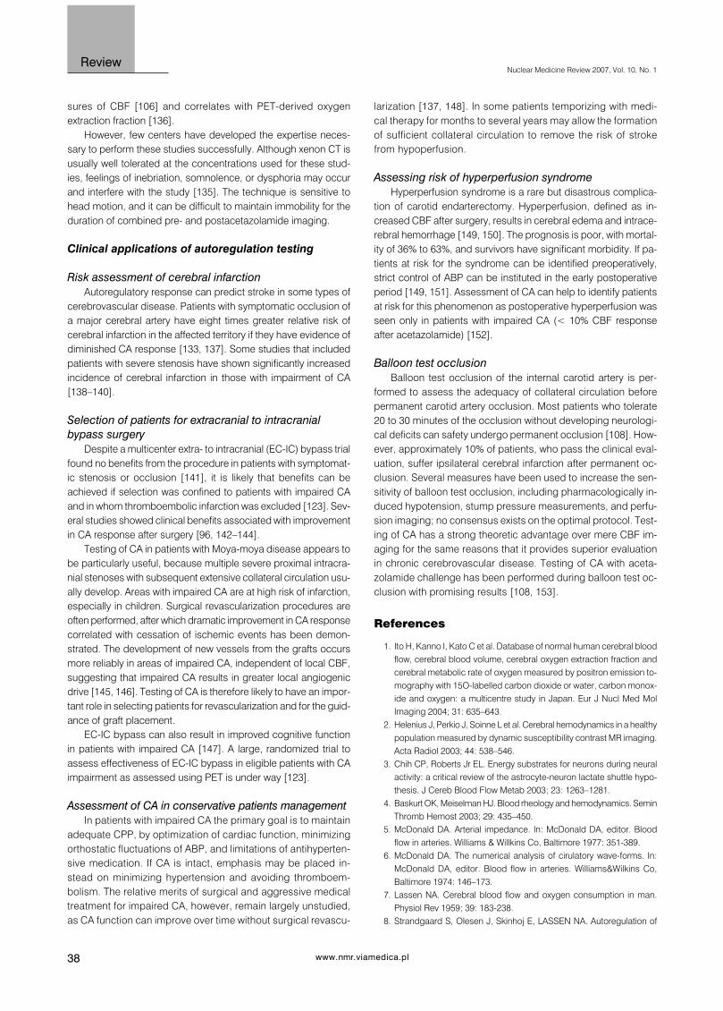
Figure 4. “Direct” and “indirect” effects of reactive oxygen species (ROS). Reactive oxygen species directly interact with DNA, proteins and lipids anddestroy them through oxidation process causing cell death (direct effect). High concentration of ROS damage structure and function of mitochondria andchange the redox state of the cell interior (indirect effect). Damage to the mitochondria leads to release of factors initiating process of programmed celldeath (apoptosis). Oxidative stress activates transcription factor NFkB (nuclear factor kB), which induce synthesis many proinflammatory cytokines andadhesion molecules. Increased expression of cytokines and adhesion molecules augments inflammatory process in neural tissue.
34
Nuclear Medicine Review 2007, Vol. 10, No. 1
www.nmr.viamedica.pl
Review
Based on these facts three very important conclusions shouldbe drawn which are relevant for clinical practice. First, there is noperfect relation between clinical signs and severity of intracranialhypertension; second, monitoring of ICP is extremely important togauge the risk of herniation; and third, maintaining CBF is thehighest priority measure to treat patients with intracranial hyper-tension. Thus, close monitoring of CBF is extremely important whentaking care of patients with intracranial pathology.
Chronic hypoperfusion and investigationsof cerebral blood flow regulation
In patients with impaired CA even slight fluctuations in ABPmay lead to cerebral ischemia or encephalopathy due to global orlocal hyperperfusion [76]. In such patients ischemia is the resultof a combination of chronic and acute factors. Typically slowlyprogressive narrowing of the large cerebral artery leads to drop inlocal CPP. The perfusion deficit is most marked at the regionsfarthest from the main supplying arteries — the arterial borderzones [76] — at the margins of the territories supplied by the an-terior, middle, and posterior cerebral arteries or between the cor-tical vessels and small penetrating arteries at the base of the brain.The baseline drop in CPP makes these areas susceptible to fluc-tuations in systemic ABP, oxygen supply, and local vascular re-sistance. When the drop in perfusion is of short of duration, tran-sient neurological deficits may occur, but when the CPP falls be-low critical levels for a sufficient duration, infarction will result. Thus,it is important to know in advance whether a patient is at high orlow risk of brain infarct caused by hypoperfusion alone.
In disease states, produced for instance by marked arterialnarrowing, the peripheral microvessels are maximally dilated, anda decrease in CPP results in a decrease of CBF. With mild de-creases in CPP, cellular function is maintained by increased oxy-gen and nutrient extraction (called often as stage II) [77]. As CPPfalls further, oxygen extraction reaches a maximum, and any de-crease in CBF are accompanied by decreased oxygen consump-tion (referred to as “stage III”). When critical levels of oxygen andnutrient delivery cannot be met, cell death occurs.
Two main approaches to predict the risk of cerebral infarctiondue to drop of CPP are employed. One is to make quantitativemeasurements of oxygen extraction, oxygen consumption andCBF. In practice, such measurements can be made using positronemission tomography (PET) with 15O2 and C15O2. An area with ex-hausted cerebrovascular reserve will show relatively decreasedCBF and increased fractional oxygen extraction. Other methodsof assessment of CA on the basis of measurements of oxygenextraction rate include arterio-venous O2 difference and near-in-frared spectroscopy. The second most often used approach is tomeasure the response of local CBF to a physiology challenge,such as pharmacologic vasodilatation, elevated CO2 level, anda challenge with decreased ABP.
Perfusion imaging becomes a standard part of stroke imag-ing to determine the ischemic penumbra in patients with ischemicstroke. In case of chronic perfusion impairment, however, a singleCBF or blood volume measurement is rather not adequate. First,interpretation of any single perfusion measurements is difficultbecause of the high physiological variability of CBF. Second, therelationship between absolute CBF and progression to infarctionvaries widely between and within patients. This is particularly im-
more sensitive to ischemia than to pressure. It has been shownthat neurons can withstand pressure several times higher thanthe atmospheric pressure [70].
Decrease in CPP lowers intraluminal ABP and leads to dilata-tion of arteries through the vasogenic mechanism. Furthermore,excessive buildup of CO2, potassium ions, and adenosine add tothe effect of hypoxia resulting from insufficient CBF to cause va-sodilatation. Unfortunately, at this point, even significant vasodila-tation does not help much improving CBF. Increased ICP con-stricts cerebral veins and is transmitted through their thin, compliantwalls so that the point of maximal resistance of cerebral vascularbed shifts from arterial to venous system [71]. In other words,CBF decreases because of a lower and lower pressure gradientbetween arterial and venous system. High pressure in venoussystem decreases venous outflow and leads to congestive brainswelling.
Under these conditions, general cerebral vasodilatation haseven more deleterious effects. Dilated arteries contain more bloodand increased cerebral blood volume further increases ICP. More-over, maximal arterial dilatation profoundly decreases stiffness ofarterial walls, which become good transmitters of intraluminal ABPinto the brain parenchyma [70]. Thus, increased cerebral bloodvolume, the transmission of intraluminal ABP into brain parenchy-ma, as well as decreased venous outflow are main determinantsof ICP increase. In this situation, the dynamic system consistingof intracranial cavity and its contents loses its compliance andbecomes “stiff” so that even small additional volume of systolicblood surge leads to huge ICP increase [72]. When ICP reachesthe level of ABP, CPP decreases to zero leading to complete ces-sation of CBF. It will restart only if ABP rises sufficiently beyondthe ICP to restore CBF. If this fails to occur, brain death occurs.
When the homodynamic disturbances caused by pathologicintracranial volume overcome physiologic mechanisms of intrac-ranial volume compensation, ICP not only increases but also ran-dom ICP fluctuations begin to occur more frequently. Statisticalanalysis of these fluctuations has allowed pattern descriptionsspecific for increased ICP [73, 74]. These fluctuations are called Awaves of Lundberg. A waves are characterized by a rapid rise inICP up to 50–100 mmHg which is followed by a variable periodduring which the ICP remains elevated and then a rapid fall tobaseline. These waves are also called “plateau” waves. It shouldbe mentioned that when A waves occur, the baseline level of ICPis already elevated. Plateau waves develop as a result of a rapidincrease in intracerebral blood volume, when sudden decreasesin CPP leads to vasodilatation. Thus, plateau waves occur in pa-tients with intact CA and reduced intracranial compliance. Thesesudden transient drops in CPP may be triggered by many, oftenpoorly characterized factors. A well known trigger of A waves ispostural change. The increase in CPP required to abort plateauwaves may be generated by the Cushing phenomenon, which isactivated when brainstem ischemia triggers systemic hyperten-sion, thus increasing CPP [75]. During plateau waves, patientsmay have severe headache, visual disturbances, impairment ofpostural or motor control, altered consciousness, bradycardia andhigh ABP. The last two symptoms are caused by activation ofCushing reflex in effort to preserve CBF. However, in many pa-tients plateau waves may not cause these symptoms and brainstem herniation ensues with minimal warning signs.
35www.nmr.viamedica.pl
ReviewWojciech Rudziński et al. Cerebral hemodynamics and investigations of CBF regulation
portant in assessing chronic rather the acute hypoperfusion, be-cause the degree of flow decrement is generally of the same or-der as that from the physiological sources of variability [78]. Third,absolute perfusion measurements can be inaccurate because themodels used to derive CBF are not sufficiently accurate. This prob-lem is most significant in methods that depend on intravasculartracers. With these methods, a perfusion value is assigned frommodel-dependent calculations based on the dispersion of a trac-er [79]. Regional perfusion, however, is only one of the factorsthat can affect this dispersion. An additional important factor isintravascular dispersion arising from convection and diffusionduring bulk flow in the large arteries between the heart and thecapillaries. This intravascular dispersion may vary substantially anddepend on the degree of turbulence within the vessels and on thepath length to the tissue of interest. Because this dispersion haseffects on the tissue concentration versus time function that aresimilar to variation from capillary flow, variation in turbulence orpath length will produce artifactual changes in the value obtainedfor tissue perfusion. Despite of these limitations assessment ofabsolute CBF is important, because an absolute decrease in CBFmay help to identify the “steal” phenomenon.
The assessment of CA is free of the above mentioned limita-tions. It can be evaluated by measuring relative blood flow chang-es in response to the change in the blood pressure using “static”or “dynamic” approaches [80]. Both approaches attempt to es-tablish whether CA is normal or impaired, using the autoregulato-ry curve of CBF in humans as a model [81, 82]. In the static meth-od, evaluation of CA is performed under steady-state conditions.That is, a first measurement of CBF obtained at a constant base-line ABP is followed by another steady-state measurement that istaken after the autoregulatory response to a change in ABP hasbeen completed. If the change in ABP (within the lower and upperlimits of CA estimated for healthy humans) does not change CBF,CA is functioning properly. If manipulation of ABP evokes signifi-cant changes in CBF, the CA is impaired. In the static method,manipulation of ABP is usually achieved using vasoactive drugs.Static methods evaluate the efficiency of the CA, that is overallchange in cerebrovascular impedance induced by changes in ABP.
Dynamic methods use rapid changes in ABP and analyze CBFand ABP during the whole autoregulatory process [80]. Systemichemodynamic changes may be induced using rapid leg cuff de-flation, progressive lower body negative pressure, Valsalva ma-neuver, deep breathing, ergometric exercise or head-down tilting.Dynamic methods are able to assess both efficiency and latencyof CA response, that is overall change in CVR as well as the timein which the change in the CVR is achieved.
The use of systemic hypotension to challenge the CA responseis limited, however, because pharmacologically induced hypoten-sion could result in permanent ischemic injury, in particular in pa-tients with already insufficient CBF. Balloon test occlusion itself isa focal challenge to CPP; but it is rather more important to knowwhether current flow is adequate than in the effect of completeocclusion. What is needed is a mild global challenge to cerebralperfusion that is unlikely to produce ischemia. Two methods ofphysiological challenge that meet these criteria and that have re-ceived substantial study for testing CA include a test with admin-istration of acetazolamide and systemic partial CO2 pressuremanipulations.
Acetazolamide is a carbonic anhydrase inhibitor that acts asa potent cerebral vasodilator. Administered intravenously, it slow-ly penetrates the blood-brain barrier, where it reversibly inhibitsthe carbonic anhydrase in brain parenchyma. Carbonic anhydrazecatalyzes the conversion of bicarbonate and hydrogen ion to wa-ter and CO2. Acetazolamide decreases the production of bicar-bonate and results in a decrease in the extracellular pH in thebrain [83, 84]. This induced acidosis results in vasodilatation ofsmall arterioles [85, 86]. Acetazolamide administration thus in-duces a considerable increase in CBF [86]. Doses used in theassessment of cerebrovascular reserve are in the 15–18 mg/kgrange; in many studies a standard total dose of 1 g is used [87].Systemic ABP, HR and respiratory rate are unaffected [88]. Theeffect on CBF may be seen within the first 5 minutes after admin-istration, and steal phenomenon is most conspicuous at about 5minutes [89]. Peak CBF augmentation occurs at approximately10 minutes after bolus intravenous administration and diminisheslittle over the next 20 minutes. With these dose parameters, a 30%to 60% increase in CBF is achieved in normal subjects [90]. Thisresponse is little diminished in the healthy aged population [91].The percentage increase in CBF after acetazolamide administra-tion may be used to define cerebrovascular reserve based on thefollowing formula:
Cerebrovascular reserve = (CBFpost – CBFpre)/CBFpreacetazolamide
Although a global increase in CBF by about 30% is acceptedas normal, a variety of criteria have been used to define an abnor-mal response to acetazolamide. The definition has varied amongthe major studies in which it was used, including such criteria asgreater than 5% decrement in absolute CBF, a less than 10% in-crement in absolute CBF, an absolute change of less than 10 mL//100 g/min, and a value greater than two standard deviations be-low control values.
The bolus administration of acetazolamide over several min-utes at these doses is generally well tolerated. The most com-monly reported side effects of intravenous administration includetransient perioral numbness, paresthesias, and headaches [88].The theoretic concern of induced ischemia as results of the “steal”phenomenon has not been borne out by clinical experience [90].
Manipulation of arterial CO2 tension (PaCO2) has also beenused to test cerebrovascular reserve. Low level hypercarbia re-sults in a reproducible increase in CBF of 0.01–0.02 mL/g/min foreach 1 mm Hg rise in PaCO2. The effect is rapid and quickly re-versible. It is mediated through a change in extracellular pH aswell as NO and cyclic guanosine monophosphate and results fromchanges in vascular smooth muscle tone [92]. CO2 manipulationhas been performed with a number of schemes, including breathholding or hyperventilation, rebreathing, and inhalation of 3% to5% CO2. The effect of altered CO2 tension on CBF has been quan-tified either as a fixed effect for particular CO2 manipulation (sim-ilar to the quantification of acetazolamide effect) or as the slope ofthe CO2 — CBF relationship according to the formula [93]:
Cerebrovascular reserve = [(CBFpost- CO2 – CBFpre- CO2)//(CBFpre-CO2)] ¥ [100/(Papre- CO2 – Papost- CO2)]
In clinical practice, the magnitude of CBF changes is smaller
36
Nuclear Medicine Review 2007, Vol. 10, No. 1
www.nmr.viamedica.pl
Review
than that achieved with acetazolamide. The average increase incerebrovascular reserve (percentage change in blood flow permm Hg change in PaCO2) is 1.1% to 2.9%. With 5% CO2 inhala-tion, the CBF response is approximately half of the response toacetazolamide. Furthermore, although CO2 manipulation and ac-etazolamide in theory both produce vasodilatation and alter CBFin similar fashion, the correlation between CBF changes producedby the two has varied from poor to moderate [94–96]. CO2 ma-nipulation also has the disadvantage of itself altering systemicABP — mean ABP increase of about 10 mm Hg with 5% CO2
— an effect that may blunt the CBF response [94]. However, CO2
manipulation is easily performed, generally well tolerated, and hasno long-term effects. The rapid response to CO2 makes it particu-larly suitable for transcranial Doppler (TCD) measurements.
An important phenomenon affecting the results of CA testingis that of “steal”. In regions under stage II or III conditions, it iscommon to observe a decrease in CBF after vasodilator adminis-tration [90, 97]. This phenomenon is created by the interactionbetween regions of different cerebrovascular response and thesystemic effects of the agents used for physiologic or pharmaco-logic challenge. Areas with proper CA will show vasodilatation,lowering the pressure in the larger blood vessels supplying bothnormal and abnormal areas. This lower pressure results in a dropin blood flow in the regions that are already maximally dilated.
In assessment of CA spatial and temporal resolution (the de-gree of CA impairment can be regionally and tissue specific), si-multaneous imaging of parenchymal and vascular structures (sta-tus of parenchyma and large arteries is important in interpretationof autoregulatory test's results), invasiveness, cost and availabili-ty should be taken into account when one selects a testing tech-nique.
Physiological techniques to assess functionality of CAPhysiological techniques include TCD, perfusion CT (pCT) and
MR (pMR) imaging with dynamic contrast bolus administration,arterial spin-labeling MR perfusion (ASL MR), blood oxygenationlevel-dependent MR imaging (BOLD). More invasive techniquessuch as positron emission tomography (PET), single-photon emis-sion CT, and Xenon CT are not reviewed here.
Transcranial Doppler ultrasonography (TCD)The most popular technique used to assess CA using both
static and dynamic methods is the measurement of blood flowvelocity (CBFV) changes in cerebral arteries with TCD. Implemen-tation of this technique is based on the fact that changes in CBFwill be reflected by changes in CBFV. If CA is intact, drop or in-crease in ABP will not affect CBFV. This is because any change inABP will be compensated by adequate change in diameter of smallresistance arterioles. Thus, CPP, CBF and CBFV will remain un-affected.
Many investigators tried to formulate objective criteria to clas-sify CA usually as normal/impaired using different approaches.For static methods, most commonly used measures of CA arecorrelation coefficient (r) between CBF and mean ABP as well as“index of static autoregulation” (aARi), which is expressed asa ratio of percentage change in CVR to percentage change inABP (sARi = %CVR/%ABP) [98, 80]. The percentage change inCVR is calculated as the ratio of ABP to CBF (CVR = ABP/CBF).
For perfect CA, correlation coefficient would equal 0 and aARiwould be 1, while complete lack of CA would yield r and sARi of1 and 0, respectively. Most commonly proposed threshold be-tween normal and impaired CA for both r and sARi is 0.5 [98, 99].Evaluation of CA in dynamic methods is based on “dynamic CAindex” (dARi). This is the dynamic equivalent of sARi and is de-fined as dARi = (DCVR/DT)/DABP, where DT is the time when CAresponse occurs [100, 80]. Thus, the dynamic CA index evalu-ates not only overall change in CBF induced by change in ABP,but also quantifies time-dependence of the CA response. A valueof dARi = 0 represents the absence of CA, while value = 9 corres-ponds to a very efficient CA response. The proposed thresholdvalue between normal and impaired CA is about 5 [80, 101]. Staticand dynamic methods of evaluation of CA yield similar results [80].
TCD can only measure velocities in large arteries. Regionalareas of impaired CA in an artery distribution may be missed whenaveraged with areas that are better perfused. Furthermore, corre-lation between flow velocities and CBF is weak in some patients,mostly because of collateral circulation [102, 103]. Also, TCD cannot be performed in some patients due to absence of acousticwindows [104].
Perfusion CT and MR imaging with dynamic contrastbolus administration
The use of this technology for acute stroke evaluation is in-creasing. The bolus is tracked during the passage through cere-bral circulation in order to calculate cerebral blood volume andCBF using models that are less robust than those used for freelydiffusible tracers [105, 106]. In assessment of CA, however, thisproblem is minimized as preacetazolamide study serves as a con-trol for postacetazolamide study, both can be performed at onesession. The spatial resolution is high and the flow data can beeasily mapped to the anatomic images. Furthermore, MR or CTangiography can be performed at the same time. The radiationdose, limited coverage of the brain and allergic reaction for con-trast are disadvantages of the CT perfusion, whereas in MR perfu-sion is difficult to obtain reliable absolute quantitation because ofthe complex relationship between concentration and signal.CT perfusion has received limited but promising use in assess-ment of CA [107, 108]. The experience with MR perfusion is moreextensive and promising [109, 110].
Arterial spin-labeling MR perfusion (ASL MR)Experience with this method in evaluation of CA is limited. The
effect of magnetic saturation or inversion of the protons in arterialblood on the MR signal is used to derive quantitative CBF mea-surements. This technique does not require exogenous contrastand provides good spatial resolution and repeatability. However,signal-to-noise ratio is poor, and imaging times of 3-6 minuteshave been needed to obtain satisfactory results for even single-slice imaging. Though water is a diffusible tracer, the arterial tran-sit time and limited diffusibility of water have effects on CBF mea-surements with this technique [111], a particular important prob-lem when studying patients with chronic cerebrovascular disease.Experience is limited, but expected and reproducible CBF chan-ges are seen when the technique is applied, in conjunction withacetazolamide or CO2 manipulation, to healthy adults [111, 112],and those with cerebrovascular disease [113]. A study showed
37www.nmr.viamedica.pl
ReviewWojciech Rudziński et al. Cerebral hemodynamics and investigations of CBF regulation
that ASL MR imaging with acetazolamide challenge produced re-sults comparable to those of 123I-IMP SPECT in patients with arte-rial stenosis [114].
Blood oxygenation level-dependent MR imaging (BOLD)BOLD imaging, used primarily for functional activation stu-
dies, has been applied to assess CA, despite the BOLD does notmeasure CBF directly. BOLD exploits the magnetic susceptibilitydifferences between oxidative measures of regional oxygenation.The BOLD signal depends on several factors, including local CBF,cerebral blood volume, oxygen delivery, oxygen consumption,hemoglobin level, and pH [115]. BOLD signal has been shownalso to change in response to CO2 alteration or acetazolamidechallenge, although there is substantial variability in the response[116–118]. The same maneuvers that increase CBF generally re-sult in an elevated regional relative proportion of oxy-hemoglobin.However, the effects of these challenge maneuvers in the settingof chronic flow impairment are not well established. Complex in-terplay exists between the physiological factors listed earlier, andBOLD signal will depend heavily on the degree and timing of the-ses changes. Limited studies demonstrated BOLD imaging res-ponse to vasodilatory challenge in patients with CA impairment[115, 118–120]. The response correlated moderately with SPECTresponse after acetazolamide [115], however, the response didnot reliably match the TCD response [120].
Positron emission tomography (PET)PET methods, which are used to assess CA, vary depending
on the technique [121]. In general PET employs the juxtaposedmeasurement of CBF and oxygen extraction to determine wheth-er perfusion pressure has fallen below levels where the oxygendemand of tissue can be met by changes in cerebrovascular re-sistance rather than measure the vascular response to a pharma-cologic or physiologic challenge. Theoretically, an increased oxy-gen extraction fraction should correspond to the level where cere-brovascular resistance is depleted when assessed by a challengetest. In practice, however, it has been shown that these two mea-sures are strongly related but not the same [93, 122]. Althoughoxygen extraction fraction has been chosen by some as the mea-sure of choice for CA [123], it remains to be seen whether theoxygen extraction fraction or pharmacologic challenge of CA issuperior in identifying patients at high risk of stroke.
In clinical practice, PET has disadvantages that currently limitits application. PET measurements require isotopes, chiefly 15O,with a very short half-life; it therefore requires an on-site cyclotron.The availability of this equipment is limited. PET studies of thereserve are cumbersome and expensive, thus they are used morein clinical research than in routine practice. The spatial resolutionof PET has improved substantially, but the studies still require longacquisition times and are sensitive to patient motion.
Single-photon emission CT (SPECT)SPECT has been used in conjunction with several radiophar-
maceutical agents to measure CBF and test cerebrovascular re-sistance [124]. These agents include technetium-99m-hexame-thyl-propylenamine oxime (99mTc-HMPAO), 99mTc-ethylene cysteinedimmer (99mTc-ECD), and N-isopropyl-[iodine-123]-iodoamphet-amine (123I-IMP). They are extracted by the brain in proportion to
local CBF. They have a long half-life, measured in hours, whichallows time separation of injection and imaging long enough toobtain high signal-to-6noise images. The assessment of CA withthese agents can be performed with both qualitative and quanti-tative CBF measurements. The qualitative methods are less ex-pensive and more simply performed but only allow assessmentof relative CBF (i.e. comparison of the two hemispheres or com-parison of cerebral hemispheres with cerebellum). Although thepresence of impaired CA can be detected by these methods [125,126], the results vary substantially [127], and have comparedunfavorably with quantitative methods of flow assessment [128,129]. These results are probably due in part to the fact the qualita-tive assessment of CA does not permit direct detection of the“steal” phenomenon. A degree of quantitation has been achievedwith 123I-IMP using a single arterial blood sample [129, 130]. Withthis method, it has been shown that acetazolamide-induced CBFchange measured with these agents correlates with quantitativeCBF response [129].
SPECT, performed with these agents, has several disadvan-tages that limit its clinical use. The long half-life of the injectedagents necessitates either a several-day delay between the pre-and postacetazolamide studies, the use of different radiophar-maceuticals, or the use of only a small tracer does for the pre-acetazolamide study; each of theses machinations introducespotential errors when the two studies are compared. Imaging timeitself is long. The nominal spatial resolution of SPECT studies isinferior to that of the other imaging modalities, and co-registrationwith CT or MR imaging is needed when intrahemispheric variabil-ity in CA is observed.
SPECT has also been performed with xenon-133 (133Xe),a rapidly diffusible, inert radiotracer that is delivered by inhalation[131]. This method produces a quantitative CBF measurements[132, 133]. Although it is free of some of the difficulties of theinjectable SPECT agents, the imaging times are long, and thespatial resolution is even less than that achieved with the otheragents. Furthermore, the substantial attenuation of the low-ener-gy gamma article emitted degrades the imaging of deep struc-tures.
Xenon CTXenon CT also provides quantitative assessment of CBF and
CA [90]. An approximately 70:30 mixture of oxygen and non-ra-dioactive xenon gas is delivered by inhalation for several minutes,and serial CT scans are acquired. Because the X-ray attenuationof xenon exceeds that of cerebral tissue, the accumulation of thistracer can be measured over time. Using the Kety-Schmidt mod-el, CBF can be delivered from this time course of oxygen accu-mulation over 5 to 6 minutes. This method provides reliable CBFquantitation [128] combined with high-resolution imaging meth-ods [128, 134]. Although xenon inhalation at these concentrationsis itself known to increase CBF after several minutes, the phe-nomenon has minimal effect with well-designed protocols [135].The study can be performed before and after acetazolamide ad-ministration in the same session, because the xenon washes outof the brain rapidly.
These qualities make xenon CT well suited for assessment ofCA, and this technique has received substantial clinical use andstudy. It has been validated against the “golden” standard mea-
38
Nuclear Medicine Review 2007, Vol. 10, No. 1
www.nmr.viamedica.pl
Review
sures of CBF [106] and correlates with PET-derived oxygenextraction fraction [136].
However, few centers have developed the expertise neces-sary to perform these studies successfully. Although xenon CT isusually well tolerated at the concentrations used for these stud-ies, feelings of inebriation, somnolence, or dysphoria may occurand interfere with the study [135]. The technique is sensitive tohead motion, and it can be difficult to maintain immobility for theduration of combined pre- and postacetazolamide imaging.
Clinical applications of autoregulation testing
Risk assessment of cerebral infarctionAutoregulatory response can predict stroke in some types of
cerebrovascular disease. Patients with symptomatic occlusion ofa major cerebral artery have eight times greater relative risk ofcerebral infarction in the affected territory if they have evidence ofdiminished CA response [133, 137]. Some studies that includedpatients with severe stenosis have shown significantly increasedincidence of cerebral infarction in those with impairment of CA[138–140].
Selection of patients for extracranial to intracranialbypass surgery
Despite a multicenter extra- to intracranial (EC-IC) bypass trialfound no benefits from the procedure in patients with symptomat-ic stenosis or occlusion [141], it is likely that benefits can beachieved if selection was confined to patients with impaired CAand in whom thromboembolic infarction was excluded [123]. Sev-eral studies showed clinical benefits associated with improvementin CA response after surgery [96, 142–144].
Testing of CA in patients with Moya-moya disease appears tobe particularly useful, because multiple severe proximal intracra-nial stenoses with subsequent extensive collateral circulation usu-ally develop. Areas with impaired CA are at high risk of infarction,especially in children. Surgical revascularization procedures areoften performed, after which dramatic improvement in CA responsecorrelated with cessation of ischemic events has been demon-strated. The development of new vessels from the grafts occursmore reliably in areas of impaired CA, independent of local CBF,suggesting that impaired CA results in greater local angiogenicdrive [145, 146]. Testing of CA is therefore likely to have an impor-tant role in selecting patients for revascularization and for the guid-ance of graft placement.
EC-IC bypass can also result in improved cognitive functionin patients with impaired CA [147]. A large, randomized trial toassess effectiveness of EC-IC bypass in eligible patients with CAimpairment as assessed using PET is under way [123].
Assessment of CA in conservative patients managementIn patients with impaired CA the primary goal is to maintain
adequate CPP, by optimization of cardiac function, minimizingorthostatic fluctuations of ABP, and limitations of antihyperten-sive medication. If CA is intact, emphasis may be placed in-stead on minimizing hypertension and avoiding thromboem-bolism. The relative merits of surgical and aggressive medicaltreatment for impaired CA, however, remain largely unstudied,as CA function can improve over time without surgical revascu-
larization [137, 148]. In some patients temporizing with medi-cal therapy for months to several years may allow the formationof sufficient collateral circulation to remove the risk of strokefrom hypoperfusion.
Assessing risk of hyperperfusion syndromeHyperperfusion syndrome is a rare but disastrous complica-
tion of carotid endarterectomy. Hyperperfusion, defined as in-creased CBF after surgery, results in cerebral edema and intrace-rebral hemorrhage [149, 150]. The prognosis is poor, with mortal-ity of 36% to 63%, and survivors have significant morbidity. If pa-tients at risk for the syndrome can be identified preoperatively,strict control of ABP can be instituted in the early postoperativeperiod [149, 151]. Assessment of CA can help to identify patientsat risk for this phenomenon as postoperative hyperperfusion wasseen only in patients with impaired CA (< 10% CBF responseafter acetazolamide) [152].
Balloon test occlusionBalloon test occlusion of the internal carotid artery is per-
formed to assess the adequacy of collateral circulation beforepermanent carotid artery occlusion. Most patients who tolerate20 to 30 minutes of the occlusion without developing neurologi-cal deficits can safety undergo permanent occlusion [108]. How-ever, approximately 10% of patients, who pass the clinical eval-uation, suffer ipsilateral cerebral infarction after permanent oc-clusion. Several measures have been used to increase the sen-sitivity of balloon test occlusion, including pharmacologically in-duced hypotension, stump pressure measurements, and perfu-sion imaging; no consensus exists on the optimal protocol. Test-ing of CA has a strong theoretic advantage over mere CBF im-aging for the same reasons that it provides superior evaluationin chronic cerebrovascular disease. Testing of CA with aceta-zolamide challenge has been performed during balloon test oc-clusion with promising results [108, 153].
References
1. Ito H, Kanno I, Kato C et al. Database of normal human cerebral bloodflow, cerebral blood volume, cerebral oxygen extraction fraction andcerebral metabolic rate of oxygen measured by positron emission to-mography with 15O-labelled carbon dioxide or water, carbon monox-ide and oxygen: a multicentre study in Japan. Eur J Nucl Med MolImaging 2004; 31: 635–643.
2. Helenius J, Perkio J, Soinne L et al. Cerebral hemodynamics in a healthypopulation measured by dynamic susceptibility contrast MR imaging.Acta Radiol 2003; 44: 538–546.
3. Chih CP, Roberts Jr EL. Energy substrates for neurons during neuralactivity: a critical review of the astrocyte-neuron lactate shuttle hypo-thesis. J Cereb Blood Flow Metab 2003; 23: 1263–1281.
4. Baskurt OK, Meiselman HJ. Blood rheology and hemodynamics. SeminThromb Hemost 2003; 29: 435–450.
5. McDonald DA. Arterial impedance. In: McDonald DA, editor. Bloodflow in arteries. Williams & Willkins Co, Baltimore 1977: 351-389.
6. McDonald DA. The numerical analysis of cirulatory wave-forms. In:McDonald DA, editor. Blood flow in arteries. Williams&Wilkins Co,Baltimore 1974: 146–173.
7. Lassen NA. Cerebral blood flow and oxygen consumption in man.Physiol Rev 1959; 39: 183-238.
8. Strandgaard S, Olesen J, Skinhoj E, LASSEN NA. Autoregulation of
39www.nmr.viamedica.pl
ReviewWojciech Rudziński et al. Cerebral hemodynamics and investigations of CBF regulation
brain circulation in severe arterial hypertension. Br Med J 1973; 1:507–510.
9. Waldemar G, Schmidt JF, Andersen AR, Vorstrup S, Ibsen H, Paulson OB.Angiotensin converting enzyme inhibition and cerebral blood flow au-toregulation in normotensive and hypertensive man. J Hypertens 1989;7: 229–235.
10. Wallis SJ, Firth J, Dunn WR. Pressure-induced myogenic responsesin human isolated cerebral resistance arteries. Stroke 1996; 27: 2287––2290.
11. Garcia-Roldan JL, Bevan JA. Flow-induced constriction and dilationof cerebral resistance arteries. Circ Res 1990; 66: 1445–1448.
12. Thorin-Trescases N, Bevan JA. High levels of myogenic tone antago-nize the dilator response to flow of small rabbit cerebral arteries. Stroke1998; 29: 1194–1200.
13. McDonald DA. Blood flow in arteries. Williams & Willkins Co, Balti-more 1977.
14. Slish DF, Welsh DG, Brayden JE. Diacylglycerol and protein kinase Cactivate cation channels involved in myogenic tone. Am J Physiol HeartCirc Physiol 2002; 283: H2196–H2201.
15. Kitamura K, Xiong Z, Teramoto N, Kuriyama H. Roles of inositol tris-phosphate and protein kinase C in the spontaneous outward currentmodulated by calcium release in rabbit portal vein. Pflugers Arch 1992;421: 539–551.
16. McCarron JG, Crichton CA, Langton PD, MacKenzie A, Smith GL.Myogenic contraction by modulation of voltage-dependent calciumcurrents in isolated rat cerebral arteries. J Physiol 1997; 498 (Part 2):371–379.
17. Ngai AC, Winn HR. Modulation of cerebral arteriolar diameter by in-traluminal flow and pressure. Circ Res 1995; 77: 832–840.
18. Faraci FM. Role of nitric oxide in regulation of basilar artery tone invivo. Am J Physiol 1990; 259: H1216–H1221.
19. Lee JE, Kwak J, Suh CK, Shin JH. Dual effects of nitric oxide on thelarge conductance calcium-activated potassium channels of rat brain.J Biochem Mol Biol 2006; 39: 91–96.
20. Miyahara K, Kawamoto T, Sase K et al. Cloning and structural charac-terization of the human endothelial nitric-oxide-synthase gene. EurJ Biochem 1994; 223: 719–726.
21. Kontos HA, Raper AJ, Patterson JL. Analysis of vasoactivity of localpH, PCO2 and bicarbonate on pial vessels. Stroke 1977; 8: 358–360.
22. Horiuchi T, Dietrich HH, Hongo K, Goto T, Dacey RG, Jr. Role of en-dothelial nitric oxide and smooth muscle potassium channels in cere-bral arteriolar dilation in response to acidosis. Stroke 2002; 33: 844––849.
23. Kovecs K, Komjati K, Marton T, Skopal J, Sandor P, Nagy Z. Hyper-capnia stimulates prostaglandin E(2) but not prostaglandin I(2) re-lease in endothelial cells cultured from microvessels of human fetalbrain. Brain Res Bull 2001; 54:387–390.
24. Lindauer U, Vogt J, Schuh-Hofer S, Dreier JP, Dirnagl U. Cerebrovas-cular vasodilation to extraluminal acidosis occurs via combined acti-vation of ATP-sensitive and Ca2+-activated potassium channels.J Cereb Blood Flow Metab 2003; 23: 1227–1238.
25. Toda N, Hatano Y, Mori K. Mechanisms underlying response to hy-percapnia and bicarbonate of isolated dog cerebral arteries. AmJ Physiol 1989; 257: H141–H146.
26. Simpson RE, Phillis JW. Adenosine deaminase reduces hypoxic andhypercapnic dilatation of rat pial arterioles: evidence for mediation byadenosine. Brain Res 1991; 553: 305–308.
27. Fredricks KT, Liu Y, Rusch NJ, Lombard JH. Role of endothelium andarterial K+ channels in mediating hypoxic dilation of middle cerebralarteries. Am J Physiol 1994; 267: H580–H586.
28. Horiuchi T, Dietrich HH, Hongo K, Dacey RG, Jr. Mechanism of extra-cellular K+-induced local and conducted responses in cerebral penet-rating arterioles. Stroke 2002; 33: 2692–2699.
29. Di Tullio MA, Tayebati SK, Amenta F. Identification of adenosine A1and A3 receptor subtypes in rat pial and intracerebral arteries. Neuro-sci Lett 2004; 366: 48–52.
30. Kleppisch T, Nelson MT. Adenosine activates ATP-sensitivepotassium channels in arterial myocytes via A2 receptors and cAMP--dependent protein kinase. Proc Natl Acad Sci USA 1995; 92: 12441––12445.
31. Li G, Cheung DW. Modulation of Ca(2+)-dependent K(+) currents inmesenteric arterial smooth muscle cells by adenosine. Eur J Pharma-col 2000; 394: 35–40.
32. Bleys RL, Cowen T. Innervation of cerebral blood vessels: morpholo-gy, plasticity, age-related, and Alzheimer's disease-related neurode-generation. Microsc Res Tech 2001; 53: 106–118.
33. Baffi J, Gorcs T, Slowik F et al. Neuropeptides in the human superiorcervical ganglion. Brain Res 1992; 570: 272–278.
34. Baumbach GL, Heistad DD. Effects of sympathetic stimulation andchanges in arterial pressure on segmental resistance of cerebralvessels in rabbits and cats. Circ Res 1983; 52: 527–533.
35. Goadsby PJ, Uddman R, Edvinsson L. Cerebral vasodilatation in thecat involves nitric oxide from parasympathetic nerves. Brain Res 1996;707: 110–118.
36. Hara H, Hamill GS, Jacobowitz DM. Origin of cholinergic nerves to therat major cerebral arteries: coexistence with vasoactive intestinalpolypeptide. Brain Res Bull 1985; 14: 179–188.
37. Morita-Tsuzuki Y, Hardebo JE, Bouskela E. Inhibition of nitric oxidesynthase attenuates the cerebral blood flow response to stimulationof postganglionic parasympathetic nerves in the rat. J Cereb BloodFlow Metab 1993; 13: 993–997.
38. Edvinsson L, Ekman R, Jansen I, McCulloch J, Uddman R. Calcitoningene-related peptide and cerebral blood vessels: distribution andvasomotor effects. J Cereb Blood Flow Metab 1987; 7: 720–728.
39. Edvinsson L, Brodin E, Jansen I, Uddman R. Neurokinin A in cerebralvessels: characterization, localization and effects in vitro. Regul Pept1988; 20: 181–197.
40. Uddman R, Goadsby PJ, Jansen I, Edvinsson L. PACAP, a VIP-likepeptide: immunohistochemical localization and effect upon cat pialarteries and cerebral blood flow. J Cereb Blood Flow Metab 1993; 13:291–297.
41. Edvinsson L, Jansen O, I, Kingman TA, McCulloch J, Uddman R.Modification of vasoconstrictor responses in cerebral blood vesselsby lesioning of the trigeminal nerve: possible involvement of CGRP.Cephalalgia 1995; 15: 373–383.
42. Lassen LH, Haderslev PA, Jacobsen VB, Iversen HK, Sperling B,Olesen J. CGRP may play a causative role in migraine. Cephalalgia2002; 22: 54–61.
43. Weber JR, Angstwurm K, Bove GM et al. The trigeminal nerve andaugmentation of regional cerebral blood flow during experimentalbacterial meningitis. J Cereb Blood Flow Metab 1996; 16: 1319–1324.
44. Kalaria RN, Stockmeier CA, Harik SI. Brain microvessels are innerva-ted by locus ceruleus noradrenergic neurons. Neurosci Lett 1989; 97:203–208.
45. Moreno MJ, Lopez dP, Conde MV, Marco EJ. Cat cerebral arteriesare functionally innervated by serotoninergic fibers from central andperipheral origins. Stroke 1995; 26: 271–275.
46. Sato A, Sato Y, Uchida S. Activation of the intracerebral cholinergicnerve fibers originating in the basal forebrain increases regional cere-bral blood flow in the rat's cortex and hippocampus. Neurosci Lett2004; 361: 90–93.
47. Cauli B, Tong XK, Rancillac A et al. Cortical GABA interneurons inneurovascular coupling: relays for subcortical vasoactive pathways.J Neurosci 2004; 24: 8940–8949.
48. Mulligan SJ, MacVicar BA. Calcium transients in astrocyte endfeetcause cerebrovascular constrictions. Nature 2004; 431: 195–199.
40
Nuclear Medicine Review 2007, Vol. 10, No. 1
www.nmr.viamedica.pl
Review
49. White RP, Hindley C, Bloomfield PM et al. The effect of the nitric oxidesynthase inhibitor L-NMMA on basal CBF and vasoneuronal couplingin man: a PET study. J Cereb Blood Flow Metab 1999; 19: 673–678.
50. Belliveau JW, Kennedy DN, Jr., McKinstry RC et al. Functional map-ping of the human visual cortex by magnetic resonance imaging.Science 1991; 254: 716–719.
51. Zonta M, Angulo MC, Gobbo S et al. Neuron-to-astrocyte signaling iscentral to the dynamic control of brain microcirculation. Nat Neurosci2003; 6: 43–50.
52. Bezzi P, Carmignoto G, Pasti L et al. Prostaglandins stimulate calci-um-dependent glutamate release in astrocytes. Nature 1998; 391: 281––285.
53. Bhardwaj A, Northington FJ, Carhuapoma JR et al. P-450 epoxygen-ase and NO synthase inhibitors reduce cerebral blood flow responseto N-methyl-D-aspartate. Am J Physiol Heart Circ Physiol 2000; 279:H1616–H1624.
54. Gebremedhin D, Lange AR, Lowry TF et al. Production of 20-HETEand its role in autoregulation of cerebral blood flow. Circ Res 2000;87: 60–65.
55. Christopherson KS, Hillier BJ, Lim WA, Bredt DS. PSD-95 assemblesa ternary complex with the N-methyl-D-aspartic acid receptor anda bivalent neuronal NO synthase PDZ domain. J Biol Chem 1999;274: 27467–27473.
56. Pelligrino DA, Gay RL, III, Baughman VL, Wang Q. NO synthase inhi-bition modulates NMDA-induced changes in cerebral blood flow andEEG activity. Am J Physiol 1996; 271: H990–H995.
57. Alonso-Galicia M, Drummond HA, Reddy KK, Falck JR, Roman RJ.Inhibition of 20-HETE production contributes to the vascular respon-ses to nitric oxide. Hypertension 1997; 29:320–325.
58. Sun CW, Falck JR, Okamoto H, Harder DR, Roman RJ. Role of cGMPversus 20-HETE in the vasodilator response to nitric oxide in rat cere-bral arteries. Am J Physiol Heart Circ Physiol 2000; 279: H339-H350.
59. Kara T, Narkiewicz K, Somers VK. Chemoreflexes — physiology andclinical implications. Acta Physiol Scand 2003; 177: 377–384.
60. Heistad DD, Marcus ML, Ehrhardt JC, Abboud FM. Effect of stimula-tion of carotid chemoreceptors on total and regional cerebral bloodflow. Circ Res 1976; 38: 20–25.
61. Mitchell RA. Respiratory chemosensitivity in the medulla oblongata.J Physiol 1969; 202: 3P–4P.
62. Jordan J, Shannon JR, Diedrich A et al. Interaction of carbon dioxideand sympathetic nervous system activity in the regulation of cerebralperfusion in humans. Hypertension 2000; 36: 383–388.
63. Baumbach GL, Heistad DD. Effect of sympathetic stimulation andchanges in arterial pressure on segmental resistance of cerebral ves-sels in rabitts and cats. Cir Res 1983; 52: 527–33.
63. Roatta S, Micieli G, Bosone D et al. Effect of generalised sympatheticactivation by cold pressor test on cerebral haemodynamics in healthyhumans. J Auton Nerv Syst 1998; 71: 159–166.
64. Heistad DD, Marcus ML. Total and regional cerebral blood flow dur-ing stimulation of carotid baroreceptors. Stroke 1976; 7: 239–243.
65. Rapela CE, Green HD, Denison AB, Jr. Baroreceptor reflexes andautorregulation of cerebral blood flow in the dog. Circ Res 1967; 21:559–568.
66. Bondar RL, Kassam MS, Stein F, Dunphy PT, Fortney S, Riedesel ML.Simultaneous cerebrovascular and cardiovascular responses duringpresyncope. Stroke 1995; 26: 1794–1800.
67. Mokri B. The Monro-Kellie hypothesis - Applications in CSF volumedepletion. Neurology 2001; 56: 1746–1748.
68. Miller JD. Intracranial pressure. Curr Opin Neurol Neurosurg 1989; 2:15–20.
69. Kingman TA, Mendelow AD, Graham DI, Teasdale GM. Experimentalintracerebral mass: time-related effects on local cerebral blood flow.J Neurosurg 1987; 67: 732–738.
70. Sahay KB, Mehrotra R, Sachdeva U, Banerji AK. Elastomechanicalcharacterization of brain-tissues. Journal of Biomechanics 1992; 25:319–26.
71. Johnston IH, Rowan JO. Raised intracranial pressure and cerebralblood flow. Venous outflow tract pressures and vascular resistancesin experimental intracranial hypertension. J Neurol Neurosurg Psy-chiatry 1974; 37: 392–402.
72. Ikeyama A, Maeda S, Ito A, Banno K, Nagai H, Furuse M. The analysisof the intracranial pressure by the concept of the driving pressurefrom the vascular system. Neurochirurgia 1978; 21: 43–53.
73. Swiercz M, Mariak Z, Krejza J, Lewko J, Szydlik P. Intracranial pres-sure processing with artificial neural networks: prediction of ICP trends.Acta Neurochir (Wien) 2000; 142: 401–406.
74. Mariak Z, Swiercz M, Krejza J, Lewko J, Lyson T. Intracranial pressureprocessing with artificial neural networks: classification of signal pro-perties. Acta Neurochir (Wien) 2000; 142: 407-411; discussion 411––412.
75. Ungerboeck K, Tenckhoff D, Heimann A, Wagner W, Kempski OS.Transcranial Doppler and cortical microcirculation at increased intrac-ranial pressure and during the Cushing response: An experimentalstudy on rabbits. Neurosurgery 1995; 36: 147–157.
76. Derdeyn CP, Grubb RL, Powers WJ. Cerebral hemodynamic impair-ment. Neurology 1999; 53: 251–259.
77. Powers WJ. Cerebral hemodynamics in ischemic cerebrovascular-dis-ease. Ann Neurol 1991; 29: 231–240.
78. Rohl L, Ostergaard L, Simonsen CZ, Vestergaard-Poulsen P, Andersen G,Sakoh M et al. Viability thresholds of ischemic penumbra of hyper-acute stroke defined by perfusion-weighted MRI and apparent diffu-sion coefficient. Stroke 2001; 32: 1140–1146.
79. Bassingt JB. Concurrent flow model for extraction during transcapil-lary passage. Circ Res 1974; 35: 483–503.
80. Tiecks FP, Lam AM, Aaslid R, Newell DW. Comparison of static anddynamic cerebral autoregulation measurements. Stroke 1995; 26:1014–1019.
81. Aaslid R, Lash SR, Bardy GH, Gild WH, Newell DW. Dynamic pres-sure-flow velocity relationships in the human cerebral circulation. Stroke2003; 34: 1645–1649.
82. Paulson OB, Strandgaard S, Edvinsson L. Cerebral autoregulation.Cerebrovasc Brain Metab Rev 1990; 2: 161–192.
83. Heuser D, Astrup J, Lassen NA, Betz E. Brain carbonic-acid acidosisafter acetazolamide. Acta Physiol Scand 1975; 93: 385–390.
84. Vorstrup S, Henriksen L, Paulson OB. Effect of acetazolamide on ce-rebral blood-flow and cerebral metabolic-rate for oxygen. J Clin Invest1984; 74: 1634–1639.
85. Okazawa H, Yamauchi H, Sugimoto K, Toyoda H, Kishibe Y,Takahashi M. Effects of acetazolamide on cerebral blood flow, bloodvolume, and oxygen metabolism: A positron emission tomographystudy with healthy volunteers. J Cereb Blood Flow Metab 2001; 21:1472–1479.
86. Sorteberg W, Lindegaard KF, Rootwelt K et al. Effect of acetazola-mide on cerebral-artery blood velocity and regional cerebral blood-flow in normal subjects. Acta Neurochir 1989; 97: 139–145.
87. Grossmann WM, Koeberle B. The dose-response relationship of ace-tazolamide on the cerebral blood flow in normal subjects. CerebrovascDis 2000; 10: 65–69.
88. Sullivan HG, Kingsbury TB, Morgan ME et al. The rCBF response todiamox in normal subjects and cerebrovascular-disease patients.J Neurosurg 1987; 67: 525–534.
89. Kuwabara Y, Ichiya Y, Sasaki M, Yoshida T, Masuda K. Time depen-dency of the acetazolamide effect on cerebral hemodynamics in pa-tients with chronic occlusive cerebral-arteries — early steal phenome-non demonstrated by [O-15]H2O positron emission tomography.Stroke 1995; 26: 1825–1829.

41www.nmr.viamedica.pl
ReviewWojciech Rudziński et al. Cerebral hemodynamics and investigations of CBF regulation
90. Yonas H, Pindzola RR. Physiological determination of cerebrovascu-lar reserves and its use in clinical management. Cerebrovasc BrainMetab Rev 1994; 6: 325–340.
91. Petrella JR, Decarli C, Dagli M et al. Age-related vasodilatory responseto acetazolamide challenge in healthy adults: A dynamic contrast-en-hanced MR study. Am J Neurorad 1998; 19: 39–44.
92. Brian JE. Carbon dioxide and the cerebral circulation. Anesthesiology1998; 88: 1365–1386.
93. Kanno I, Uemura K, Higano S et al. Oxygen extraction fraction at maxi-mally vasodilated tissue in the ischemic brain estimated from the re-gional co2 responsiveness measured by Positron Emission Tomo-graphy. J Cereb Blood Flow Metab 1988; 8: 227–235.
94. Kazumata K, Tanaka N, Ishikawa T, Kuroda S, Houkin K, Mitsumori K.Dissociation of vasoreactivity to acetazolamide and hypercapnia— Comparative study in patients with chronic occlusive major cere-bral artery disease. Stroke 1996; 27: 2052–2058.
95. Gambhir S, Inao S, Tadokoro M, Nishino M, Ito K, Ishigaki T et al.Comparison of vasodilatory effect of carbon dioxide inhalation andintravenous acetazolamide on brain vasculature using positron emis-sion tomography. Neurol Res 1997; 19: 139–144.
96. Ringelstein EB, Vaneyck S, Mertens I. Evaluation of cerebral vasomo-tor reactivity by various vasodilating stimuli — comparison of CO2 toacetazolamide. J Cereb Blood Flow Metab 1992; 12: 162-168.
97. Vorstrup S, Lassen NA. Evaluation of the cerebral vasodilatory capa-city before extracranial-intracranial bypass-surgery. Acta Neurol Scand1986; 73: 538.
98. Lam JM, Hsiang JN, Poon WS. Monitoring of autoregulation usinglaser Doppler flowmetry in patients with head injury. J Neurosurg 1997;86: 438–445.
99. Bouma GJ, Muizelaar JP. Relationship between cardiac output andcerebral blood flow in patients with intact and with impaired autore-gulation. J Neurosurg 1990; 73: 368–374.
100. Aaslid R, Lindegaard KF, Sorteberg W, Nornes H. Cerebral autoregu-lation dynamics in humans. Stroke 1989; 20: 45–52.
101. Tiecks FP, Douville C, Byrd S, Lam AM, Newell DW. Evaluation ofimpaired cerebral autoregulation by the Valsalva maneuver. Stroke1996; 27: 1177–1182.
102. Brauer P, Kochs E, Werner C, Bloom M, Policare R, Pentheny S et al.Correlation of transcranial Doppler sonography mean flow velocity withcerebral blood flow in patients with intracranial pathology. J Neuro-surg Anesthesiol 1998; 10: 80–85.
103. Demolis P, Dinh YRT, Giudicelli JF. Relationships between cerebralregional blood flow velocities and volumetric blood flows and theirrespective reactivities to acetazolamide. Stroke 1996; 27: 1835–1839.
104. Krejza J, Swiat M, Pawlak M et al. Suitability of temporal bone acous-tic window: conventional TCD versus transcranial color-coded duplexsonography. J Neuroimaging 2007 (in press).
105. Ostergaard L, Weisskoff RM, Chesler DA, Gyldensted C, Rosen BR.High resolution measurement of cerebral blood flow using intravas-cular tracer bolus passages. 1. Mathematical approach and statisti-cal analysis. Magn Reson Med 1996; 36: 715–725.
106. Latchaw RE, Yonas H, Hunter GJ et al. Guidelines and recommenda-tions for perfusion imaging in cerebral ischemia — A Scientific State-ment for Healthcare Professionals by the Writing Group on PerfusionImaging, from the Council on Cardiovascular Radiology of the Amer-ican Heart Association. Stroke 2003; 34: 1084–1104.
107. Eastwood JD, Alexander MJ, Petrella JR, Provenzale JM. Dynamic CTperfusion imaging with acetazolamide challenge for the preprocedur-al evaluation of a patient with symptomatic middle cerebral artery oc-clusive disease. Am J Neuroradiol 2002; 23: 285–297.
108. Jain R, Hoeffner EG, Deveikis JP, Harrigan MR, Thompson BG, Mukherji SK.Carotid perfusion CT with balloon occlusion and acetazolamide chal-lenge test: Feasibility. Radiology 2004; 231: 906–913.
109. Guckel FJ, Brix G, Schmiedek P et al. Cerebrovascular reserve ca-pacity in patients with occlusive cerebrovascular disease: Assessmentwith dynamic susceptibility contrast-enhanced MR imaging and theacetazolamide stimulation test. Radiology 1996; 201: 405–412.
110. Schreiber WG, Guckel F, Stritzke P, Schmiedek P, Schwartz A, Brix G.Cerebral blood flow and cerebrovascular reserve capacity: Estima-tion by dynamic magnetic resonance imaging. J Cereb Blood FlowMetab 1998; 18: 1143–1156.
111. Yen YF, Field AS, Martin EM et al. Test-retest reproducibility of quan-titative CBF measurements using FAIR perfusion MRI and acetazola-mide challenge. Magn Reson Med 2002; 47: 921–928.
112. Kastrup A, Li TQ, Glover GH, Moseley ME. Cerebral blood flow-relat-ed signal changes during breath-holding. Am J Neurorad1999; 20:1233–1238.
113. Detre JA, Alsop DC. Perfusion magnetic resonance imaging with con-tinuous arterial spin labeling: methods and clinical applications in thecentral nervous system. Eur J Radiol 1999; 30: 115–124.
114. Arbab AS, Aoki S, Toyama K et al. Quantitative measurement of re-gional cerebral blood flow with flow-sensitive alternating inversion re-covery imaging: Comparison with [iodine123]-iodoamphetamin sin-gle photon emission CT. Am J Neurorad 2002; 23: 381–388.
115. Shiino A, Morita Y, Tsuji A et al. Estimation of cerebral perfusion re-serve by blood oxygenation level-dependent imaging: Comparisonwith single-photon emission computed tomography. J Cereb BloodFlow Metab 2003; 23: 121–135.
116. Rostrup E, Larsson HBW, Toft PB et al. Functional MRI of CO2 in-duced increase in cerebral perfusion. NMR Biomed 1994; 7: 29–34.
117. Hedera P, Lai S, Lewin JS et al. Assessment of cerebral blood flowreserve using functional magnetic resonance imaging. J Magn ResonImag 1996; 6: 718–725.
118. Bruhn H, Kleinschmidt A, Boecker H. The effect of acetazolamide onregional cerebral blood oxygenation at rest and under stimulation asassessed by MRI. J Cereb Blood Flow Metab 1994; 14: 742–748.
119. Kleinschmidt A, Steinmetz H, Sitzer M, Merboldt KD, Frahm J.Magnetic-resonance-imaging of regional cerebral blood oxygenationchanges under acetazolamide in carotid occlusive disease. Stroke1995; 26: 106–110.
120. Lythgoe DJ, Williams SCR, Cullinane M, Markus HS. Mapping of cere-brovascular reactivity using bold magnetic resonance imaging. MagnReson Imag 1999; 17: 495–502.
121. Baron JC, Frackowiak RS, Herholz K et al. Use of PET methods formeasurement of cerebral energy metabolism and hemodynamics incerebrovascular disease. J Cereb Blood Flow Metab 1989; 9: 723––742.
122. Nemoto EM, Yonas H, Kuwabara H et al. Identification of Hemody-namic compromise by cerebrovascular reserve and oxygen extrac-tion fraction in occlusive vascular disease. J Cereb Blood Flow Metab2004; 24: 1081–1089.
123. Adams HP, Jr., Powers WJ, Grubb RL, Jr., Clarke WR, Woolson RF.Preview of a new trial of extracranial-to-intracranial arterial anastomo-sis: the carotid occlusion surgery study. Neurosurg Clin N Am 2001;12: 613–661.
124. Catafau AM. Brain SPECT in clinical practice. Part 1: Perfusion. J NuclMed 2001; 42: 259–271.
125. Hirano T, Minematsu K, Hasegawa Y, Tanaka Y, Hayashida K, Yamaguchi T.Acetazolamide reactivity on I-123 Imp single-photon emission com-puted-tomography in patients with major cerebral-artery occlusive dis-ease - correlation with positron emission tomography parameters.J Cereb Blood Flow Metab 1994; 14: 763–770.
126. Imaizumi M, Kitagawa K, Hashikawa K et al. Detection of misery per-fusion with split-dose I-123-iodoamphetamine single-photon emissioncomputed tomography in patients with carotid occlusive diseases.Stroke 2002; 33: 2217–2223.
42 Nuclear Medicine Review Vol. 10, No. 1, 2007
Nuclear Medicine Review 2007, Vol. 10, No. 1Review
127. Yokota C, Hasegawa Y, Minematsu K, Yamaguchi T. Effect of aceta-zolamide reactivity and long-term outcome in patients with major ce-rebral artery occlusive diseases. Stroke 1998; 29: 640–644.
128. Yonas H, Pindzola RR, Meltzer CC, Sasser H. Qualitative versus quan-titative assessment of cerebrovascular reserves. Neurosurgery 1998;42: 1005–1010.
129. Ogasawara K, Ito H, Sasoh M et al. Quantitative measurement of re-gional cerebrovascular reactivity to acetazolamide using (123) I-N--isopropyl-p-iodoamphetamine autoradiography with SPECT: Valida-tion study using (H2O)-O-15 with PET. J Nucl Med 2003; 44: 520––525.
130. Ogasawara K, Okuguchi T, Sasoh M et al. Qualitative versus quantita-tive assessment of cerebrovascular reactivity to acetazolamide usingiodine-123-N-isopropyl-p-iodoamphetamine SPECT in patients withunilateral major cerebral artery occlusive disease. AJNR Am J Neuro-radiol 2003; 24: 1090–1095.
131. Lassen NA, Henriksen L, Paulson O. Regional cerebral blood-flow instroke by Xe-133 inhalation and emission tomography. Stroke 1981;12: 284–288.
132. Payne JK, Trivedi MH, Devous MD. Comparison of technetium-99m--HMPAO and xenon-133 measurements of regional cerebral bloodflow by SPECT. J Nucl Med 1996; 37: 1735–1740.
133. Ogasawara K, Ogawa A, Terasaki K, Shimizu H, Tominaga T,Yoshimoto T. Use of cerebrovascular reactivity in patients with symp-tomatic major cerebral artery occlusion to predict 5-year outcome:Comparison of xenon-133 and iodine-123-IMP single-photon emis-sion computed tomography. J Cereb Blood Flow Metab 2002; 22:1142–1148.
134. Yonas H, Darby JM, Marks EC, Durham SR, Maxwell C. CBF mea-sured by Xe-CT: approach to analysis and normal values. J CerebBlood Flow Metab 1991; 11: 716–725.
135. Witt JP, Holl K, Heissler HE, Dietz H. Stable Xenon Ct Cbf — effects ofblood-flow alterations on Cbf calculations during inhalation of 33-per-cent stable Xenon. AJNR Am J Neuroradiol 1991; 12: 973–975.
136. Nariai T, Suzuki R, Hirakawa K, Maehara T, Ishii K, Senda M. Vascularreserve in chronic cerebral-ischemia measured by the acetazolamidechallenge test - comparison with positron emission tomography. AJNRAm J Neuroradiol 1995; 16: 563–570.
137. Kuroda S, Houkin K, Kamiyama H, Mitsumori K, Iwasaki Y, Abe H.Long-term prognosis of medically treated patients with internal carot-id or middle cerebral artery occlusion — Can acetazolamide test pre-dict it? Stroke 2001; 32: 2110–2115.
138. Kleiser B, Widder B. Course of carotid artery occlusions with impairedcerebrovascular reactivity. Stroke 1992; 23: 171–174.
139. Vernieri F, Pasqualetti P, Passarelli F, Rossini PM, Silvestrini M. Out-come of carotid artery occlusion is predicted by cerebrovascular re-
activity. Stroke 1999; 30: 593–8.140. Settakis G, Molnar C, Kerenyi L et al. Acetazolamide as a vasodilatory
stimulus in cerebrovascular diseases and in conditions affecting thecerebral vasculature. Eur J Neurol 2003; 10: 609–620.
141. Barnett HJM. Failure of extracranial intracranial arterial bypass to re-duce the risk of ischemic stroke — results of an international random-ized trial. NEJM 1985; 313: 1191–200.
142. Karnik R, Valentin A, Ammerer HP, Donath P, Slany J. Evaluation ofvasomotor reactivity by transcranial Doppler and acetazolamide testbefore and after extracranial-intracranial bypass in patients with inter-nal carotid artery occlusion. Stroke 1992; 23: 812–817.
143. Kuroda S, Kamiyama H, Abe H et al. Acetazolamide test in detectingreduced cerebral perfusion reserve and predicting long-term progno-sis in patients with internal carotid-artery occlusion. Neurosurgery 1993;32: 912–919.
144. Schmiedek P, Piepgras A, Leinsinger G, Kirsch CM, Einhaupl K.Improvement of cerebrovascular reserve capacity by EC-IC arterialbypass-surgery in patients with ICA occlusion and hemodynamic ce-rebral-ischemia. J Neurosurg 1994; 81: 236–244.
145. Nariai T, Suzuki R, Matsushima Y et al. Surgically induced angiogen-esis to compensate for hemodynamic cerebral-ischemia. Stroke 1994;25: 1014–1021.
146. Iwama T, Hashimoto N, Takagi Y, Tsukahara T, Hayashida K. Predict-ability of extracranial/intracranial bypass function: A retrospective studyof patients with occlusive cerebrovascular disease. Neurosurgery 1997;40: 53–59.
147. Sasoh M, Ogasawara K, Kuroda K et al. Effects of EC-IC bypass sur-gery on cognitive impairment in patients with hemodynamic cerebralischemia. Surg Neurol 2003; 59: 455–460.
148. Widder B, Kleiser B, Krapf H. Course of cerebrovascular reactivity inpatients with carotid-artery occlusions. Stroke 1994; 25: 1963–1977.
149. Henderson RD, Phan TG, Piepgras DG, Wijdicks EFM. Mechanismsof intracerebral hemorrhage after carotid endarterectomy. J Neuro-surg 2001; 95: 964–979.
150. Piepgras DG, Morgan MK, Sundt TM, Yanagihara T, Mussman LM.Intracerebral hemorrhage after carotid endarterectomy. J Neurosurg1988; 68: 532–536.
151. Reigel M, Hollier LH, Kazmier FJ, Obrien PC. Hypertension and latesurvival in aneurysm patients. J Am Coll Cardiol 1987; 9: A114.
152. Hosoda K, Kawaguchi T, Ishii K et al. Prediction of hyperperfusionafter carotid endarterectomy by brain SPECT analysis with semiquan-titative statistical mapping method. Stroke 2003; 34: 1187–1193.
153. Okudaira Y, Arai H, Sato K. Cerebral blood flow alteration by aceta-zolamide during carotid balloon occlusion — parameters reflectingcerebral perfusion pressure in the acetazolamide test. Stroke 1996;27: 617–621.