cells
DESCRIPTION
cells. The cell is a unit of organization. Units of life. Cell video. http://www.youtube.com/watch?v=1Z9pqST72is&feature=youtube_gdata_player. Cell vocabulary. vocabulary: Section 1 1. Cell, 2. Cell theory, 3. Plasma membrane, 4. Organelle, 5. Eukaryotic cell, 6. Nucleus, - PowerPoint PPT PresentationTRANSCRIPT

cells
Units of life
The cell is a unit of organization

Cell video
• http://www.youtube.com/watch?v=1Z9pqST72is&feature=youtube_gdata_player

Cell vocabulary• vocabulary: Section 1
• 1. Cell,
• 2. Cell theory,
• 3. Plasma membrane,
• 4. Organelle,
• 5. Eukaryotic cell,
• 6. Nucleus,
• 7. Prokaryotic cell

History of studying cells
• Because of the limitations of the human eye, much of the early biological research concentrated on developing tools to help us see very small things.

• As imaging technology became more sophisticated, biological discoveries abounded.
• Next is a timeline detailing some of those major events in biology.
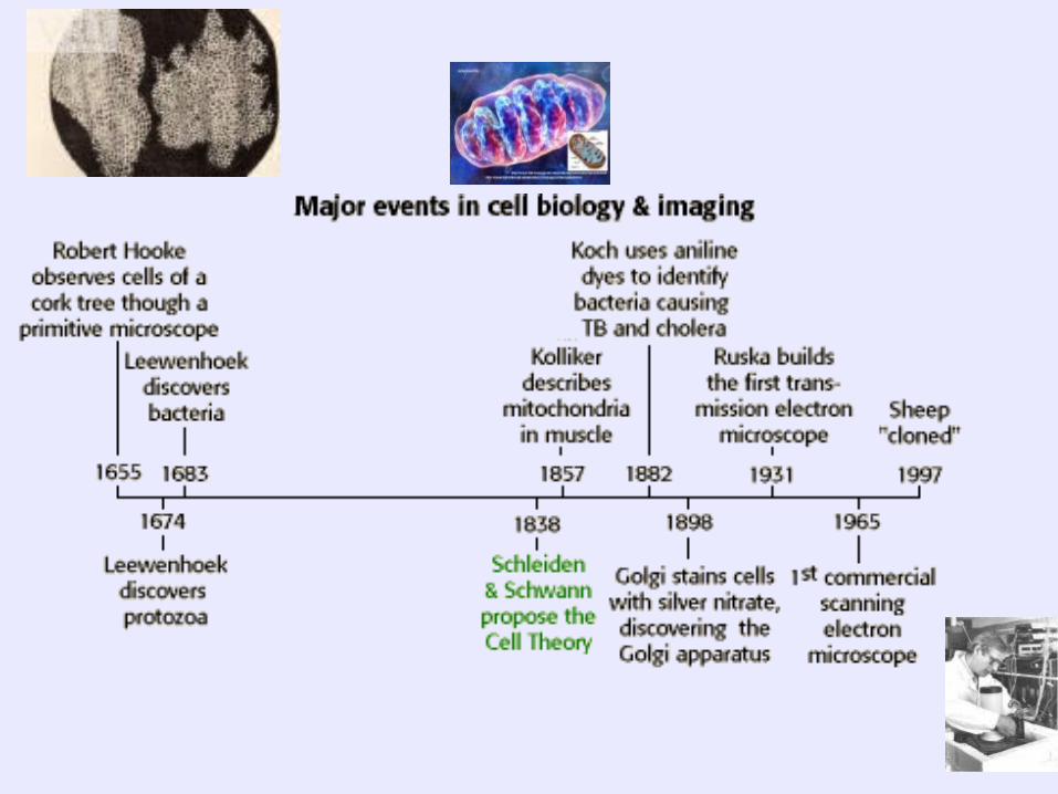

1665 Robert Hooke
• Robert Hooke observed “cellulae” or little rooms when he observed pieces of cork.
• From his work came the term cell. the basic unit of structure and function for all living things!


• What are the respective sizes of a virus and a plant cell?
• A.3 mm, 30 mm
• B.30 nm, 30 µm
• C.30 µm, 30 nm
• D.3 cm, 30 cm

B.30 nm, 30 µm

The Cell Theory • When Schleiden and Schwann proposed the
cell theory in 1838, cell biology research was forever changed. The cell theory states that:

SC.912.L.14.1
• Ch 7.*
• Describe the scientific theory of cells (cell theory) and relate the history of its discovery to the process of science.

Cell theory
• All life forms are made from one or more cells.
• Cells only arise from pre-existing cells.
• The cell is the smallest form of life.

• It took the work of several scientist and likely many more to develop the cell theory. Like all theories, it takes support by many experiments over a long period of time to develop a theory!

• The cell theory also provides us with an operational definition of "life".

• Which of the following is not a part of the cell theory?
• A.All animals are formed by cells.
• B.Reproduction requires vegetative duplication or the sexual mixing of gametes.
• C.Cells are the smallest form of life.
• D.Abnormal cells self destruct by apoptosis.

• D. Abnormal cells self destruct by apoptosis

• 1. How many scientists are listed in your book as contributing towards developing the cell theory? _______3__
• What were there names? __Schleiden, Schwann and Virchow___________________,
• 2. Why do you think there were 3 scientist involved in this process? It takes the work of many scientists to develop a theory _over a long period of time and much testing must happen.
•
• Could there have been others that contributed? ____yes________ Did this happen over a year or likely more time? ______more_______________. What does it take for an idea in science to become a theory? ___repeated tests with no evidence against the theory.______
• 3. What are the 3 main parts of the cell theory?
• 1. All life forms are made from one or more cells.
2. Cells only arise from pre-existing cells.
3. The cell is the smallest form of life.
• 4. Can cells appear spontaneously without genetic material from previous cells? no__
• 5. How was the cell theory developed? By combining the research from several scientists over several years.

Microscopes
• SC.912.L.14.4 Compare and contrast the different types of microscopes (as N 1.1)

Dissection microscope
• 3 D image,
• Living or dead specimen
• 20-40 X usually
• Limited by size of scope

Dissection scope

Compound Microscope
• 10 to 1000X
• Living and dead specimens can be viewed.
• Slides of thin stained specimens must be made.

• thin sections that light can pass through
• Use two lenses to magnify the object. Multiply the power of each lens with the second to get the total power.
• Can view living or dead specimens.

Transmission Electron Microscope
• Magnify up to 500,000 X
• Thin specimens treated with metal must be dead!
• In a vacuum chamber!

Organelles can be seen with a TEM

Scanning Electron Microscope• 3D images
• No lenses
• Electrons instead of light
• Only non living specimens!
• 10X to 500,000X magnification!!!

Human blood, pollen and a spider on SEM

• 1. Which type of microscope uses 2 lenses and visible light to magnify and view thin sections of specimens by passing light through the specimen? Compound light
• 2. How do you determine the power of a compound microscope? Multiply the 2 lenses together
• 3. What type of microscope uses a beam of electrons and a magnet to produce a black and white image of the specimen? Transmission electron microscope_
• 4. What is a problem with transmission microscopes with regard to viewing live specimens? The specimen must be in a vacuum chamber and prepped with metals so it must be dead!
• 5. What type of microscope produces a 3D image of the specimen? The scanning electron microscope

• Compare the 4 types of microscopes: compound light, dissecting, TEM and SEM, include the following information
Microscope type Magnification range
Method used to view? Lenses? electrons
Image produced?
view of living dead organisms
Dissecting Microscope
Compound light Microscope
Transmission Electron microscope
Scanning Electron Microscope
SC.912.L.14.3
Compare and contrast the general structures of prokaryotic and eukaryotic cells.(MC AA)

• Living things are classified in six kingdoms based on structure.
• Eubacteria Prokaryotic
• Archaea Prokaryotic
• Protista Eukaryotic
• Plantae Eukaryotic
• Fungae Eukaryotic
• Animalia Eukaryotic

Within prokaryotes
• which appeared 3.5 billion years ago, are the kingdoms
• Eubacteria and Archaea.

Within eukaryotes
• which evolved 1.5 billion years ago, are the kingdoms
• Protista, Plantae, Fungae, Animalia.

• Cells are also defined according the need for energy.
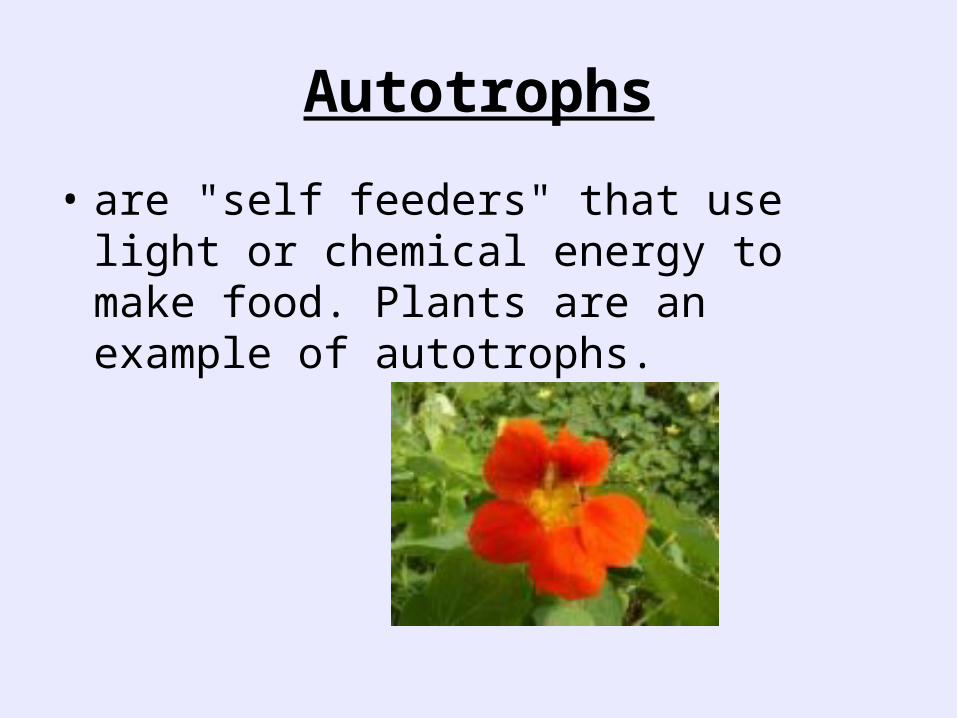
Autotrophs
• are "self feeders" that use light or chemical energy to make food. Plants are an example of autotrophs.

heterotrophs
• In contrast, heterotrophs ("other feeders") obtain energy from other autotrophs or heterotrophs. Many bacteria and animals are heterotrophs.

Multicellular Organisms
• Multicellular organisms are created from a complex organization of cooperating cells.
• There must be new mechanisms for cell to cell communication and regulation.

• There also must be unique mechanisms for a single fertilized egg to develop into all the different kinds of tissues of the body.
• In humans, there are 1014 cells comprising 200 kinds of tissues!

• Cells are classified by fundamental units of structure and by the way they obtain energy.
• Cells are classified as prokaryotes or eukaryotes

Prokaryotes
• prokaryotes include the kingdoms of Eubacteria (simple bacteria) and Archaea.

• Simply stated, prokaryotes are molecules surrounded by a membrane and cell wall.
•
• Prokaryotic cells lack characteristic eukaryotic sub cellular membrane enclosed "organelles", but may contain membrane systems inside a cell wall.

• Eukaryotes • Basic structure The basic eukaryotic cell
contains the following: • plasma membrane • glycocalyx (components external to the
plasma membrane) • cytoplasm (semi fluid) • cytoskeleton - microfilaments and
microtubules that suspend organelles, give shape, and allow motion
• presence of characteristic membrane enclosed subcellular organelles

organelles

1. What is the main purpose of the plasma membrane? To control what goes in and out of the cell. It is selectively permeable!
2. What is the structure of the plasma membrane? It is a bilayer of phospholipids with some proteins embedded.
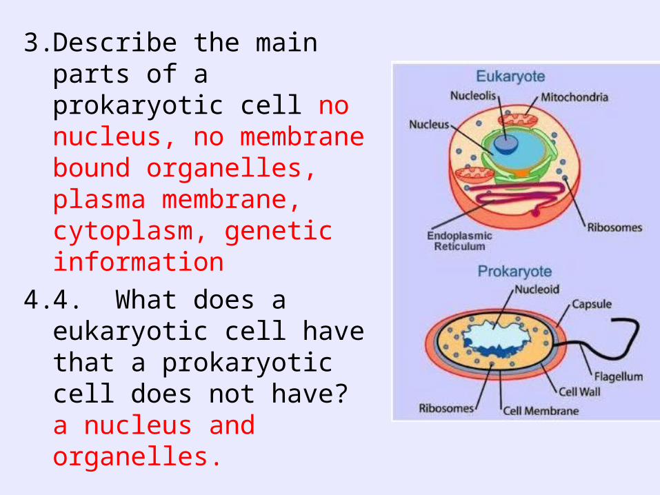
3. Describe the main parts of a prokaryotic cell no nucleus, no membrane bound organelles, plasma membrane, cytoplasm, genetic information
4. 4. What does a eukaryotic cell have that a prokaryotic cell does not have? a nucleus and organelles.

• 5. What is the current theory as to why there are two types of cells, prokaryotic and eukaryotic? According to the endosymbiotic theory the prokaryotic cells started living inside each other and developed into the eukaryotic cells.

6. A cell with the DNA located in a nuclear membrane, mitochondria, ribosomes, and endoplasmic reticulum is most likely what type of cell? eukaryotic.
• 7. Cells that lack a membrane bound nucleus and organelles are most likely what type of cells? prokaryotic

Section 2
• Read your reading essentials book section 2 or text book section 2!
Vocabulary:
1. Selective permeability,
2. Phospholipid bilayer,
3. Transport protein,
4. Fluid mosaic model

SC.912.L.14.2
• Relate structure to function for the components of plant and animal cells.
• Explain the role of cell membranes as a highly selective barrier (passive and active transport) (as 14.3)

Selective permeability
Allows some substances to pass through and keeps others out.
• This can change at different times! • This is not due to the size of the holes in the
membrane!• Some things require energy to get them
across• Some things are small enough to pass by
themselves others need helper molecules.

• Plasma Membrane
• A lipid/protein/carbohydrate complex, providing a barrier and containing transport and signaling systems.

Floating around in the cell membrane are different kinds of proteins. These are generally globular proteins. They are not held in any fixed pattern but instead float around in the phospholipid layer.
There are carrier proteins that regulate transport and diffusion

Cell membrane - Function • The cell membrane's function, in general,
revolves around is membrane proteins. General functions include:
• Receptor proteins which allow cells to communicate,
• transport proteins regulate what enters or leaves the cell,
• and marker proteins which identify the cell

Cell membrane - Function - Regulation of transport
• Transport Proteins come in two forms: Carrier proteins are peripheral proteins which do not extend all the way through the membrane. They move specific molecules through the membrane one at a time.

Channel proteins extend through the bilipid layer. They form a pore through the membrane that can
move molecules in several ways.

Cell membrane - Function - Carrier Proteins
• These are carrier proteins. They do not extend through the membrane. They bond and drag molecules through the bilipid layer and release them on the opposite side.

Symports also use the process of diffusion. In this case a molecule that is moving naturally into the cell through
diffusion is used to drag another molecule into the cell. In this
example glucose hitches a ride with sodium.

• The cell membrane can also engulf structures that are much too large to fit through the pores in the membrane proteins.
• This process is known as endocytosis.
• In this process the membrane itself wraps around the particle and pinches off a vesicle inside the cell.

Endocytosis

Exocytosis
• The opposite of endocytosis is exocytosis.
• Large molecules that are manufactured in the cell are released through the cell membrane.

Proteins in the membrane• There are carrier proteins that
regulate transport and diffusion
• Marker proteins that identify the cell to other cells
• And receptor proteins that allow the cell to receive instructions
• 1. What is the difference between a fish net and a cell membrane in terms of the cell membrane being selectively permeable? (What can a cell membrane do and a fish net not do?) A cell membrane can change what it lets in and does not let in from day to day and minute to minute an fish net can not do this!
• 2. Why are phospholipids the perfect organic molecule to make up the call membrane? They have a polar head and a non polar tail making them a water resistant barrier to the inside of the cell. things can pass between them using the proteins that are embedded in them or between them if they are small enough.
• 3. What is the role of the protein carrier in moving things across the cell membrane? They can provide a channel to pass through or they can move the item across by changing position.
• 4. How do transport proteins contribute to the selective permeability of the plasma membrane? They can turn on or off depending on the chemical present and the “needs” of the cell.

• 5. What else do proteins assist the cell membrane with? They transmit signals to the cell, provide support to the structure giving it shape.
• 6. Why is the plasma membrane referred to as a fluid mosaic? Because the phospholipids and proteins can move around in the structure and are not set permanently in one location, they move like a fluid.
• • 7. The cell component that acts as a highly selective
barrier and controls the movements of items into and out of the cell is the what? The plasma membrane or cell membrane

Draw a section of the cell membrane

The plasma membrane
• http://www.youtube.com/watch?v=moPJkCbKjBs&feature=youtube_gdata_player

Section 3 Vocabulary:
1. Cytoplasm, 8. Lysosomes,
2. Cytoskeleton 9. Centriole,
3. Ribosome 10. Mitochondrion,
4. Nucleolus, 11. Chloroplast
5. Endoplasmic reticulum 12. Cell wall,
6. Golgi apparatus 13. Cilium
7. Vacuole 14. Flagellum

SC.912.L.14.2:
• Relate structure to function for the components of plant and animal cells.
• Compare and contrast the general structures of prokaryotic and eukaryotic cells.

Cytoplasm: semi fluid material in all cells.
In prokaryotes all chemical processes occur
Here!
In eukaryotes the organelles are located with in the cytoplasm along with lost of chemicals needed for the chemical reactions that happen in the cell.!

Cytoplasm
• The term cytoplasm refers to everything between the cell membrane and the nuclear envelope. It consists of primarily of water. It also contains various organelles as well as salts, dissolved gasses and nutrients.

cytoskeleton
• Microfilaments and micro fibrils ( proteins) help hold the shape of the cell. They are part of the cytoplasm and make up the compartments where organelles are found.

Nucleus• Double membrane surrounding the
chromosomes and the nucleolus. Pores allow specific communication with the cytoplasm. The nucleolus is a site for synthesis of RNA making up the ribosome

NUCLEUS
• The nucleus is the headquarters of the cell.
• It regulates all cell activity.
• It consists of a nuclear envelope, (the outer membrane) and nucleoplasm.
• In the nucleoplasm you can see chromatin and the nucleolus.

• The nuclear envelope is a double membrane. Is has 4 phospholipid layers. It is also has large pores through which materials pass back and forth.

nucleus

nucleolus

• Within the nucleus are found chromatin and a structure called the nucleolus.
• Chromatin is DNA in its active form. It consists of DNA looped around histone proteins.
• The nucleolus is a knot of chromatin.
• It is the nucleolus that manufactures ribosomes

Nucleus - Function • The nucleus regulates all cell activity.
• It does this by controlling the enzymes present.

• The chromatin is composed of DNA.
• DNA contains the information for the production of proteins.
• This information is encoded in the 4 DNA bases.
• Adenine, thymine, cytocine, and guanine.
• The specific sequence of these bases tells the cell what order to put the amino acids.

• There are three processes that enable the cell to manufacture protein:
• Replication allows the nucleus to make exact copies of its DNA
•Transcription allows the cell to make RNA working copies of its DNA
• In translation the Messenger RNA is used to line up amino acids into a protein molecule

The Endoplastic Reticulum
• Spreading throughout the cytoplasm is the endoplasmic reticulum. It is a folded system of membranes that loop back and forth giving it a very large surface area. This membrane provides a surface area for cell reactions. It is also the Site of lipid production.

• Smooth E.R. has no ribosomes associated with it The rough E.R. has ribosomes

ER
• Endoplasmic Reticulum 194

Rough endoplasmic reticulum (RER)
• A network of interconnected membranes forming channels within the cell. Covered with ribosomes (causing the "rough" appearance) which are in the process of synthesizing proteins for secretion or localization in membranes.

endoplasmic reticulum

• Smooth endoplasmic reticulum (SER)
• A network of interconnected membranes forming channels within the cell. A site for synthesis and metabolism of lipids. Also contains enzymes for detoxifying chemicals including drugs and pesticides.
•
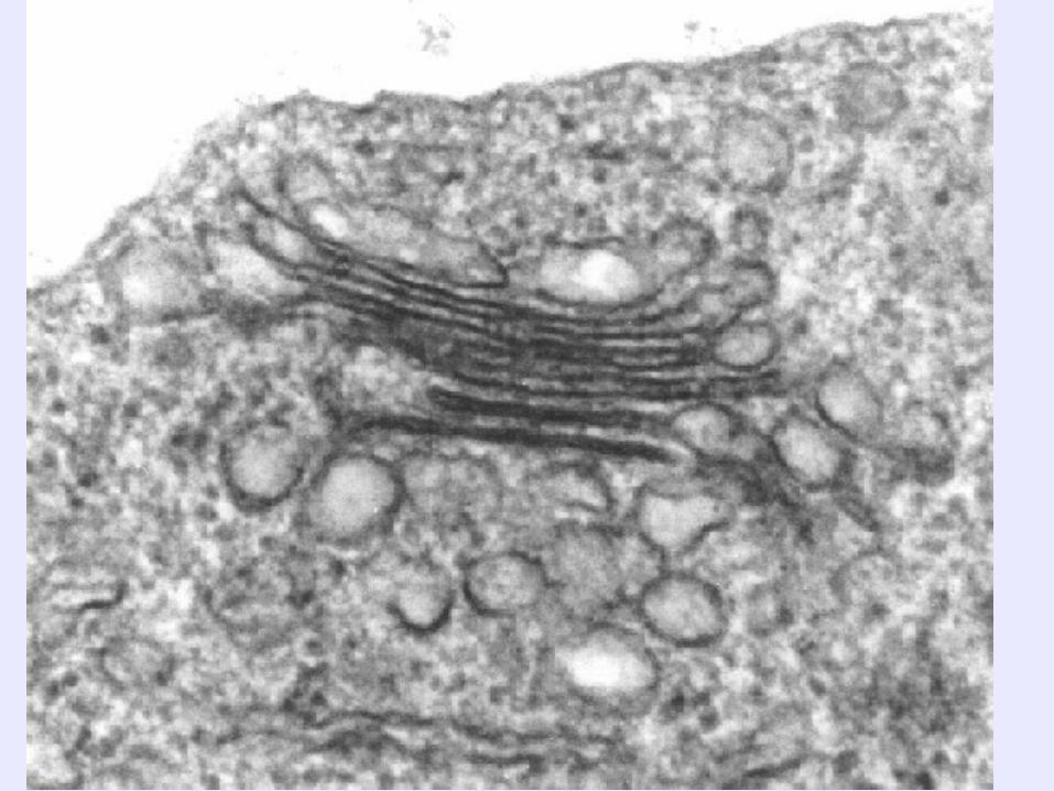
Golgi apparatus• A series of stacked membranes. Vesicles
(small membrane surrounded bags) carry materials from the RER to the Golgi apparatus. Vesicles move between the stacks while the proteins are "processed" to a mature form. Vesicles then carry newly formed membrane and secreted proteins to their final destinations including secretion or membrane localization.


The Golgi Apparatus
• The golgi body is responsible for packaging proteins for the cell. Once the proteins are produced by the rough E.R. they pass into the sack like cisternae that are the main part of the golgi body. These proteins are then squeezed off into the little blebs which drift off into the cytoplasm.

Vacuoles • Vacuoles are large empty appearing areas
found in the cytoplasm.
• They are usually found in plant cells where they store waste.
• As a plant cell ages they get larger.
• In mature cells they occupy most of the cytoplasm.

Contractile Vacuoles
• These organelles are critical in enabling protozoa to combat the effects of osmosis. Protozoa must constantly excrete the water that enters through their membranes.

Lysosymes
• A membrane bound organelle that is responsible for degrading proteins and membranes in the cell, and also helps degrade materials ingested by the cell.

Centrioles
• Centrioles are found only in animal cells. They function in cell division. Notice the 9 sets of 3 arrangement of the protein fibers

• Mitochondria
• Surrounded by a double membrane with a series of folds called cristae. Functions in energy production through metabolism. Contains its own DNA, and is believed to have originated as a captured bacterium.
•

mitochondria

Chloroplasts (plastids)• Surrounded by a double membrane, containing
stacked thylacoid membranes. Responsible for photosynthesis, the trapping of light energy for the synthesis of sugars. Contains DNA, and like mitochondria is believed to have originated as a captured bacterium.
•

chloroplasts

Plastids
• Plastids are large organelles found on plants and some protists but not in animals or fungi. They can easily be seen through a light microscope. Chloroplasts represent one group of plastids called chromoplasts (colored plastids). The other plastids are called leucoplasts (colorless plastids); they usually store food molecules. Included in this group are amyloplasts or starch plastids shown here in potato root cell.

Ribosomes• Protein and RNA complex responsible for
protein synthesis.
• VacuolesMembrane surrounded "bags" that contain water and storage materials in plants.
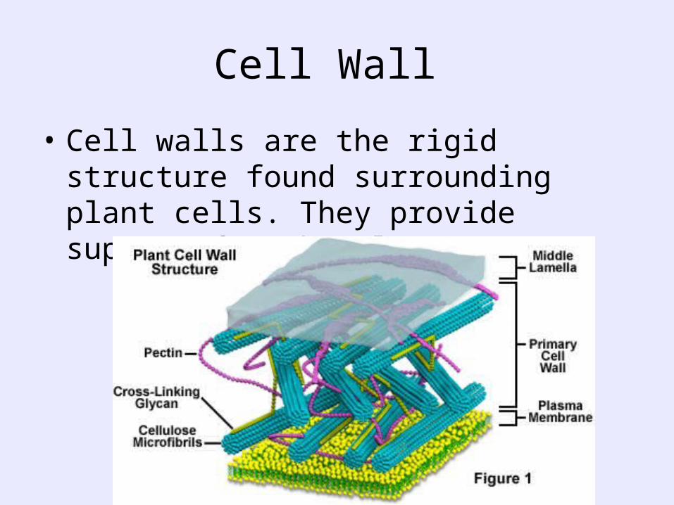
Cell Wall
• Cell walls are the rigid structure found surrounding plant cells. They provide support for the plant

Cell wall
• Plants have a rigid cell wall in addition to their cell membranes.

Cillia and Flagella
• These are hair like extensions off of the cell membrane. Their structures are similar except that cillia tend to be small and numerous and flagella tent to be large and fewer. Their they beat back and forth rhythmically. In unicellular organisms their job is locomotion. In large multicellular organisms their role is to move fluid past the cell. Notice the 9+2 arrangement of the microtubules
Parts of the cell rap
• http://www.youtube.com/watch?v=-zafJKbMPA8&feature=youtube_gdata_player

Plant and animal cells

Eukaryotic Plant cell u tube
• http://www.youtube.com/watch?v=8Wa_JaYhMls&feature=youtube_gdata_player

Animal cell
• http://www.youtube.com/watch?v=Fzj6TRnXmps&feature=youtube_gdata_player

3. The organelle with a green pigment and membranes with enzymes for photosynthesis is the chloroplast
4. The jelly-like substance inside of a cell is called cltoplasm.
5. The cytoskeleton __ is a network on protein fibers that anchor organelles and provide structure for the cell.
6. The _nucleus____ manages the functions of the cell and contains a eukaryotic cell’s DNA.
7. Proteins are produced in small protein and RNA containing structures called _ribosomes_____.
8. The _nucleolus__ is a structure located in the nucleus where ribosomes are produced.

9. The Endoplasmic reticulum___ is a system of membranes that transport materials from the nucleus to the Golgi apparatus. Two kinds of ER are rough ___, with ribosomes, and ____smooth __, without ribosomes.
10.After proteins are produced on the ER, they are transported to the _golgi Apparatus which modifies, sorts, and repackages them into vesicles.
11.What happens to proteins after they are placed into vesicles on the Golgi apparatus? They are exported by exocytosis or saved for later use by the cell
12.What materials are stored in vacuoles? Water , food enzymes sometimes waste products

13. Which organelles are responsible for digesting excess or worn out organelles and food in the cell? Lysosomes
14. Why are centrioles located near the nucleus? They are involved in cell replication and lining up the chromosomes that are found in the nucleus
15. Why are the inner folds of mitochondria highly folded? To increase the surface area for respiration that occurs there
16. What organelles are the sites of photosynthesis? Chloroplasts
17. How is the cell wall different from the cell membrane? It is made up of cellulose not phospholipids and does not control what goes in and out of the cell but provides a support structure

18.Differentiate between cilia and flagella. Cilia:_usually many and small and short Flagella:_often only one and long thicker in diameter
19.What three structures are found in plant cells but not in animal cells? Large central vacuole, chloroplast and cell wall. Explain why they are not needed in animal cells. No need to support using water ( also used in photosynthesis , no need for chlorophyll because no photosynthesis occurs and animal cells are more flexible having only a cell membrane, no cell wall.
20.Describe the structures that proteins pass through, beginning with their production and ending with their exit from the cell. Ribosomes, re and golgi apparatus_

Specialty organelles • these structures are usually found only in certain kinds
of cells.
Animal cells generally contain centrioles.
• Plant cells generally contain storage vacuoles, cell walls, and plastids.
• Cilia and flagella are found in many different life forms.
Be aware that there are many other kinds of living things besides plants and animals.

Peroxisomes or Microbodies
•Produce and degrade hydrogen peroxide, a toxic compound that can be produced during metabolism

Lysosomes
• Lysosomes are called suicide sacks. They are produced by the golgi body. They consist of a single membrane surrounding powerful digestive enzymes.

Those organelles involved in protein production
• Ribosomes
• The endoplastic reticulum
• The golgi apparatus
• Lysosomes
Those organelles involved in energy production
• Mitochondria
• Chloroplasts

Cell characteristics • Size:
• 1. smallest cells are bacteria a. 0.2 um in diameter
• 2. longest cells in mammals
a. nerve cells, giraffe neck

Typical sizes
• 1. bacteria (prokaryotes)
a. diameter 1-5 um
• 2. higher plant & animal cells (eukaryotes) a. 10-50 um
• 3. eukaryotic cells are about 1000x larger in volume than prokaryotic cells

surface area

Why are cells these sizes?
• surface area/volume ratio (SA/V) everything entering cell comes
through the plasma membrane (PM)
An increased cytoplasmic volume means more stuff must come thru
-more glucose -more amino acids -more ions -more of everything needed in the cell

Surface area to volume ratio

Bigger cell means
An increased volume => relative decline in Surface area to volume ratio
Plasma Membrane loses ability to transport enough stuff in/out of cell

Solutions to the surface Area Problem
• (1) infoldings (increases SA) • (2) outward protrusions (increases SA) • note the projections that increase surface
area w/o increasing volume much
• e.g.: microvilli of • intestinal mucosa • cells

Other solutions
large vacuoles => decreases cytoplasm volume
(a) Elodea cell
with vacuole
indicated

membrane

ribosomes

prokaryotic cells

eukaryotic cells
• Plant, animal, fungal and Protista cells


Biotechnology video
• http://www.youtube.com/watch?v=o1GQyciJaTA&feature=youtube_gdata_player
Giant Plant Cell
– nucleus and nucleolus – mitochondrion – chloroplast – rough endoplasmic reticulum (focus on function of ribosome) – smooth endoplasmic reticulum (focus on function of ER) – Golgi complex – lysosomes – peroxisomes – microtubules and microfilaments – vacuole
• _ cytoskeleton

• 1. Students research the structure and function of their chosen organelle.
• 2. Students produce a blueprint describing how they will construct their model and its size.
• 3. Students prepare a class presentation.

Organelle Nucleus
Function1. stores the cell's
hereditary material, or DNA
2. coordinates the cell's activities, which include growth, intermediary metabolism, protein synthesis, and reproduction (cell division).
Structure
Nucleolus manufacture the subunits that combine to form ribosomes

Organelle
Plasma Membrane
Function
acts as a boundary, holding the cell together and keeping other substances from entering.
Structure

OrganelleMicrofilament
Function
These filaments are primarily structural in function and are an important component of the cytoskeleton.
structure Microfilaments are solid rods made of globular proteins
Microtubules are found throughout the cytoplasm of all eukaryotic cells (prokaryotes don't have them) and carry out functions, ranging from transport to structural support.
straight, hollow cylinders
Organelle function Structure
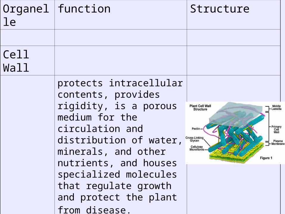
Cell Wall
protects intracellular contents, provides rigidity, is a porous medium for the circulation and distribution of water, minerals, and other nutrients, and houses specialized molecules that regulate growth and protect the plant from disease.

Organelle function Structure
Mitochondrion break down carbohydrate and sugar molecules to provide energy
oblong shaped organelles

OrganelleChloroplast
Function
contains the pigment chlorophyll. It absorbs light energy needed for photosynthesis to occur.
Structure

Organelle Endoplasmic Reticulum
Function
manufactures, processes, and transports chemical compounds for use inside and outside of the cell.
It is connected to the double-layered nuclear envelope, providing a pipeline between the nucleus and the cytoplasm.
structure
network of sacs

Organelle Ribosome
Function
serve as the protein production machinery for the cell
structure composed of approximately 60 percent RNA and 40 percent
protein.
Vacuole
50-80% of volume
plant cells have a large, single vacuole that stores things, helps in plant growth, and plays an important structural role for the plant.

Organelle function StructurePeroxisomes spherical and bound
by a single membrane. There are several types of microbodies but peroxisomes are the most common.
CILLIA/ Flagella locomotion of organisms. In multicellular organisms, cilia function to move fluid or materials past an immobile cell as well as moving
a cell or group of cells.

• Creating the Organelles
• The Giant Plant Cell is shaped like a giant cube that is 3 meters (300 cm) on each edge. You have been provided with a chart of actual cell organelle sizes.
• You must, using ratios, calculate the size of your giant organelle so that it is proportionately correct for the huge cell.
• You also need to research the function of the organelle in order to build it so that it can carry out its role in the giant cell.
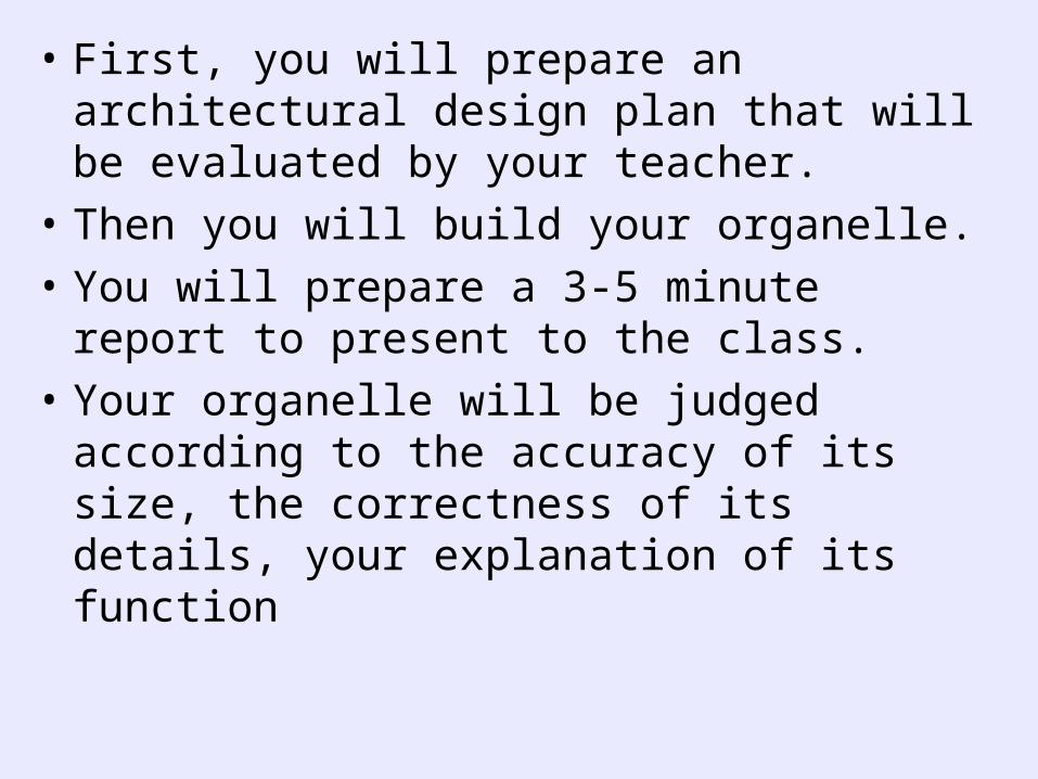
• First, you will prepare an architectural design plan that will be evaluated by your teacher.
• Then you will build your organelle.
• You will prepare a 3-5 minute report to present to the class.
• Your organelle will be judged according to the accuracy of its size, the correctness of its details, your explanation of its function

Equation for determining size of organelle:
• Size of giant organelle =Actual size of organelle
• Size of giant cell (300 cm) =Actual size of average plant cell (30mm)
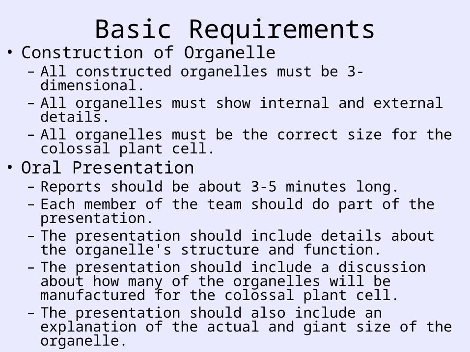
Basic Requirements• Construction of Organelle
– All constructed organelles must be 3-dimensional. – All organelles must show internal and external details. – All organelles must be the correct size for the colossal plant
cell.• Oral Presentation
– Reports should be about 3-5 minutes long. – Each member of the team should do part of the
presentation. – The presentation should include details about the organelle's
structure and function. – The presentation should include a discussion about how
many of the organelles will be manufactured for the colossal plant cell.
– The presentation should also include an explanation of the actual and giant size of the organelle.

• CHART OF ACTUAL ORGANELLE SIZES
• Cell or Organelle Size in x (10-3 mm
Average Plant Cell 30 x
Nucleus 7.5-10 x
Nucleolus 2.5 x
Plasma Membrane 0.009 x thick
Mitochondrion 0.2-1 x wide x 3-10 x long
Chloroplast (other plastids are similar sizes) 2 x 5 x
Ribosome 0.025 x
Endoplasmic Reticulum (in most plant cells) 0.5 x thick (2 membranes of 0.009 mm with 0.03 mm compartment between them)
Golgi Complex 1 x 1 x (membranes have thickness of ER)
Vacuole (central) sometimes serves as lysosome 50-80% of volume of cell
Microtubules 0.02 x diameter
Microfilaments 0.007 0.5-1 x diameter
Lysosomes (in some plant cells) 0.2-2 x
Peroxisomes 3 x
Cell Wall 1-2 x thick
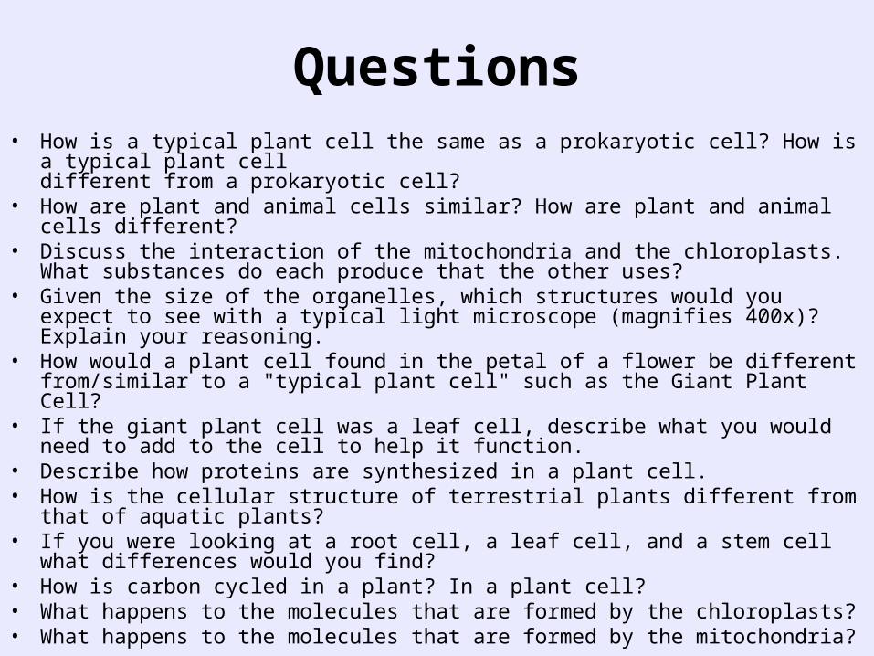
Questions• How is a typical plant cell the same as a prokaryotic cell? How is a typical plant cell
different from a prokaryotic cell?• How are plant and animal cells similar? How are plant and animal cells different?• Discuss the interaction of the mitochondria and the chloroplasts. What substances do
each produce that the other uses?• Given the size of the organelles, which structures would you expect to see with a
typical light microscope (magnifies 400x)? Explain your reasoning.• How would a plant cell found in the petal of a flower be different from/similar to a
"typical plant cell" such as the Giant Plant Cell?• If the giant plant cell was a leaf cell, describe what you would need to add to the cell
to help it function.• Describe how proteins are synthesized in a plant cell.• How is the cellular structure of terrestrial plants different from that of aquatic plants?• If you were looking at a root cell, a leaf cell, and a stem cell what differences would
you find?• How is carbon cycled in a plant? In a plant cell?• What happens to the molecules that are formed by the chloroplasts?• What happens to the molecules that are formed by the mitochondria?