cell proliferation activity and prognostic index in...
TRANSCRIPT

Analytical Cellular Pathology 16 (1998) 233–242 233IOS Press
Cell proliferation activity and prognosticindex in squamous cell lung carcinoma
José Antonio Alvarez-Riesgoa, Andrés Sampedrob,∗, Radhamés Hernándeza,María Victoria Folguerasc, Ana Salas-Bustamanteb and Antonio CuetoaaDepartamento de Medicina, Universidad de Oviedo, Facultad de Medicina, C/ Julián Clavería s/n,33006 Oviedo, Spainb Servicio de Citometría, Hospital Central de Asturias, Bloque Polivante A, 1a Planta, C/ CelestinoVillamil s/n, 33006 Oviedo, Spainc Servicio de Anatomía Patolögica II, Hospital Central de Asturias, C/ Celestino Villamil s/n,33006 Oviedo, Spain
Received 16 December 1997Revised 14 April 1998Accepted 5 June 1998
Abstract. Flow Cytometry (FC) has been incorporated into cancer research in relation to its prognostic value together withhistological parameters and TNM stages. We have studied by means of FC the cell cycle of 132 samples from male patientswith Squamous Cell Lung Carcinoma (SQCLC). All of the patients received curative surgery and the clinical follow-up was60 months. The clinical and cytometric parameters were evaluated in order to predict the patients’ outcome. The presence oftumoural recurrence and the tumoural stage showed statistical significance associated with survival. The multivariant analysisreveals radiotherapy (p = 0.004) as protective variable and the high S-phase fraction (SPF) (p = 0.001) and stage IIIA(p = 0.012) as risk factors. The SPF appears as an independent prognostic factor for overall survival time. We can build aprognostic index representative of different prognostic groups, which allows us to improve the individual monitoring of thesepatients.
Keywords: Squamous cell lung carcinoma, flow cytometry, cell proliferation, DNA ploidy, prognosis
1. Introduction
Lung cancer is an important public health problem in our region, with an incidence rate in malesof 112.40 per 100,000 and a male/female occurrence ratio of 10/1 [10]. From the histological point ofview, 45% of the non-small cell lung carcinomas (NSCLC) are squamous carcinomas. At present, theanatomic description of dissemination and tumoural aggressiveness precision are the two main sets offactors which allow us to estimate the prognosis of the patients [12]. However, the variability in thesurvival rate within the same stages, proves that we still need to find other prognostic factors which candetermine which patients are most at risk to suffer a recurrence of the disease [5,23].
Technological development has had a great impact on the precision of tumoural stage and on the overallsurvival rate of patients [11,31]. The use increasingly sensitive image analysis methods has facilitated the
* Address for correspondence: Dr. A. Sampedro, Servicio de Citometría, Hospital Central de Asturias, Bloque Polivante A,1a Planta, C/ Celestino Villamil s/n, 33006 Oviedo, Spain. Tel.: +34 8 510 36 60; Fax: +34 8 510 36 58; E-mail:[email protected].
0921-8912/98/$8.00 1998 – IOS Press. All rights reserved

234 J.A. Alvarez-Riesgo et al. / Cell proliferation activity and prognostic index
improvement of the survival rate in SQCLC due to a more precise identification of the subsidiary surgicaltreatment of these patients [22]. As the long-term intervention results and treatment responses are basedon inherent biological factors within the tumoural cells, it is becoming more and more important for usto concentrate our research efforts on the tumoural biology of lung cancer [6].
DNA ploidy and tumour cell proliferation activity estimated by FC have been considered potentialindicators of survival and treatment response in SQCLC [1,32,36,39]. In some of the SQCLC series,overall survival varies depending on the ploidy, regardless of the stage [16,26,35]. Tumoural recurrencein SCQLC has been reported 50% lower than in other NSCLC [33].
The reported variability of the prevalence and prognostic significance of DNA aneuploidy could berelated to the number of samples analysed, the methodological differences and the histological types [8,16,17,32].
The prognostic value of the DNA aneuploidy can be completed with the analysis of the cell pro-liferation activity estimated using FC according to the percentage of cells in S-phase. The increasingproliferation activity has a negative effect on the survival rate [35]. In this way, the neoplasias with ahigh rate of cells in S-phase are more likely to develop metastasis, regardless of the histological differen-tiation, stage of the disease, or the type of treatment [37]. The cytometric information obtained from thecell cycle analysis can be combined with the information provided by the tumoural extension and othervariables so as to build up a Prognostic Index (PI) [14].
Because of the male/female incidence and the histologic frequency, we carried out a retrospectivestudy on 132 male patients with SQCLC who have received curative surgery to establish the prognosticsignificance of these cytometric variables. The final aim of our study was to establish objective andreproducible follow-up parameters, complementary to the clinicopathological ones which allow us toassess, in each case, the potential aggressiveness of the neoplasia and the corresponding therapeuticmonitoring.
2. Materials and methods
2.1. Patients
One hundred and thirty-two males with SQCLC treated by curative surgery, were studied and includedin the tumour archives at the Hospital General de Asturias (HGA) from January 1, 1985 to December 31,1989. The surgery was planned as curative in patients in stages I, II and IIIA, according to the TNMclassification of the Fourth World Wide Lung Cancer Conference [19]. A further reclassification wascarried out after pathological study. The original diagnosis was established by radiography, cytologyof sputum and/or bronchoscopy. The information was completed with preoperative and postoperativepathological examinations.
Regarding the complementary treatment, radiotherapy decision was taken according to the pathologicstudy, incorporating the clinical staging parameters such as tumour size, grading of differentiation, vas-cular and pleural invasion and tumour necrosis. Chemotherapy was not given to the patients.
The follow-up of the patients continued for 60 months after the date of surgery. A complementaryprocedure was established for those patients for whom we had no information during the follow-up pe-riod. At first, they were investigated by means of the program of oncological patients treated with opiatederivatives, whose mandatory register is kept by the regional medical authorities. Finally, the RegionalStatistical Decease Bulletins (RSDB) were assessed during a 5 year period after surgery, recording the

J.A. Alvarez-Riesgo et al. / Cell proliferation activity and prognostic index 235
cause of death in order to complete the follow-up. The patients lost in our study were 9/132 (6.8%) dis-tributed in a uniform way during the periods, 3 (1985), 2 (1986), 2 (1987), 2 (1989), and the cause is theloss of clinical history information.
The tumoural recurrence was confirmed by biopsy, cytology or fine needle aspiration. The relapse freetime was obtained by subtracting the surgical resection date from the recurrence date.
2.2. Flow cytometry
The flow cytometric analysis was carried out by the Cytometry Service of the University of Oviedofrom the material embedded in paraffin and archived in the Pathology Service of the HGA. A detaileddescription of the technique used has been published [27]. To select the blocks we revised for eachpatient all the slides available stained with hematoxylin–eosin, selecting those with a greater proportionof tumour and a lower rate of inflammatory component. When possible, sections from two or more blockswere taken. Three 50µm thick sections were obtained from each block of tissue. The deparaffination wascarried out with xylene for 35–40 min at 70◦C, and left at room temperature for 10 min. Ethanol wasused in decreasing concentration (100, 96, 70%) for 10 min in each step, later washing the sample witha PBS buffer and centrifuging it at 300× g for 10 min. After centrifugation, enzymatic disaggregationwas performed in pepsin solution (Pepsin A, EC 3.4.23.1, Sigma, St. Louis, MO, USA) for 30 min in ashaking water bath at 37◦C. In addition, the samples were mechanically dissociated using a 5 ml syringeand with a 20 gauge needle, drawn in and out of the tube several times. The single cells and nucleiobtained were then filtered through a nylon mesh (70µm) and stained with propidium iodide accordingto the Vindelöv technique [34].
A FacScan cellular analyser from Becton Dickinson was used which was equipped with an argon-ionlaser light source, with excitation at a wave length of 488 nm, and three fluorescence detectors, selectingthe FL2 with a fluorescent emission band of about 585 nm. To obtain and analyze the data we used theCellfit software 2.0.2 from Becton Dickinson. Tumour sample acquisition was carried out together witha DNA diploid control, using lymphocytes from tonsil embedded in paraffin and processed at the sametime as the tumoural suspensions. Should the presence of aneuploid tumours be questioned with morethan one population, a mixed sample of tumour and control was prepared. From each sample, 10,000–15,000 cells were analysed [4] and the CV was always found to be lower than 6%. The number of eventswere those obtained after the exclusion of debris and aggregates. The histograms obtained were theninterpreted by an analyst with no prior knowledge of the clinical characteristics of the neoplasia. Thefollowing were taken as cytometric variables: DNA ploidy, DNA Index (DI) and SPF. The adjustment ofdebris and aggregates were established by an electronic gate. In all the DNA diploid cases RFIT modelwas used in the assessment of S-phase, and POLY model in the DNA aneuploid ones. The cases withDI between 0.9 and 1.1 were considered as DNA diploids, less than 0.9 or greater than 1.10 as DNAaneuploids, and between 1.9 and 2.1 as DNA tetraploids.
2.3. Statistical analysis
The data was analysed using the SPSS/PC+ version 4.01 and Egret version 0.26.6 Pecan. The qualita-tive variable analysis was carried out using the chi square test. The Cox model [7] was used for regressionanalysis. The formula applied wash(t,xi) = h0(t)eβx, whereh(t,xi) represents the hazard function orhazard rate, with values in the explicative variables (x) in the time (t), h0(t) is the function of the refer-ence hazard rate (“baseline” or “underlying hazard function”) and eβx represents an exponential function
236 J.A. Alvarez-Riesgo et al. / Cell proliferation activity and prognostic index
whose exponent represents the linear combination of the explicative variables (xi). Thep value to enterwas established at 0.05, andp value to remove at 0.15. The prognostic index was built from the mul-tivariant model. The results of this analysis have been used to define a PI, expressed by the formula:PI =
∑i βiXi. The PI expressed as a continuous quantitative variable, changes into a negative scale,
and was thus converted into a positive scale in order to aid understanding. To represent the survival ac-cording to the PI, the results were categorized by quartiles (Q), and the differences were analysed withthe log-rank technique. From the variables examined in the Cox regression models, the relapse free time,measured in months, and the rate of cells in S-phase were considered as continuous quantitative variables.The tumoural recurrence and the application of radiotherapy were expressed as dichotomic variables, andthe tumoural stage as an ordinal variable.
3. Results
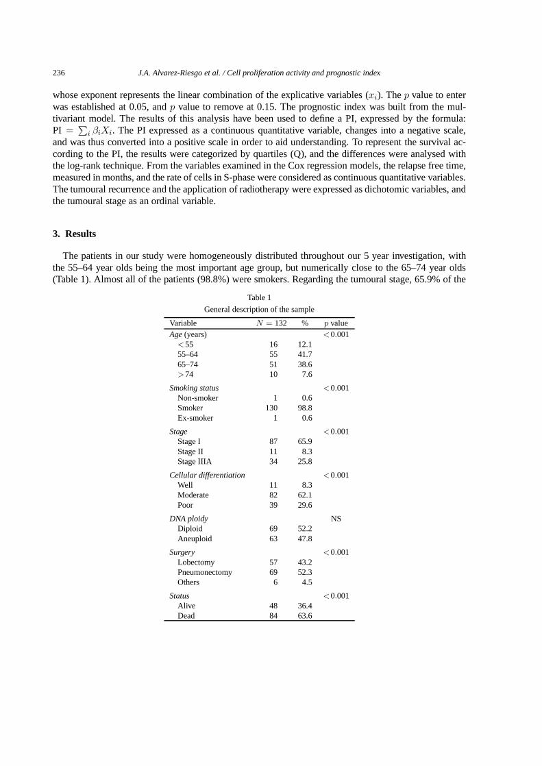
The patients in our study were homogeneously distributed throughout our 5 year investigation, withthe 55–64 year olds being the most important age group, but numerically close to the 65–74 year olds(Table 1). Almost all of the patients (98.8%) were smokers. Regarding the tumoural stage, 65.9% of the
Table 1
General description of the sample
Variable N = 132 % p valueAge(years) <0.001<55 16 12.155–64 55 41.765–74 51 38.6>74 10 7.6
Smoking status <0.001Non-smoker 1 0.6Smoker 130 98.8Ex-smoker 1 0.6
Stage <0.001Stage I 87 65.9Stage II 11 8.3Stage IIIA 34 25.8
Cellular differentiation <0.001Well 11 8.3Moderate 82 62.1Poor 39 29.6
DNA ploidy NSDiploid 69 52.2Aneuploid 63 47.8
Surgery <0.001Lobectomy 57 43.2Pneumonectomy 69 52.3Others 6 4.5
Status <0.001Alive 48 36.4Dead 84 63.6
J.A. Alvarez-Riesgo et al. / Cell proliferation activity and prognostic index 237
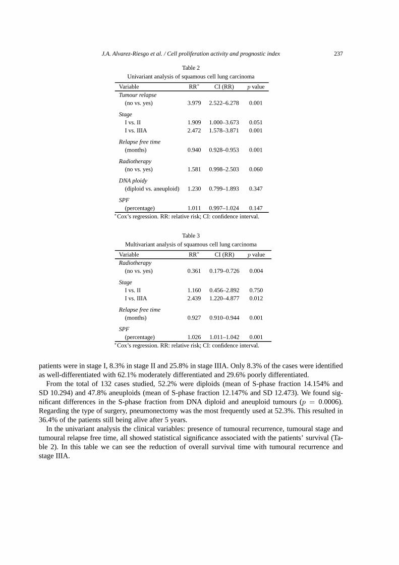
Table 2
Univariant analysis of squamous cell lung carcinoma
Variable RR∗ CI (RR) p valueTumour relapse
(no vs. yes) 3.979 2.522–6.278 0.001
StageI vs. II 1.909 1.000–3.673 0.051I vs. IIIA 2.472 1.578–3.871 0.001
Relapse free time(months) 0.940 0.928–0.953 0.001
Radiotherapy(no vs. yes) 1.581 0.998–2.503 0.060
DNA ploidy(diploid vs. aneuploid) 1.230 0.799–1.893 0.347
SPF(percentage) 1.011 0.997–1.024 0.147
∗Cox’s regression. RR: relative risk; CI: confidence interval.
Table 3
Multivariant analysis of squamous cell lung carcinoma
Variable RR∗ CI (RR) p valueRadiotherapy
(no vs. yes) 0.361 0.179–0.726 0.004
StageI vs. II 1.160 0.456–2.892 0.750I vs. IIIA 2.439 1.220–4.877 0.012
Relapse free time(months) 0.927 0.910–0.944 0.001
SPF(percentage) 1.026 1.011–1.042 0.001
∗Cox’s regression. RR: relative risk; CI: confidence interval.
patients were in stage I, 8.3% in stage II and 25.8% in stage IIIA. Only 8.3% of the cases were identifiedas well-differentiated with 62.1% moderately differentiated and 29.6% poorly differentiated.
From the total of 132 cases studied, 52.2% were diploids (mean of S-phase fraction 14.154% andSD 10.294) and 47.8% aneuploids (mean of S-phase fraction 12.147% and SD 12.473). We found sig-nificant differences in the S-phase fraction from DNA diploid and aneuploid tumours (p = 0.0006).Regarding the type of surgery, pneumonectomy was the most frequently used at 52.3%. This resulted in36.4% of the patients still being alive after 5 years.
In the univariant analysis the clinical variables: presence of tumoural recurrence, tumoural stage andtumoural relapse free time, all showed statistical significance associated with the patients’ survival (Ta-ble 2). In this table we can see the reduction of overall survival time with tumoural recurrence andstage IIIA.
238 J.A. Alvarez-Riesgo et al. / Cell proliferation activity and prognostic index
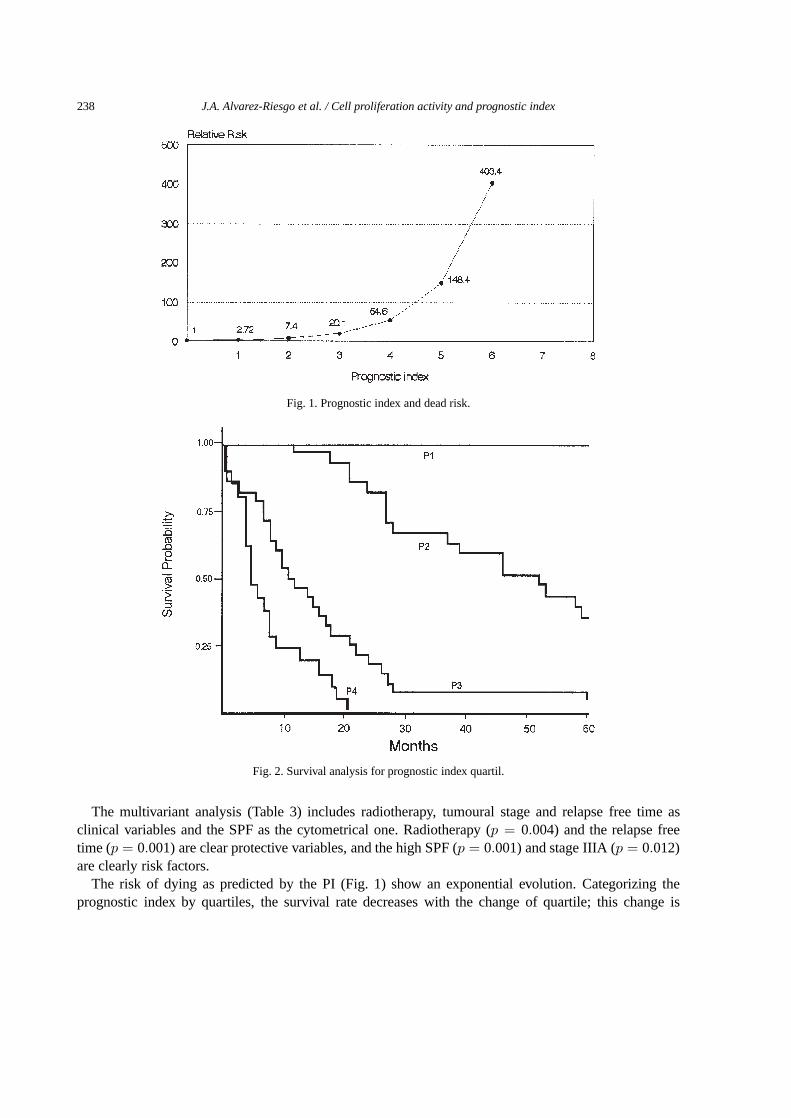
Fig. 1. Prognostic index and dead risk.
Fig. 2. Survival analysis for prognostic index quartil.
The multivariant analysis (Table 3) includes radiotherapy, tumoural stage and relapse free time asclinical variables and the SPF as the cytometrical one. Radiotherapy (p = 0.004) and the relapse freetime (p = 0.001) are clear protective variables, and the high SPF (p = 0.001) and stage IIIA (p = 0.012)are clearly risk factors.
The risk of dying as predicted by the PI (Fig. 1) show an exponential evolution. Categorizing theprognostic index by quartiles, the survival rate decreases with the change of quartile; this change is

J.A. Alvarez-Riesgo et al. / Cell proliferation activity and prognostic index 239
Fig. 3. Survival and prognostic index at 6, 12 and 24 months.
statistically significant (p < 0.001) (Fig. 2). The results in the evaluation of survival probability at 6, 12and 24 months confirm the previous results (Fig. 3).
4. Discussion
Of the total number of patients, 98.8% were smokers, which represents a very similar proportion tothe 95–98% obtained by other authors [20,21]. In the tracheobronchial area, 90% of the tumours areattributable to smoking, estimating the time of empirical induction to be between 20–40 years fromthe beginning of smoking to the maximum disease incidence [3,24,30]. Thus, our cases correspond topatients starting in their 50s and 60s, with an accumulated incidence of 53.8% of the patients at the ageof 64.
In our series, 65.9% of the patients were in stage I of the disease. The definition of the degree of cellu-lar differentiation is complex and a controversial subject among pathologists, with a tendency to establisha high rate of tumours as moderately differentiated. We found 62.1% of SQCLC to be moderately dif-ferentiated, similar to the results obtained by other authors [15], and only 8.3% to be well differentiatedtumours.
At present, it is agreed that the main factors affecting the prognosis of patients with lung canceris the TNM staging with complementary information on the differentiation degree, vascular and pleuralinvasion, and growth rate. In the univariant analysis (Table 2) only some clinical variables show statisticalsignificance. Our multivariant model (Table 3) includes clinical variables and incorporates the SPF resultsobtained by FC. The TNM and the SPF become risk factors, increasing the risk of death in 1.026 perunit increase in SPF. This is only evident for the TNM in stage IIIA (2.439). The similarities betweenthe rate of death in stages I and II of the TNM can be related to the problems of staging [18,25]. Thetumoural stage has shown an independent prognostic significance in different investigations related toSQCLC [23] and the whole of NSCLC [9].
The multivariant analysis of prognostic factors contributes to the understanding of the interrelationbetween them, identifying subgroups of patients who can take advantage of the different therapeutic

240 J.A. Alvarez-Riesgo et al. / Cell proliferation activity and prognostic index
strategies [28,29]. In some cases, variables which are not significant in the univariant analysis are in-cluded in the multivariant analysis, due to the adjusting of the effect of other variables included in themodel. This is also the case with the SPF, the inclusion of which improves the precision of the effect ofvariables included in the model (Table 3). The increasing relapse free time and the application of radio-therapy acted as protective variables in our patients. Although the benefit of radiotherapy application inlung cancer stages II and III is contradictory [38] in our cases, this proves to be a protective variable,reducing the risk of death by two thirds.
In the formulation of a model, spurious results can be obtained if we include a large number of vari-ables, with the subsequent loss of accuracy and efficiency [13]. This risk is low if we consider as pre-dictive variables 5–10% of the events. Our model considers 4 prognostic variables, representing 4.7% ofdeaths following the recommendations of different authors. Since none of the selected variables in themultivariate analysis contradict the clinical knowledge or the known biological principles on SQCLC,we accept that their inclusion in a prognostic model can be reasonable, because there is no randomassociation with the survival of any variables of the model.
The prognostic factors observed in this study fulfil the main essential properties for their applica-tion [12]: they show a high correlation with the patient’s prognosis and a good methodological repro-ducibility. Furthermore, these factors can be used to predict individual prognosis and formulate thera-peutic strategies [13].
One of the final objectives of building regression models is the obtaining of a prognostic index whichcan summarize all the information and represent the survival probability, or any other result of interestby combining different prognostic factors. In this way, its use could be studied in the implementation oftreatment plans, clinical trial, or the design of control strategies in the follow-up of the patients [2].
From the explanatory variables of our multivariate model we have built a PI which enables us toappreciate that an increase in its value is related to an increase in the relative risk of mortality of 2.72 foreach prognostic unit (Fig. 1). Low values on the PI are due to a greater importance of protective variables:application of radiotherapy, relapse free time, the first stages of the TNM and the low rate of SPF.
By dividing the PI into quartiles (Fig. 2), significant statistical differences (p < 0.001) are marked inrelation to survival. A clear decrease in the survival in higher quartiles can be seen.
In Fig. 3 we represent the survival probability at 6, 12 and 24 months, and so we could establish threeprognostic groups with cut-off points at PI 2 and 3.5, and term them good, regular and bad prognosis. Thispossibility of building independent prognostic groups improves the efficiency of the patients’ control,establishing different standards for the observation depending on the expected evolution of the disease.
Acknowledgements
This work was supported in part by a grant from the Vicerrectorado de Investigación, Universidad deOviedo, Project DF/93/219/60. The oral presentation at the 5th ESACP Congress in Oslo, May 25–29,1997, was partly based on the results of this paper.
References
[1] S. Abe, Y. Tsuneta, K. Makimura and Y. Kawakami, Nuclear DNA content as an indicator of chemosensitivity in small-cellcarcinoma of the lung,Anal. Quant. Cytol. Histol.9 (1987), 425–428.
[2] P.K. Andersen, Survival analysis 1982–1991: the second decade of the proportional hazards regression model,Stat. Med.10 (1990), 1931–1941.

J.A. Alvarez-Riesgo et al. / Cell proliferation activity and prognostic index 241
[3] C.E. Bartecchi, T.D. Mackenzie and R.W. Schrier, The human costs of tobacco use (first of two parts),New Engl. J. Med.330(1994), 907–912.
[4] E. Bergers, P.J. van Diest and J.P.A. Baak, Reliable DNA histogram interpretation. Number of nuclei requiring measure-ment with flow cytometry,Anal. Quant. Cytol. Histol. 19 (1997), 277–284.
[5] F.A. Carey, U.S. Prasard, W.S. Walkers, E.W.J. Cameron, D. Lamb and C.C. Nird, Prognostic significance of tumour de-oxyribonucleic acid content in surgically resected small-cell carcinoma of lung,J. Thorac. Cardiovasc. Surg.103(1992),1214–1217.
[6] D.N. Carney, The biology of lung cancer,Curr. Opin. Oncol.4 (1992), 292–298.[7] D.R. Cox, Regression models and life-tables (with discussion),J. Roy. Stat. Soc. Ser. B34 (1972), 187–220.[8] H. Dazzi, N. Thacher, P.S. Hasleton and R. Swindell, DNA analysis by flow cytometry in nonsmall cell lung cancer:
relationship to epidermal growth factor receptor, histology, tumour stage and survival,Respir. Med.84 (1990), 217–223.[9] J. Deslauriers et al., Carcinoma of the lung. Evaluation of satellite nodules as a factor influencing prognosis after resection,
J. Thorac. Cardiovasc. Surg.97 (1989), 504–512.[10] M. Echeverria and R. Alonso de la Torre, Incidencia del cáncer en Asturias en 1988–1989, Consejería de Servicios
Sociales, Principado de Asturias, 1994 (unpublished).[11] A.R. Feinstein, D. Sonin and C.K. Wells, The Will Rogers phenomenon. Stage migration and the new diagnostic tech-
niques as a source of misleading statistics for survival cancer,New Engl. J. Med.312(1985), 1604–1608.[12] P. Fielding, C. Fenoglio-Preiser and L. Freedman, The future of prognostic factors in outcome prediction for patients with
cancer,Cancer70 (1992), 2367–2377.[13] F. Harrel, K. Lee, D. Matchar and T. Reichert, Regression models for prognostic prediction: advantages, problems and
suggested solutions,Cancer Treat. Rep.69 (1985), 1071–1077.[14] P. Hermaneck, R.V. Hutter and L.H. Sobin, Prognostic grouping: the next step in tumour classification,J. Cancer Res.
Clin. Oncol.116(1990), 513–516.[15] Y. Ichinose et al., Is T factor of the TNM staging system a predominant prognostic factor in pathologic stage I non-small-
cell lung cancer?,J. Thorac. Cardiovasc. Surg.106(1993), 90–94.[16] H. Isobe, H. Miyamoto, T. Shimizu, H. Haneda, M. Hashimoto, K. Inoue, S. Mizuno and Y. Kawakami, Prognostic and
therapeutic significance of the flow cytometric nuclear DNA content in non-small cell lung cancer,Cancer65 (1990),1391–1395.
[17] K. Kayser, M. Altiner, H. Dieneman and H.J. Gabius, Changes during the last decade in clinical parameters of oper-ated lung carcinoma patients of a Center of Thoracic Surgery and the prognostic significance of TNM, morphometric,cytometric and glycohistochemical properties,Thorac. Cardiovasc. Surg.45 (1997), 196–199.
[18] A. Little, T. Demester and H. MacMahon, The staging of lung cancer,Sem. Oncol.10 (1983), 56–70.[19] C.F. Mountain, A new international staging system for lung cancer,Chest89 (1986), 225S–233S.[20] J.M. Oramas et al., Estudio de 244 casos de cáncer de pulmón: frecuencia, distribución y supervivencia en Santa Cruz de
Tenerife,Neoplasia6 (1989), 51–56.[21] K.E. Ossan, H. Anton-Culver, T. Kurosaki and T. Taylor, Sex differences in lung-cancer risk associated with cigarette
smoking,Int. J. Cancer54 (1993), 44–48.[22] G. Pearson, Current status of surgical resection for lung cancer,Chest106(1994), 337S–339S.[23] T. Rice, T. Baver, G. Gephardt, S. Menendorp, D. McLain and T. Kirby, Prognostic significance of flow cytometry in
non-small-cell lung cancer,J. Thorac. Cardiovasc. Surg.106(1993), 210–217.[24] K. Rothman, Induction and latent periods,Am. J. Epidemiol.114(1981), 253–259.[25] J. Ruckdeschel, Therapeutic options for the treatment of small cell and non-small cell lung cancer,Curr. Opin. Oncol.5
(1993), 323–334.[26] A.A. Sahin, J.Y. Ro, El-Naggar, J.S. Lee, A.G. Ayala, K. Feague and W.K. Hong, Flow cytometric analysis of DNA
content of non-small-cell lung cancer: ploidy as a significant prognostic indicator in squamous cell carcinoma of the lung,Cancer65 (1990), 530–537.
[27] A. Sampedro and A. Orfao,DNA Cytometric Analysis, Servicio de Publicaciones de la Universidad de Oviedo, Oviedo,Spain, 1993.
[28] H. Sather, The use of prognostic factors in clinical trial,Cancer58 (1986), 461–467.[29] P. Schlichting, E. Chistensen and P.K. Andersen, Prognostic factors in cirrhosis identified by Cox’s regression model,
Hepatology3 (1983), 889–895.[30] R. Severson, Cigarette smoking and leukemia,Cancer60 (1987), 141–144.[31] F.A. Shepherd, Screening, diagnosis and staging of the lung,Curr. Opin. Oncol.5 (1993), 310–312.[32] D. Tirindelli-Danessi, L. Teodori, F. Mauro, C. Modini, C. Botti and F. Cicconetti, Prognostic significance of flow cytom-
etry in lung cancer. A 5-year study,Cancer60 (1987), 844–851.[33] P.C. Van Bodegom, J.P.A. Baak, C. Stroet-Van Galeen, N. Schipper, E. Wise-Brekelmans, R. Vanderschueren and S.S. Wa-
genaar, The percentage of aneuploid cells is significant correlated with survival in accurately staged with stage I resectedsquamous cell lung cancer and long-term follow-up,Cancer63 (1989), 143–147.

242 J.A. Alvarez-Riesgo et al. / Cell proliferation activity and prognostic index
[34] L. Vindelöv and J. Christensen, A review of techniques and results obtained in one laboratory by an integrated system ofmethods designed for routine clinical flow cytometric DNA analysis,Cytometry11 (1990), 753–770.
[35] M. Volm, H. Bak and E.W. Hahn, DNA and S-phase distribution and incidence of metastasis in human primary lungcancer,Cytometry9 (1988), 183–188.
[36] M. Volm, P. Drings, J. Mattern, J. Sonka, I. Vogt-Moykoff and K. Wayss, Prognostic significance of DNA and resistance-predictive test in non-small cell lung carcinoma,Cancer56 (1985), 1396–1403.
[37] M. Volm, E.W. Hahn, J. Matthew, I. Vogt-Moykopf and E. Weber, Five-year follow-up study of independent clinicaland flow cytometric prognostic factors for survival of patients with non-small cell lung cancer,Cancer Res.48 (1988),2923–2928.
[38] T.H. Weissemburger, Effects of postoperative mediastinal radiation on completely resected stage II and stage III epider-moid cancer of the lung,Chest106(1994), 297S–301S.
[39] P. Zimmerman, M. Bint, G. Hawson and P. Pardons, Ploidy as a prognostic determinant in surgically treated lung cancer,Lancet2 (1987), 530–533.

Submit your manuscripts athttp://www.hindawi.com
Stem CellsInternational
Hindawi Publishing Corporationhttp://www.hindawi.com Volume 2014
Hindawi Publishing Corporationhttp://www.hindawi.com Volume 2014
MEDIATORSINFLAMMATION
of
Hindawi Publishing Corporationhttp://www.hindawi.com Volume 2014
Behavioural Neurology
EndocrinologyInternational Journal of
Hindawi Publishing Corporationhttp://www.hindawi.com Volume 2014
Hindawi Publishing Corporationhttp://www.hindawi.com Volume 2014
Disease Markers
Hindawi Publishing Corporationhttp://www.hindawi.com Volume 2014
BioMed Research International
OncologyJournal of
Hindawi Publishing Corporationhttp://www.hindawi.com Volume 2014
Hindawi Publishing Corporationhttp://www.hindawi.com Volume 2014
Oxidative Medicine and Cellular Longevity
Hindawi Publishing Corporationhttp://www.hindawi.com Volume 2014
PPAR Research
The Scientific World JournalHindawi Publishing Corporation http://www.hindawi.com Volume 2014
Immunology ResearchHindawi Publishing Corporationhttp://www.hindawi.com Volume 2014
Journal of
ObesityJournal of
Hindawi Publishing Corporationhttp://www.hindawi.com Volume 2014
Hindawi Publishing Corporationhttp://www.hindawi.com Volume 2014
Computational and Mathematical Methods in Medicine
OphthalmologyJournal of
Hindawi Publishing Corporationhttp://www.hindawi.com Volume 2014
Diabetes ResearchJournal of
Hindawi Publishing Corporationhttp://www.hindawi.com Volume 2014
Hindawi Publishing Corporationhttp://www.hindawi.com Volume 2014
Research and TreatmentAIDS
Hindawi Publishing Corporationhttp://www.hindawi.com Volume 2014
Gastroenterology Research and Practice
Hindawi Publishing Corporationhttp://www.hindawi.com Volume 2014
Parkinson’s Disease
Evidence-Based Complementary and Alternative Medicine
Volume 2014Hindawi Publishing Corporationhttp://www.hindawi.com