cell il-6 differentiation via stimulation of dendritic adenosine
TRANSCRIPT
of April 1, 2018.This information is current as
Cell IL-6Differentiation via Stimulation of Dendritic
Adenosine Receptor Promotes Th172BThe A
Peter B. ErnstWilliam G. Ross, Mohammed S. Alam, Joel Linden and Jeffrey M. Wilson, Courtney C. Kurtz, Steven G. Black,
http://www.jimmunol.org/content/186/12/6746doi: 10.4049/jimmunol.1100117May 2011;
2011; 186:6746-6752; Prepublished online 18J Immunol
Referenceshttp://www.jimmunol.org/content/186/12/6746.full#ref-list-1
, 22 of which you can access for free at: cites 46 articlesThis article
average*
4 weeks from acceptance to publicationFast Publication! •
Every submission reviewed by practicing scientistsNo Triage! •
from submission to initial decisionRapid Reviews! 30 days* •
Submit online. ?The JIWhy
Subscriptionhttp://jimmunol.org/subscription
is online at: The Journal of ImmunologyInformation about subscribing to
Permissionshttp://www.aai.org/About/Publications/JI/copyright.htmlSubmit copyright permission requests at:
Email Alertshttp://jimmunol.org/alertsReceive free email-alerts when new articles cite this article. Sign up at:
Print ISSN: 0022-1767 Online ISSN: 1550-6606. Immunologists, Inc. All rights reserved.Copyright © 2011 by The American Association of1451 Rockville Pike, Suite 650, Rockville, MD 20852The American Association of Immunologists, Inc.,
is published twice each month byThe Journal of Immunology
by guest on April 1, 2018
http://ww
w.jim
munol.org/
Dow
nloaded from
by guest on April 1, 2018
http://ww
w.jim
munol.org/
Dow
nloaded from
The Journal of Immunology
The A2B Adenosine Receptor Promotes Th17 Differentiationvia Stimulation of Dendritic Cell IL-6
Jeffrey M. Wilson,* Courtney C. Kurtz,* Steven G. Black,* William G. Ross,*
Mohammed S. Alam,* Joel Linden,† and Peter B. Ernst*
Adenosine is an endogenous metabolite produced during hypoxia or inflammation. Previously implicated as an anti-inflammatory
mediator in CD4+ T cell regulation, we report that adenosine acts via dendritic cell (DC) A2B adenosine receptor (A2BAR) to
promote the development of Th17 cells. Mouse naive CD4+ T cells cocultured with DCs in the presence of adenosine or the stable
adenosine mimetic 59-(N-ethylcarboximado) adenosine resulted in the differentiation of IL-17– and IL-22–secreting cells and
elevation of mRNA that encode signature Th17-associated molecules, such as IL-23R and RORgt. The observed response was
similar when DCs were generated from bone marrow or isolated from small intestine lamina propria. Experiments using
adenosine receptor antagonists and cells from A2BAR2/2 or A2AAR
2/2/A2BAR2/2 mice indicated that the DC A2BAR promoted
the effect. IL-6, stimulated in a cAMP-independent manner, is an important mediator in this pathway. Hence, in addition to
previously noted direct effects of adenosine receptors on regulatory T cell development and function, these data indicated that
adenosine also acts indirectly to modulate CD4+ T cell differentiation and suggested a mechanism for putative proinflammatory
effects of A2BAR. The Journal of Immunology, 2011, 186: 6746–6752.
Discrete populations of CD4+ T lymphocytes with uniquecytokine-expression signatures are important for hosthealth and immunity in the face of a diverse array of
pathogenic and commensal microorganisms. Classically dividedinto just two subsets (1), more recent findings indicated a broaderdiversity in CD4+ T cell heterogeneity. For instance, it is now wellaccepted that, in addition to Th1 and Th2 cells, regulatory T cells(Tregs) and Th17 cells represent functionally unique cells thatdevelop from a common naive precursor (2, 3). Development intothese subsets is controlled largely by cues derived from APCsduring activation of Ag-inexperienced CD4+ T cells. Elaboratingthe factors and processes that govern the differentiation of CD4+
T cells remains a fundamental goal of cellular immunology.Adenosine is a purine nucleoside with multitudinous physio-
logical effects. Accumulating during hypoxia and inflammation, itis now clear that adenosine acts on both innate and adaptive im-mune cells, with the outcome depending on the combinatorialexpression of four unique G protein-coupled receptors (reviewed inRefs. 4–6). Signaling via the Gs-coupled A2A adenosine receptor(A2AAR), adenosine regulates T cell activation directly (7–11) andhas been implicated in the development and function of Tregs(12–14). Indeed, the protective effect of adenosine analogs inmany inflammatory disease models has been largely attributed to
anti-inflammatory actions on T cells (15, 16). Adenosine receptor(AR) expression in APCs is more diverse, although A2AAR andthe Gs/Gq-coupled A2BAR predominate (17–19). Previous reportssuggested that the effects of adenosine on APCs are also largelyanti-inflammatory, as exemplified by attenuated TNF-a and aug-mented IL-10 (18, 20). Because Gs-coupled responses inducecAMP in both T cells and APCs, current hypotheses suggest thatthe accumulation of cAMP accounts for many of the inhibitoryeffects of adenosine (6, 21, 22). However, in some in vivo models,interference with the A2BAR protected animals from variousforms of intestinal inflammation, suggesting that these receptorsare proinflammatory (23, 24). These potential proinflammatoryeffects of adenosine are not well understood.Because adenosine acts on both APCs and T cells and is
abundant at inflammatory sites, it could represent a physiologicalmediator that influences CD4+ T cell differentiation. Previousreports indicated that adenosine acts directly on T cells to inhibitTh1 cytokine responses (25) and promote Treg development (14).However, the role of adenosine in controlling CD4+ T cell dif-ferentiation in the context of APCs has not been comprehensivelyexamined, especially in light of the broader heterogeneity of CD4+
T cell responses now appreciated. To determine the effects ofadenosine on the fate of APC-primed naive CD4+ T cells, highlypurified FACS-sorted CD4+CD62LhiCD44loCD25lo T cells werecocultured with dendritic cells (DCs) in the presence of adenosineor a stable nonselective adenosine mimetic. In this article, wereport that under these conditions, adenosine preferentially fa-vored CD4+ T cells with a Th17 cytokine and transcription factorsignature. Induction of Th17 cells was predominantly controlledby DC A2BAR and was conserved, even in the presence of TGF-b1. IL-6 is an important mediator of the response, as revealed byneutralizing Ab to IL-6–gp130. A2BAR stimulation of DC IL-6required the phospholipase C (PLC)b/protein kinase C (PKC)pathway, but cAMP was dispensable. These data collectively in-dicated that, under some circumstances, adenosine acts viaA2BARs expressed on DCs to alter the cytokine microenviron-ment and favor the development of Th17 cells. Thus, adenosine
*Department of Medicine, University of Virginia, Charlottesville, VA 22908; and†La Jolla Institute of Allergy and Immunology, La Jolla, CA 92037
Received for publication January 13, 2011. Accepted for publication April 11, 2011.
This work was supported by National Institutes of Health Grants AI 070491 and AI079145 (to P.B.E.).
Address correspondence and reprint requests to Dr. Peter B. Ernst, Department ofMedicine, University of Virginia, P.O. Box 100708, Charlottesville, VA 22908.E-mail address: [email protected]
Abbreviations used in this article: A2AAR, A2A adenosine receptor; AR, adenosinereceptor; BMDC, bone marrow-derived dendritic cell; DC, dendritic cell; EPAC,exchange proteins directly activated by cAMP; NECA, 59-(N-ethylcarboximado)adenosine; PKC, protein kinase C; PLC, phospholipase C; SFB, segmented filamen-tous bacteria; Treg, regulatory T cell.
Copyright� 2011 by TheAmericanAssociation of Immunologists, Inc. 0022-1767/11/$16.00
www.jimmunol.org/cgi/doi/10.4049/jimmunol.1100117
by guest on April 1, 2018
http://ww
w.jim
munol.org/
Dow
nloaded from
represents a biological mediator that controls CD4+ T cell dif-ferentiation.
Materials and MethodsMice
C57BL/6 mice were purchased from The Jackson Laboratory. A2AAR2/2,
A2BAR2/2, and A2AAR
2/2/A2BAR2/2 double knockout mice on
a C57BL/6 background were bred and maintained at the University ofVirginia (17). All mice were housed and handled in accordance with theInstitutional Animal Care and Use Committee of the University of Vir-ginia.
Reagents
Adenosine, the adenosine mimetic 59-(N-ethylcarboximado) adenosine(NECA), the cAMP inhibitor protein kinase inhibitor, and the exchangeproteins directly activated by cAMP (EPAC) agonist 8-pCPT-29-O-Me-cAMP were purchased from Sigma. The AR antagonists ZM241385 andSCH58261 were purchased from Tocris, and ATL692 was kindly providedby PGxHealth, a division of Clinical Data (Charlottesville, VA). The fol-lowing cell-signaling inhibitors were purchased from Santa Cruz Bio-technology: 29, 59-dideoxyadenosine, PD98059, SB203580, U-73122,LY294002, and bisindolylmaleimide I.
Cell purification
Naive CD4+ T cells were isolated from spleen by physical disaggrega-tion, followed by RBC lysis with ACK buffer and enrichment with CD4microbeads and LS columns (Miltenyi Biotec), according to the manu-facturer’s instructions. Resultant cells were stained with anti-mouse CD4-FITC (BD Biosciences), CD44-PE (eBioscience), CD62L-PE/Cy5.5(eBioscience), and CD25-allophycocyanin (BD Biosciences), and CD4+
CD44loCD62LhiCD25lo cells were sorted with a BD FACSVantage cellsorter. In some cases, CD4+ or CD4+CD62L+ T cells were enriched withCD4+ or CD4+CD62L+ MACS kits (Miltenyi Biotec), according to themanufacturer’s instructions.
Bone marrow-derived DCs (BMDCs) were generated as describedpreviously (17). Briefly, bone marrow was flushed from femurs and tibiasand RBCs were lysed with ACK buffer and cultured in RPMI 1640 sup-plemented with 10% heat-inactivated FBS, 100 U/ml penicillin, 100 mg/mlstreptomycin, 50 mM 2-ME, 25 mM HEPES, 2 mM sodium pyruvate, and20 ng/ml GM-CSF (PeproTech). After 6 d, nonadherent/semiadherent cellswere harvested and enriched with CD11c microbeads and LS columns(Miltenyi Biotec). Cells were incubated overnight with 5 ng/ml GM-CSFand used the following day in coculture experiments.
APCs from small intestine lamina propria were isolated, as describedpreviously (17, 26). Briefly, small intestines were removed from eutha-nized donor c57BL/6 mice, opened longitudinally to flush out feces, cutinto 5-mm pieces, and incubated for 20 min at 37˚C on a shaker in HBSSsupplemented with 5% heat-inactivated FBS and 2 mM EDTA. Afterpassing the preparation through a metal filter, intestinal fragments werecollected, and the step was repeated. Then intestinal fragments wereminced and incubated for 20 min at 37˚C on a shaker in HBSS supple-mented with 5% heat-inactivated FBS and 1 mg/ml type VIII collagenase(Sigma). After straining debris and washing once with complete DC me-dia, cells were incubated with FcBlock, and APCs were enriched bya MACS kit using both CD11b and CD11c microbeads. Resultant cellswere stained with CD11b (eBioscience) and CD11c (BD Biosciences) Ab,and APCs were sorted with a BD FACSVantage cell sorter.
CD4+ T cell stimulation
DCs (1 3104) and naive CD4+ T cells (1 3 105) were cultured in 200 mlT cell media in 96-well plates in the presence of 1 mg/ml soluble anti-CD3ε. Cells were treated with 500 nM NECA or vehicle control and, insome cases, with 5 or 100 mM adenosine. Other experiments were con-ducted with TGF-b1 (R&D Systems), 5 mg/ml neutralizing Ab to IL-6–gp130 (R&D Systems), or 250 nM AR antagonists. Supernatant and celllysates for real-time RT-PCR were collected at 72 h. Supernatant wasassayed using commercial kits, according to manufacturer’s instructions,for IL-17 (BioLegend), IFN-g (BD Biosciences), IL-22 (R&D Systems),IL-10 (BD Biosciences), and IL-6 (BD Biosciences).
Intracellular cell staining and flow cytometry
For flow cytometric analysis of intracellular markers, cells were harvestedon day 4 and washed three times. After resting overnight in complete T cellmedia, cells were stimulated with 50 ng/ml PMA, 750 ng/ml ionomycin, and
GolgiStop (BD Biosciences) for 4–5 h. Cells were incubated for 15 minwith FcBlock, stained with anti-CD4 (BD Biosciences), permeabilized,and stained with anti–IL-17, anti–IFN-g, and anti-Foxp3 using a Cytofix/Cytoperm kit (BD Biosciences). Samples were assayed with a BDFACSCalibur flow cytometer, and data were analyzed with FlowJo(TreeStar) software.
Semiquantitative real-time RT-PCR
Cells from coculture were collected and lysed, and total RNAwas isolatedwith Qiagen mini-prep kits and reverse transcribed into cDNA with Su-perscript II (Invitrogen), according to the manufacturer’s recommendations.Transcripts were measured by real-time RT-PCR with primer and dual-labeled probes (Il4, Mm00445259_m1; Il6, Mm00446190_m1; Il10,Mm00439616_m1; Il17a, Mm00439619_m1; Il21, Mm00517640_m1;Il22, Mm00444241_m1; Il23r, Mm00519943_m1; Tnf, Mm00443258_m1;Ifng Mm00801778_m1; Rorc, Mm01261022_m1; and Foxp3,Mm00475156_m1) (all from Applied Biosystems) detected in a SmartCycler (Cepheid) and normalized against 18s rRNA. The fold change inthe expression of a given gene of interest was determined using the DDCT
method (27).
Statistical analysis
Data are reported as the mean 6 SEM. Unless indicated, all data werepooled from three or more independent experiments. The two-tailed Stu-dent t test, assuming unequal variance, was performed using MicrosoftExcel software. A p value , 0.05 was considered significant.
ResultsAdenosine promotes differentiation of Th17 cells
To determine the role of ARs in controlling CD4+ T cell differ-entiation, naive CD4+ T cells were activated with DCs and anti-CD3, treated with a stable nonselective adenosine mimetic(NECA) or vehicle control, and assayed for factors associatedwith different Th lineages. Real-time RT-PCR indicated thatNECA preferentially upregulated Il17 compared with transcriptsassociated with other Th cell subsets (Fig. 1A). This IL-17 re-sponse was confirmed by ELISA analysis of 72-h supernatant andintracellular cytokine staining following restimulation (Fig. 1B,1C). IFN-g responses were modestly augmented by NECA, but toa lesser extent than IL-17. Importantly, adenosine itself also in-creased the frequency of IL-17–expressing CD4+ T cells (Fig.1D). Together, these results demonstrated that, in the presence ofDCs, adenosine favors the development of IL-17–secreting CD4+
T cells. Further studies were conducted to determine whetherthese cells had characteristics consistent with a bona fide Th17cell. Indeed, IL-22 protein, as well as Il21, Il22, Il23r, and Rorc(RORgt), was significantly elevated (Fig. 2).Published studies suggest that adenosine is an important mol-
ecule in Treg development and function (6). To assess whether ARspromote Tregs in the DC coculture model, we assessed Foxp3transcript and intracellular protein in cells treated with NECA orvehicle control. NECA treatment had little effect on either Foxp3levels or intracellular Foxp3 protein in CD4+ T cells (Figs. 1A, 3).To address the possibility that adenosine acts in concert with TGF-b1 to favor Tregs, some coculture experiments were conductedwith exogenous TGF-b1. Although TGF-b1 alone is sufficient topromote the development of Foxp3-expressing cells, this responsewas impaired in the presence of NECA (Fig. 3). Moreover, IL-17–expressing CD4+ T cells emerged concomitantly with the dimi-nution of TGF-b1–induced Foxp3+ Tregs.
Th17 response depends on DC A2BAR
Adenosine signals via four G protein-coupled receptors, A1AR,A2AAR, A2BAR, and A3AR, which are all expressed, albeit un-equally, by different hematopoietic-derived cells. We conductedexperiments using various pharmacological antagonists to de-termine which receptor(s) contributed to the observed Th17
The Journal of Immunology 6747
by guest on April 1, 2018
http://ww
w.jim
munol.org/
Dow
nloaded from

response. ZM241385, an antagonist to both A2AAR and A2BAR,effectively abolished the NECA-mediated IL-17 induction, thusindicating that some combination of the two receptors is likelyinvolved (Fig. 4A). The response was similarly impaired byATL692, an A2BAR selective antagonist, whereas the A2AARselective inhibitor SCH58261 had little effect, showing that theTh17 response is predominantly mediated via A2BAR. Becauseprevious work demonstrated an important role for A2BAR in thecontrol of DC function, coculture experiments were conductedwith DCs derived from A2BAR
2/2 mice or mice lacking bothA2BAR and A2AAR. NECA induction of IL-17 was minimal in
these cocultures, suggesting that Th17 differentiation in thismodel requires A2BAR expressed by the DCs (Fig. 4B). In con-trast, NECA still promoted a significant IL-17 response incocultures of wild-type DCs with CD4+ T cells lacking A2AAR orboth A2BAR and A2AAR. Further reinforcing the importance ofDCs in this system, NECA did not affect IL-17 when naive CD4+
T cells were stimulated with anti-CD3/28 in the absence of APCs(data not shown).
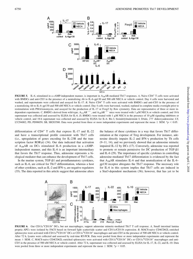
A2BAR promotes IL-6 to favor Th17, and IL-6 response iscAMP independent
The development of Th17 cells is controlled largely via factorsderived from innate immune cells, such as IL-6 and TGF-b1.Previous reports showed that adenosine could promote IL-6 ina variety of cell types, including DCs (28, 29). To assess the roleof IL-6 in the A2BAR-mediated Th17 response, cocultures wereconducted in the presence of a neutralizing Ab to the IL-6–gp130receptor. NECA induction of IL-17 was abrogated in the presenceof the IL-6–gp130 inhibitor, thus demonstrating that IL-6 is indeedan important mediator of the Th17 response (Fig. 5A, 5B). Al-though NECA decreased the number of cells expressing Foxp3 asTh17 cells increased, the decrease in Foxp3 did not occur whenIL-6-gp130 was blocked. These findings suggested that IL-6concentrations are a key determinant of the effects of adenosinein the reciprocal control of Th17 and Treg development (Fig. 5B).Experiments with a neutralizing Ab to TGF-b1 also impairedTh17 development, thus suggesting that TGF-b1 is also importantin this system (data not shown).To test whether IL-6 in this system is derived from DC A2BAR,
we conducted experiments with DCs generated from wild-type,A2AAR
2/2, or A2BAR2/2 mice. A2BAR
2/2 DCs produced littleIL-6 upon NECA treatment, whereas wild-type and A2AAR
2/2
DCs produced significant amounts of IL-6 in response to NECA(Fig. 5C). Importantly, A2BAR
2/2 DCs did not have an intrinsicdefect in IL-6 production, because LPS provoked abundant IL-6(data not shown). Pharmacologic blockade with AR-selectiveantagonists corroborated that the IL-6 response depended onA2BAR (data not shown). A role for A2BAR in TGF-b1 inductionwas also assessed, but NECA treatment of DCs did not elevateTGF-b1 protein or mRNA, as assayed by ELISA or real-time RT-PCR (data not shown). This suggested that low levels of TGF-b1are constitutively produced by DCs or, alternatively, that TGF-b1is residually present in the FBS used in cocultures.As introduced previously, the anti-inflammatory effects of
adenosine are often attributed to signaling pathways involvingA2AAR and the intracellular mediator cAMP. This is consistentwith the general anti-inflammatory effects of other cAMP-elevating agents, such as prostaglandin E2 and vasoactive in-testinal peptide (30). However, in our model, which incorporatesboth CD4+ T cells and APCs, we describe a putative proin-flammatory effect of adenosine on CD4+ T cells that is A2BARdependent. A2BAR differs from A2AAR and the other ARs in thatit can couple to both Gs and Gq, enabling it to activate bothadenylyl cyclase and PLC, respectively (6). Thus, we sought todetermine whether A2BAR relied on cAMP or whether othersignaling modalities were important.To elucidate key pathways that control the NECA-mediated
responses observed in our study, we treated DCs with NECA inthe presence of various pharmacologic inhibitors known to inhibitG protein pathways and assessed IL-6 production. At the con-centrations studied, the inhibitors did not impair DC viability, asassessed by trypan blue exclusion (data not shown). Inhibition oftwo distinct targets of the adenylyl cyclase/cAMP axis had no effecton NECA-mediated IL-6 induction, whereas inhibition of the PLC/
FIGURE 1. AR activation promotes differentiation of IL-17–secreting
CD4+ T cells. A, Naive CD62LhiCD44loCD25loCD4+ T cells were acti-
vated with BMDCs and anti-CD3 in the presence of 500 nM NECA or
vehicle control. After 72 h, cells were harvested, and mRNAwas assessed
by real-time RT-PCR, or supernatant was collected and assessed by ELISA
for IL-17 and IFN-g (B). Data are pooled from three or more independent
experiments and represent the mean 6 SEM. *p , 0.05. C, Day 4 cells
were harvested, washed, and replated in complete media overnight prior to
restimulation with PMA/ionomycin. Dot plots show intracellular IFN-g
and IL-17 in CD4+ T cells. D, Naive CD4+ T cells were activated with
BMDCs and anti-CD3 in the presence of vehicle control or 5 or 100 mM
adenosine. Day 4 cells were harvested, washed, and replated in complete
media overnight prior to restimulation with PMA/ionomycin. Dot plots
show intracellular IFN-g and IL-17 in CD4+ T cells. Dot plots are repre-
sentative of three or more independent experiments.
FIGURE 2. An adenosine mimetic promotes development of bona fide
Th17 cells. Naive CD4+ T cells were activated with BMDCs and anti-CD3
in the presence of 500 nM NECA or vehicle control. A, After 72 h, cells
were harvested, and mRNA was assessed by real-time RT-PCR, or su-
pernatant was collected and assessed by ELISA for IL-22 (B). Data were
pooled from three or more independent experiments and represent the
mean 6 SEM. *p , 0.05.
6748 ADENOSINE PROMOTES Th17 DEVELOPMENT
by guest on April 1, 2018
http://ww
w.jim
munol.org/
Dow
nloaded from

PKC axis with either U-73122 or bisindolylmaleimide I impairedthe IL-6 response significantly (Fig. 5D). Because MAPKs, in-cluding p38 and ERK, also play a role in mediating A2BAR effectsin some cells, additional inhibitors targeting other pathways wereused. We found that the ERK inhibitor PD98509 and the PI3Kinhibitor LY294002 both blocked NECA-mediated IL-6, but thep38 inhibitor SB203580 had little effect. Lastly, treating DCs withthe cAMP-elevating agent forskolin or the EPAC agonist 8-pCPT-
29-O-Me-cAMP provoked little IL-6, suggesting that stimulatingcAMP or EPAC pathways is not sufficient to drive DC productionof IL-6 (data not shown). Taken together, this indicated thatNECA promoted IL-6 in DCs independently of cAMP but thatPLC/PKC, ERK, and PI3K are involved, and it explains whyA2BAR, but not A2AAR, stimulates IL-6.
Intestinal DCs support NECA-mediated Th17 response
Th17 cells are especially abundant at mucosal sites, such as thesmall intestine, where they are important in controlling neutrophilaccumulation and epithelial barrier integrity (31). The intestineis subject to frequent hypoxia and, thus, is thought to be anadenosine-rich site (32). Therefore, to model the effects of aden-osine on T cell activation in the gut, we isolated highly purifiedsubsets of gut APCs and cultured them with naive CD4+ T cells(Fig. 6). We found that the adenosine mimetic NECA significantlyenhanced IL-17 in cocultures containing a population of CD11chi
CD11b+ gut DCs, a subset previously associated with Th17 re-sponses (26). Additionally Il22 was significantly upregulated byNECA treatment with this DC subset. Conversely, NECA had nosuch effect in cultures with a population of CD11cloCD11b+
macrophages, which is consistent with previous studies suggestingthis population of APCs promotes Tregs (26). Foxp3 was not al-tered in any of the NECA-treated groups.
DiscussionPrevious work showed that direct engagement of A2AAR on T cellsinhibited proliferation, as well as Th1 and Th2 responses (9–11).A2AAR also contributes to the generation and function of Tregs(10, 12–14, 33). However, T cell activation and differentiation donot occur in isolation but require APCs and a cadre of extracel-lular factors. Moreover, the accumulation of endogenous adeno-sine at inflammatory sites does not exclusively target A2AAR;it stimulates other available AR subtypes. To more faithfullymodel how adenosine impacts CD4+ T cell differentiation, westudied the effects of a nonselective adenosine mimetic in cocul-tures with DCs and highly purified naive CD4+ T cells. In thisstudy, we found that adenosine preferentially promoted de novo
FIGURE 3. An adenosine mimetic favors Th17 cells in the presence of TGF-b1. Naive CD4+ T cells were activated with BMDCs and anti-CD3 in the
presence of TGF-b1 and 500 nM NECA or vehicle control. Day 4 cells were harvested, washed, and replated in complete media overnight prior to
restimulation with PMA/ionomycin. Dot plots show intracellular IL-17 and Foxp3 in CD4+ T cells. Dot plots (A) are representative of three or more
independent experiments, and compiled data (B) were pooled from three or more independent experiments and represent the mean 6 SEM. *p , 0.05.
FIGURE 4. Th17 response depends on DC A2BARs. Naive CD4+ T cells
were activated with BMDCs and anti-CD3 in the presence of 500 nM
NECA or vehicle control. Seventy-two–hour supernatant was collected and
assessed by ELISA for IL-17. A, AR-selective antagonists or vehicle
control was added to cultures at a concentration of 250 nM. Data were
pooled from two independent experiments conducted in duplicate and
represent the mean 6 SEM. BMDCs (B) or CD4+ T cells (C) were derived
from A2BAR2/2 or A2AAR
2/2/A2BAR2/2 mice, where indicated. Data
were pooled from three or more independent experiments and represent the
mean 6 SEM. *p , 0.05.
The Journal of Immunology 6749
by guest on April 1, 2018
http://ww
w.jim
munol.org/
Dow
nloaded from
differentiation of CD4+ T cells that express IL-17 and IL-22and have a transcriptional profile consistent with Th17 cells(i.e., upregulation of genes encoding for IL-23R and the tran-scription factor RORgt) (34). Our data indicated that activationof A2BAR on DCs stimulated IL-6 production in a cAMP-independent manner, and this IL-6 is an important intermediarythat favors the Th17 response. Thus, adenosine represents a bi-ological mediator that can enhance the development of Th17 cells.In the murine system, TGF-b1 and proinflammatory cytokines,
such as IL-6, are critical for Th17 differentiation, whereas a hostof other cytokines, such as IL-2 and IFN-g, are negative regulators(35). The data reported in this article suggest that adenosine alters
the balance of these cytokines in a way that favors Th17 differ-entiation at the expense of Treg development. For instance, ade-nosine directly impairs IL-2 and IFN-g production by Th cells(9–11, 14), and we previously showed that an adenosine mimeticimpaired IL-12 by DCs (17). Conversely, adenosine was reportedto promote or remain permissive for DC production of TGF-b1and IL-6 (29). The importance of specific cytokines in controllingadenosine-mediated Th17 differentiation is evidenced by the factthat A2BAR stimulates IL-6 and that neutralization of the IL-6–gp130 receptor abrogates the Th17 response. The necessary rolefor IL-6 in this system implies that Th17 cells are induced ina Stat3-dependent mechanism (36); however, that has yet to be
FIGURE 5. IL-6, stimulated in a cAMP-independent manner, is important in A2BAR-mediated Th17 responses. A, Naive CD4+ T cells were activated
with BMDCs and anti-CD3 in the presence of a neutralizing Ab to IL-6–gp130 and 500 nM NECA or vehicle control. Day 4 cells were harvested and
washed, and supernatants were collected and assayed for IL-17. B, Naive CD4+ T cells were activated with BMDCs and anti-CD3 in the presence of
a neutralizing Ab to IL-6–gp130 and 500 nM NECA or vehicle control. Day 4 cells were harvested, washed, replated in complete media overnight prior to
restimulation with PMA/ionomycin, and assayed for the production of IL-17 or Foxp3 by flow cytometry. Data are representative of three or more in-
dependent experiments. C, BMDCs derived from wild-type, A2AAR2/2, and A2BAR
2/2 mice were treated with 1 mM NECA or vehicle control, and 18-h
supernatant was collected and assessed by ELISA for IL-6. D, BMDCs were treated with 1 mM NECA in the presence of 10 mM signaling inhibitors or
vehicle control, and 18-h supernatant was collected and assessed by ELISA for IL-6. Bis I, bisindolylmaleimide I; DAdo, 2959 dideoxyadenosine; LY,
LY294002; PD, PD98059; SB, SB203580. Data were pooled from three or more independent experiments and represent the mean 6 SEM. *p , 0.05.
FIGURE 6. Gut CD11c+CD11b+ DCs, but not macrophages, support adenosine mimetic-mediated Th17 T cell response. A, Small intestinal lamina
propria APCs were isolated by FACS based on forward light scatter/side scatter and CD11c/CD11b expression. B, MACS-naive CD4CD62L-enriched
splenocytes were activated with CD11chiCD11b+ DCs or CD11cloCD11b+ macrophages and anti-CD3 in the presence of 500 nM NECA or vehicle control.
After 72 h, lysates were collected and assessed by real-time RT-PCR. Data were pooled from three or more independent experiments and represent the
mean 6 SEM. C, MACS-naive CD4CD62L-enriched splenocytes were activated with CD11chiCD11b+ DCs or CD11cloCD11b+ macrophages and anti-
CD3 in the presence of 500 nM NECA or vehicle control. After 72 h, supernatant was collected and assessed by ELISA for IL-17, IL-22, and IL-10. Data
were pooled from three or more independent experiments and represent the mean 6 SEM. *p , 0.05.
6750 ADENOSINE PROMOTES Th17 DEVELOPMENT
by guest on April 1, 2018
http://ww
w.jim
munol.org/
Dow
nloaded from
confirmed. The role of TGF-b1 in this system is less clear, be-cause we did not find direct evidence that A2BAR stimulation wassufficient to generate TGF-b1. However, the fact that neutrali-zing Ab to TGF-b1 diminished IL-17 production indicated thatadenosine-mediated Th17 differentiation requires the presence ofTGF-b1.Although NECA favors Th17 cells and not Tregs, upon IL-6
neutralization, the opposite result was observed: Th17 expansionwas abrogated as was the decrease in cells expressing Foxp3. Thesedata suggested that IL-6 induced by NECA was predominantly, ifnot solely, responsible for the decrease in Foxp3-producing Th cellsand that it is a key intermediate controlling the effects of adeno-sine on Th17 and Treg development. Stimulation of A2AAR wasimplicated in the expansion of Tregs (14) when T cells werestimulated in the absence of APCs. However, A2AAR activation didnot inhibit Th17 cells when a source of APCs was provided (14).The finding that A2BAR acts on DCs to promote Th17 cell
development conflicts with the conclusions of other investigatorsthat adenosine signaling favors a Th2-biasing DC (37, 38). Thesegroups studied human monocyte-derived DCs and restricted theirinvestigation to Th1 and Th2 markers in MLR, specifically IFN-gand IL-4 and/or IL-5 (37, 38). Thus, these investigators studiedDCs with characteristics unique from BMDCs and did not assessmarkers associated with other Th phenotypes, such as IL-17 or IL-22. A potentially important distinction between the studies is thedifference in the human and mouse systems. However, in additionto species variation, there are other factors that may be important.In the single study that compared the effects of adenosine, in thepresence or absence of LPS, on DC-polarizing capacity, anti-Th1/pro-Th2 skewing was only observed in the group with LPS (37,38). In our model system, no LPS was required to achieve thestriking upregulation of IL-17 and IL-22.The data obtained with BMDCs were consistent with observa-
tions made using DCs isolated from the intestinal lamina propria,a site rich in Th17 cells. When used in coculture experiments, theCD11c+CD11b+ DC subset preferentially increased IL-17 and IL-22 mRNA in the presence of the adenosine mimetic. These find-ings indicated that ARs modulate CD11c+CD11b+ lamina propriaDCs and suggested that endogenous adenosine acts via laminapropria DCs to control intestinal Th responses, which agrees withother studies on intestinal DCs (26). The difference in A2BAR-mediated effects between these two intestinal APC populationsmay reflect the fact that resting macrophages express fewerA2BAR (17), and this receptor leads to the production of IL-6,whereas the A2AAR does not. Although we showed that adeno-sine is one factor that can promote Th17 development, there areclearly redundant pathways that contribute to the in vivo response.For instance, A2BAR
2/2 mice have numbers of Th17 cells insmall intestine lamina propria comparable to wild-type controls;moreover, the abundance of Th17 cells in our A2BAR
2/2 micecould be augmented by manipulating commensal flora (data notshown), presumably due to segmented filamentous bacteria (SFB),which persist in some, but not all, mouse facilities (39). Althoughruling out a necessary role for A2BAR in the SFB-mediated Th17response, this does not discount the possibility that local accu-mulation of adenosine can act to promote Th17 cells.The role of A2BAR in the promotion of Th17 cells, and possibly
inflammation, is somewhat surprising but not entirely unexpected.For example, some studies implicated A2BAR in pulmonary in-flammation (40). This is at least consistent with other evidencesuggesting that intestinal inflammation is reduced in the absenceof normal A2BAR function (23, 24). However, it is important tonote that another group investigating adenosine in the gut foundthe exact opposite (41). One interpretation of the conflicting
results of A2BAR regulation on intestinal inflammation is thatthe microbiome differs in the animal facilities of the respectiveinvestigators. Although the A2BAR-dependent induction of Th17could be consistent with these receptors promoting inflammation,emerging thought suggests that Th17 cells may confer protective/anti-inflammatory responses. For example, mice harboring SFBhave a marked increase in intestinal Th17 cells but no disease(39). In addition, the coexpression of IL-22 by Th17 cells can beprotective (42). Similarly, IL-17R knockout mice have worse in-testinal disease in some models (43). Because Th1, Th2, and Th17cells contribute to the protection of the host, which is why theyexist, their contribution to the pathogenesis of disease may requirethe presence of other factors. For example, in Crohn’s disease,Th17 responses are implicated in the pathogenesis but usually arethe result of their heightened activation by accessory cytokines,such as IL-23 (44). Thus, the presence of IL-17–producing cells inthe absence of other cues may not be sufficient for inflammation toensue.Factors that calibrate the magnitude and quality of CD4+ T cell
responses are critical for host health. Although previous workemphasized an inhibitory role for adenosine in controlling CD4+
Th cells, the current study suggests that adenosine promotes Th17cells. Thus, adenosine represents a biological mediator that reg-ulates CD4+ T cell development and function, albeit in a mannerthat was not predicted. Adenosine shares its pleiotropic functionswith other factors, such as IL-2 and TGF-b1. For example, IL-2drives the expansion of effector T cells, but it is required for themaintenance of Tregs (45). Similarly, TGF-b1 is important for theinduction of Foxp3 and the accumulation of induced Tregs, and itcontributes to Th17 cell development (46). The relative concen-tration of a nucleoside, the types of cells that it targets, and thespecific ARs available for engagement may be important deter-minants of the final outcome. The current study is significantbecause it adds to the regulatory effects of adenosine and expandsthe current thinking on its role in the control of host responses.
AcknowledgmentsWe thank Elizabeth Wiznerowicz, Joanne Lannigan, and Michael Solga for
technical assistance.
DisclosuresJ.L. has a financial interest in PGxHealth. P.B.E. is the principal investi-
gator on a grant from the National Institutes of Health that includes a sub-
contract with PGxHealth.
References1. Mosmann, T. R., H. Cherwinski, M. W. Bond, M. A. Giedlin, and R. L. Coffman.
1986. Two types of murine helper T cell clone. I. Definition according to profilesof lymphokine activities and secreted proteins. J. Immunol. 136: 2348–2357.
2. Harrington, L. E., R. D. Hatton, P. R. Mangan, H. Turner, T. L. Murphy,K. M. Murphy, and C. T. Weaver. 2005. Interleukin 17-producing CD4+ effectorT cells develop via a lineage distinct from the T helper type 1 and 2 lineages.Nat. Immunol. 6: 1123–1132.
3. Park, H., Z. Li, X. O. Yang, S. H. Chang, R. Nurieva, Y. H. Wang, Y. Wang,L. Hood, Z. Zhu, Q. Tian, and C. Dong. 2005. A distinct lineage of CD4 T cellsregulates tissue inflammation by producing interleukin 17. Nat. Immunol. 6:1133–1141.
4. Hasko, G., and B. N. Cronstein. 2004. Adenosine: an endogenous regulator ofinnate immunity. Trends Immunol. 25: 33–39.
5. Sitkovsky, M. V., D. Lukashev, S. Apasov, H. Kojima, M. Koshiba, C. Caldwell,A. Ohta, and M. Thiel. 2004. Physiological control of immune response andinflammatory tissue damage by hypoxia-inducible factors and adenosine A2Areceptors. Annu. Rev. Immunol. 22: 657–682.
6. Ernst, P. B., J. C. Garrison, and L. F. Thompson. 2010. Much ado about aden-osine: adenosine synthesis and function in regulatory T cell biology. J. Immunol.185: 1993–1998.
7. Huang, S., S. Apasov, M. Koshiba, and M. Sitkovsky. 1997. Role of A2a ex-tracellular adenosine receptor-mediated signaling in adenosine-mediated in-hibition of T-cell activation and expansion. Blood 90: 1600–1610.
The Journal of Immunology 6751
by guest on April 1, 2018
http://ww
w.jim
munol.org/
Dow
nloaded from

8. Ohta, A., and M. Sitkovsky. 2001. Role of G-protein-coupled adenosine recep-tors in downregulation of inflammation and protection from tissue damage.Nature 414: 916–920.
9. Koshiba, M., D. L. Rosin, N. Hayashi, J. Linden, and M. V. Sitkovsky. 1999.Patterns of A2A extracellular adenosine receptor expression in different func-tional subsets of human peripheral T cells. Flow cytometry studies with anti-A2A receptor monoclonal antibodies. Mol. Pharmacol. 55: 614–624.
10. Naganuma, M., E. B. Wiznerowicz, C. M. Lappas, J. Linden, M. T. Worthington,and P. B. Ernst. 2006. Cutting edge: Critical role for A2A adenosine receptors inthe T cell-mediated regulation of colitis. J. Immunol. 177: 2765–2769.
11. Lappas, C. M., J. M. Rieger, and J. Linden. 2005. A2A adenosine receptor in-duction inhibits IFN-gamma production in murine CD4+ T cells. J. Immunol.174: 1073–1080.
12. Kobie, J. J., P. R. Shah, L. Yang, J. A. Rebhahn, D. J. Fowell, andT. R. Mosmann. 2006. T regulatory and primed uncommitted CD4 T cells ex-press CD73, which suppresses effector CD4 T cells by converting 59-adenosinemonophosphate to adenosine. J. Immunol. 177: 6780–6786.
13. Deaglio, S., K. M. Dwyer, W. Gao, D. Friedman, A. Usheva, A. Erat, J. F. Chen,K. Enjyoji, J. Linden, M. Oukka, et al. 2007. Adenosine generation catalyzed byCD39 and CD73 expressed on regulatory T cells mediates immune suppression.J. Exp. Med. 204: 1257–1265.
14. Zarek, P. E., C. T. Huang, E. R. Lutz, J. Kowalski, M. R. Horton, J. Linden,C. G. Drake, and J. D. Powell. 2008. A2A receptor signaling promotes peripheraltolerance by inducing T-cell anergy and the generation of adaptive regulatoryT cells. Blood 111: 251–259.
15. Okusa, M. D., J. Linden, T. Macdonald, and L. Huang. 1999. Selective A2Aadenosine receptor activation reduces ischemia-reperfusion injury in rat kidney.Am. J. Physiol. 277: F404–F412.
16. Lappas, C. M., Y. J. Day, M. A. Marshall, V. H. Engelhard, and J. Linden. 2006.Adenosine A2A receptor activation reduces hepatic ischemia reperfusion injuryby inhibiting CD1d-dependent NKT cell activation. J. Exp. Med. 203: 2639–2648.
17. Wilson, J. M., W. G. Ross, O. N. Agbai, R. Frazier, R. A. Figler, J. Rieger,J. Linden, and P. B. Ernst. 2009. The A2B adenosine receptor impairs thematuration and immunogenicity of dendritic cells. J. Immunol. 182: 4616–4623.
18. Kreckler, L. M., T. C. Wan, Z. D. Ge, and J. A. Auchampach. 2006. Adenosineinhibits tumor necrosis factor-alpha release from mouse peritoneal macrophagesvia A2A and A2B but not the A3 adenosine receptor. J. Pharmacol. Exp. Ther.317: 172–180.
19. Yang, D., Y. Zhang, H. G. Nguyen, M. Koupenova, A. K. Chauhan, M. Makitalo,M. R. Jones, C. St Hilaire, D. C. Seldin, P. Toselli, et al. 2006. The A2Badenosine receptor protects against inflammation and excessive vascular adhe-sion. J. Clin. Invest. 116: 1913–1923.
20. Csoka, B., Z. H. Nemeth, L. Virag, P. Gergely, S. J. Leibovich, P. Pacher,C. X. Sun, M. R. Blackburn, E. S. Vizi, E. A. Deitch, and G. Hasko. 2007. A2Aadenosine receptors and C/EBPbeta are crucially required for IL-10 productionby macrophages exposed to Escherichia coli. Blood 110: 2685–2695.
21. Hasko, G., B. Csoka, Z. H. Nemeth, E. S. Vizi, and P. Pacher. 2009. A(2B) adenosinereceptors in immunity and inflammation. Trends Immunol. 30: 263–270.
22. Sitkovsky, M. V., and A. Ohta. 2005. The ‘danger’ sensors that STOP the im-mune response: the A2 adenosine receptors? Trends Immunol. 26: 299–304.
23. Kolachala, V., B. Ruble, M. Vijay-Kumar, L. Wang, S. Mwangi, H. Figler,R. Figler, S. Srinivasan, A. Gewirtz, J. Linden, et al. 2008. Blockade of aden-osine A2B receptors ameliorates murine colitis. Br. J. Pharmacol. 155: 127–137.
24. Kolachala, V. L., M. Vijay-Kumar, G. Dalmasso, D. Yang, J. Linden, L. Wang,A. Gewirtz, K. Ravid, D. Merlin, and S. V. Sitaraman. 2008. A2B adenosinereceptor gene deletion attenuates murine colitis. Gastroenterology 135: 861–870.
25. Csoka, B., L. Himer, Z. Selmeczy, E. S. Vizi, P. Pacher, C. Ledent, E. A. Deitch,Z. Spolarics, Z. H. Nemeth, and G. Hasko. 2008. Adenosine A2A receptor ac-tivation inhibits T helper 1 and T helper 2 cell development and effector func-tion. FASEB J. 22: 3491–3499.
26. Denning, T. L., Y. C. Wang, S. R. Patel, I. R. Williams, and B. Pulendran. 2007.Lamina propria macrophages and dendritic cells differentially induce regulatoryand interleukin 17-producing T cell responses. Nat. Immunol. 8: 1086–1094.
27. Livak, K. J., and T. D. Schmittgen. 2001. Analysis of relative gene expressiondata using real-time quantitative PCR and the 2(-Delta Delta C(T)) Method.Methods 25: 402–408.
28. Sun, Y., F. Wu, F. Sun, and P. Huang. 2008. Adenosine promotes IL-6 release inairway epithelia. J. Immunol. 180: 4173–4181.
29. Novitskiy, S. V., S. Ryzhov, R. Zaynagetdinov, A. E. Goldstein, Y. Huang,O. Y. Tikhomirov, M. R. Blackburn, I. Biaggioni, D. P. Carbone, I. Feoktistov,and M. M. Dikov. 2008. Adenosine receptors in regulation of dendritic celldifferentiation and function. Blood 112: 1822–1831.
30. Koga, K., G. Takaesu, R. Yoshida, M. Nakaya, T. Kobayashi, I. Kinjyo, andA. Yoshimura. 2009. Cyclic adenosine monophosphate suppresses the tran-scription of proinflammatory cytokines via the phosphorylated c-Fos protein.Immunity 30: 372–383.
31. Kolls, J. K., P. B. McCray, Jr., and Y. R. Chan. 2008. Cytokine-mediated regu-lation of antimicrobial proteins. Nat. Rev. Immunol. 8: 829–835.
32. Eltzschig, H. K., J. Rivera-Nieves, and S. P. Colgan. 2009. Targeting the A2Badenosine receptor during gastrointestinal ischemia and inflammation. ExpertOpin. Ther. Targets 13: 1267–1277.
33. Friedman, D. J., B. M. Kunzli, Y. I. A-Rahim, J. Sevigny, P. O. Berberat,K. Enjyoji, E. Csizmadia, H. Friess, and S. C. Robson. 2009. From the Cover:CD39 deletion exacerbates experimental murine colitis and human poly-morphisms increase susceptibility to inflammatory bowel disease. Proc. Natl.Acad. Sci. USA 106: 16788–16793.
34. Zhu, J., and W. E. Paul. 2010. Heterogeneity and plasticity of T helper cells. CellRes. 20: 4–12.
35. Bettelli, E., T. Korn, and V. K. Kuchroo. 2007. Th17: the third member of theeffector T cell trilogy. Curr. Opin. Immunol. 19: 652–657.
36. Yang, X. O., A. D. Panopoulos, R. Nurieva, S. H. Chang, D. Wang,S. S. Watowich, and C. Dong. 2007. STAT3 regulates cytokine-mediated gen-eration of inflammatory helper T cells. J. Biol. Chem. 282: 9358–9363.
37. Panther, E., S. Corinti, M. Idzko, Y. Herouy, M. Napp, A. la Sala, G. Girolomoni,and J. Norgauer. 2003. Adenosine affects expression of membrane molecules,cytokine and chemokine release, and the T-cell stimulatory capacity of humandendritic cells. Blood 101: 3985–3990.
38. Yang, M., C. Ma, S. Liu, Q. Shao, W. Gao, B. Song, J. Sun, Q. Xie, Y. Zhang,A. Feng, et al. 2010. HIF-dependent induction of adenosine receptor A2b skewshuman dendritic cells to a Th2-stimulating phenotype under hypoxia. Immunol.Cell Biol. 88: 165–171.
39. Ivanov, I. I., Rde. L. Frutos, N. Manel, K. Yoshinaga, D. B. Rifkin, R. B. Sartor,B. B. Finlay, and D. R. Littman. 2008. Specific microbiota direct the differen-tiation of IL-17-producing T-helper cells in the mucosa of the small intestine.Cell Host Microbe 4: 337–349.
40. Sun, C. X., H. Zhong, A. Mohsenin, E. Morschl, J. L. Chunn, J. G. Molina,L. Belardinelli, D. Zeng, and M. R. Blackburn. 2006. Role of A2B adenosinereceptor signaling in adenosine-dependent pulmonary inflammation and injury.J. Clin. Invest. 116: 2173–2182.
41. Frick, J. S., C. F. MacManus, M. Scully, L. E. Glover, H. K. Eltzschig, andS. P. Colgan. 2009. Contribution of adenosine A2B receptors to inflammatoryparameters of experimental colitis. J. Immunol. 182: 4957–4964.
42. Zenewicz, L. A., G. D. Yancopoulos, D. M. Valenzuela, A. J. Murphy, M. Karow,and R. A. Flavell. 2007. Interleukin-22 but not interleukin-17 provides protectionto hepatocytes during acute liver inflammation. Immunity 27: 647–659.
43. O’Connor, W., Jr., M. Kamanaka, C. J. Booth, T. Town, S. Nakae, Y. Iwakura,J. K. Kolls, and R. A. Flavell. 2009. A protective function for interleukin 17A inT cell-mediated intestinal inflammation. Nat. Immunol. 10: 603–609.
44. Kobayashi, T., S. Okamoto, T. Hisamatsu, N. Kamada, H. Chinen, R. Saito,M. T. Kitazume, A. Nakazawa, A. Sugita, K. Koganei, et al. 2008. IL23 dif-ferentially regulates the Th1/Th17 balance in ulcerative colitis and Crohn’sdisease. Gut 57: 1682–1689.
45. Nelson, B. H. 2004. IL-2, regulatory T cells, and tolerance. J. Immunol. 172:3983–3988.
46. Wahl, S. M. 2007. Transforming growth factor-beta: innately bipolar. Curr. Opin.Immunol. 19: 55–62.
6752 ADENOSINE PROMOTES Th17 DEVELOPMENT
by guest on April 1, 2018
http://ww
w.jim
munol.org/
Dow
nloaded from